Safety and Efficacy of Colchicine in the Treatment of Recurrent Aphthous Stomatitis

Celiac Disease 2016Grand Rounds
Regions Hospital
Ahsan M Bhatti MD, FACP
Bhatti Gastroenterology Consultants

I have nothing to disclose

Case Introduction
• 35 year old female referred for IBS. H/O 1-3 episodes of diarrhea. Excessive gas and nocturnal symptoms.
• Glossitis and angular stomatitis
• Patient had H/O diarrhea when baby and in teen age years.
• H/O Iron deficiency, hypothyroidism.
• On supplements and MVI. H/O iron supplements
• H/O miscarriage and irregular menstrual cycle.

Educational Objectives• Identify the signs and symptoms of celiac disease for
clinicians.
• Understand the Testing and utilization of blood work and Pathology.
• Management plan and long term follow up.
• Appreciate conditions associated with CD.
• Conditions similar to celiac disease• Non Gluten wheat allergies
• Gluten Intolerance

INTRODUCTION
• A small bowel disorder characterized by mucosal inflammation, villous atrophy, and crypt hyperplasia, which occur upon exposure to dietary gluten and which demonstrate improvement after withdrawal of gluten from the diet.
• Celiac disease (also called gluten-sensitive enteropathy and non-tropical sprue) was first described by Samuel Gee in 1888

Celiac Disease
• CD is an immune response to Gliadins in the diet in susceptible patients that lead to inflammatory response and damage in the small bowel.
• Highest prevalence in European. ( Finland 2.4%)
• Female to male ratio is 2:1
• Disease of all ages ( >20% older than 60)
• North American Prevalence• General 1:100
• Symptomatic 1:50
• FDR 1:10


The Changing Face of Celiac
PAST PRESENTRare Much more commonMore in kids All agesHigh mortality Low mortalityClassic features: Protean features:
+ Diarrhea + GI features+ Weight loss + Extra-intestinal + Failure to thrive features

Pathogenesis• CD patients ingest glutens which are
resistant to proteases
• Antigen presenting cell ( HLA DQ2/8) present gliadin to T cells.
• T cells recruit other immune cells, cytokines are released.
• tTG is released which make residue of gliadin more antigenic
• tTG targeted by humoral response creates antibodies
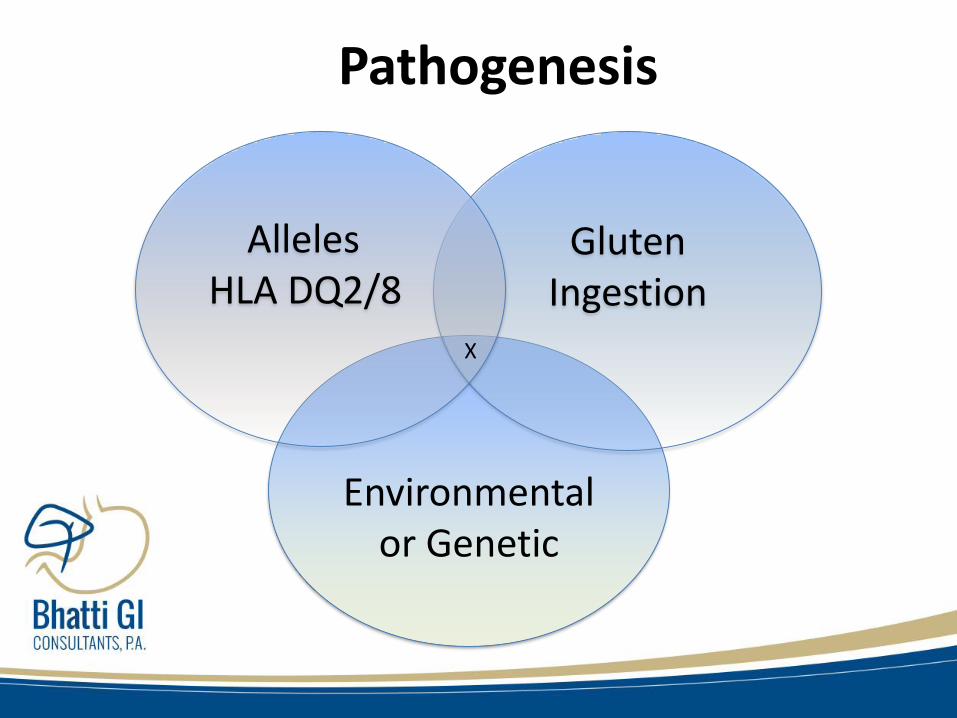
Environmentalor Genetic
GlutenIngestion
AllelesHLA DQ2/8
X
Pathogenesis

Change in Awareness
• Patients with symptoms of diarrhea
– Before 1981: 91.3%
– In 1990’s: 58.3%
– After 2000: 37.0%
• Average # of visit to pediatrician before diagnosis is 8.
• Average duration of symptoms ( except diarrhea) is 4 year.


Genetics OF celiac disease
• HLA DQ2 or DQ8 required. (prevalence about 30-
40%)
• Family risk of CD
– Monozygotic twins 80%
– Siblings 10%
– Kids 5-10%
• Syndromes associated with CD• Down’s, Turner's, William's

Diagnosis
• History and supportive clinical features
• Supportive Serologies
• Small bowel Biopsies characteristics.• Gold slandered for Dx.
• Clinical response to Gluten free diet.

Celiac Disease
Atypical Celiac
Disease
Asymptomatic (silent)
Celiac Disease
Latent Celiac
Disease
Classic Disease

Manifestations of Celiac Disease
General Gastrointestinal
Short Stature Diarrhea, steatorrhea
Weight loss* Flatulence, distension
Failure to thrive Abdominal discomfort
Lethargy Anorexia, nausea, vomiting
Delayed puberty Constipation**
Edema Aphthous stomatitis
*10%+ obese** 20% constipated
Rubio-Tapia A, Murray JA. Curr Opin Gastroenterol 2010; 26; 116-22


Non Classical CD
Category Examples
Hematologic Anemia ( Iron,B12,folate), Functional Asplenia (H-J bodies)
Other Labs Vit. Deficiencies, Abnormal Liver Function tests
Musculoskeletal Osteopenia, osteoporosis, osteomalacia, arthropathies
Neuro Seizures, ataxia, neuropathy
Reproductive Infirtility, Miscarriages
Skin Dermatitis Herpetiformis

Autoimmune Diseases
• Type 1 Diabetes, Addison’s disease, Thyroiditis
• IBD, Microscopic colitis ( 70 times more likely)
• IgA deficiency
• Autoimmune Hepatitis, PBC,PSC
• RA, Sjogren’s
• IgA mesangial nephropathy.

Iron Deficiency Anemia
• CD prevalence in patients with IDA • (3-9% with no sx)
• (10-15% with sx)
• 33% of patient with CD also had other source of IDA on EGD.
• Most overlooked in Menstruating women
• During EGD Small bowel Biopsies should be done on patient with IDA
– Grisolano S et al J Clincal Gastro 2004

Abnormal LFT and CD
• ALT and AST
– Autoimmune hepatitis, NAFLD, Reactive hepatitis
• Alkaline Phosphatase
– PBC,PSC, Osteomalacia ( vit D deficiency)

Autoimmune Liver Disease in CD
30 patients with CD and liver biopsy19 with autoimmune cause, (26% with CD sx)
Autoimmune hepatitis (9)PSC (7)PBC (3)
11 with other causes, (82% with CD sx)Cryptogenic, hep C, fat, sarcoid, lymphoma
Mounaijed, et al. Am J Clin Pathol 2011; 136: 128-37

Dermatitis Herpetiformis (DH)
• DH represents intestinal sensitivity to gluten – not from skin contact
• Pruritic papulovesicular
• Extensor surfaces
• Symmetrical
• 80-90% no GI symptoms, but 75% with villous atrophy

Dermatitis Herpetiformis (DH)• When a person with celiac disease consumes gluten, the
mucosal immune system in the intestine responds by producing a type of antibody called immunoglobulin A (IgA)
• As IgA enters the bloodstream, it can collect in small blood vessels under the skin, triggering further immune reactions that result in the blistering rash of DH.
https://celiac.org/celiac-disease/dermatitis-herpetiformis/
• Biopsies for diagnosis are taken from the affected area AND normal skin adjacent to the affected area

CLINICAL MANIFESTATIONS
• Gastrointestinal manifestations
• Non-gastrointestinal manifestations• Neuropsychiatric disease
• Arthritis
• Iron deficiency
• Metabolic bone disease
• Hyposplenism
• Kidney disease
• Idiopathic pulmonary hemosiderosis

WHO SHOULD BE TESTED?
• Those with gastrointestinal symptoms including
• Chronic or recurrent diarrhea,
• Malabsorption, weight loss,
• Abdominal distension or bloating.
• Irritable bowel syndrome or severe lactose intolerance.

WHO SHOULD BE TESTED?
• Individuals without other explanations for signs and symptoms such as iron deficiency anemia, folate or vitamin B12 deficiency, persistent elevation in serum ALT, short stature, delayed puberty, recurrent fetal loss, low birthweight infants, reduced fertility, persistent aphthous stomatitis, dental enamel hypoplasia, idiopathic peripheral neuropathy, nonhereditary cerebellar ataxia, or recurrent migraine headaches.
• Patients with type 1 diabetes mellitus, and first-degree relatives of individuals with celiac disease if they have signs, symptoms, or laboratory evidence of possible celiac disease

CELIAC DISEASE DIAGNOSIS
• Blood tests
• Small intestine biopsy
• Genetic testing

When to test for celiac Disease
• Before testing Patient should be on Gluten containing diet. Sensitivity of serologies drop significantly on gluten free diet.
• Gluten challenge
– 2 slices of wheat bread/day for 2 weeks
• If poor tolerance, test than
• If tolerable, than 6 more weeks, than test
– Leffler D et al, gut 20129 Epub)

SERUM ANTIBODY ASSAYS• AGA
– Anti gliadin antibody (IgA/IgG)
• EMA
– IgA endomysial antibody (IgA EMA)
• Tissue Transglutamenase:
– IgA tissue transglutaminase antibody (IgA tTG)
– IgG tissue transglutaminase antibody (IgG tTG)
• Deaminated Gliadin Peptide:
– IgA deaminated gliadin peptide (IgA DGP)
– IgG deaminated gliadin peptide (IgG DGP

Assay sensitivity and specificity
• IgA endomysial antibodies – sensitivity 90to 98 % ; specificity 97 to 100 %
• IgA tissue transglutaminase antibodies – sensitivity 90 to 98 %; specificity 95 to 97 %
• IgA deaminated gliadin peptide (DAP)– sensitivity 74-84 %; specificity 99 %
• IgG deaminated gliadin peptide(DAP) – sensitivity 92 %; specificity 100 %

When To do EGD and Biopsy
• All patients with celiac disease should have biopsy before GFD.
• Allows to confirm diagnosis.
• Allows baseline for future progression.
• Should be done in those with:• Positive serologies.
• Strong symptoms
• Iron deficiency anemia


Marsh Modified
Histologic criterion Corazza
Increased intraepithelial lymphocytes*
Crypt hyperplasia Villous atrophy
Type 0 No No No None
Type 1 Yes No No Grade A
Type 2 Yes Yes No
Type3a Yes Yes Yes (partial) Grade B1
Type 3b Yes Yes Yes (subtotal)
Type 3c Yes Yes Yes (total) Grade B2
Summary of histologic classifications frequently used for celiac disease
*> 40 intraepithelial lymphocytes per 100enterocytes for Marsh modified (Oberhuber); > 25 intraepitheliallymphocytes per 100 enterocytes for Corazza


Biopsy for Pediatrics?
Recent guideline from the European Society of Pediatric Gastroenterology, Hepatology, and Nutrition (ESPGHAN) proposed that it may be possible to avoid any intestinal biopsy in children who meet the following criteria:
• Characteristic symptoms of CD• TTG IgA levels >10x upper limit of normal
(confirmed with + EMA in different blood sample)• Positive HLA-DQ 2/8
Rubio-Tapia, A; Am Journal of Gastroenterology, May 2013 volume 108

D/D of Biopsy
• NSAIDs
• SIBO
• IBD
• H-Pylori,
• Giardia
• CVID
• Lymphoma
• Viral Gastroenteritis
• Tropical sprue,
• Whipple’s Disease
• Non-gluten proteins
• Drugs (Olmasaten, Benicar)

HLA testing
• HLA DQ- 2 (95%) HLA DQ- 8 (5%)
• 30-40% of the population
• Necessary, But not enough
• Negative predictive value 99-100%
• Celiac Disease prevalence 1%


Celiac disease testing algorithm

Treatment• Gluten free diet life long
• Skilled Dietician.
• Labs.
– CBC, ( ferritin, B12,folate)
– LFT, Vit D, TSH, Ca++
– INR, Vit A, E, copper, zinc, etc.
• Bone Density.
• Vaccine for encapsulated • May be functionally A splenic


Diet Compliance
• Lower Compliance
– Diagnosis at older age
– Teens
– Asymptomatic
– Detection via family screening.

Clinical response time line
• Symptom relief is quick:• 67% notice complete response in 6 months.
• Mean time is 4 weeks
• Serologies:• Significant by 6 months
• Histology:• May take 6-24 months, Slower in adults

Follow up
• Baseline• Dietician, labs
• 3-6 months• Assess symptoms and repeat serologies
• Yearly Follow-up
• Repeat biopsies?

Celiac Disease Complications
• Nonresponsive celiac disease
• Refractory celiac disease
• Ulcerative jejunitis
• Lymphoma
• Skin conditions

Refractory sprue
• Type 1: – Normal population of intraepithelial lymphocytes.
• Type 2: – Aberrant or premalignant population of intraepithelial lymphocytes
based upon clonality analysis of T-cell receptors and immunopheno typing.
– Type 2 can progress to enteropathy-associated T-cell lymphoma, which may present clinically as ulcerative jejunitis. The diagnosis can be established on biopsy; CT, MRI, and 18F-FDG PET scans can help identify suspicious areas

Summary
• Mono symptomatic or non classic features are more common than classic Celiac disease
• Clinical History, serologies and Small bowel biopsy substantiate the diagnosis.
• GFD is life long, challenging and expensive.
• If not responding than verify the diagnosis, check compliance, consider complication
• Screening at risk population. Mass screening is not endorsed


Professional level information
– Academy of Nutrition and Dietetics (formerly American Dietetic Association)
– (www.eatright.org)
– American Celiac Disease Alliance
– (www.americanceliac.org)
– American Gastroenterological Association
– (http://www.gastro.org/info_for_patients/2013/6/6/understanding-celiac-disease)
– Celiac Disease Foundation
– (www.celiac.org)
– Gluten Intolerance Group of North America
– (www.gluten.net)
– National Foundation for Celiac Awareness (NFCA)
– (www.celiaccentral.org)
– National Institute of Diabetes and Digestive and Kidney Diseases
– (www.niddk.nih.gov)
– National Library of Medicine
– (www.nlm.nih.gov/medlineplus/celiacdisease.html)
– North American Society for the Study of Celiac Disease
– (www.nasscd.org)
– US Food and Drug Administration
– (http://www.fda.gov/Food/GuidanceRegulation/GuidanceDocumentsRegulatoryInformation/ucm059116.htm)
– FDA and USDA labeling:
– •Thompson T. ADA Pocket Guide to Gluten-Free Strategies for Clients with Multiple Diet Restrictions, American Dietetic Association, Chicago 2011.
– •http://www.glutenfreedietitian.com/labeling-of-usda-regulated-foods

PATHOGENESIS
• Genetic factors
– The frequent intrafamilial occurrence and the remarkably close association with the HLA-DQ2 and/or DQ8 gene loci provide the basis of our current understanding of celiac disease
– HLA typing for DQ2 (DQA1*05; DQB1*02) and DQ8 (DQA1*03; DQB1*0302) may be useful in individuals with equivocal small bowel histologic findings since celiac disease is unlikely if neither is present
– Homozygosity for HLA DQ2 has been associated with an increased risk for celiac disease and
enteropathy-associated T-cell lymphoma