Cbl presentation
32
BME 150 Challenge Based Learning Group 26 Amanda Hong Ronald Luu Roland Moder Tony Nguyen Calvin Song
-
Upload
roland-moder -
Category
Technology
-
view
309 -
download
3
Transcript of Cbl presentation
BME 150 Challenge Based LearningGroup 26
Amanda HongRonald Luu
Roland ModerTony Nguyen
Calvin Song
64-year-old male. Coming into the Emergency Department with chief complaint of abdominal swelling.
Patient X seems to have a yellow pigment which he says is abnormal.
Upon physical examination, patient has an enlarged liver.
Past medical history indicates patient has compensated cirrhosis.
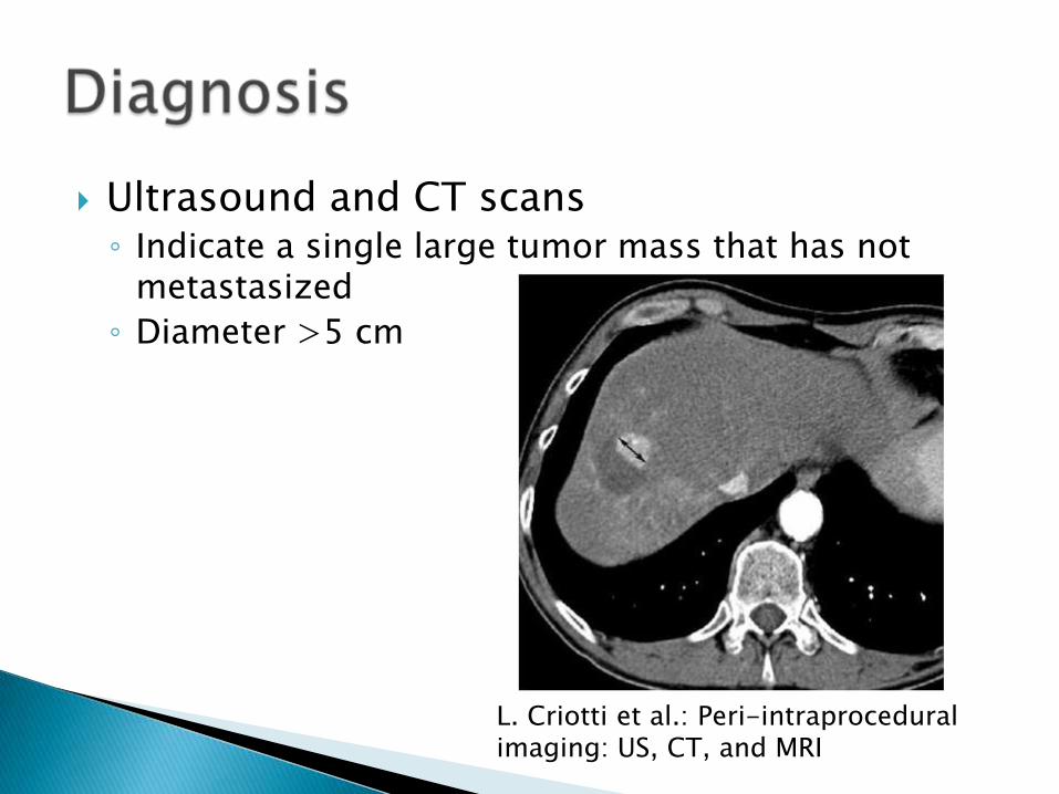
Ultrasound and CT scans◦ Indicate a single large tumor mass that has not
metastasized
◦ Diameter >5 cm
L. Criotti et al.: Peri-intraproceduralimaging: US, CT, and MRI
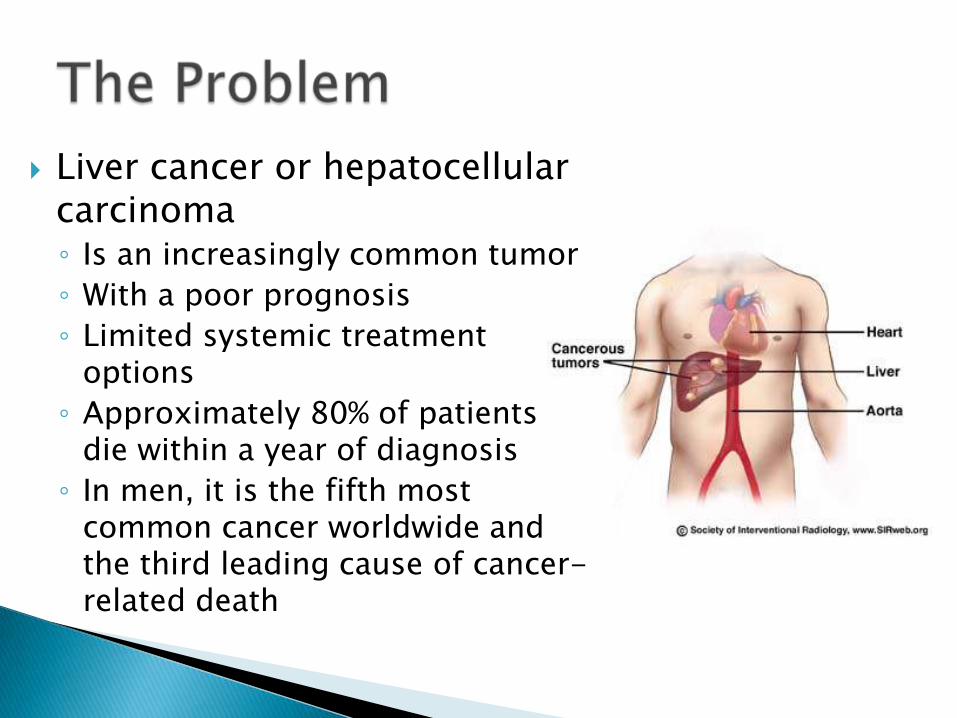
Liver cancer or hepatocellular carcinoma◦ Is an increasingly common tumor
◦ With a poor prognosis
◦ Limited systemic treatment options
◦ Approximately 80% of patients die within a year of diagnosis
◦ In men, it is the fifth most common cancer worldwide and the third leading cause of cancer-related death
Describes the severity of the cancer – size, metastasis, etc.
Tells physicians survival rates and cancer treatment
Many staging systems for hepatocellular carninoma including:◦ Cancer of the Liver Italian Program Score
◦ Chinese University Prognostic Index
◦ Barcelona Liver Clinic
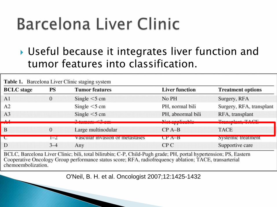
Useful because it integrates liver function and tumor features into classification.
O'Neil, B. H. et al. Oncologist 2007;12:1425-1432
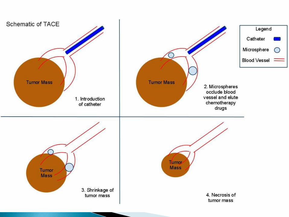
Model the drug transport from the microsphere into the tumor mass.
Decide whether TACE is an appropriate treatment over traditional chemotherapy.
Treat patient X successfully and thoroughly.
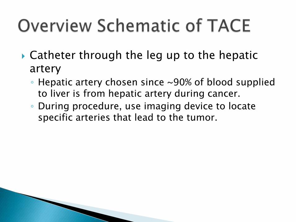
Catheter through the leg up to the hepatic artery◦ Hepatic artery chosen since ~90% of blood supplied
to liver is from hepatic artery during cancer.
◦ During procedure, use imaging device to locate specific arteries that lead to the tumor.
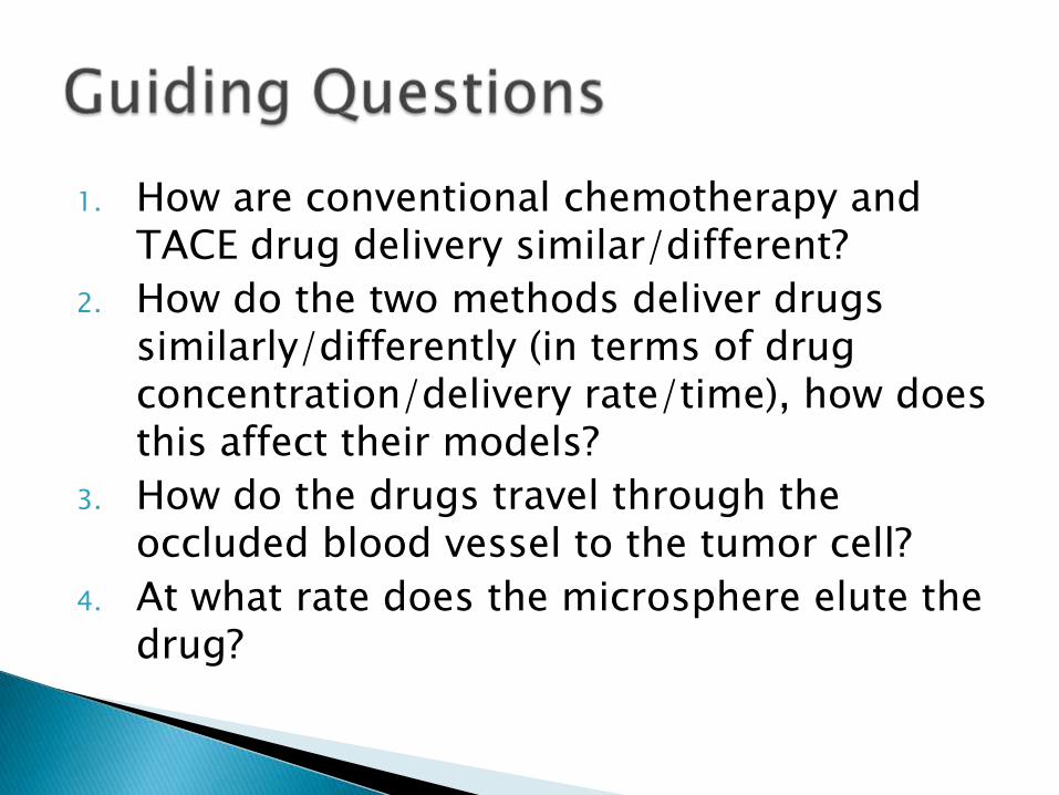
1. How are conventional chemotherapy and TACE drug delivery similar/different?
2. How do the two methods deliver drugs similarly/differently (in terms of drug concentration/delivery rate/time), how does this affect their models?
3. How do the drugs travel through the occluded blood vessel to the tumor cell?
4. At what rate does the microsphere elute the drug?
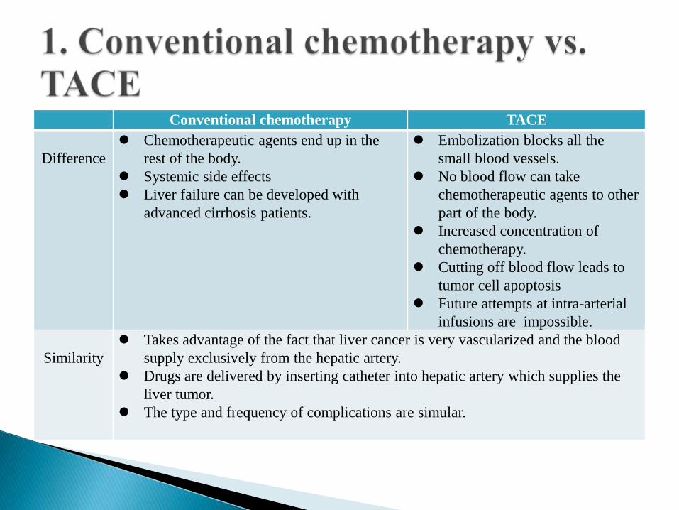
Conventional chemotherapy TACE
Difference
Chemotherapeutic agents end up in the
rest of the body.
Systemic side effects
Liver failure can be developed with
advanced cirrhosis patients.
Embolization blocks all the
small blood vessels.
No blood flow can take
chemotherapeutic agents to other
part of the body.
Increased concentration of
chemotherapy.
Cutting off blood flow leads to
tumor cell apoptosis
Future attempts at intra-arterial
infusions are impossible.
Similarity
Takes advantage of the fact that liver cancer is very vascularized and the blood
supply exclusively from the hepatic artery.
Drugs are delivered by inserting catheter into hepatic artery which supplies the
liver tumor.
The type and frequency of complications are simular.
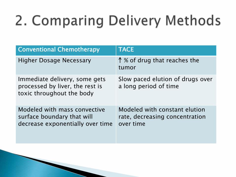
Conventional Chemotherapy TACE
Higher Dosage Necessary % of drug that reaches the tumor
Immediate delivery, some gets processed by liver, the rest is toxic throughout the body
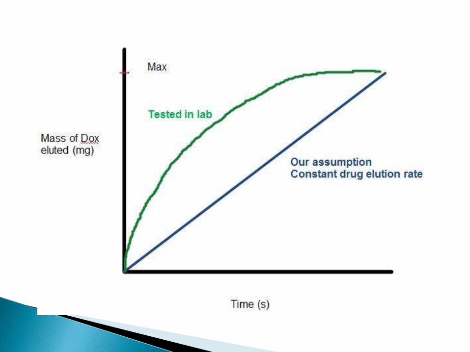
Slow paced elution of drugs over a long period of time
Modeled with mass convective surface boundary that will decrease exponentially over time
Modeled with constant elution rate, decreasing concentration over time
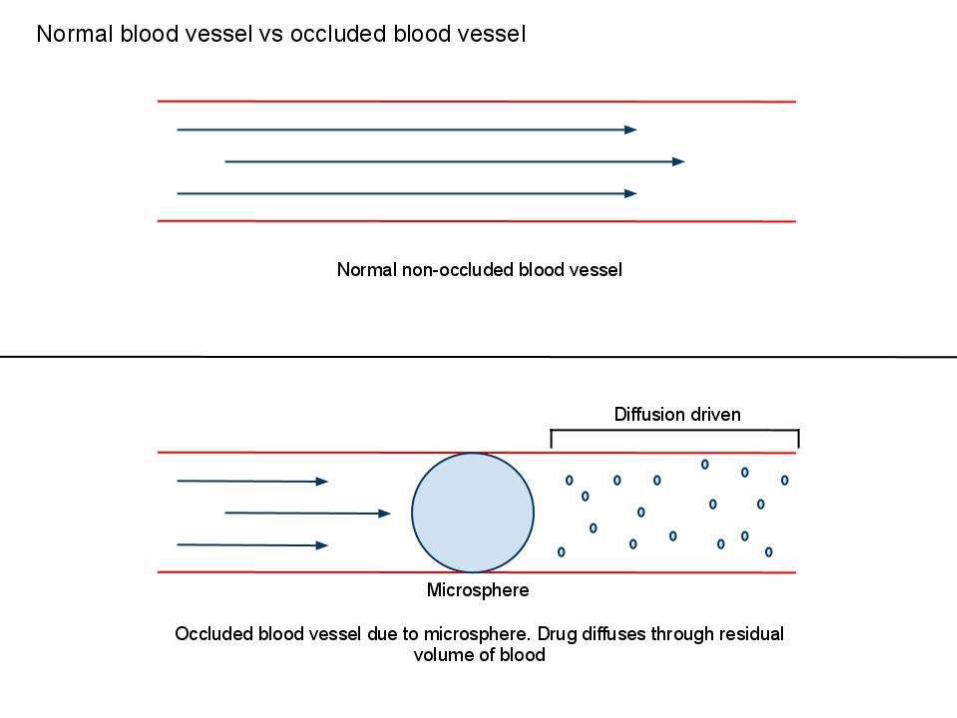
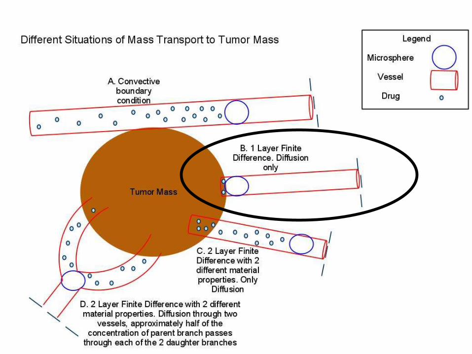
Occluded blood vessel: a blood vessel that is closed or stopped due to an obstruction
Assume that the blood after the microsphere is not moving.
Model can be simplified, drug travels through blood follows diffusion principles.
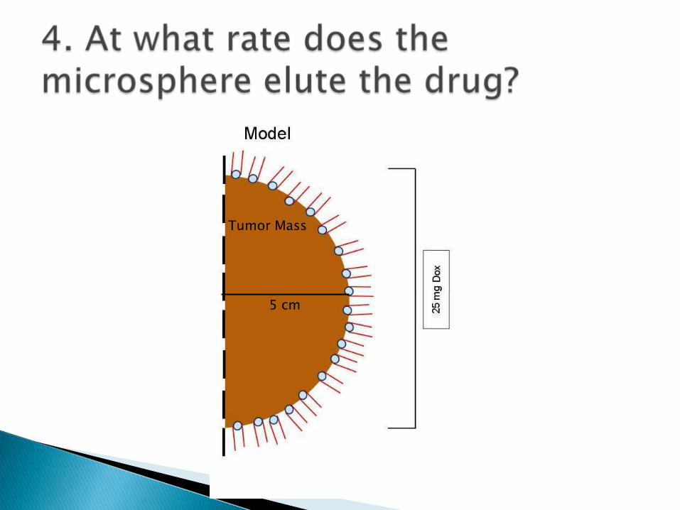
Tumor Mass
5 cm
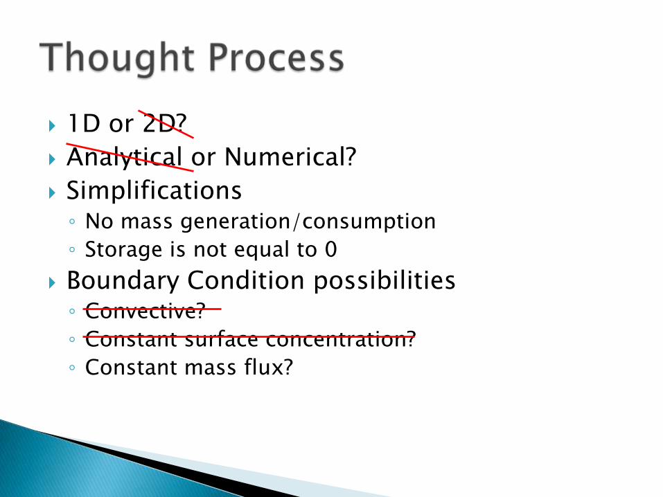
1D or 2D?
Analytical or Numerical?
Simplifications◦ No mass generation/consumption
◦ Storage is not equal to 0
Boundary Condition possibilities◦ Convective?
◦ Constant surface concentration?
◦ Constant mass flux?
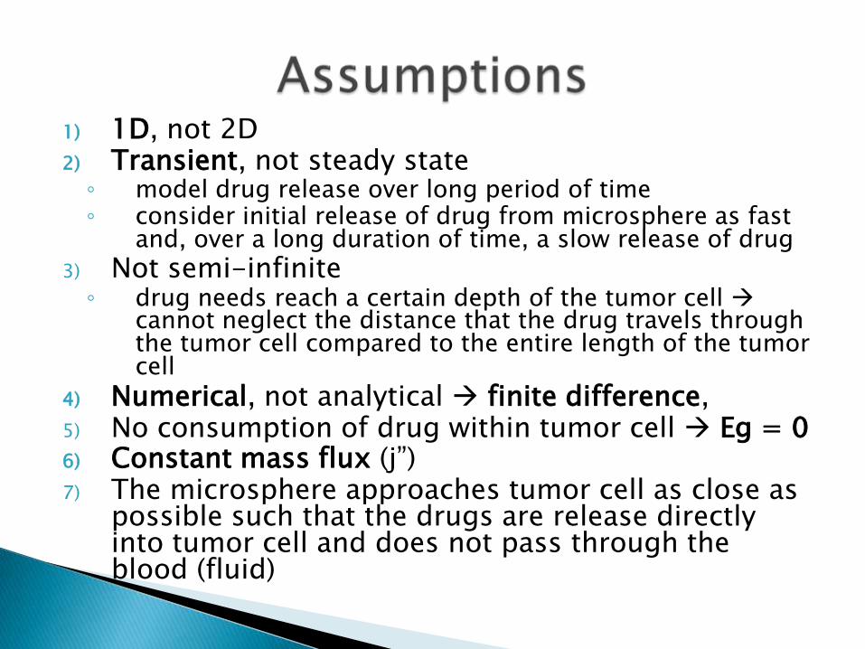
1) 1D, not 2D2) Transient, not steady state
◦ model drug release over long period of time◦ consider initial release of drug from microsphere as fast
and, over a long duration of time, a slow release of drug
3) Not semi-infinite◦ drug needs reach a certain depth of the tumor cell
cannot neglect the distance that the drug travels through the tumor cell compared to the entire length of the tumor cell
4) Numerical, not analytical finite difference,5) No consumption of drug within tumor cell Eg = 06) Constant mass flux (j‖) 7) The microsphere approaches tumor cell as close as
possible such that the drugs are release directly into tumor cell and does not pass through the blood (fluid)
Therefore, no diffusion occurs through the layer of blood in the vessel, but directly to the tumor cell from the microsphere.
This is the best case scenario for maximum drug diffusion.
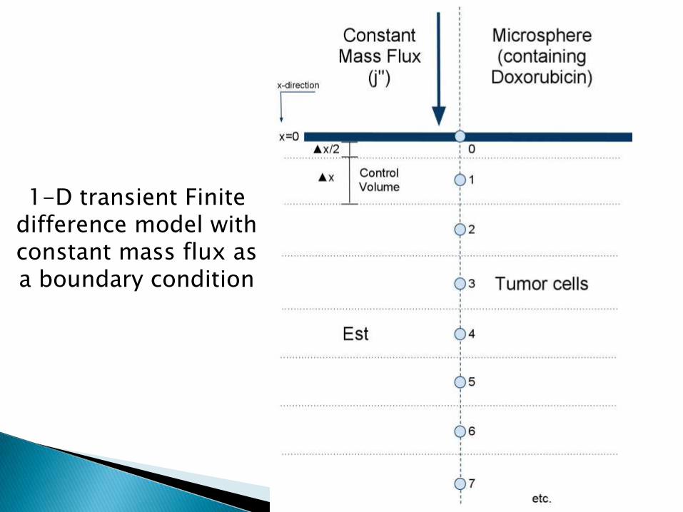
1-D transient Finite difference model with constant mass flux as a boundary condition
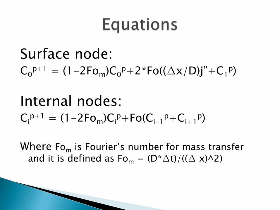
Surface node:C0
p+1 = (1-2Fom)C0p+2*Fo((∆x/D)j‖+C1
p)
Internal nodes:Ci
p+1 = (1-2Fom)Cip+Fo(Ci-1
p+Ci+1p)
Where Fom is Fourier’s number for mass transfer
and it is defined as Fom = (D*∆t)/((∆ x)^2)
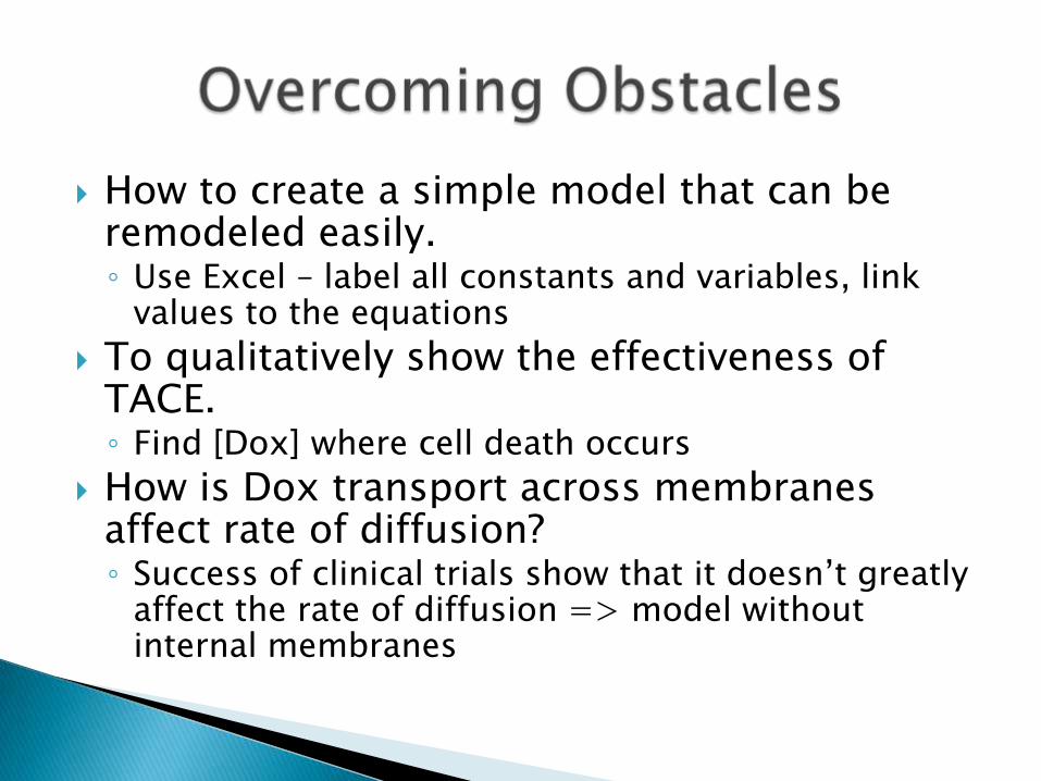
How to create a simple model that can be remodeled easily.◦ Use Excel – label all constants and variables, link
values to the equations
To qualitatively show the effectiveness of TACE.◦ Find [Dox] where cell death occurs
How is Dox transport across membranes affect rate of diffusion?◦ Success of clinical trials show that it doesn’t greatly
affect the rate of diffusion => model without internal membranes
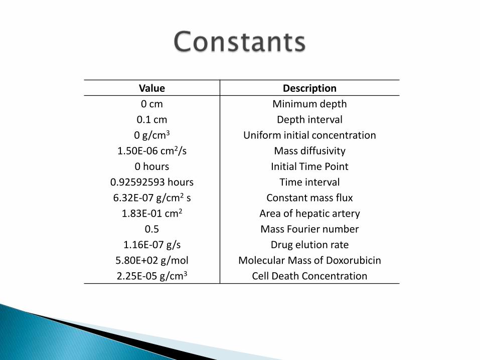
Value Description
0 cm Minimum depth
0.1 cm Depth interval
0 g/cm3 Uniform initial concentration
1.50E-06 cm2/s Mass diffusivity
0 hours Initial Time Point
0.92592593 hours Time interval
6.32E-07 g/cm2 s Constant mass flux
1.83E-01 cm2 Area of hepatic artery
0.5 Mass Fourier number
1.16E-07 g/s Drug elution rate
5.80E+02 g/mol Molecular Mass of Doxorubicin
2.25E-05 g/cm3 Cell Death Concentration

0
0.05
0.1
0.15
0.2
0.25
0.3
0.35
0.4
0.45
0.5
0 24 48 72 96 120 144
Co
nce
ntr
atio
n (
g/c
m^3
)
Time (Hours)
1D Transient Finite Difference Constant Mass Flux Model
0.000
0.200
0.500
0.900
1.400
2.000
178
Depth (cm)
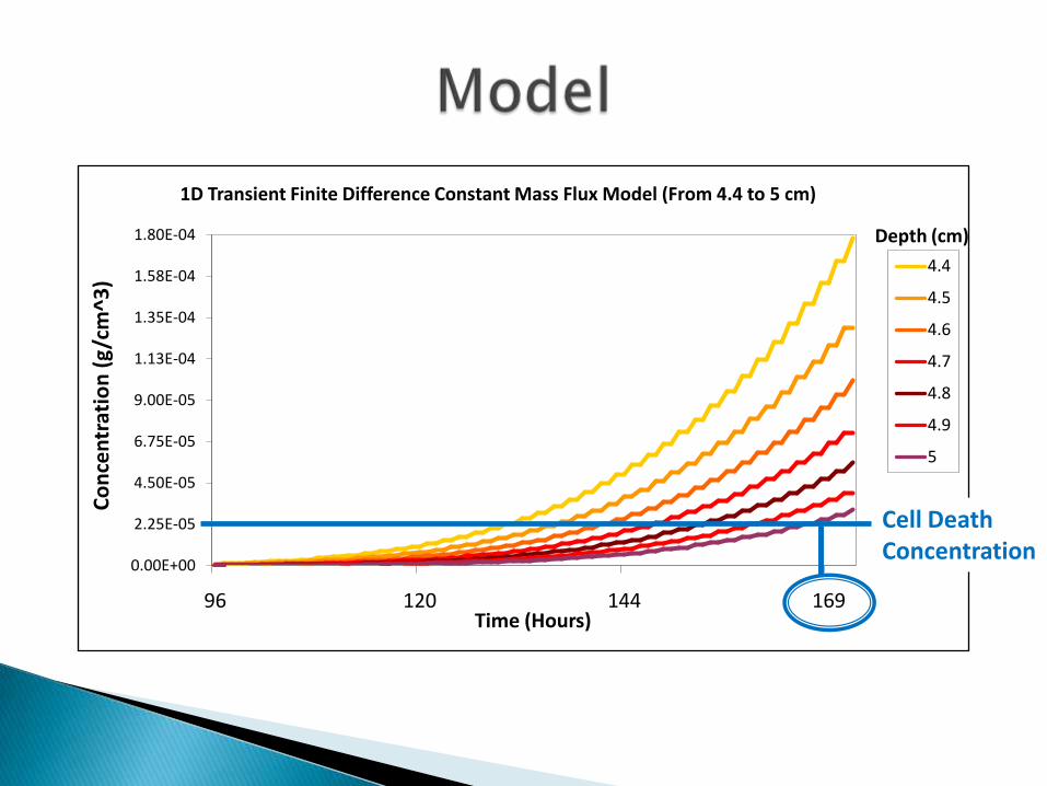
0.00E+00
2.25E-05
4.50E-05
6.75E-05
9.00E-05
1.13E-04
1.35E-04
1.58E-04
1.80E-04
96 120 144 169
Co
nce
ntr
atio
n (
g/c
m^3
)
Time (Hours)
1D Transient Finite Difference Constant Mass Flux Model (From 4.4 to 5 cm)
4.4
4.5
4.6
4.7
4.8
4.9
5
Cell DeathConcentration
Depth (cm)
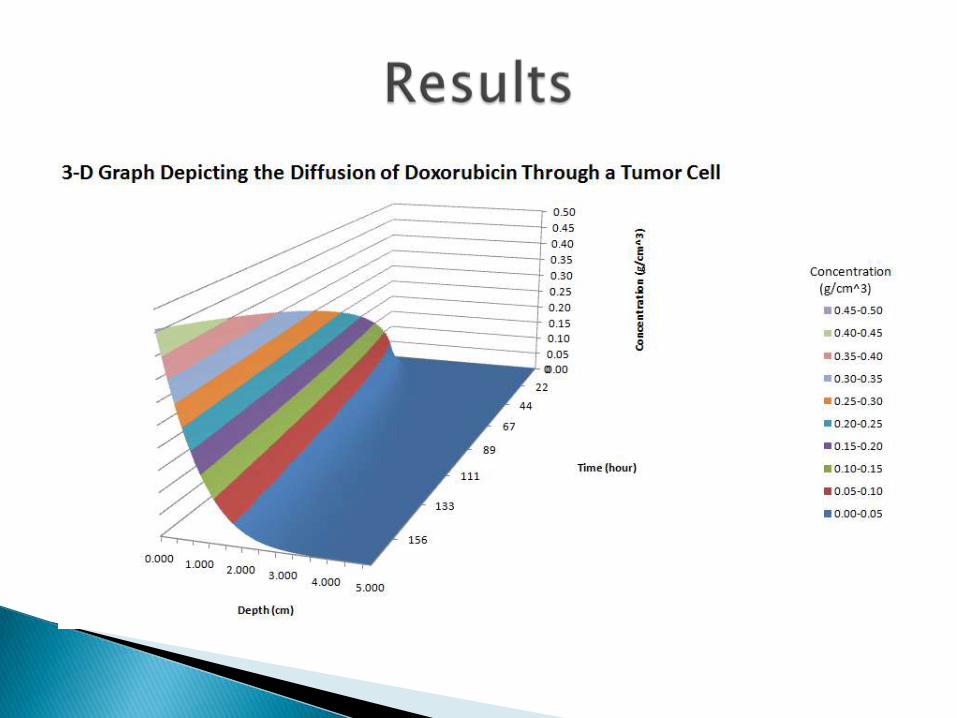
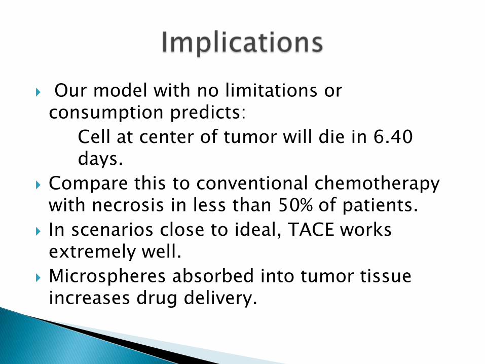
Our model with no limitations or consumption predicts:
Cell at center of tumor will die in 6.40 days.
Compare this to conventional chemotherapy with necrosis in less than 50% of patients.
In scenarios close to ideal, TACE works extremely well.
Microspheres absorbed into tumor tissue increases drug delivery.
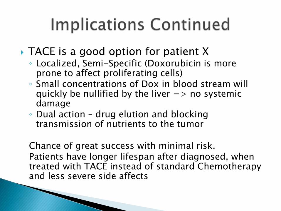
TACE is a good option for patient X◦ Localized, Semi-Specific (Doxorubicin is more
prone to affect proliferating cells)◦ Small concentrations of Dox in blood stream will
quickly be nullified by the liver => no systemic damage
◦ Dual action – drug elution and blocking transmission of nutrients to the tumor
Chance of great success with minimal risk.Patients have longer lifespan after diagnosed, when treated with TACE instead of standard Chemotherapy and less severe side affects
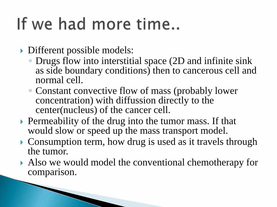
Different possible models: ◦ Drugs flow into interstitial space (2D and infinite sink
as side boundary conditions) then to cancerous cell and normal cell.
◦ Constant convective flow of mass (probably lower concentration) with diffussion directly to the center(nucleus) of the cancer cell.
Permeability of the drug into the tumor mass. If that would slow or speed up the mass transport model.
Consumption term, how drug is used as it travels through the tumor.
Also we would model the conventional chemotherapy for comparison.
1. Qian, Jun. "Application of poly-lactide-co-glycolide-microspheres in the transarterial chemoembolization in an animal model of hepatocellular carcinoma." World Journal of Gastroenterology 9.1 (2003): 94-98. Web. 25 May 2011.
2. Leen, E. "The use of duplex sonography in the detection of colorectal hepatic metastases." British Journal of Cancer 63. (1991): 323-325. Web. 25 May 2011
3. Lewis, Andrew L. "Doxorubicin eluting beads – 1: Effects of drug loading on bead characteristics and drug distribution." Journal of Material Science: Material Medicine 18. (2007): 1691-1699. Web. 25 May 2011.
4. Gonzalez, m. Victoria. "Doxorubicin eluting beads—2: methods for evaluating drug elution and in-vitro:in-vivo correlation." Journal of Material Science: Material Medicine 19. (2008): 767-775. Web. 25 May 2011.
5. Kettenbach, Joachim. "Drug-Loaded Microspheres for the Treatment of Liver Cancer: Review of Current Results." Cardiovascular and Intervention Radiology 31. (2008): 468-476. Web. 25 May 2011.
6. Kos, Sebastian. "Elution Characteristics of Doxorubicin-loaded Microspheres Differ by Drug-loading Method and Microsphere Size."Journal of Vascular and Interventional Radiology 22.3 (2011): 361-369. Web. 25 May 2011.
7. Hafeli, Urs O, Casillas S, Dietz D. "Hepatic Tumor Radioembolization in a Rat Model Using Radioactive Rhenium Glass Microspheres." J. Radiation Oncology Biol. Phys. 44. (1999): 189-199. Web. 25 May 2011.
8. Altrekruse S, Katherine M, Reichman, M. "Hepatocellular Carcinoma Incidence, Mortality, and Survival Trends in the United States From 1975 to 2005." J. Clinical Oncology 27. (2008): 1485-1491. Web. 25 May 2011.
9. Jackson T. "Intracellular Accumulation and Mechanism of Action of Doxorubicin in a Spatio-temporal Tumor Model." J. theor. Biol.220. (2003): 201-213. Web. 25 May 2011.
10. Tzafiri A R, Lerner E I, Flashner-Barak M. "Mathematical Modeling and Optimization of Drug Delivery from Intratumorally Injected Microspheres ." Clin Cancer Res 11. (2005): 826-834. Web. 25 May 2011.
11. Hong K, Khwaja A, Liapi E. "New Intra-arterial Drug Delivery System for the Treatment of Liver Cancer: Preclinical Assessment in a Rabbit Model of Liver Cancer." Clin Cancer Res 12. (2006): 2563-2567. Web. 25 May 2011.
12. Lee K, Liapi E A, Cornell C. "Doxorubicin-Loaded QuadraSphere Microspheres: Plasma Pharmacokinetics and Intratumoral Drug Concentration in an Animal Model of Liver Cancer." Cardiovasc Intervent Radiol 33. (2010): 576-582. Web. 25 May 2011.
13. Dalmark M, Hoffmann E K. "Doxorubicin (Adriamycin) transport in Ehrlich ascites tumour cells: comparison with transport in human red blood cells.." Scand J Clin Lab Invest 43. (1983): 241-248.
14. Hassan F, Morikawa A , Islam S. ―Lipopolysaccharide augments the in vivo lethal action of doxorubicin against mice via hepatic damage‖ Clin Exp Immunol. 151 (2008). 334-340. Web. 25 May 2011
15. Aliberti C, Tilli M . "Trans-arterial Chemoembolization (TACE) of Liver Metastases from Colorectal Cancer Using Irinotecan-Eluting Beads: Preliminary Results ." Anticancer Research 26. (2006): 3793-3795. Web. 25 May 2011.