causes of neural specialization for letters perception of ...
21
This article was downloaded by: [Indiana University Libraries] On: 03 December 2013, At: 10:19 Publisher: Routledge Informa Ltd Registered in England and Wales Registered Number: 1072954 Registered office: Mortimer House, 37-41 Mortimer Street, London W1T 3JH, UK Cognitive Neuropsychology Publication details, including instructions for authors and subscription information: http://www.tandfonline.com/loi/pcgn20 The role of sensorimotor learning in the perception of letter-like forms: Tracking the causes of neural specialization for letters Karin H. James a & Thea P. Atwood a a Department of Psychological and Brain Sciences , Indiana University , Bloomington, IN, USA Published online: 07 May 2009. To cite this article: Karin H. James & Thea P. Atwood (2009) The role of sensorimotor learning in the perception of letter-like forms: Tracking the causes of neural specialization for letters, Cognitive Neuropsychology, 26:1, 91-110, DOI: 10.1080/02643290802425914 To link to this article: http://dx.doi.org/10.1080/02643290802425914 PLEASE SCROLL DOWN FOR ARTICLE Taylor & Francis makes every effort to ensure the accuracy of all the information (the “Content”) contained in the publications on our platform. However, Taylor & Francis, our agents, and our licensors make no representations or warranties whatsoever as to the accuracy, completeness, or suitability for any purpose of the Content. Any opinions and views expressed in this publication are the opinions and views of the authors, and are not the views of or endorsed by Taylor & Francis. The accuracy of the Content should not be relied upon and should be independently verified with primary sources of information. Taylor and Francis shall not be liable for any losses, actions, claims, proceedings, demands, costs, expenses, damages, and other liabilities whatsoever or howsoever caused arising directly or indirectly in connection with, in relation to or arising out of the use of the Content. This article may be used for research, teaching, and private study purposes. Any substantial or systematic reproduction, redistribution, reselling, loan, sub-licensing, systematic supply, or distribution in any form to anyone is expressly forbidden. Terms & Conditions of access and use can be found at http://www.tandfonline.com/page/terms-and-conditions
Transcript of causes of neural specialization for letters perception of ...

This article was downloaded by: [Indiana University Libraries]On: 03 December 2013, At: 10:19Publisher: RoutledgeInforma Ltd Registered in England and Wales Registered Number: 1072954 Registered office:Mortimer House, 37-41 Mortimer Street, London W1T 3JH, UK
Cognitive NeuropsychologyPublication details, including instructions for authors and subscriptioninformation:http://www.tandfonline.com/loi/pcgn20
The role of sensorimotor learning in theperception of letter-like forms: Tracking thecauses of neural specialization for lettersKarin H. James a & Thea P. Atwood aa Department of Psychological and Brain Sciences , Indiana University ,Bloomington, IN, USAPublished online: 07 May 2009.
To cite this article: Karin H. James & Thea P. Atwood (2009) The role of sensorimotor learning in theperception of letter-like forms: Tracking the causes of neural specialization for letters, CognitiveNeuropsychology, 26:1, 91-110, DOI: 10.1080/02643290802425914
To link to this article: http://dx.doi.org/10.1080/02643290802425914
PLEASE SCROLL DOWN FOR ARTICLE
Taylor & Francis makes every effort to ensure the accuracy of all the information (the “Content”)contained in the publications on our platform. However, Taylor & Francis, our agents, and ourlicensors make no representations or warranties whatsoever as to the accuracy, completeness, orsuitability for any purpose of the Content. Any opinions and views expressed in this publicationare the opinions and views of the authors, and are not the views of or endorsed by Taylor &Francis. The accuracy of the Content should not be relied upon and should be independentlyverified with primary sources of information. Taylor and Francis shall not be liable for anylosses, actions, claims, proceedings, demands, costs, expenses, damages, and other liabilitieswhatsoever or howsoever caused arising directly or indirectly in connection with, in relation to orarising out of the use of the Content.
This article may be used for research, teaching, and private study purposes. Any substantialor systematic reproduction, redistribution, reselling, loan, sub-licensing, systematic supply, ordistribution in any form to anyone is expressly forbidden. Terms & Conditions of access and usecan be found at http://www.tandfonline.com/page/terms-and-conditions
The role of sensorimotor learning in the perceptionof letter-like forms: Tracking the causes
of neural specialization for letters
Karin H. James and Thea P. AtwoodDepartment of Psychological and Brain Sciences, Indiana University, Bloomington, IN, USA
Functional specialization in the brain is considered a hallmark of efficient processing. It is thereforenot surprising that there are brain areas specialized for processing letters. To better understand thecauses of functional specialization for letters, we explore the emergence of this pattern of responsein the ventral processing stream through a training paradigm. Previously, we hypothesized that thespecialized response pattern seen during letter perception may be due in part to our experience inwriting letters. The work presented here investigates whether or not this aspect of letter proces-sing—the integration of sensorimotor systems through writing—leads to functional specializationin the visual system. To test this idea, we investigated whether or not different types of experienceswith letter-like stimuli (“pseudoletters”) led to functional specialization similar to that which existsfor letters. Neural activation patterns were measured using functional magnetic resonance imaging(fMRI) before and after three different types of training sessions. Participants were trained to recog-nize pseudoletters by writing, typing, or purely visual practice. Results suggested that only afterwriting practice did neural activation patterns to pseudoletters resemble patterns seen for letters.That is, neural activation in the left fusiform and dorsal precentral gyrus was greater when participantsviewed pseudoletters than other, similar stimuli but only after writing experience. Neural activationalso increased after typing practice in the right fusiform and left precentral gyrus, suggesting thatin some areas, any motor experience may change visual processing. The results of this experimentsuggest an intimate interaction among perceptual and motor systems during pseudoletter perceptionthat may be extended to everyday letter perception.
Keywords: Letters; fMRI; Pseudoletters; Writing; Development; Neural activation.
Functional specialization, the propensity for neuralsystems to be active more to one category of stimu-lus than to a seemingly similar category, has beenthought to reflect efficient, sometimes expert,
neural processing (Downing, Jiang, Shuman, &Kanwisher, 2001; Gauthier, Williams, Tarr, &Tanaka, 1997; Tanaka & Gauthier, 1997).Functional specialization has been shown to exist
Correspondence should be addressed to Karin H. James, Department of Psychological and Brain Sciences, Indiana University,
1101 East 10th Street, Bloomington IN, 47405, USA (E-mail: [email protected]).
The authors would like to acknowledge Thomas James for helpful comments on the manuscript and for scholarly discussion on
this topic.
# 2008 Psychology Press, an imprint of the Taylor & Francis Group, an Informa business 91http://www.psypress.com/cogneuropsychology DOI:10.1080/02643290802425914
COGNITIVE NEUROPSYCHOLOGY, 2009, 26 (1), 91–110
Dow
nloa
ded
by [
Indi
ana
Uni
vers
ity L
ibra
ries
] at
10:
19 0
3 D
ecem
ber
2013

for several stimulus categories includingfaces (Kanwisher, 2000; Kanwisher, Chun, &McDermott, 1996), body parts (Downing et al.,2001), places (Kanwisher, 2000), biologicalmotion (Grossman & Blake, 2002), words (Cohenet al., 2000), and individual letters (Flowers et al.,2004; James, James, Jobard, Wong, & Gauthier,2005; Garrett et al., 2000; Longcamp, Anton,Roth, & Velay, 2003). In one survey of functionalspecialization of 20 stimulus categories in thevisual cortex, Downing, Chan, Peelen, Dodds, andKanwisher (2006) found that there were a smallnumber of regions that were actually category selec-tive, indicating that this is not a typical responseprofile for the ventral visual stream (Downinget al., 2006). Functional specialization was revealedonly for faces, body parts, and places—categories ofstimuli with which we are very efficient at proces-sing. Another category of stimuli (that was nottested in the aforementioned work) that we learnto recognize extremely efficiently is composed ofthe written characters of our native writingsystem. Written characters are a particularly inter-esting category of stimuli because the recency ofsuch stimuli in our evolutionary history suggeststhat there has been no change in our innate neuralarchitecture that would support stimulus-specificefficiency of processing. Nonetheless, with sufficientexposure and training, individuals become extre-mely efficient at processing their native charactersin many formats over the course of just a fewyears. Functional specialization for the written char-acter has been shown in a number of studies (e.g.,Flowers et al., 2004; Garrett et al., 2000; Jameset al., 2005). However, why this specialized neuralresponse pattern occurs is still a topic of speculation.First we consider a set of theories that are notnecessarily mutually exclusive, but that intend tooffer explanations as to why functional specializ-ation for letters occurs. We then show evidencethat suggests that motor learning may play a partin the emergence of neural specialization forletters in the ventral stream.
There are several theories that have been pro-posed to explain why different regions becomespecialized for processing particular categories ofstimuli. One attempt to account for functional
specialization in high-level visual areas concernsthe eccentricity biases associated with differentobject categories (Hasson, Harel, Levy, &Malach, 2003; Hasson, Levy, Behrmann,Hendler, & Malach, 2002; Levy, Hasson,Avidan, Hendler, & Malach, 2001; Levy, Hasson,Harel, & Malach, 2004; Malach, Levy, &Hasson, 2002). Based on results from a variety offunctional magnetic resonance imaging (fMRI)studies, the authors suggested that functionalspecialization is a result of different resolutiondemands associated with different object cat-egories. Face perception is associated with centre-biased visual areas for the detailed discriminationrequired, while perception of buildings is associatedwith periphery-biased visual areas for the large-scale integration involved. Words and letters,because of their small size, represent the extremecase of object perception requiring high resolutionand foveation.
Another proposal based on stimulus processingrequirements contends that specialized processingin the ventral visual processing stream may also bedue to the level of categorical analysis that isrequired for a given task. For example, duringface perception one often has to identify the faceof an individual (e.g., Al Gore). Such discrimi-nation within a homogeneous class (e.g., amongother faces) requires consideration of not onlythe fine-grain, metric differences of features (e.g.,lip thickness), but also the second-order relationsamong face features (e.g., interocular distance;Diamond & Carey, 1986). However, face identifi-cation at this level does not require coarse dis-crimination, for example whether a feature ispresent or absent—that is, we do not have todiscern whether or not Al Gore has a nose inorder to distinguish him from other people.Letter perception, in contrast, relies on the useof coarse information like feature presence/absence (e.g., an oblique stroke in “N”) and first-order relations (e.g., the oblique stroke is inbetween the two vertical strokes in “N”), as wellas second-order relations (the angle of theoblique line is also important to distinguish an“N” from an “H”). Analyses of the stimulus prop-erties of characters across different languages have
92 COGNITIVE NEUROPSYCHOLOGY, 2009, 26 (1)
JAMES AND ATWOOD
Dow
nloa
ded
by [
Indi
ana
Uni
vers
ity L
ibra
ries
] at
10:
19 0
3 D
ecem
ber
2013

also shown that a high level of redundancy is intro-duced in their creation, such that one can perceiveonly part of a character and be able to distinguish itfrom the other alternatives (Changizi & Shimojo,2004). Therefore, letter identification involvesprocessing of different types of stimulus infor-mation when compared with other stimuluscategories.
Other theories of ventral visual stream organiz-ation and specialization couch the problem interms of a continuous object-form topography,such that neighbouring neural substrates tend torepresent features that are more similar to eachother (Carlson, Schrater, & He, 2003; Cox &Savoy, 2003; Haxby et al., 2001; Ishai,Ungerleider, Martin, Schouten, & Haxby, 1999;O’Toole, Jiang, Abdi, & Haxby, 2005). Animportant idea of this theory is that there is noabsolute neural specialization for certain objectcategories. Instead, there is relative preference ofcertain substrates for certain categories based onfeatures, and object information is widely distribu-ted and overlapping across the high-level visualareas. Support for this theory comes from findingthat activity in category-specific areas (e.g., face-selective regions in the fusiform gyrus) containsdiagnostic information for the categorization ofnonpreferred categories (Haxby et al., 2001), andactivity patterns over a wide region of the inferiortemporal cortex correlate with physical propertiesof objects (O’Toole et al., 2005). It should benoted, however, that object representations inthe occipito-temporal cortex are probably notcompletely distributed, as some regions (e.g., theface- and scene-selective areas) still enjoy agreater discriminatory power for their preferredcategories than other objects (O’Toole et al.,2005; Spiridon & Kanwisher, 2002).
Another theory maintains that different parts ofthe ventral visual processing stream are suited fordifferent processes. The observed object selectivityis a result of the prolonged recruitment of differentsubstrates to fulfil specific recognition demands fordifferent objects (Bukach, Gauthier, & Tarr, 2006;Gauthier, Skudlarski, Gore, & Anderson, 2000;Tarr & Gauthier, 2000). Accordingly, “recog-nition demand” and “experience” are therefore
the key factors determining the use of differentsubstrates for different categories. This theorygains support from findings of recruitment of theface-selective areas for other types of objects likecars, dogs, birds, fingerprints, and novel objects,which are claimed to require a common perceptualdemand of fine-grained discrimination andcommon holistic manner of processing thatresults from extensive experience (Gauthier et al.,2000; Xu, 2005).
These ideas allow speculation as to why theventral visual stream may demonstrate functionalspecialization for letters, but how would thisdevelop? We propose that functional specializ-ation for letters may be caused by the way thatwe learn to recognize letters and, more specifically,that specialization for letters may reflect the sen-sorimotor integration that is required when welearn to write letters ( James & Gauthier, 2006;Longcamp et al., 2003). Sensorimotor experiencein the form of learning to print and write lettersallows the interplay between motor productionand visual perception to broaden the stored rep-resentation of letters. That is, motor constructionof forms may lead to motor programmes that arestored with visual information. Because, duringwriting, these programmes are variable, they mayserve to augment visual information, allowingvery different-looking exemplars to be categorizedas the same letter. Grouping visually dissimilarletters into a single category occurs before childrenare exposed visually to a large variety of handwrit-ing styles or fonts, but after they learn to print. Insupport of the idea that sensorimotor experiencemay lead to functional specialization in the visualprocessing stream, recent work has shown thatwhen participants view letters, a sensorimotornetwork becomes engaged ( James & Gauthier,2006). That is, areas in the ventral visual proces-sing stream become active during visual perceptionbut, more interestingly, so do motor and premotorregions of the brain (perhaps part of the dorsalstream of visual processing). This finding wasinterpreted as showing that neural circuits forwriting letters (premotor cortex) were automati-cally activated upon seeing letters, implying thatmotor experiences were stored and reactivated
COGNITIVE NEUROPSYCHOLOGY, 2009, 26 (1) 93
SENSORIMOTOR LEARNING AND LETTERS
Dow
nloa
ded
by [
Indi
ana
Uni
vers
ity L
ibra
ries
] at
10:
19 0
3 D
ecem
ber
2013
upon subsequent encounters. In addition, ventralvisual areas—specifically, the left fusiformgyrus—were engaged when participants wroteletters (without seeing them), but not when theydrew shapes ( James & Gauthier, 2006). Thus,coactivation of brain regions that process bothvisual and motor information during either per-ceptual or motor interaction with familiar letterstimuli was shown, suggesting a sensorimotor rep-resentation of letters.
Additional evidence for coactivation of thevisual processing areas and motor regions comesfrom a study showing common repetition suppres-sion profiles in ventral and dorsal stream regions(Mahon et al., 2007). In this study, repetition sup-pression (RS), the tendency for neural activity tobe reduced upon repeated stimulus presentation(Grill-Spector, Henson, & Martin, 2006), wasshown to emerge for “tools” in areas in theventral visual processing stream as well as indorsal stream structures. In addition, the authorsuse a functional connectivity analysis to showthat areas in the left middle temporal gyrus andthe left inferior parietal lobule both show RS fortool stimuli. Here, “tools” were defined as objectswhose function (how to interact with them) wasapparent in their structure—arguably, objectswith “affordances” (Gibson, 1979). Interestingly,objects that did not have a systematic relationshipbetween structure and function, termed “arbitrarilymanipulated” objects (e.g., book, envelope), over-lapped in their RS functions with “tools” in righthemisphere ventral (middle fusiform) and dorsalstream (right caudal inferior parietal lobule)systems, but not in left hemisphere systems. Thiswork suggests that motor information (how tointeract with objects) is associated with visualobject processing.
This idea is supported by behavioural work aswell. There is now a substantial body of evidencethat motor experience—that is, our history of inter-actions with some objects—can facilitate visualrecognition (Harman, Humphrey, & Goodale,1999; James, Humphrey, & Goodale, 2001; Jameset al., 2002) and mental rotation (Wexler, Kosslyn,& Berthoz, 1998; Wohlschlager & Wohlschlager,1998), as well as the development of spatial maps
(Bai & Bertenthal, 1992; Campos et al., 2000; forreview see Wexler & Boxtel, 2005). For example,visual recognition of novel objects is facilitated byactively moving the objects compared to watchingthe same movement performed by another person(Harman et al., 1999; James et al., 2001, 2002).Such behavioural facilitation supports the idea thatthe dorsalmotor and ventral visual systems are inter-acting during object processing.
In addition, several neuroimaging studies havefound that motor systems are automatically activatedupon visual perception of some objects (Chao &Martin, 2000; Gerlach, Law, Gade, & Paulson,2002; Grezes & Decety, 2002; James & Gauthier,2006; Longcamp et al., 2003; Longcamp, Anton,Roth, & Velay, 2005a; Longcamp, Zerbato-Poudou, & Velay, 2005b; Mecklinger, Gruenewald,Besson, & von Cramon, 2002). This literaturesuggests that motor systems are active when wevisually perceive objects that we regularly interactwith motorically—for example, tools and utensils(Chao & Martin, 2000; Mahon et al., 2007;Mecklinger et al., 2002). Interestingly, these objectsalso invoke, by their appearance alone, specific waysto interact with them. That is, they contain “affor-dances” (Gibson, 1979) that can be used to specifymotor interactions (potentially independent of ourexperience), and these affordances are visually per-ceptible. By investigating the effects of prior motorexperience on visual recognition of objects withoutaffordances, one can more directly attribute therecruitment of motor systems to experience.
Letters, by their appearance alone, do not“suggest” how we must interact with them—inthis way, they are similar to the arbitrarily manip-ulable objects in the Mahon et al. (2007) studies.Perceiving letters, however, does seem to invokethe associated history of letter-specific motoricinteractions. For instance, in a series of studies,Freyd and colleagues (Babcock & Freyd, 1988;Freyd, 1983) found that the way that a subjectis taught to write a letter-like symbol directlyaffects their subsequent recognition of thatsymbol. In addition, writing experience can alterthe perception of movement illusions in writtensymbols (Tse & Cavanagh, 2000), and knowledgeof cursive stroke directions affects anticipated
94 COGNITIVE NEUROPSYCHOLOGY, 2009, 26 (1)
JAMES AND ATWOOD
Dow
nloa
ded
by [
Indi
ana
Uni
vers
ity L
ibra
ries
] at
10:
19 0
3 D
ecem
ber
2013

letter identity (Orliaguet, Kandel, & Bois, 1997).Longcamp et al. (2005b) have demonstrated thatchildren recognize letters more efficiently afterbeing trained to print letters versus beingtrained to type letters. This latter study suggeststhat motor experience may be generative—themotor experience that is important for visual rec-ognition is through constructing the form of theletter, not by the simple motor act of typing. Astudy conducted by Cunningham and Stanovich(1990) found similar results. They used threedifferent modalities to teach children how tospell words. Children were presented with aword both visually and orally and were asked tospell words by writing, arranging a set of tiles,or typing the word. After spending 30 minutestraining over the course of four days, childrenwere asked to spell out each of the words on asheet of paper, by using a computer keyboardand by arranging tiles. Results demonstratedthat there were significantly more correctresponses for those words learned in the writingcondition versus the typing and tile conditions,and this effect emerged regardless of the formatin which the children spelled the words in thetest session.
Furthermore, research on individuals with lit-eracy disabilities has also suggested a link amongmotor and visual systems in letter processing.Writing movements can facilitate letter recog-nition in patients with pure alexia—the inabilityto identify letters and words (Bartolomeo,Bachoud-Levi, Chokron, & Degos, 2002; Seki,Yajima, & Sugishita, 1995). Although thesepatients cannot recognize a letter visually, if theyare allowed hand movements while they arelooking at the letter, they will often trace out theshape of the letter as if writing it—and this move-ment (that is unseen by them) facilitates theirvisual recognition of letters. In addition, somedyslexic adults are delayed in motor tasks; it ispossible that in such cases motor difficultiesaffected their letter-learning ability (Stoodley,Fawcett, Nicolson, & Stein, 2005). Similarly, chil-dren exhibiting developmental dyspraxia, a dis-order that can manifest in reduced fine motorskills (including writing), often have difficulty in
letter identification and in learning to read—thisdisorder is highly comorbid with dyslexia (e.g.,Portwood, 2000). Furthermore, a patient hasbeen reported with agraphia (inability to write)with alexia that has resulted from damage to theleft premotor cortex (Anderson, Damasio, &Damasio, 1990). Although this patient cannotread or identify letters and cannot write letters orwords, she can draw complex shapes, and she canwrite numbers. This case provides some evidencethat damage to the motor system can affect notonly writing, but also visual processing of letters.It also shows that the motor deficit can be very cir-cumscribed to one particular category of stimuli.
The neuroimaging, patient, and behaviouralresults outlined above have revealed an interestingaspect of letter recognition—while we ultimatelyspend a lot of time reading words, it is the isolatedletter that we need to learn first, and we learn thisstimulus by seeing and writing. The work presentedhere investigates whether this aspect of letterprocessing—the integration of sensorimotorsystems—leads to functional specialization in thevisual system. The left fusiform gyrus of the literateadult is already specialized for processing lettersmore than other, similar characters (Flowers et al.,2004; Garrett et al., 2000; James & Gauthier,2006; James et al., 2005). Therefore, it is difficultto assess the effects of writing experience on thedevelopment of this specialization. We thereforeconducted a training study that directly comparedthe effects of writing experience, typing experience,and visual-only experience on visual “letter” proces-sing. Because adults have already learned letters,however, we trained them on a group of letter-likecharacters, referred to here as “pseudoletters”. Inthis way we investigated the effects of motor experi-ence on visual recognition and on neural processing.
Method
ParticipantsA total of 18 participants gave informed consentaccording to the guidelines of the IndianaUniversity Human Participants Review Boardand were paid for their participation. Allwere undergraduate or graduate students enrolled
COGNITIVE NEUROPSYCHOLOGY, 2009, 26 (1) 95
SENSORIMOTOR LEARNING AND LETTERS
Dow
nloa
ded
by [
Indi
ana
Uni
vers
ity L
ibra
ries
] at
10:
19 0
3 D
ecem
ber
2013

at Indiana University. All participants wereright-handed and reported normal or corrected-to-normal vision, had English as their firstlanguage, and had no known history of neurologi-cal or psychiatric disorders. A total of 10 femalesand 8 males participated, and they were betweenthe ages of 21 to 31 years with a median age of23.5 years.
StimuliStimuli were 96 � 96 pixels and were presented inisolation, in the centre of a computer screen.Stimuli included a group of 18 capital letters (H,A, F, C, S, U, K, N, T, B, D, G, R, J, L, P, Z, Y),18 shapes and symbols (e.g., clover, heart, percen-tage sign, pound sign, treble clef, star, etc.), 18pseudoletters (studied), and another set of 18pseudoletters (unstudied; see Figure 1). Each ofthese groups had four more subsets—an Arialfont group, a serif-type font group, a cursive-typefont group, and a rotation group (consisting ofeach stimulus rotated 0, 45, 90, and 180degrees). Stimuli that lacked noticeable differencewhen rotated were not used—for example, theletter “O” and the shape of a square were notused because each of these symbols, when rotated180 degrees, are no different from their counter-parts oriented at 0 degrees rotation. Differentformats of the stimuli were used to add variationto the stimulus sets and to attempt to make
matching tasks more difficult in the behaviouralportion of the experiment. All orientations andfont types were used in the matching task, bothwithin the scanning environment and outside.
General procedureThere were four sessions in total, and each sessionwas separated by one day of rest: First, there was apretraining imaging session (a “pretrain scan”),which helped to determine the participant’sinitial blood-oxygen-level-dependent (BOLD)activation to the novel (pseudoletter) alphabet aswell as to letters and simple shapes. Participantsthen took part in two training sessions, separatedby one day. The training sessions consisted oftraining procedures, followed by behaviouraltesting. We included testing in these training ses-sions to (a) assess whether or not the training hadany effect from Day 1 to Day 2 and (b) to motiv-ate the participants to learn the stimuli. Alltesting procedures involved visual tasks only.After training, participants took part in a finalposttraining imaging session (a “posttrain scan”)to determine whether any change in BOLD acti-vation patterns had occurred as a result of thetraining sessions.
Scanning protocol. Both pre- and posttrainingscanning sessions proceeded in the samemanner. All stimuli were back-displayed with aMitsubishi XL30 projector onto a screen thatwas viewed through a mirror from the bore ofthe Siemens Trio 3T scanner. Stimuli were pre-sented with SuperLab Pro 2.0.4 software withDell Inspiron 6000 laptops. Each scanningsession consisted of six runs. The first five runswere functional scans that measured activationto our stimulus conditions, and the final run wasa high-resolution anatomical scan. Conditionswere presented in a blocked design: Each blockcontained 16 different presentations of the stimu-lus condition. Each run consisted of the samestimulus blocks, but the order of the blocks wasrandomized across runs. Order of runs wasvaried across participants. Each run began andended with a 16-s fixation cross and, in addition,consisted of 16 blocks of experimental trials: 4
Figure 1. A: Example of pseudoletter stimuli. B: Font types: sansserif, serif, cursive.
96 COGNITIVE NEUROPSYCHOLOGY, 2009, 26 (1)
JAMES AND ATWOOD
Dow
nloa
ded
by [
Indi
ana
Uni
vers
ity L
ibra
ries
] at
10:
19 0
3 D
ecem
ber
2013

stimulus types (letters, studied pseudoletters,unstudied pseudoletters, and shapes) � 2 changeconditions (font or rotation) � 2 repetitions.Within each block, participants were required toperform a one-back matching task by pressing abutton with their right index finger when twostimuli presented consecutively possessed thesame category identity (an identity that is sharedregardless of font type or orientation). That is,participants had to detect whether an “A” wasan “A” regardless of font or orientation. Stimuliin each block were pseudorandomized such thatthere were at least two repetitions (one-backmatch) of a stimulus in each block. The one-back perceptual match task was used to maintainattention. Participants pressed a button withtheir right index finger when an image wasrepeated irrespective of font or orientation. Thistask was described to the participant prior toscanning. Stimuli were presented in the centreof the screen for 500 ms, followed by a Gaussiannoise mask presented for 500 ms. There were 16presentations of stimuli within each block, andthe ratio of repetitions to nonrepeats was 1 : 7(making this a very easy task). The stimuli thatwere presented within a block were randomlyselected such that not every Rotation � Stimuluscombination was presented, the only requirementbeing that an average of two repetitions occurredin each block. Each block was separated by a 10-s fixation cross.
Training Sessions 1 and 2Both training sessions were run in the samemanner, although the order of the behaviouraltests varied. All stimulus presentations were runon a Dell Optiplex GX 280 desktop computer,via SuperLab Pro 2.0.4 software, and responsetime and accuracy data were collected via a com-puter keyboard. Participants were randomlyassigned to one of three conditions: a writing con-dition, a typing condition, or a visual condition.Each condition consisted of four trainingexposures and six behavioural tests. The trainingexposure varied according to the condition(writing, typing, or visual). The six behaviouraltests were constant for all participants, although
the order of the presentation of the tests variedper training session (1 or 2). A training sessionconsisted of a training exposure, followed by tworecognition tests, followed by another trainingexposure, until a total of four training exposureswere completed.
Training exposure conditions. For the writing con-dition, participants were given a pad of paperand a pen and were asked to copy pseudoletterspresented on the computer screen to the best oftheir ability. For the typing condition, partici-pants were asked to find and type the pseudoletterthat was presented on the screen. A keyboard wasmodified for this purpose (pictures of the pseudo-letters were affixed to a regular keyboard). Whenthe participant typed the key, the presentationof the pseudoletter on the screen disappearedand was not replaced. Thus, the screen wasblank after the participant typed their response.For the visual condition, participants were askedto look at the presented stimulus and to try tomemorize its form. All participants were notifiedthat they would be tested on these stimuli aftertraining. No feedback was given for any of thetraining conditions. In each training task, eachof the 18 stimuli were presented a total of threetimes in their proper orientation, for a total of54 stimulus presentations per training task. Thiswas repeated four times per training day (for atotal of eight repetitions). Each stimulus waspresented for the same amount of time (4seconds) for each training condition. Thisresulted in the visual condition having a greateramount of visual exposure to each stimulus,because they were not required to divert theirgaze to write or type. However, in the typing con-dition, participants would find and then see thepseudoletter on the keyboard. In the writing con-dition, they would write and then see the writtenpseudoletter. For each training group the stimuluswas presented on the screen for the entire trial.Although we did not measure the amount oftime that each person required to type or write,both conditions were easily completed in thetime allotted, and one did not appear to takelonger than any other.
COGNITIVE NEUROPSYCHOLOGY, 2009, 26 (1) 97
SENSORIMOTOR LEARNING AND LETTERS
Dow
nloa
ded
by [
Indi
ana
Uni
vers
ity L
ibra
ries
] at
10:
19 0
3 D
ecem
ber
2013

Behavioural testing. The tests did not change fromparticipant to participant, although the order ofthe tests did vary. A total of six tests were pre-sented in one training session. Three of thesewere standard visual search tests. The participantwas presented with a target (trained pseudoletter)stimulus, followed by a Gaussian noise mask, fol-lowed by a field of stimuli. The fields were 2 � 2(small array), 3 � 3 (medium array), or 6 � 6(large array). The participant’s task was toquickly decide whether the target stimulus waspresented in the field of stimuli. The participantpressed the “/” key if the target was present and“z” if the target was absent. Stimuli were presentedin roman font and in an upright orientation. Thevisual search tests were included as a measure oflearning that did not require explicit recognitionof the target stimulus. However, visual searchhas been shown to be sensitive to learning effectsand even as an effective measure of automaticityin processing (Czerwinski, Lightfoot, & Shiffrin,1992). We were therefore curious to see whetheror not visual search ability became more efficientwith our training paradigm.
Two tests required matching of stimuli. Theparticipant was presented with a target stimulus,followed by a Gaussian noise mask, and then asecond stimulus, followed by a brief fixationcross. The participant’s task was to decidewhether the two stimuli were the same or different(serial match task). The first match test incorpor-ated font changes between the two stimuli; thesecond match test changed orientation of thefirst and second stimulus presentations. Thesetwo matching tasks consisted completely of thetrained pseudoletters. Participants pressed the “/”key if the stimuli were the same and the “z” ifthe stimuli were different. This task was includedto assess implicit processing of the pseudolettersand how this may change over time. In addition,this task was a close approximation of the one-back task that was performed in the imagingenvironment, except that during this task alllevels of orientation changes and font changeswere used.
The final test was an old/new recognition test:Participants decided whether a presented stimulus
was one that had been previously studied, or ifthe stimulus was new and had not been studied.Again, this test consisted completely of the trainedpseudoletters, as well as 18 novel pseudoletterstimuli (the “new” stimuli). Font and orientationwere changed randomly throughout this pro-cedure; thus, not all levels of orientation andfont were necessarily used. Participants pressedthe “/” key if the pseudoletter had been previouslystudied and “z” if the pseudoletter was new. Thenovel pseudoletters presented in the old–newtest decision were presented only within the con-fines of this task. Of the three tasks, this was theonly test that required explicit recollection of thestudied pseudoletter stimuli.
Because this type of training exposure is vir-tually untested, we were unsure how it wouldaffect performance. For this reason, we includedseveral behavioural tests that would potentiallyallow us to determine how our training affecteddifferent types of performance (e.g., implicit vs.explicit recognition).
Imaging parameters. Imaging was performed usinga 3-T Siemens Magnetom Trio whole-body MRIsystem and a phased-array eight-channel head coil,located at the Indiana University Psychologicaland Brain Sciences department. The field of viewwas 22 � 22 � 12.5 cm, with an in-plane resol-ution of 64 � 64 pixels and 25 slices per volumethat were 4 mm thick with a 1.0-mm gap amongthem. These parameters allowed us to collectdata from the entire brain. The resulting voxelsize was 3.4 mm � 3.4 mm � 5.0 mm. Imageswere acquired using an echo-planar technique(echo time, TE ¼ 30 ms; time to repetition,TR ¼ 2,000 ms; flip angle ¼ 708) for BOLDbased imaging. High-resolution T1-weightedanatomical volumes were acquired using a 3DTurbo-flash acquisition. Functional data under-went slice time correction, 3D motion correction,linear trend removal, and Gaussian spatial blurring(FWHM 4 mm) using the analysis tools in BrainVoyagerTM. Individual functional volumes werecoregistered to anatomical volumes with anintensity-matching, rigid-body transformationalgorithm. Individual anatomical volumes were
98 COGNITIVE NEUROPSYCHOLOGY, 2009, 26 (1)
JAMES AND ATWOOD
Dow
nloa
ded
by [
Indi
ana
Uni
vers
ity L
ibra
ries
] at
10:
19 0
3 D
ecem
ber
2013

normalized to the stereotactic space of Talairachand Tournoux (1988) using an eight-parameteraffine transformation, with parameters selectedby visual inspection of anatomical landmarks.Applying the same affine transformation tothe coregistered functional volumes placed thefunctional data in a common brain space, allowingcomparisons across participants. Voxel size ofthe normalized functional volumes was standar-dized at 1 mm � 1 mm � 1 mm using trilinearinterpolation.
fMRI data analysis procedures. The functional datawere further analysed with a random effectsgeneral linear model (GLM) using BrainVoyager’sTM multisubject GLM procedure. TheGLM analysis allows for the correlation of predic-tor variables or functions with the recorded acti-vation data (criterion variables) across scanningsessions. The predictor functions were based onthe blocked stimulus presentation paradigm ofthe particular run being analysed and representan estimate of the predicted haemodynamicresponse during that run. Regions of interestwere determined based on group statistical para-metric maps (SPMs) that were considered abovethreshold if they met the following criteria in ourrandom-effects analysis: (a) significant atp , .001, uncorrected, with a cluster threshold of270 contiguous 1-mm isometric voxels; (b) peakstatistical probability within a cluster at leastp , .0001, uncorrected.
To localize regions of the brain that wereengaged during letter processing, we performed aletters versus fixation contrast in the group datacombined across the pretrain and posttrain scans.Results of this contrast are presented in Figure 2and produced 12 regions of interest (ROIs). Wethen extracted percentage BOLD signal changevalues for all participants within these ROIs forboth pretrain and posttrain scans. Using peak acti-vation from each participant as our dependentmeasure, we then performed an omnibus analysisof variance (ANOVA) for each ROI. However,we only compared trained and untrained pseudo-letters with one another, these being our stimuli
of interest (that is, we did not extract BOLDsignal change to shapes).
Results and discussion
Behavioural resultsTwo separate 2 � 3 mixed measures ANOVAs(one for reaction times and one for accuracy)were performed for each test type with trainingday (1 or 2) as a within-subjects variable and train-ing condition (write, type, or visual) as a between-subjects variable.
Reaction times. For each test type there was no sig-nificant effect of training group: Match 1 (fontchange), F(2, 15) ¼ 1.4, ns; Match 2 (orientationchange), F(2, 15) ¼ 0.93, ns; Visual Search 1,F(2, 15) ¼ 1.6, ns; Visual Search 2, F(2, 15) ¼1.03, ns; recognition, F(2, 15) ¼ 0.85, ns. Thus,the type of training did not affect speed of responsein any of these measures. However, each group didimprove their performance over training days inthe match tasks and in the large-array visualsearch task: Match 1, F(1, 15) ¼ 23.4, p,.0001; Match 2, F(1, 15) ¼ 13.9, p , .002;Visual Search 2, F(1, 15) ¼ 8.7, p , .01. Therewas no improvement with training in the small-array visual search task, F(1, 15) ¼ 1.8, ns; andonly a trend towards significance in the recog-nition task, F(1, 15) ¼ 3.9, p , .06. There wereno significant interactions between training dayand training condition.
Accuracy. There were no significant effects on accu-racy as a function of training, or training group (allFs, 2.8, ns). This is a surprising result given theexpectation that motor training would facilitateboth reaction time and accuracy. Accuracy,however, was quite high in these tests prior toany training, which may have contributed to theinsignificant change in performance. In addition,it is not unusual to show reaction time effectswithout accuracy effects in many cognitive tasks(e.g., Harman et al., 1999; Prinzmetal, McCool,& Park, 2005). Another possible explanation forthe absence of accuracy differences is the smallsample size: In each condition there were only 6
COGNITIVE NEUROPSYCHOLOGY, 2009, 26 (1) 99
SENSORIMOTOR LEARNING AND LETTERS
Dow
nloa
ded
by [
Indi
ana
Uni
vers
ity L
ibra
ries
] at
10:
19 0
3 D
ecem
ber
2013

Figure 2.Regions of interest (ROIs) resulting from the contrast of letters greater than fixation baseline combined across pretrain and posttrain
scans. Statistical parametric maps (SPMs) depict averaged group data (pre-and postscan data are collapsed for each participant) viewed at an
uncorrected statistical threshold of p, .0001 (see Table 1 for Talairach coordinates, cluster size, t-values, and p-values for each ROI). Data
are depicted in radiological coordinates. Top row, left: bilateral inferior occipital gyrus; centre: bilateral posterior fusiform gyrus; right: bilateral
middle fusiform gyrus. Middle row, left: bilateral middle occipital gyrus; centre: bilateral precentral gyrus; right: left dorsal precentral gyrus
(seen dorsal to the precentral activation). Bottom row: left medial precentral gyrus (seen medial to bilateral precentral activation). To view a
colour version of this figure, please see the online issue of the Journal.
100 COGNITIVE NEUROPSYCHOLOGY, 2009, 26 (1)
JAMES AND ATWOOD
Dow
nloa
ded
by [
Indi
ana
Uni
vers
ity L
ibra
ries
] at
10:
19 0
3 D
ecem
ber
2013
participants. Studies that have shown effects ofmotor training on behavioural performance haveused much larger sample sizes (e.g., Longcampet al., 2005a, 2005b). More days of training mayhave facilitated changes in accuracy as well. Priorstudies on training participants on novel objectshave required 7 hours of training to result in abehavioural change (Gauthier et al., 1997). Thus,the behavioural methods that we used may nothave been sensitive enough to reveal the under-lying neural changes that were occurring.
fMRI resultsBehavioural results from imaging session.Behavioural performance in the one-back percep-tual matching task was not analysed beyond com-puting descriptive statistics, because of ceilingeffects. Note that there was an average of only tworepetitions of a stimulus per block; therefore, thistask was very easy for the participants. Meanresponses in the letter task for each group wasat ceiling (99 + 0.5% for motor group;98.3 + 1.1% for typing group, and 97.5 + 2.1%for visual group), as was performance for studiedpseudoletters (motor group, 97.3 + 0.5%; typegroup, 98.0 + 2.8%; visual group, 96.3 + 1%),unstudied pseudoletters (motor group, 94.4+2.3%; type group, 98.9 + 1.7%; visual group99 + 1.3%), and simple shapes (motor group99.9 + 0.5%; type group, 94.5 + 2.7%; visualgroup, 95.5 + 1.5%).
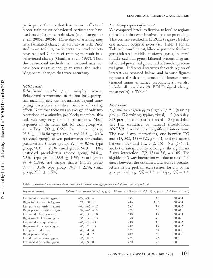
Localizing regions of interestWe compared letters to fixation to localize regionsof the brain that were involved in letter processing.This contrast resulted in 12 ROIs (Figure 2): bilat-eral inferior occipital gyrus (see Table 1 for allTalairach coordinates), bilateral posterior fusiformgyrus,bilateral middle fusiform gyrus, bilateralmiddle occipital gyrus, bilateral precentral gyrus,left dorsal precentral gyrus, and left medial precen-tral gyrus. Inferential statistics for each region ofinterest are reported below, and because figuresrepresent the data in terms of difference scores(trained minus untrained pseudoletters), we alsoinclude all raw data (% BOLD signal changemean peaks) in Table 2.
ROI resultsLeft inferior occipital gyrus (Figure 3). A 3 (traininggroup, TG: writing, typing, visual) � 2 (scan day,SD: pretrain scan, posttrain scan) � 2 (pseudolet-ter, PL: untrained or trained) mixed-modelANOVA revealed three significant interactions.The two 2-way interactions, one between TGand SD, F(2, 15) ¼ 5.3, p , .01, and the secondbetween TG and PL, F(2, 15) ¼ 8.5, p, .01,are better interpreted by looking at the significant3-way interaction, F(2, 15) ¼ 3.8, p , .05. Thesignificant 3-way interaction was due to no differ-ences between the untrained and trained pseudo-letters in the pretrain scan session for any of thegroups—writing, t(5) ¼ 1.3, ns; type, t(5) ¼ 1.4,
Table 1. Talairach coordinates, cluster size, peak t value, and significance level of each region of interest
Region of interest Talairach coordinates (peak) (x, y, z) Cluster size (1-mm voxels) t(17) peak p , (uncorrected)
Left inferior occipital gyrus –29, –93, –1 353 8.2 .000003
Right inferior occipital gyrus 27, –92, –1 496 11.3 .000004
Left posterior fusiform gyrus –43, –66, –12 657 9.4 .000002
Right posterior fusiform gyrus 38, –66, –15 275 7.0 .00001
Left middle fusiform gyrus –43, –58, –10 680 8.2 .000003
Right middle fusiform gyrus 36, –59, –13 560 6.3 .00002
Left middle occipital gyrus –46, –73, –9 290 9.3 .000002
Right middle occipital gyrus 46, –73, –5 487 8.7 .000002
Left precentral gyrus –45, –4, 34 675 7.4 .000001
Right precentral gyrus 40, –4, 32 409 7.9 .000001
Left dorsal precentral gyrus –46, –8, 51 275 5.6 .0001
Left medial precentral gyrus –34, –9, 50 270 5.8 .0001
COGNITIVE NEUROPSYCHOLOGY, 2009, 26 (1) 101
SENSORIMOTOR LEARNING AND LETTERS
Dow
nloa
ded
by [
Indi
ana
Uni
vers
ity L
ibra
ries
] at
10:
19 0
3 D
ecem
ber
2013
ns; visual, t(5) ¼ 1.7, ns—with significant differ-ences between the two PL types in the postscansession for two (writing and typing) of the threegroups: writing, t(5) ¼ 2.6, p, .05; type,t(5) ¼ 2.3, p, .05; visual, t(5) ¼ 0.65, ns. Thus,in this early visual area, the PL training had aneffect on percentage BOLD signal change in themotor and typing training groups.
Right inferior occipital gyrus. The results of theANOVA in this region revealed one significantmain effect, that of the group variable, F(2,15) ¼ 11.9, p, .001. This effect was driven bylower overall peak percentage signal change inthe visual group (M ¼ 0.87) than in the typinggroup (M ¼ 1.2), t(5) ¼ 5.4, p , .001, and in the
motor group (M ¼ 1.15), t(5) ¼ 4.1, p , .001.The writing and typing groups were not signifi-cantly different from one another, t(5) ¼ 1.2, ns.
Left posterior fusiform gyrus (see Figure 4). The3 � 2 � 2 ANOVA revealed several significantdifferences in this region. First, there were maineffects of SD, F(1, 15) ¼ 17.4, p , .001, andPL, F(1, 15) ¼ 5.6, p , .05; the main effect ofgroup was not significant, F(1, 15) ¼ 1.3, ns.The significant main effects must be interpretedin light of the three 2-way interactions and one3-way interaction. The three 2-way interactions,between TG and SD, F(2, 15) ¼ 5.7, p , .01,TG and PL, F(2, 15) ¼ 4.7, p , .05, andSD � PL, F(1, 15) ¼ 18.4, p , .0001, are better
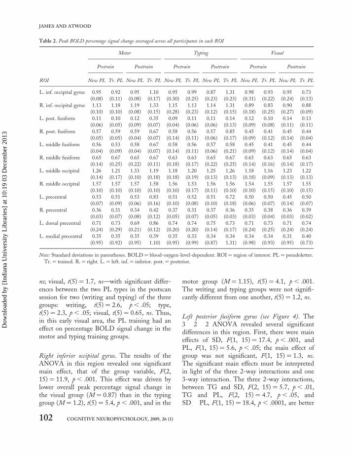
Table 2. Peak BOLD percentage signal change averaged across all participants in each ROI
Motor Typing Visual
Pretrain Posttrain Pretrain Posttrain Pretrain Posttrain
ROI New PL Tr. PL New PL Tr. PL New PL Tr. PL New PL Tr. PL New PL Tr. PL New PL Tr. PL
L. inf. occipital gyrus 0.95 0.92 0.95 1.10 0.95 0.99 0.87 1.31 0.98 0.93 0.95 0.73
(0.08) (0.11) (0.08) (0.17) (0.30) (0.25) (0.23) (0.23) (0.31) (0.22) (0.24) (0.13)
R. inf. occipital gyrus 1.13 1.18 1.19 1.33 1.15 1.13 1.14 1.31 0.89 0.83 0.90 0.88
(0.10) (0.10) (0.08) (0.15) (0.28) (0.23) (0.12) (0.15) (0.18) (0.25) (0.27) (0.09)
L. post. fusiform 0.11 0.10 0.12 0.35 0.09 0.11 0.11 0.14 0.12 0.10 0.14 0.13
(0.06) (0.05) (0.09) (0.07) (0.04) (0.06) (0.06) (0.13) (0.09) (0.08) (0.11) (0.11)
R. post. fusiform 0.57 0.59 0.59 0.67 0.58 0.56 0.57 0.85 0.45 0.41 0.45 0.44
(0.05) (0.05) (0.04) (0.07) (0.14) (0.11) (0.06) (0.17) (0.09) (0.12) (0.14) (0.04)
L. middle fusiform 0.56 0.53 0.58 0.67 0.58 0.56 0.57 0.58 0.45 0.41 0.45 0.44
(0.04) (0.09) (0.04) (0.07) (0.14) (0.11) (0.06) (0.21) (0.09) (0.12) (0.14) (0.04)
R. middle fusiform 0.65 0.67 0.65 0.67 0.63 0.63 0.65 0.67 0.65 0.63 0.65 0.63
(0.14) (0.25) (0.22) (0.11) (0.18) (0.17) (0.22) (0.25) (0.14) (0.16) (0.14) (0.17)
L. middle occipital 1.26 1.21 1.33 1.19 1.18 1.20 1.25 1.26 1.18 1.16 1.23 1.22
(0.14) (0.17) (0.10) (0.18) (0.18) (0.19) (0.13) (0.13) (0.18) (0.09) (0.13) (0.13)
R. middle occipital 1.57 1.57 1.57 1.58 1.56 1.53 1.56 1.56 1.54 1.55 1.57 1.55
(0.10) (0.10) (0.10) (0.10) (0.10) (0.17) (0.11) (0.10) (0.10) (0.15) (0.10) (0.15)
L. precentral 0.53 0.51 0.53 0.83 0.51 0.52 0.51 0.72 0.50 0.50 0.45 0.50
(0.07) (0.09) (0.06) (0.16) (0.10) (0.08) (0.10) (0.18) (0.06) (0.07) (0.14) (0.07)
R. precentral 0.36 0.31 0.34 0.42 0.37 0.31 0.37 0.36 0.35 0.38 0.36 0.39
(0.03) (0.07) (0.08) (0.12) (0.05) (0.07) (0.05) (0.03) (0.03) (0.04) (0.03) (0.02)
L. dorsal precentral 0.71 0.73 0.69 0.86 0.74 0.74 0.75 0.73 0.71 0.73 0.71 0.74
(0.24) (0.29) (0.21) (0.12) (0.20) (0.20) (0.14) (0.17) (0.24) (0.25) (0.24) (0.24)
L. medial precentral 0.35 0.35 0.35 0.39 0.35 0.33 0.34 0.34 0.34 0.34 0.31 0.40
(0.95) (0.92) (0.95) 1.10) (0.95) (0.99) (0.87) 1.31) (0.98) (0.93) (0.95) (0.73)
Note: Standard deviations in parentheses. BOLD ¼ blood-oxygen-level-dependent. ROI ¼ region of interest. PL ¼ pseudoletter.
Tr. ¼ trained. R. ¼ right. L. ¼ left. inf. ¼ inferior. post. ¼ posterior.
102 COGNITIVE NEUROPSYCHOLOGY, 2009, 26 (1)
JAMES AND ATWOOD
Dow
nloa
ded
by [
Indi
ana
Uni
vers
ity L
ibra
ries
] at
10:
19 0
3 D
ecem
ber
2013

understood when we consider the three-way inter-action, F(2, 15) ¼ 13.2, p , .0001. The 3-wayinteraction revealed that for the writing traininggroup, there was a significant difference only inthe posttraining scan between trained anduntrained pseudoletters, t(5) ¼ 4.02, p , .005,but not between trained and untrained PLs priorto training, t(5) ¼ 1.9, ns. In the typing traininggroup, there were no significant differencesbetween trained and untrained PLs during thepretrain scan, t(5) ¼ 0.21, ns, or during the post-train scan, t(5) ¼ 0.26, ns. For the visual traininggroup, again, there were no significant differencesbetween the trained and untrained PLs in the pre-train scan, t(5) ¼ 1.97, ns, or in the posttrain con-dition, t(5) ¼ 0.47, ns. Thus, in the left posteriorfusiform gyrus, we find that percentage BOLDsignal change increases as a function of training,but only when the training involves writing thepseudoletters, not after typing or purely visual
training. When letters are compared to othervisual stimuli, the left fusiform gyrus has beenshown to be engaged more during letter processingthan during processing of other, similar shapes(Flowers et al., 2004; James et al., 2005;Longcamp et al., 2003). Here we show that acti-vation before and after training with pseudoletterschanged activation patterns in this region,suggesting that the sensitivity to letters in thisregion may be due to sensorimotor interactionsas well.
Right posterior fusiform gyrus (Figure 5). Another3 � 2 � 2 mixed-model ANOVA was run on thedata from the right fusiform gyrus, revealing amain effect of SD, F(1, 15) ¼ 9.8, p, .005, andof PL, F(1, 15) ¼ 9.2, p , .005. Three significantinteractions emerged as well: one between TGand PL, F(1, 15) ¼ 6.8, p , .005, one betweenSD and PL, F(1, 15) ¼ 7.0, p, .01, and a three-way interaction, F(1, 15) ¼ 3.4, p, .05. Thethree-way interaction was driven by no significantdifferences in percentage BOLD signal change
Figure 3. A depiction of the significant three-way interaction
among pseudoletters (PLs), training group, and scan day in the
left inferior occipital gyrus. All interaction graphs depict peak
blood-oxygen-level-dependent (BOLD) activation of untrained
pseudoletters subtracted from trained pseudoletters as a function of
training group and scan day. Error bars depict 95% confidence
intervals, and therefore overlap with the x-axis depicts
nonsignificance, while nonoverlap depicts a significant difference
between trained and untrained pseudoletters (see text for
inferential statistics and Table 2 for mean % BOLD signal
change values). Note that there are no differences between trained
and untrained PLs before training (light grey bars), but
significant differences after training for the motor and typing
training group.
Figure 4. A depiction of the significant three-way interaction
among pseudoletters (PLs), training group, and scan day in the
left posterior fusiform gyrus. There is no significant difference
between trained and untrained pseudoletters prior to training and
no difference in activation to these two stimulus sets after typing
and visual training. There is, however, a large difference in
blood-oxygen-level-dependent (BOLD) percentage signal change
(greater to trained than to untrained) after motor training.
COGNITIVE NEUROPSYCHOLOGY, 2009, 26 (1) 103
SENSORIMOTOR LEARNING AND LETTERS
Dow
nloa
ded
by [
Indi
ana
Uni
vers
ity L
ibra
ries
] at
10:
19 0
3 D
ecem
ber
2013

for new versus trained pseudoletters in thepretraining scan session for any of the groups (allt values , 1.9), contrasting with a differencebetween the new and trained PL conditions inthe posttraining session after typing training,t(5) ¼ 3.4, p , .005, and writing training,t(5) ¼ 2.75, p , .05, but not after visual-onlytraining, t(5) ¼ 0.45, ns.
Left middle fusiform gyrus. Another 3 � 2 � 2mixed-model ANOVA revealed a significantmain effect of TG in this region, F(1, 15) ¼ 8.6,p , .005, with the visual training group producingless percentage signal change than the writinggroup, t(5) ¼ 2.8, p , .05, and less than thetyping group, t(5) ¼ 3.1, p, .05 (writing,M ¼ 0.58, SD ¼ 0.07; typing, M ¼ 0.57,SD ¼ 0.13; visual, M ¼ 0.43, SD ¼ 0.09). Noother effects were significant (all other F values, 2.0). In contrast with the left posterior fusiform,the left middle fusiform was not affected by train-ing in this paradigm. It is curious that the visualtraining group displayed less activation overall in
this region—a result also found in the rightinferior occipital gyrus.
Right middle fusiform gyrus. There were no signifi-cant differences among any of the conditions inthis region (all F values , 2.0). This resultreinforces the idea that the middle fusiformgyrus, although letter sensitive, is not involved inthe changes due to training that are seen in themore posterior fusiform.
Bilateral middle occipital gyrus. There were nosignificant differences among any of the conditionsin these regions.
Left precentral gyrus (Figure 6). From the3 � 2 � 2 ANOVA, we see significant maineffects of all three conditions: TG, F(1,15) ¼ 8.8, p , .005; SD, F(1, 15) ¼ 8.0, p , .01;and PL, F(1, 15) ¼ 15.7, p , .001. There werealso two significant interactions, one betweenTG and SD, F(1, 15) ¼ 3.7, p , .05, and onebetween SD and PL, F(1, 15) ¼ 16.3, p, .001.Simple effects demonstrated that the TG � SDinteraction was due to the writing, t(5) ¼ 2.7,
Figure 6. The three-way interaction among pseudoletters (PLs),
training group, and scan day in the left precentral gyrus. There is
no significant difference between trained and untrained
pseudoletters prior to training and, again, no difference after
visual training. However, motor and typing training both result
in greater activation to trained than to untrained PLs.
Figure 5. The significant three-way interaction among
pseudoletters (PLs), training group, and scan day in the right
posterior fusiform gyrus. There is no significant difference between
trained and untrained pseudoletters prior to training and no
difference after visual training. There is, however, a difference in
blood-oxygen-level-dependent (BOLD) percentage signal change
(greater to trained than to untrained) after motor and typing
training.
104 COGNITIVE NEUROPSYCHOLOGY, 2009, 26 (1)
JAMES AND ATWOOD
Dow
nloa
ded
by [
Indi
ana
Uni
vers
ity L
ibra
ries
] at
10:
19 0
3 D
ecem
ber
2013

p , .05, and typing, t(5) ¼ 2.1, p , .05, groupsboth showing an increase in percentage signalchange after training, whereas the visual traininggroup did not, t(5) ¼ 0.69, ns. The SD � PLinteraction was due to an increase in percentagesignal change after training in the trained pseudo-letters only, t(5) ¼ 3.7, p , .005, that was notpresent prior to training, t(5) ¼ 0.25, ns. Takentogether, after training, the two groups that inter-acted with the stimuli using motor systems bothshowed increases in percentage BOLD signalchange. The second interaction suggests that inall three groups, the trained pseudoletters wereresponded to with greater percentage signalchange than were the untrained pseudolettersonly after training (M ¼ 0.49, SD ¼ 0.10 fornew PLs, and M ¼ 0.68, SD ¼ 0.19 for trainedPLs after training sessions). The three-way inter-action approached significance, F(1, 15) ¼ 2.9,p , .08, as reflected in Figure 6—the writingand typing groups appeared to be significantlydifferent from the visual group in terms of thechange in BOLD response after training. Recentresearch has found recruitment of the leftprecentral gyrus during visual letter processing( James & Gauthier, 2006; Longcamp et al.,2003, 2005a, 2005b). The hypothesis that hasbeen brought forth by both groups is that leftmotor regions are activated because of storedmotor programmes that result from experience inwriting letters. The current results support thisclaim and extend it by showing that typing train-ing also results in activation in this region duringvisual presentation of pseudoletter stimuli. Thisis the first direct evidence supporting the claimthat neural engagement in motor areas duringvisual tasks is due to motor experience and notspecifically writing experience.
Right precentral gyrus. Prior evidence for right pre-central gyrus engagement during letter-processingtasks is less compelling than that for the left pre-central gyrus. The usual explanation for the lackof activation is that the left hemisphere processeslanguage stimuli more than does the right hemi-sphere. We did find some right precentral engage-ment during letter tasks here, though—perhaps
due to the contrast that is used (letters vs. fixation).Although the 3 � 2 � 2 ANOVA performed onthe data from this region revealed a significantmain effect of SD, F(1, 15) ¼ 7.4, p , .01, withthe posttraining scan resulting in a higher percen-tage signal change overall (M ¼ 0.37, SD ¼ 0.05)than the pretraining scan (M ¼ 0.37, SD ¼ 0.06),there were no other significant effects (allFs, 2.5).
Left dorsal precentral gyrus (Figure 7). This region issimilar to that previously found to be engagedduring letter perception in some studies ( James& Gauthier, 2006). The 3 � 2 � 2 ANOVAthat was performed on the data from this regionrevealed three effects. The first was a main effectof PL, F(1, 15) ¼ 5.7, p , .03. Perceiving newPLs resulted in a lower percentage signal change(M ¼ 0.71, SD ¼ 0.12) than did trained PLs(M ¼ 0.76, SD ¼ 0.14). There were also twointeractions, one 2-way interaction between TGand PL, F(2, 15) ¼ 3.5, p , .05, and a 3-wayinteraction among TG, SD, and PL, F(2,15) ¼ 3.8, p, .05. Simple effects analysesrevealed that there was an increase in percentagesignal change between the new and trained PLsonly after writing training, t(5) ¼ 2.7, p, .05(all other t values , 2.0; see Figure 7).
Left medial precentral gyrus. The results of ourlocalizer contrast also revealed a region in themedial portion of the precentral gyrus that wasinvolved in letter perception. A 3 � 2 � 2ANOVA in this region revealed one significantinteraction among the conditions: that betweenSD and PL, F(1, 15) ¼ 5.1, p , .05. This inter-action was due to a significant difference betweennew and trained PLs only after training,t(5) ¼ 2.5, p , .05, but not before training,t(5) ¼ 0.33, ns. Thus, there was an effect oftraining in this region, but it was not specific toa given type of training.
SummaryTo summarize these results, our localizer contrastrevealed 12 regions in the brain that were activemore to letter perception than to a fixation
COGNITIVE NEUROPSYCHOLOGY, 2009, 26 (1) 105
SENSORIMOTOR LEARNING AND LETTERS
Dow
nloa
ded
by [
Indi
ana
Uni
vers
ity L
ibra
ries
] at
10:
19 0
3 D
ecem
ber
2013

baseline. Of these 12 regions, 5 showed a three-way interaction among our conditions, wherenew versus trained pseudoletters were respondedto differently as a function of scan day and traininggroup. In 3 of these 5 regions (the left inferioroccipital gyrus, the right fusiform gyrus, and theleft precentral gyrus), writing and typing groupsshowed an increase in percentage signal changewhen perceiving trained pseudoletters after train-ing versus perceiving untrained PLs in the samesession. In 2 other regions (the left posterior fusi-form gyrus and the left dorsal precentral gyrus)there was greater neural activation during trainedPL perception than during untrained PL percep-tion only after writing training. Interestingly,these latter regions are the same areas that havebeen found previously to respond to letter percep-tion and letter writing (James & Gauther, 2006).
Thus, we have demonstrated that regions of thebrain that respond more to letters than to a fixationcross are not necessarily letter specific. Theseregions are also engaged during perception ofother letter-like stimuli after a certain type ofexperience. Here we tested how writing, typing,and visual experience with previously novelcharacters would affect neural responses in these
letter-selective regions. Interestingly, the trainingconditions served to change the response patternsin several of the regions.
GENERAL DISCUSSION
There is abundant evidence that there is neuralfunctional specialization for processing individualletters (Flowers et al., 2004; James & Gauthier,2006; James et al., 2005; Polk & Farah, 1998;Polk et al., 2002), but, until now, there has notbeen any work investigating how this specializ-ation may develop. We now present evidencethat our experience in writing letters may contrib-ute to the development of functional specializationfor letters. We demonstrate that after writingpractice, some brain regions that are engagedduring letter processing are also engaged more totrained pseudoletters than to untrained pseudolet-ters, implying that these regions increase theirresponse to these particular stimuli only after aspecific type of motor interaction. In other brainregions, activation changed both after writingand after typing training, implying that theseregions increase responses after any motor experi-ence. Interestingly, the regions that were found tochange response as a result of writing training werethe same regions as those found in previous workto respond during letter perception and duringletter writing (the left fusiform gyrus and the leftdorsal precentral gyrus; James & Gauthier, 2006).
Although performance on some of our beha-vioural measures improved with training, therewas no difference in performance in behaviouraltasks as a function of different training conditions.This null behavioural result is in marked contrastto our significant differences in BOLD activationamong training groups. Showing a difference inBOLD activation, but not a difference in beha-vioural measures, is not an uncommon patternof results (e.g., see James & Gauthier, 2006;McLaughlin, Osterhout, & Kim, 2004;Wilkinson & Halligan, 2003a, 2003b). Such aresult can be attributed to the greater sensitivityof measuring changes in BOLD response thanresponse times or accuracy measures (see also
Figure 7. The three-way interaction among pseudoletters (PLs),
training group, and scan day in the left dorsal precentral gyrus.
There is no significant difference between trained and untrained
pseudoletters prior to training and, again, no difference after
typing or visual training. However, motor training resulted in
greater activation to trained than to untrained PLs.
106 COGNITIVE NEUROPSYCHOLOGY, 2009, 26 (1)
JAMES AND ATWOOD
Dow
nloa
ded
by [
Indi
ana
Uni
vers
ity L
ibra
ries
] at
10:
19 0
3 D
ecem
ber
2013
Wilkinson & Halligan, 2003a for further discus-sion of this view). Such sensitivity is importantin revealing neural processing that can informcurrent cognitive theories and shape futureresearch endeavours.
The imaging results of this work suggest thatafter experience with certain types of stimuli—inthis case, pseudoletters—brain regions changetheir responses to the learned versus unlearnedexemplars of that stimulus set. Because neural acti-vation changes are based on training and not basedon stimulus properties (trained and untrainedpseudoletters are interchangeable), our results donot support theories of ventral stream organizationthat focus on eccentricity biases (e.g., Hassonet al., 2003) or stimulus characteristics (e.g.,Haxby et al., 2001) as organizing principles ofventral stream activation. We would argue thatthe functional specialization that has been docu-mented to letters (e.g., Flowers et al., 2004;Garrett et al., 2000; James et al., 2005) in theleft ventral stream may be based partially on ourmotor experience with the stimuli.
We know that motor interactions with objectsplay a crucial role in learning about the environ-ment, and it makes sense that the brain is orga-nized to reflect this interaction: We seecoactivation of visual and motor regions duringperception of a variety of different types ofobjects (e.g., Chao & Martin, 2000; Grezes &Decety, 2002; James & Gauthier, 2006). Thepresent results support this general idea: thatafter motor experience, seeing a pseudoletterrecruits areas of the brain that are specialized forletter processing, comprising a sensorimotornetwork. But what is it about writing experiencethat leads to this functional specialization?
Presumably, practised motor experiences set uprepresentations of motor programmes in thefrontal cortex, and these representations are acti-vated upon subsequent visual presentation of theobject. Efferent copies of such information mayproject to the ventral visual processing stream,affecting neural activation in these regions. Thisprocess may facilitate recognition of the objectby augmenting the visual information withsensorimotor information. What then is it about
the motor information that would affect visualprocessing?
We would suggest that when children learn towrite letters, they also learn a very efficient wayto group letters into “basic-level” categories(Rosch, Mervis, Gray, Johnson, & Boyes-Braem,1976) and to perhaps ignore irrelevant changesamong exemplars within that category. That is,the category of the letter “A”—presumably abasic, or “entry level” ( Jolicoeur, 1985) cat-egory—has many exemplars within it that lookvery different (e.g., A, a, a, A, etc). To become effi-cient readers, we must learn to identify exemplarsthat are in the same category, despite irrelevantchanges in appearance. Children initially learn tovisually recognize and categorize letters that arein a single font type and always appear in thesame orientation (the sans serif, capital blockalphabet). When children start to write lettersthemselves however, the motor information thatproduces that letter is variable. This variability,at first, may conflict with the intent to write acertain letter. With practice, the motor variabilitymay serve to augment visual processing by broad-ening a visual-motor representation. In thisinterpretation, motor experience changes storedrepresentations and perhaps then also changesvisual processing. In fact, recent research hasshown that preliterate children (4–5-year-olds)who are given printing practice with letters havea very different neural response to letters than dochildren who do not receive such practice. Thatis, prior to practice, the ventral visual processingstream of both groups of children responds simi-larly to visually presented letters—neural responsesto letters is the same as those to shapes. But afterprinting practice, the left fusiform gyrus shows aheightened activation to letters compared tosimple shapes ( James, 2008).
Another possibility as to why the sensorimotorsystems interact may be due simply to coactivation.Coactivation of multiple systems during learningcould lead to “wiring together” of such systems,leading to future coactivations upon subsequentencounters of the same input. More likely, andnot mutually exclusive with the former expla-nation, activation of sensorimotor systems may
COGNITIVE NEUROPSYCHOLOGY, 2009, 26 (1) 107
SENSORIMOTOR LEARNING AND LETTERS
Dow
nloa
ded
by [
Indi
ana
Uni
vers
ity L
ibra
ries
] at
10:
19 0
3 D
ecem
ber
2013

serve to potentiate a motor response. If seeingthe item previously required a motor response,then perhaps seeing it again would activate a pro-gramme associated with the response to preparefor action.
Our knowledge of the effects of learningexperience on functional brain changes inhumans is in its beginning stages. The researchpresented here demonstrates that our sensorimotorinteractions with the environment are storedduring learning. Furthermore, our motor systemsare active during visual presentations, suggestingthat the motor system is involved in visual proces-sing at some level and may contribute to functionalspecialization in the ventral stream. Activation ofthe motor system during visual processing shownhere presumably reflects the behavioural findingsthat motor information affects visual processingof letters (e.g., Freyd, 1983; Orliaguet et al.,1997; Tse & Cavanagh, 2000). Additional workis needed to uncover why motor interactionsduring learning influence visual processing andwhether this interaction is crucial for normalletter recognition ability to develop.
REFERENCES
Anderson, S. W., Damasio, A. R., & Damasio, H.(1990). Troubled letters but not numbers: Domainspecific cognitive impairments following focaldamage in frontal cortex. Brain, 113, 749–766.
Babcock, M. K., & Freyd, J. J. (1988). Perception ofdynamic information on static form. American
Journal of Psychology, 101, 111–131.Bai, D. L., & Bertenthal, B. I. (1992). Locomotor status
and the development of spatial search skills. ChildDevelopment, 63, 215–226.
Bartolomeo, P., Bachoud-Levi, A.-C., Chokron, S., &Degos, J. D. (2002). Visually- and motor- basedknowledge of letters: Evidence from a pure alexicpatient. Neuropsychologia, 40, 1363–1371.
Bukach, C. M., Gauthier, I., & Tarr, M. J. (2006).Beyond faces and modularity: The power of anexpertise framework. Trends in Cognitive Sciences,10, 159–166.
Campos, J. J., Anderson, D. I., Barbu-Roth, M. A.,Hubbard, E. M., Hertenstein, M. J., &
Witherington, D. (2000). Travel broadens themind. Infancy, 1, 149–219.
Carlson, T. A., Schrater, P., & He, S. (2003). Patternsof activity in the categorical representationsof objects. Journal of Cognitive Neuroscience, 15,704–717.
Changizi, M. A., & Shimojo, S. (2004). Character com-plexity and redundancy in writing systems overhuman history. Proceedings of the Royal Society
Britain.Chao, L. L., & Martin, A. (2000). Representation of
man-made objects in the dorsal stream.NeuroImage, 12, 478–484.
Cohen, L., Dahaene, S., Naccache, L., Lehericy, S.,Dahaene-Lambertz, G., Henaff, M. A., et al.,2000. The visual word form area: Spatial and tem-poral characterization of an initial stage of readingin normal participants and posterior split-brainpatients. Brain, 123, 291–307.
Cox, D., & Savoy, R. (2003). Functional magneticresonance imaging (fMRI) “brain reading”:Detecting and classifying distributed patterns offMRI activity in human visual cortex. NeuroImage,19, 261–270.
Cunningham, A. E., & Stanovich, K. E. (1990). Earlyspelling acquisition: Writing beats the computer.Journal of Educational Psychology, 82, 159–162.
Czerwinski, M., Lightfoot, N., & Shiffrin, R. M.(1992). Automatization and training in visualsearch. American Journal of Psychology, 105, 271–315.
Diamond, R., & Carey, S. (1986). Why faces are and arenot special: An effect of expertise. Journal of
Experimental Psychology: General, 115, 107–117.Downing, P. E., Chan, A. W.-Y., Peelen, M. V.,
Dodds, C. M., & Kanwisher, N. (2006). Domainspecificity in visual cortex. Cerebral Cortex, 16,1453–1461.
Downing, P. E., Jiang, Y., Shuman, M., &Kanwisher, N. (2001). A cortical area selective forvisual processing of the human body. Science, 293,2470–2473.
Flowers, D. L., Jones, K., Noble, K., VanMeter, J.,Zeffiro, T. A., Wood, F. B., et al. (2004).Attention to single letters activates left extrastriatecortex. NeuroImage, 21, 829–839.
Freyd, J. J. (1983). Representing the dynamics of staticform. Memory & Cognition, 11, 342–346.
Garrett, A. S., Flowers, D. L., Absher, J. R., Fahey,F. H., Gage, H. D., Keyes, J. W., et al. (2000).Cortical activity related to accuracy of letter recog-nition. NeuroImage, 11, 111–123.
108 COGNITIVE NEUROPSYCHOLOGY, 2009, 26 (1)
JAMES AND ATWOOD
Dow
nloa
ded
by [
Indi
ana
Uni
vers
ity L
ibra
ries
] at
10:
19 0
3 D
ecem
ber
2013

Gauthier, I., Skudlarski, P., Gore, J. C., & Anderson,A. W. (2000). Expertise for cars and birds recruitsbrain areas involved in face recognition. Nature
Neuroscience, 3, 191–197.Gauthier, I., Williams, P., Tarr, M. J., & Tanaka, J.
(1997). Training “greeble” experts: A frameworkfor studying expert object recognition process.Vision Research, 38, 2401–2428.
Gerlach, C., Law, I., Gade, A., & Paulson, O. B.(2002). The role of action knowledge in the compre-hension of artifacts—a PET study. NeuroImage, 18,928–937.
Gibson., J. J. (1979). The ecological approach to visual
perception. Boston: Houghton Mifflin.Grezes, J., & Decety, J. (2002). Does visual
perception of object afford action? Evidence froma neuroimaging study. Neuropsychologia, 40,212–222.
Grill-Spector, K., Henson, R., & Martin, A. (2006).Repetition and the brain: Neural models of stimulus-specific effects.Trends in Cognitive Science, 10, 18–24.
Grossman, E. D., & Blake, R. (2002). Brain activityevoked by inverted and imagined biological motion.Vision Research, 41, 1475–1482.
Harman, K. L., Humphrey, G. K., & Goodale, M. A.(1999). Active manual control of object views facili-tates recognition. Current Biology, 9, 1315–1320.
Hasson, U., Harel, M., Levy, I., & Malach, R. (2003).Large-scale mirror-symmetry organization ofhuman occipito-temporal object areas. Neuron, 37,1027–1041.
Hasson, U., Levy, I., Behrmann, M., Hendler, T., &Malach, M. (2002). Eccentricity bias as an organiz-ing principle for human high order object areas.Neuron, 34, 479–490.
Haxby, J. V., Gobbini, M. I., Furey, M. L., Ishai, A.,Schouten, J. L., & Pietrini, P. (2001). Distributedand overlapping representations of faces andobjects in ventral temporal cortex. Science, 293,2425–2430.
Ishai, A., Ungerleider, L. G., Martin, A., Schouten,J. L., & Haxby, J. (1999). Distributed representationof objects in the human ventral visual pathway.Proceedings of the National Academy of Sciences, USA,96, 9379–9384.
James, K. H. (2008). Sensori-motor experience leads to
changes in visual processing in the developing brain.Manuscript submitted for publication.
James, K., & Gauthier, I. (2006). Letter processingautomatically recruits a sensory-motor brainnetwork. Neuropsychologia, 44, 2937–2949.
James, K. H., Humphrey, G. K., & Goodale, M. A.(2001). Manipulating and recognizing visualobjects: Where the action is. Canadian Journal of
Experimental Psychology, 55, 111–120.James, K. H., Humphrey, G. K., Vilis, T., Corrie, B.,
Baddour, R., & Goodale, M. A. (2002). “Active”and “passive” learning of three-dimensional objectstructure within an immersive virtual reality environ-ment. Behavior Research Methods, Instruments &
Computers, 34, 383–390.James, K. H., James, T. W., Jobard, G., Wong, A., &
Gauthier, I. (2005). Letter processing in the visualsystem: Different activation patterns for singleletters and strings. Cognitive, Affective, &
Behavioral Neuroscience, 5, 452–466.Jolicoeur, P. (1985). The time to name disoriented
natural objects. Memory & Cognition, 13, 289–303.Kanwisher, N. (2000). Domain specificity in face
perception. Nature Neuroscience, 3, 759–763.Kanwisher, N., Chun, M. M., & McDermott, J. (1996).
fMRI in individual subjects reveals loci in extrastriatecortex differentially sensitive to faces and objects.Investigative Ophthalmology & Visual Science, 37, S193.
Levy, I., Hasson, U., Avidan, G., Hendler, T., &Malach,R. (2001). Center-periphery organization of humanobject areas. Nature Neuroscience, 4, 533–539.
Levy, I., Hasson, U., Harel, M., & Malach, R. (2004).Functional analysis of the periphery effect inhuman building related areas. Human Brain
Mapping, 22, 15–26.Longcamp, M., Anton, J. L., Roth, M., & Velay, J. L.
(2003). Visual presentation of single letters activatesa premotor area involved in writing. NeuroImage, 19,1492–1500.
Longcamp, M., Anton, J. L., Roth, M., & Velay, J. L.(2005a). Premotor activations in response to visuallypresented single letters depend on the hand used towrite: A study on left-handers. Neuropsychologia,43, 1801–1809.
Longcamp, M., Zerbato-Poudou, M., & Velay, J. L.(2005b). The influence of writing practice on letterrecognition in preschool children: A comparisonbetween handwriting and typing. Acta Psychologica,119, 67–79.
Mahon, B. Z., Milleville, S., Negri, G. A. L., Rumiati, R.I.,Martin,A.,&Caramazza,A. (2007).Action-relatedproperties of objects shape object representations inthe ventral stream. Neuron, 55, 507–520.
Malach, R., Levy, I., & Hasson, U. (2002). The topo-graphy of high-order human object areas. Trends inCognitive Sciences, 6, 176–184.
COGNITIVE NEUROPSYCHOLOGY, 2009, 26 (1) 109
SENSORIMOTOR LEARNING AND LETTERS
Dow
nloa
ded
by [
Indi
ana
Uni
vers
ity L
ibra
ries
] at
10:
19 0
3 D
ecem
ber
2013

McLaughlin, J., Osterhout, L., & Kim, A. (2004).Neural correlates of second-language word learning:Minimal instruction produces rapid change. Nature
Neuroscience, 7, 703–704.Mecklinger, A., Gruenewald, C., Besson, M. N., & von
Cramon, D. Y. (2002). Separable neuronal circuitsfor manipulable and non-manipulable objects inworking memory. Cerebral Cortex, 12, 1115–1123.
Orliaguet, J.-P., Kandel, S., & Bois, L.-J. (1997). Visualperception of motor anticipation in cursive hand-writing: Influence of spatial and movement infor-mation on the perception of forthcoming letters.Perception, 26, 905–912.
O’Toole, A. J. O., Jiang, F., Abdi, H., & Haxby, J. V.(2005). Partially distributed representations ofobjects and faces in ventral temporal cortex. Journalof Cognitive Neuroscience, 17, 580–590.
Polk, T. A., & Farah, M. J. (1998). The neural develop-ment and organization of letter recognition:Evidence from functional neuroimaging, compu-tational modeling, and behavioral studies.Proceedings of the National Academy of Sciences, USA,95, 847–852.
Polk, T. A., Stallcup, M., Aguirre, G. K., Alsop, D. C.,D’Esposito, M., Detre, J. A., et al. (2002). Neuralspecialization for letter recognition. Journal of
Cognitive Neuroscience, 14, 145–159.Portwood, M. (2000). Developmental dyspraxia.
London: David Fulton Publishing.Prinzmetal, W., McCool, C., & Park, S. (2005).
Attention: Reaction time and accuracy reveal differ-ent mechanisms. Journal of Experimental Psychology:General, 134, 73–92.
Rosch,E.,Mervis,C.B.,Gray,W.D., Johnson,D.M.,&Boyes-Braem, P. (1976). Basic objects in naturalcategories. Cognitive Psychology, 8, 382–439.
Seki, K., Yajima, M., & Sugishita, M. (1995). The effi-cacy of kinesthetic reading treatment for pure alexia.Neuropsychologia, 33, 595–609.
Spiridon, M., & Kanwisher, N. (2002). How distribu-ted is visual category information in human
occipito-temporal cortex? An fMRI study. Neuron,35, 1157–1165.
Stoodley, C. J., Fawcett, A. J., Nicolson, R. I., & Stein,J. F. (2005). Impaired balancing ability in dyslexicchildren. Experimental Brain Research, 167,370–380.
Talairach, J., & Tournoux, P. (1988). Co-planar stereo-taxic atlas of the human brain. New York: Thieme.
Tanaka, J. W., & Gauthier, I. (1997). Expertisein object and face recognition. In R. L. Goldstone,D. L. Medin, & P. G. Schyns (Eds.), Psychology of
learning and motivation: Vol. 36. Perceptual mechan-
isms of learning (pp. 83–125). San Diego, CA:Academic Press.
Tarr, M. J., & Gauthier, I. (2000). FFA: A flexiblefusiform area for subordinate-level visual processingautomatised for expertise. Nature Neuroscience, 3,764–769.
Tse, P. E., & Cavanagh, P. (2000). Chinese andAmericans see opposite apparent motions in aChinese character. Cognition, 74, B27–B32.
Wexler, M., & Boxtel, J. J. A. (2005). Depth perceptionby the active observer. Trends in Cognitive Science, 9,431–438.
Wexler, M., Kosslyn, S. M., & Berthoz, A. (1998).Motor processes in mental rotation. Cognition, 68,77–94.
Wilkinson, D., & Halligan, P. W. (2003a). The rel-evance of behavioral measures for functional-imaging studies of cognition. Nature Reviews,
Neuroscience, 5, 67–73.Wilkinson, D., & Halligan, P. W. (2003b). Stimulus
symmetry affects the bisection of figures but notlines: Evidence from event-related fMRI.NeuroImage, 20, 1756–1764.
Wohlschlager, A., & Wohlschlager, A. (1998). Mentaland manual rotation. Journal of Experimental
Psychology: Human Perception and Performance, 24,397–412.
Xu, Y. (2005). Revisiting the role of the fusiform face areain visual expertise. Cerebral Cortex, 15, 1234–1242.
110 COGNITIVE NEUROPSYCHOLOGY, 2009, 26 (1)
JAMES AND ATWOOD
Dow
nloa
ded
by [
Indi
ana
Uni
vers
ity L
ibra
ries
] at
10:
19 0
3 D
ecem
ber
2013