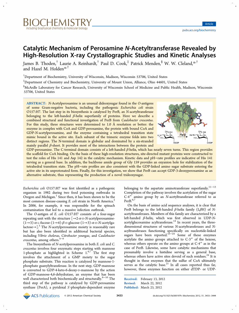
Catalytic Mechanism of Perosamine N-Acetyltransferase ...
12
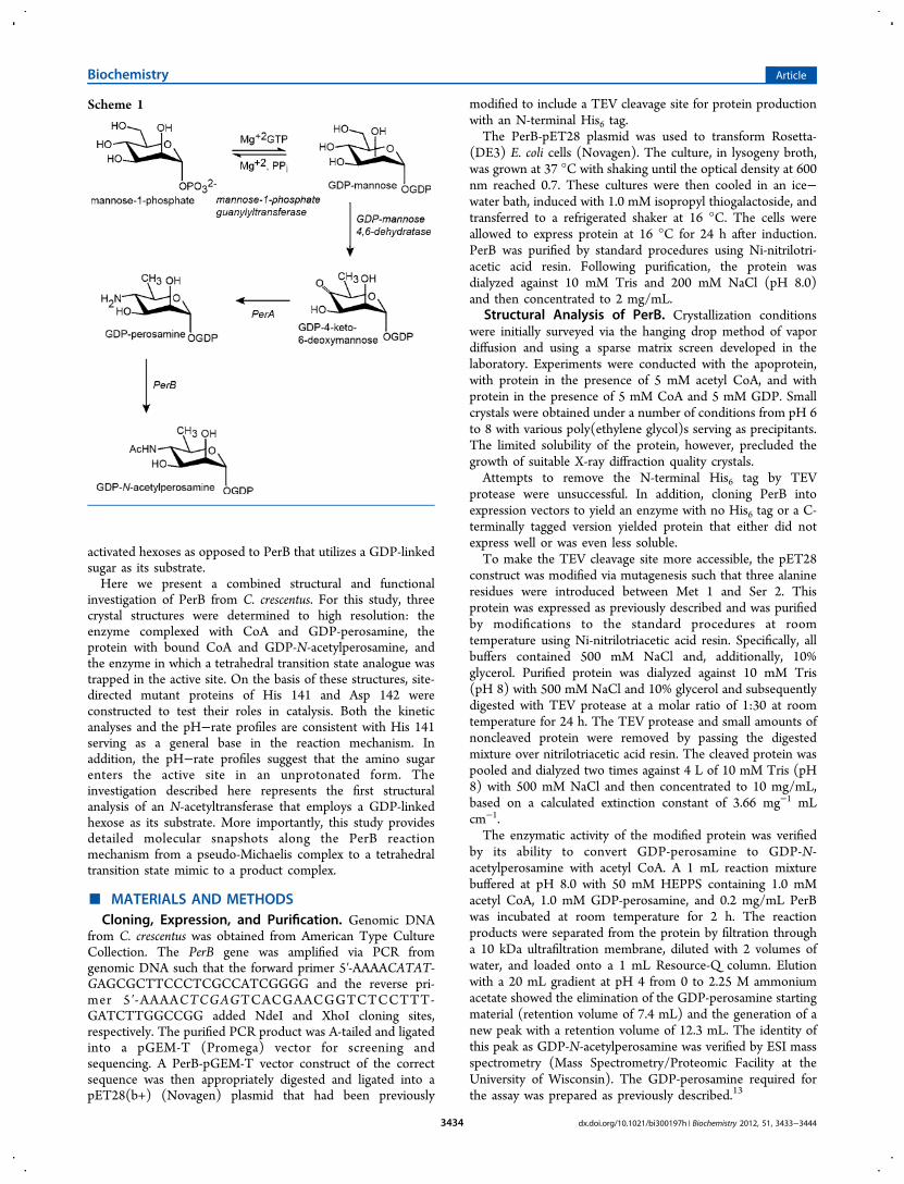
Catalytic Mechanism of Perosamine N-Acetyltransferase Revealed by High-Resolution X-ray Crystallographic Studies and Kinetic Analyses James B. Thoden, † Laurie A. Reinhardt, † Paul D. Cook, ‡ Patrick Menden, § W. W. Cleland,* ,† and Hazel M. Holden* ,† † Department of Biochemistry, University of Wisconsin, Madison, Wisconsin 53706, United States ‡ Department of Chemistry and Biochemistry, University of Mount Union, Alliance, Ohio 44601, United States § McArdle Laboratory for Cancer Research, University of Wisconsin School of Medicine and Public Health, Madison, Wisconsin 53706, United States ABSTRACT: N-Acetylperosamine is an unusual dideoxysugar found in the O-antigens of some Gram-negative bacteria, including the pathogenic Escherichia coli strain O157:H7. The last step in its biosynthesis is catalyzed by PerB, an N-acetyltransferase belonging to the left-handed β-helix superfamily of proteins. Here we describe a combined structural and functional investigation of PerB from Caulobacter crescentus. For this study, three structures were determined to 1.0 Å resolution or better: the enzyme in complex with CoA and GDP-perosamine, the protein with bound CoA and GDP-N-acetylperosamine, and the enzyme containing a tetrahedral transition state mimic bound in the active site. Each subunit of the trimeric enzyme folds into two distinct regions. The N-terminal domain is globular and dominated by a six-stranded mainly parallel β-sheet. It provides most of the interactions between the protein and GDP-perosamine. The C-terminal domain consists of a left-handed β-helix, which has nearly seven turns. This region provides the scaffold for CoA binding. On the basis of these high-resolution structures, site-directed mutant proteins were constructed to test the roles of His 141 and Asp 142 in the catalytic mechanism. Kinetic data and pH−rate profiles are indicative of His 141 serving as a general base. In addition, the backbone amide group of Gly 159 provides an oxyanion hole for stabilization of the tetrahedral transition state. The pH−rate profiles are also consistent with the GDP-linked amino sugar substrate entering the active site in its unprotonated form. Finally, for this investigation, we show that PerB can accept GDP-3-deoxyperosamine as an alternative substrate, thus representing the production of a novel trideoxysugar. Escherichia coli O157:H7 was first identified as a pathogenic organism in 1982 during two food poisoning outbreaks in Oregon and Michigan. 1 Since then, it has been shown to be the most common disease-causing E. coli strain in North America. 2 In 2006, for example, it was responsible for the spinach contamination that led to a massive infection outbreak. The O-antigen of E. coli O157:H7 consists of a four-sugar repeating unit with the structure [→2-α-D-N-acetylperosamine- (1→3)-α-L-fucose-(1→4)-β-D-glucose-(1→3)-α-D-N-acetylga- lactose→]. 3 The N-acetylperosamine moiety is reasonably rare but has also been identified in additional bacterial species, including Vibrio cholerae, Citrobacter youngae, and Caulobacter crescentus, among others. 4−6 The biosynthesis of N-acetylperosamine in both E. coli and C. crescentus involves four enzymatic steps starting with mannose 1-phosphate as highlighted in Scheme 1. 6,7 The first step involves the attachment of a GMP moiety to the sugar phosphate substrate. This reaction is catalyzed by mannose-1- phosphate guanylyltransferase. In the next step, GDP-mannose is converted to GDP-4-keto-6-deoxy-D-mannose by the action of GDP-mannose-4,6-dehydratase, an enzyme that has been well characterized both biochemically and structurally. 8−10 The third step of the pathway is catalyzed by GDP-perosamine synthase (PerA), a pyridoxal 5′-phosphate-dependent enzyme belonging to the aspartate aminotransferase superfamily. 11−13 Completion of the pathway involves the acetylation of the sugar C-4‴ amino group by an N-acetyltransferase referred to as PerB. 6,7 On the basis of amino acid sequence analyses, it is clear that PerB belongs to the left-handed β-helix family (LβH) of N- acetyltransferases. Members of this family are characterized by a left-handed β-helix, which was first observed in UDP-N- acetylglucosamine acyltransferase. 14 In recent years, the three- dimensional structures of various N-acetyltransferases and N- acyltransferases functioning specifically on nucleotide-linked sugars have been reported. 15−24 Some of these enzymes acetylate the amino groups attached to C-3‴ of the hexose, whereas others operate on the amino groups at C-4‴ as in the case of PerB. Likewise, some have catalytic mechanisms that presumably involve a histidine serving as a general base, whereas others have active sites devoid of such residues. 24 It is thought in these enzymes that the sulfur of CoA ultimately serves as the catalytic base. 21 In all cases reported thus far, however, these enzymes function on either dTDP- or UDP- Received: February 13, 2012 Revised: March 22, 2012 Published: March 23, 2012 Article pubs.acs.org/biochemistry © 2012 American Chemical Society 3433 dx.doi.org/10.1021/bi300197h | Biochemistry 2012, 51, 3433−3444
Transcript of Catalytic Mechanism of Perosamine N-Acetyltransferase ...
Catalytic Mechanism of Perosamine N-Acetyltransferase Revealed byHigh-Resolution X-ray Crystallographic Studies and Kinetic AnalysesJames B. Thoden,† Laurie A. Reinhardt,† Paul D. Cook,‡ Patrick Menden,§ W. W. Cleland,*,†
and Hazel M. Holden*,†
†Department of Biochemistry, University of Wisconsin, Madison, Wisconsin 53706, United States‡Department of Chemistry and Biochemistry, University of Mount Union, Alliance, Ohio 44601, United States§McArdle Laboratory for Cancer Research, University of Wisconsin School of Medicine and Public Health, Madison, Wisconsin53706, United States
ABSTRACT: N-Acetylperosamine is an unusual dideoxysugar found in the O-antigensof some Gram-negative bacteria, including the pathogenic Escherichia coli strainO157:H7. The last step in its biosynthesis is catalyzed by PerB, an N-acetyltransferasebelonging to the left-handed β-helix superfamily of proteins. Here we describe acombined structural and functional investigation of PerB from Caulobacter crescentus.For this study, three structures were determined to 1.0 Å resolution or better: theenzyme in complex with CoA and GDP-perosamine, the protein with bound CoA andGDP-N-acetylperosamine, and the enzyme containing a tetrahedral transition statemimic bound in the active site. Each subunit of the trimeric enzyme folds into twodistinct regions. The N-terminal domain is globular and dominated by a six-strandedmainly parallel β-sheet. It provides most of the interactions between the protein andGDP-perosamine. The C-terminal domain consists of a left-handed β-helix, which has nearly seven turns. This region providesthe scaffold for CoA binding. On the basis of these high-resolution structures, site-directed mutant proteins were constructed totest the roles of His 141 and Asp 142 in the catalytic mechanism. Kinetic data and pH−rate profiles are indicative of His 141serving as a general base. In addition, the backbone amide group of Gly 159 provides an oxyanion hole for stabilization of thetetrahedral transition state. The pH−rate profiles are also consistent with the GDP-linked amino sugar substrate entering theactive site in its unprotonated form. Finally, for this investigation, we show that PerB can accept GDP-3-deoxyperosamine as analternative substrate, thus representing the production of a novel trideoxysugar.
Escherichia coli O157:H7 was first identified as a pathogenicorganism in 1982 during two food poisoning outbreaks inOregon and Michigan.1 Since then, it has been shown to be themost common disease-causing E. coli strain in North America.2
In 2006, for example, it was responsible for the spinachcontamination that led to a massive infection outbreak.The O-antigen of E. coli O157:H7 consists of a four-sugar
repeating unit with the structure [→2-α-D-N-acetylperosamine-(1→3)-α-L-fucose-(1→4)-β-D-glucose-(1→3)-α-D-N-acetylga-lactose→].3 The N-acetylperosamine moiety is reasonably rarebut has also been identified in additional bacterial species,including Vibrio cholerae, Citrobacter youngae, and Caulobactercrescentus, among others.4−6
The biosynthesis of N-acetylperosamine in both E. coli and C.crescentus involves four enzymatic steps starting with mannose1-phosphate as highlighted in Scheme 1.6,7 The first stepinvolves the attachment of a GMP moiety to the sugarphosphate substrate. This reaction is catalyzed by mannose-1-phosphate guanylyltransferase. In the next step, GDP-mannoseis converted to GDP-4-keto-6-deoxy-D-mannose by the actionof GDP-mannose-4,6-dehydratase, an enzyme that has beenwell characterized both biochemically and structurally.8−10 Thethird step of the pathway is catalyzed by GDP-perosaminesynthase (PerA), a pyridoxal 5′-phosphate-dependent enzyme
belonging to the aspartate aminotransferase superfamily.11−13
Completion of the pathway involves the acetylation of the sugarC-4‴ amino group by an N-acetyltransferase referred to asPerB.6,7
On the basis of amino acid sequence analyses, it is clear thatPerB belongs to the left-handed β-helix family (LβH) of N-acetyltransferases. Members of this family are characterized by aleft-handed β-helix, which was first observed in UDP-N-acetylglucosamine acyltransferase.14 In recent years, the three-dimensional structures of various N-acetyltransferases and N-acyltransferases functioning specifically on nucleotide-linkedsugars have been reported.15−24 Some of these enzymesacetylate the amino groups attached to C-3‴ of the hexose,whereas others operate on the amino groups at C-4‴ as in thecase of PerB. Likewise, some have catalytic mechanisms thatpresumably involve a histidine serving as a general base,whereas others have active sites devoid of such residues.24 It isthought in these enzymes that the sulfur of CoA ultimatelyserves as the catalytic base.21 In all cases reported thus far,however, these enzymes function on either dTDP- or UDP-
Received: February 13, 2012Revised: March 22, 2012Published: March 23, 2012
Article
pubs.acs.org/biochemistry
© 2012 American Chemical Society 3433 dx.doi.org/10.1021/bi300197h | Biochemistry 2012, 51, 3433−3444
activated hexoses as opposed to PerB that utilizes a GDP-linkedsugar as its substrate.Here we present a combined structural and functional
investigation of PerB from C. crescentus. For this study, threecrystal structures were determined to high resolution: theenzyme complexed with CoA and GDP-perosamine, theprotein with bound CoA and GDP-N-acetylperosamine, andthe enzyme in which a tetrahedral transition state analogue wastrapped in the active site. On the basis of these structures, site-directed mutant proteins of His 141 and Asp 142 wereconstructed to test their roles in catalysis. Both the kineticanalyses and the pH−rate profiles are consistent with His 141serving as a general base in the reaction mechanism. Inaddition, the pH−rate profiles suggest that the amino sugarenters the active site in an unprotonated form. Theinvestigation described here represents the first structuralanalysis of an N-acetyltransferase that employs a GDP-linkedhexose as its substrate. More importantly, this study providesdetailed molecular snapshots along the PerB reactionmechanism from a pseudo-Michaelis complex to a tetrahedraltransition state mimic to a product complex.
■ MATERIALS AND METHODSCloning, Expression, and Purification. Genomic DNA
from C. crescentus was obtained from American Type CultureCollection. The PerB gene was amplified via PCR fromgenomic DNA such that the forward primer 5′-AAAACATAT-GAGCGCTTCCCTCGCCATCGGGG and the reverse pri-mer 5 ′-AAAACTCGAGTCACGAACGGTCTCCTTT-GATCTTGGCCGG added NdeI and XhoI cloning sites,respectively. The purified PCR product was A-tailed and ligatedinto a pGEM-T (Promega) vector for screening andsequencing. A PerB-pGEM-T vector construct of the correctsequence was then appropriately digested and ligated into apET28(b+) (Novagen) plasmid that had been previously
modified to include a TEV cleavage site for protein productionwith an N-terminal His6 tag.The PerB-pET28 plasmid was used to transform Rosetta-
(DE3) E. coli cells (Novagen). The culture, in lysogeny broth,was grown at 37 °C with shaking until the optical density at 600nm reached 0.7. These cultures were then cooled in an ice−water bath, induced with 1.0 mM isopropyl thiogalactoside, andtransferred to a refrigerated shaker at 16 °C. The cells wereallowed to express protein at 16 °C for 24 h after induction.PerB was purified by standard procedures using Ni-nitrilotri-acetic acid resin. Following purification, the protein wasdialyzed against 10 mM Tris and 200 mM NaCl (pH 8.0)and then concentrated to 2 mg/mL.
Structural Analysis of PerB. Crystallization conditionswere initially surveyed via the hanging drop method of vapordiffusion and using a sparse matrix screen developed in thelaboratory. Experiments were conducted with the apoprotein,with protein in the presence of 5 mM acetyl CoA, and withprotein in the presence of 5 mM CoA and 5 mM GDP. Smallcrystals were obtained under a number of conditions from pH 6to 8 with various poly(ethylene glycol)s serving as precipitants.The limited solubility of the protein, however, precluded thegrowth of suitable X-ray diffraction quality crystals.Attempts to remove the N-terminal His6 tag by TEV
protease were unsuccessful. In addition, cloning PerB intoexpression vectors to yield an enzyme with no His6 tag or a C-terminally tagged version yielded protein that either did notexpress well or was even less soluble.To make the TEV cleavage site more accessible, the pET28
construct was modified via mutagenesis such that three alanineresidues were introduced between Met 1 and Ser 2. Thisprotein was expressed as previously described and was purifiedby modifications to the standard procedures at roomtemperature using Ni-nitrilotriacetic acid resin. Specifically, allbuffers contained 500 mM NaCl and, additionally, 10%glycerol. Purified protein was dialyzed against 10 mM Tris(pH 8) with 500 mM NaCl and 10% glycerol and subsequentlydigested with TEV protease at a molar ratio of 1:30 at roomtemperature for 24 h. The TEV protease and small amounts ofnoncleaved protein were removed by passing the digestedmixture over nitrilotriacetic acid resin. The cleaved protein waspooled and dialyzed two times against 4 L of 10 mM Tris (pH8) with 500 mM NaCl and then concentrated to 10 mg/mL,based on a calculated extinction constant of 3.66 mg−1 mLcm−1.The enzymatic activity of the modified protein was verified
by its ability to convert GDP-perosamine to GDP-N-acetylperosamine with acetyl CoA. A 1 mL reaction mixturebuffered at pH 8.0 with 50 mM HEPPS containing 1.0 mMacetyl CoA, 1.0 mM GDP-perosamine, and 0.2 mg/mL PerBwas incubated at room temperature for 2 h. The reactionproducts were separated from the protein by filtration througha 10 kDa ultrafiltration membrane, diluted with 2 volumes ofwater, and loaded onto a 1 mL Resource-Q column. Elutionwith a 20 mL gradient at pH 4 from 0 to 2.25 M ammoniumacetate showed the elimination of the GDP-perosamine startingmaterial (retention volume of 7.4 mL) and the generation of anew peak with a retention volume of 12.3 mL. The identity ofthis peak as GDP-N-acetylperosamine was verified by ESI massspectrometry (Mass Spectrometry/Proteomic Facility at theUniversity of Wisconsin). The GDP-perosamine required forthe assay was prepared as previously described.13
Scheme 1
Biochemistry Article
dx.doi.org/10.1021/bi300197h | Biochemistry 2012, 51, 3433−34443434
Crystallization conditions for modified PerB were againsurveyed via the hanging drop method of vapor diffusion witheither the apoprotein or protein incubated with 5 mM CoA and5 mM GDP. X-ray diffraction quality crystals were subsequentlygrown by mixing in a 1:1 ratio the protein incubated with CoAand GDP and 24−30% monomethylether poly(ethylene glycol)5000 at pH 7.5. These crystals grew to maximal dimensions of0.8 mm × 0.8 mm × 0.4 mm and belonged to the space groupI23 with the following unit cell dimensions: a = b = c = 115.0 Å.The asymmetric unit contained one subunit.Prior to X-ray data collection, all crystals were transferred to
a cryoprotectant solution containing 30% monomethyletherpoly(ethylene glycol) 5000, 600 mM NaCl, 5 mM GDP, 5 mMCoA, and 12% ethylene glycol. The PerB structure was initiallydetermined by single isomorphous replacement using a crystalcomplexed with GDP and CoA and soaked in a solutioncontaining 1 mM methylmercury acetate for 1 day. X-ray datasets from crystals of the native protein or the native proteinsoaked in mercury were measured at 100 K using a Bruker AXS
Platinum 135 CCD detector controlled with the Proteumsoftware suite (Bruker AXS Inc.). The X-ray source was Cu Kαradiation from a Rigaku RU200 X-ray generator equipped withMontel optics and operated at 50 kV and 90 mA. These X-raydata were processed with SAINT version 7.06A (Bruker AXSInc.) and internally scaled with SADABS version 2005/1(Bruker AXS Inc.). Four mercury binding sites were identifiedwith the program SOLVE,25 giving an overall figure of merit of0.33 to 1.7 Å resolution. Solvent flattening with RESOLVE26,27
generated an interpretable electron density map, which allowedconstruction of a preliminary model using the software packageCOOT.28 This structure, refined with REFMAC,29 served asthe search model for the subsequent structural analyses of thevarious complexes described below via molecular replacementwith the software package PHASER.30
All point mutations of the modified PerB-pET28 plasmidconstruct were created via the Stratagene QuikChange methodand sequenced to verify that no other changes had beenintroduced into the gene. The three mutant proteins that were
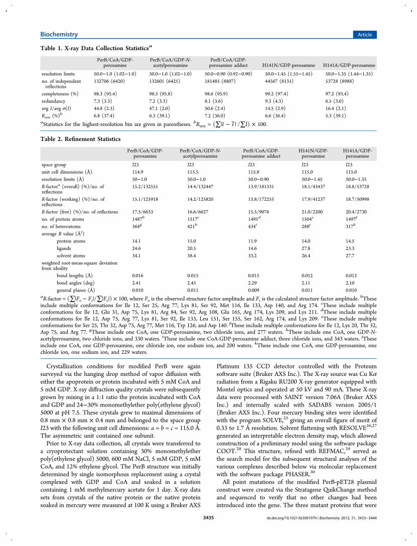
Table 1. X-ray Data Collection Statisticsa
PerB/CoA/GDP-perosamine
PerB/CoA/GDP-N-acetylperosamine
PerB/CoA/GDP-perosamine adduct H141N/GDP-perosamine H141A/GDP-perosamine
resolution limits 50.0−1.0 (1.02−1.0) 50.0−1.0 (1.02−1.0) 50.0−0.90 (0.92−0.90) 50.0−1.45 (1.55−1.45) 50.0−1.35 (1.44−1.35)no. of independentreflections
132706 (6420) 132601 (6425) 181485 (8807) 44567 (8131) 53728 (8988)
completeness (%) 98.3 (95.4) 98.5 (95.8) 98.6 (95.9) 99.2 (97.4) 97.2 (93.4)redundancy 7.3 (3.3) 7.2 (3.3) 8.1 (3.6) 9.3 (4.3) 6.5 (3.0)avg I/avg σ(I) 44.8 (2.3) 47.1 (2.0) 50.6 (2.4) 14.5 (2.9) 16.4 (2.1)Rsym (%)b 6.8 (37.4) 6.3 (39.1) 7.2 (36.0) 6.6 (36.4) 5.3 (39.1)aStatistics for the highest-resolution bin are given in parentheses. bRsym = (∑|I − I | /∑I) × 100.
Table 2. Refinement Statistics
PerB/CoA/GDP-perosamine
PerB/CoA/GDP-N-acetylperosamine
PerB/CoA/GDP-perosamine adduct
H141N/GDP-perosamine
H141A/GDP-perosamine
space group I23 I23 I23 I23 I23unit cell dimensions (Å) 114.9 115.5 115.8 115.0 115.0resolution limits (Å) 50−1.0 50.0−1.0 50.0−0.90 50.0−1.45 50.0−1.35R-factora (overall) (%)/no. ofreflections
15.2/132551 14.4/132447 13.9/181331 18.1/43437 18.8/53728
R-factor (working) (%)/no. ofreflections
15.1/125918 14.2/125820 13.8/172255 17.9/41237 18.7/50998
R-factor (free) (%)/no. of reflections 17.5/6633 16.6/6627 15.5/9076 21.0/2200 20.4/2730no. of protein atoms 1487b 1517c 1495d 1504e 1497f
no. of heteroatoms 364g 421h 434i 288j 317k
average B value (Å2)protein atoms 14.1 15.0 11.9 14.0 14.5ligands 24.6 20.5 14.6 27.8 23.3solvent atoms 34.1 38.4 33.2 26.4 27.7
weighted root-mean-square deviationfrom ideality
bond lengths (Å) 0.016 0.015 0.015 0.012 0.012bond angles (deg) 2.41 2.45 2.29 2.11 2.10general planes (Å) 0.010 0.011 0.009 0.011 0.010
aR-factor = (∑|Fo − Fc|/∑|Fo|) × 100, where Fo is the observed structure factor amplitude and Fc is the calculated structure factor amplitude. bTheseinclude multiple conformations for Ile 12, Ser 25, Arg 77, Lys 81, Ser 92, Met 116, Ile 133, Asp 140, and Arg 174. cThese include multipleconformations for Ile 12, Glu 31, Asp 75, Lys 81, Arg 84, Ser 92, Arg 108, Glu 165, Arg 174, Lys 209, and Lys 211. dThese include multipleconformations for Ile 12, Asp 75, Arg 77, Lys 81, Ser 92, Ile 133, Leu 151, Ser 155, Ser 162, Arg 174, and Lys 209. eThese include multipleconformations for Ser 25, Thr 32, Asp 75, Arg 77, Met 116, Trp 126, and Asp 140. fThese include multiple conformations for Ile 12, Lys 20, Thr 32,Asp 75, and Arg 77. gThese include one CoA, one GDP-perosamine, two chloride ions, and 277 waters. hThese include one CoA, one GDP-N-acetylperosamine, two chloride ions, and 330 waters. iThese include one CoA-GDP-perosamine adduct, three chloride ions, and 343 waters. jTheseinclude one CoA, one GDP-perosamine, one chloride ion, one sodium ion, and 200 waters. kThese include one CoA, one GDP-perosamine, onechloride ion, one sodium ion, and 229 waters.
Biochemistry Article
dx.doi.org/10.1021/bi300197h | Biochemistry 2012, 51, 3433−34443435
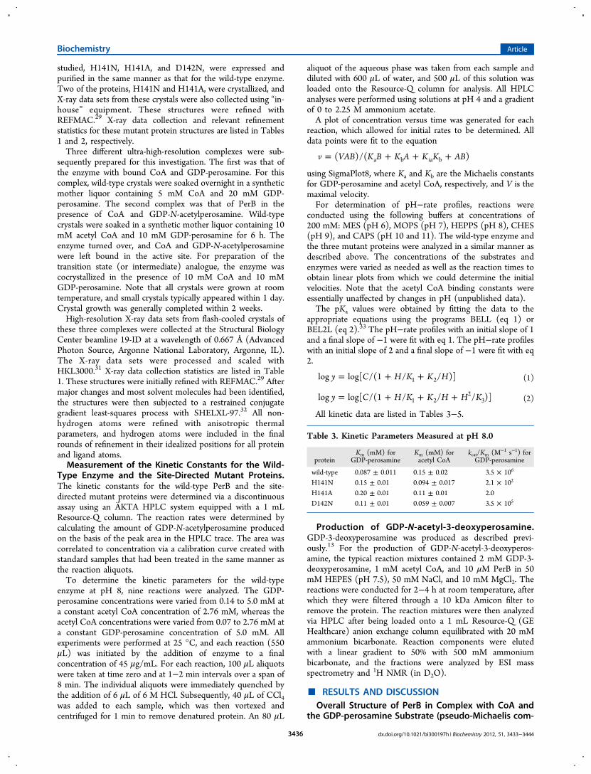
studied, H141N, H141A, and D142N, were expressed andpurified in the same manner as that for the wild-type enzyme.Two of the proteins, H141N and H141A, were crystallized, andX-ray data sets from these crystals were also collected using “in-house” equipment. These structures were refined withREFMAC.29 X-ray data collection and relevant refinementstatistics for these mutant protein structures are listed in Tables1 and 2, respectively.Three different ultra-high-resolution complexes were sub-
sequently prepared for this investigation. The first was that ofthe enzyme with bound CoA and GDP-perosamine. For thiscomplex, wild-type crystals were soaked overnight in a syntheticmother liquor containing 5 mM CoA and 20 mM GDP-perosamine. The second complex was that of PerB in thepresence of CoA and GDP-N-acetylperosamine. Wild-typecrystals were soaked in a synthetic mother liquor containing 10mM acetyl CoA and 10 mM GDP-perosamine for 6 h. Theenzyme turned over, and CoA and GDP-N-acetylperosaminewere left bound in the active site. For preparation of thetransition state (or intermediate) analogue, the enzyme wascocrystallized in the presence of 10 mM CoA and 10 mMGDP-perosamine. Note that all crystals were grown at roomtemperature, and small crystals typically appeared within 1 day.Crystal growth was generally completed within 2 weeks.High-resolution X-ray data sets from flash-cooled crystals of
these three complexes were collected at the Structural BiologyCenter beamline 19-ID at a wavelength of 0.667 Å (AdvancedPhoton Source, Argonne National Laboratory, Argonne, IL).The X-ray data sets were processed and scaled withHKL3000.31 X-ray data collection statistics are listed in Table1. These structures were initially refined with REFMAC.29 Aftermajor changes and most solvent molecules had been identified,the structures were then subjected to a restrained conjugategradient least-squares process with SHELXL-97.32 All non-hydrogen atoms were refined with anisotropic thermalparameters, and hydrogen atoms were included in the finalrounds of refinement in their idealized positions for all proteinand ligand atoms.Measurement of the Kinetic Constants for the Wild-
Type Enzyme and the Site-Directed Mutant Proteins.The kinetic constants for the wild-type PerB and the site-directed mutant proteins were determined via a discontinuousassay using an AKTA HPLC system equipped with a 1 mLResource-Q column. The reaction rates were determined bycalculating the amount of GDP-N-acetylperosamine producedon the basis of the peak area in the HPLC trace. The area wascorrelated to concentration via a calibration curve created withstandard samples that had been treated in the same manner asthe reaction aliquots.To determine the kinetic parameters for the wild-type
enzyme at pH 8, nine reactions were analyzed. The GDP-perosamine concentrations were varied from 0.14 to 5.0 mM ata constant acetyl CoA concentration of 2.76 mM, whereas theacetyl CoA concentrations were varied from 0.07 to 2.76 mM ata constant GDP-perosamine concentration of 5.0 mM. Allexperiments were performed at 25 °C, and each reaction (550μL) was initiated by the addition of enzyme to a finalconcentration of 45 μg/mL. For each reaction, 100 μL aliquotswere taken at time zero and at 1−2 min intervals over a span of8 min. The individual aliquots were immediately quenched bythe addition of 6 μL of 6 M HCl. Subsequently, 40 μL of CCl4was added to each sample, which was then vortexed andcentrifuged for 1 min to remove denatured protein. An 80 μL
aliquot of the aqueous phase was taken from each sample anddiluted with 600 μL of water, and 500 μL of this solution wasloaded onto the Resource-Q column for analysis. All HPLCanalyses were performed using solutions at pH 4 and a gradientof 0 to 2.25 M ammonium acetate.A plot of concentration versus time was generated for each
reaction, which allowed for initial rates to be determined. Alldata points were fit to the equation
= + + +v VAB K B K A K K AB( )/( )a b ia b
using SigmaPlot8, where Ka and Kb are the Michaelis constantsfor GDP-perosamine and acetyl CoA, respectively, and V is themaximal velocity.For determination of pH−rate profiles, reactions were
conducted using the following buffers at concentrations of200 mM: MES (pH 6), MOPS (pH 7), HEPPS (pH 8), CHES(pH 9), and CAPS (pH 10 and 11). The wild-type enzyme andthe three mutant proteins were analyzed in a similar manner asdescribed above. The concentrations of the substrates andenzymes were varied as needed as well as the reaction times toobtain linear plots from which we could determine the initialvelocities. Note that the acetyl CoA binding constants wereessentially unaffected by changes in pH (unpublished data).The pKa values were obtained by fitting the data to the
appropriate equations using the programs BELL (eq 1) orBEL2L (eq 2).33 The pH−rate profiles with an initial slope of 1and a final slope of −1 were fit with eq 1. The pH−rate profileswith an initial slope of 2 and a final slope of −1 were fit with eq2.
= + +y C H K K Hlog log[ /(1 / / )]1 2 (1)
= + + +y C H K K H H Klog log[ /(1 / / / )]1 22
3 (2)
All kinetic data are listed in Tables 3−5.
Production of GDP-N-acetyl-3-deoxyperosamine.GDP-3-deoxyperosamine was produced as described previ-ously.13 For the production of GDP-N-acetyl-3-deoxyperos-amine, the typical reaction mixtures contained 2 mM GDP-3-deoxyperosamine, 1 mM acetyl CoA, and 10 μM PerB in 50mM HEPES (pH 7.5), 50 mM NaCl, and 10 mM MgCl2. Thereactions were conducted for 2−4 h at room temperature, afterwhich they were filtered through a 10 kDa Amicon filter toremove the protein. The reaction mixtures were then analyzedvia HPLC after being loaded onto a 1 mL Resource-Q (GEHealthcare) anion exchange column equilibrated with 20 mMammonium bicarbonate. Reaction components were elutedwith a linear gradient to 50% with 500 mM ammoniumbicarbonate, and the fractions were analyzed by ESI massspectrometry and 1H NMR (in D2O).
■ RESULTS AND DISCUSSIONOverall Structure of PerB in Complex with CoA and
the GDP-perosamine Substrate (pseudo-Michaelis com-
Table 3. Kinetic Parameters Measured at pH 8.0
proteinKm (mM) for
GDP-perosamineKm (mM) foracetyl CoA
kcat/Km (M−1 s−1) forGDP-perosamine
wild-type 0.087 ± 0.011 0.15 ± 0.02 3.5 × 10⁶
H141N 0.15 ± 0.01 0.094 ± 0.017 2.1 × 10²
H141A 0.20 ± 0.01 0.11 ± 0.01 2.0D142N 0.11 ± 0.01 0.059 ± 0.007 3.5 × 10⁵
Biochemistry Article
dx.doi.org/10.1021/bi300197h | Biochemistry 2012, 51, 3433−34443436

plex). The crystals used for this analysis diffracted to 1.0 Åresolution and belonged to the space group I23. PerB functionsas a trimer, and in this crystal form, the biological unit packed
along a crystallographic 3-fold rotational axis resulting in onesubunit per asymmetric unit. Each subunit of the trimercontains 215 amino acid residues.6 With the exception of thefirst two N-terminal residues, the loop between Pro 41 and Arg43, and the final three C-terminal residues, the electron densitycorresponding to the polypeptide chain backbone was wellordered. The Ramachandran plot statistics for the model, ascalculated with PROCHECK,34 were excellent with 90.4 and9.6% of the ϕ and ψ angles lying within the core and allowedregions, respectively.
Table 4. Kinetic Parameters Measured from pH 6.0 to 11.0
protein pH 6.0 pH 7.0 pH 8.0 pH 9.0 pH 10.0 pH 11.0
wild-typea,b 0.27 ± 0.03 0.32 ± 0.05 0.087 ± 0.011 0.21 ± 0.03 0.84 ± 0.06 9.7 ± 0.41.9 × 104 4.3 × 10⁵ 3.5 × 10⁶ 2.3 × 10⁶ 6.9 × 10⁵ 9.6 × 10⁴
H141Na,b 7.2 ± 0.3 0.27 ± 0.04 0.15 ± 0.01 0.097 ± 0.006 0.32 ± 0.03 6.5 ± 1.72.0 4.1 × 10 2.1 × 10² 4.3 × 10² 2.5 × 10² 2.5 × 10
H141Aa,b ndc ndc 0.20 ± 0.01 ndc ndc ndc
2.0D142Na,b 0.39 ± 0.05 0.30 ± 0.03 0.11 ± 0.01 0.074 ± 0.004 0.27 ± 0.04 3.5 ± 0.3
8.4 × 10³ 8.0 × 10⁴ 3.5 × 10⁵ 3.9 × 10⁵ 1.6 × 10⁵ 4.2 × 10⁴
aKm (mM) for GDP-perosamine, top line. bkcat/Km (M−1 s−1), bottom line. cNot determined.
Table 5. pKa Values Determined from pH−Rate Profiles
protein pKa1 pKa2 pKa3
wild-typea 7.8 ± 0.3 9.3 ± 0.2 6.5 ± 0.5H141Na 8.1 ± 0.2 9.7 ± 0.2 6.0 ± 0.5D142Nb 7.7 ± 0.1 9.9 ± 0.1
aFit with BEL2L. bFit with BELL.
Figure 1. Ribbon representation of PerB in a complex with CoA and GDP-perosamine. The three subunits of the PerB trimer are highlighted in blue,green, and pink, respectively, in (a). The CoA and GDP-perosamine ligands are depicted in stick representations. A close-up view of one subunit ofthe trimer is displayed in (b). All figures were prepared with the software package PyMOL.38
Biochemistry Article
dx.doi.org/10.1021/bi300197h | Biochemistry 2012, 51, 3433−34443437
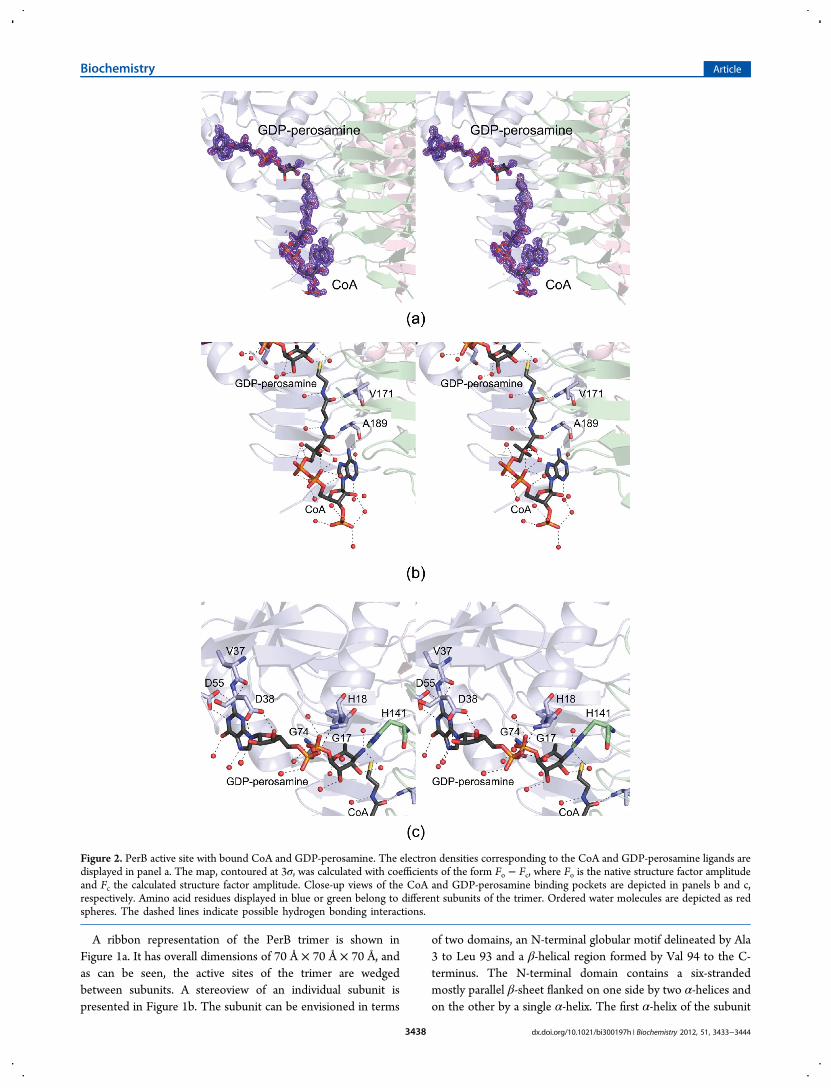
A ribbon representation of the PerB trimer is shown inFigure 1a. It has overall dimensions of 70 Å × 70 Å × 70 Å, andas can be seen, the active sites of the trimer are wedgedbetween subunits. A stereoview of an individual subunit ispresented in Figure 1b. The subunit can be envisioned in terms
of two domains, an N-terminal globular motif delineated by Ala3 to Leu 93 and a β-helical region formed by Val 94 to the C-terminus. The N-terminal domain contains a six-strandedmostly parallel β-sheet flanked on one side by two α-helices andon the other by a single α-helix. The first α-helix of the subunit
Figure 2. PerB active site with bound CoA and GDP-perosamine. The electron densities corresponding to the CoA and GDP-perosamine ligands aredisplayed in panel a. The map, contoured at 3σ, was calculated with coefficients of the form Fo − Fc, where Fo is the native structure factor amplitudeand Fc the calculated structure factor amplitude. Close-up views of the CoA and GDP-perosamine binding pockets are depicted in panels b and c,respectively. Amino acid residues displayed in blue or green belong to different subunits of the trimer. Ordered water molecules are depicted as redspheres. The dashed lines indicate possible hydrogen bonding interactions.
Biochemistry Article
dx.doi.org/10.1021/bi300197h | Biochemistry 2012, 51, 3433−34443438
is situated such that the positive end of its helix dipole momentpoints toward the pyrophosphoryl moiety of GDP-perosamine.The sixth β-strand of the N-terminal domain serves as a bridgeto the β-helix domain, which contains nearly seven turns anddisplays the characteristic hexapeptide repeat that is a hallmarkfor this family of N-acetyltransferases.35 Whereas the N-terminal domain provides most of the interactions betweenthe protein and the GDP-perosamine ligand, the C-terminaldomain serves to anchor the CoA moiety into the active site.Pro 207 adopts the cis conformation and is situated near theadenine ring of CoA.Electron density corresponding to the bound ligands is
presented in Figure 2a. The density for the CoA is very strong,as is that for the GDP portion of the substrate. The electrondensity for the hexose portion of GDP-perosamine is somewhatweaker, however. This most likely is a result of the short crystalsoaking times employed for this structural analysis. As discussedbelow, if the PerB crystals were soaked in CoA and GDP-perosamine for extended periods of time, a covalent adductformed between the two ligands.A close-up view of the CoA binding pocket is provided in
Figure 2b. The cofactor is anchored in place simply by watermolecules, the backbone amide groups of Val 171 and Ala 189,and the carbonyl group of Ala 189. The sulfur of CoA is 3.6 Åfrom the sugar C-4‴ amino group. The protein regionsurrounding the GDP-perosamine ligand is displayed in Figure2c. The guanine base is held in place by two water molecules,the carboxylate side chain of Asp 55, and the carbonyl group ofVal 37. The carboxylate group of Asp 38 bridges the ribose C-2′and C-3′ hydroxyls. In addition to numerous water molecules,the backbone amide groups of Gly 17, His 18, and Gly 74participate in hydrogen bonding interactions with thephosphoryl oxygens. With the exception of His 141, the hexosemoiety of GDP-perosamine does not interact with proteinbackbone atoms or side chain groups but rather simply withwater molecules. His 141, which is provided by a neighboringsubunit, is positioned at 2.7 Å from the hexose C-4‴ aminogroup.Given the lack of protein side chain interactions between
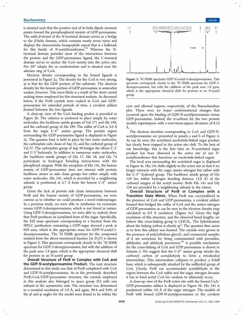
PerB and the hexose moiety of GDP-perosamine, we werecurious as to whether we could produce a novel trideoxysugar.In a previous study, we were able to synthesize via enzymaticmeans GDP-3-deoxyperosamine, which is not found in nature.Using GDP-3-deoxyperosamine, we were able to, indeed, showthat PerB produces an acetylated form of the sugar. Specifically,the ESI mass spectrum corresponding to a fraction from theHPLC purification containing a GDP-sugar showed a peak at629 amu, which is the appropriate mass for GDP-N-acetyl-3-deoxyperosamine. The 1H NMR spectrum for the compoundisolated from the above-mentioned fraction (in D2O) is shownin Figure 3. This spectrum corresponds closely to the 1H NMRspectrum for GDP-3-deoxyperosamine, but with the addition ofthe peak near 1.8 ppm, which is the appropriate chemical shiftfor protons in an N-acetyl group.Overall Structure of PerB in Complex with CoA and
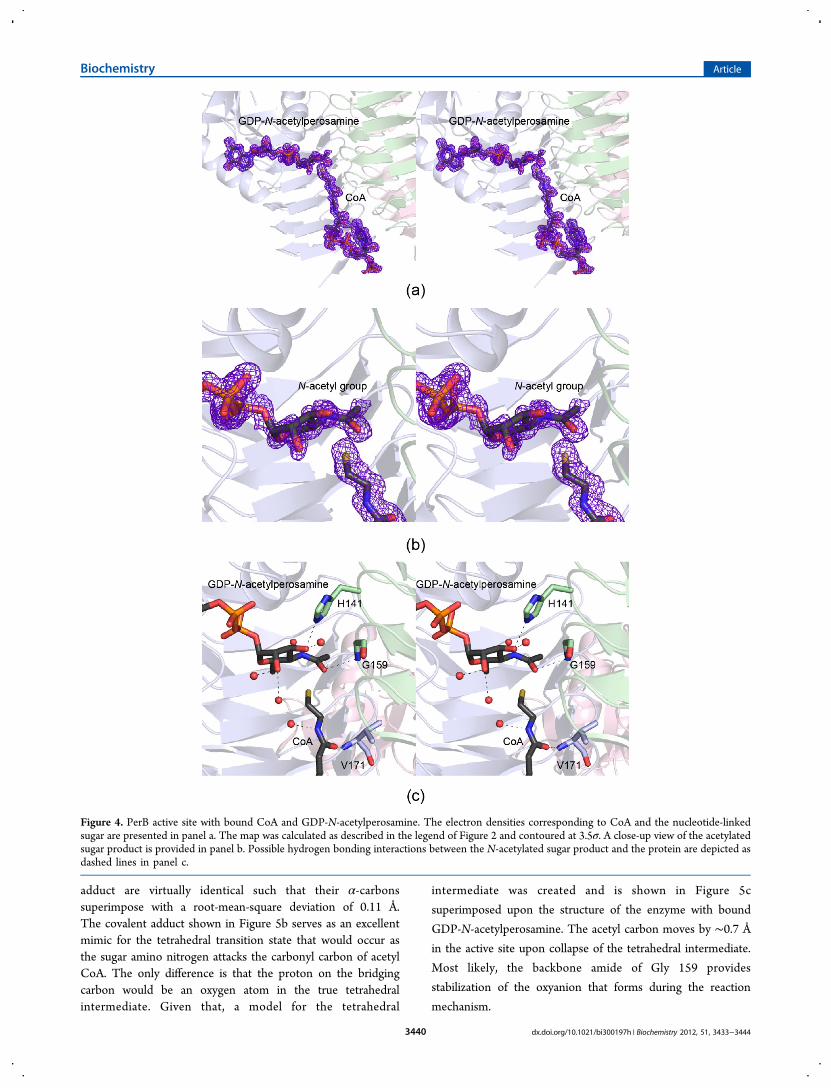
the GDP-N-acetylperosamine Product. The next structuredetermined in this study was that of PerB complexed with CoAand GDP-N-acetylperosamine. As in the previously describedPerB/CoA/GDP-perosamine structure, the crystals employedin this analysis also belonged to space group I23 with onesubunit in the asymmetric unit. The structure was determinedto a nominal resolution of 1.0 Å, and again, 90.4 and 9.6% ofthe ϕ and ψ angles for the model were found to lie within the
core and allowed regions, respectively, of the Ramachandranplot. There were no major conformational changes thatoccurred upon the binding of GDP-N-acetylperosamine versusGDP-perosamine. Indeed, the α-carbons for the two proteinmodels superimpose with a root-mean-square deviation of 0.13Å.The electron densities corresponding to CoA and GDP-N-
acetylperosamine are presented in panels a and b of Figure 4.As can be seen, the acetylated nucleotide-linked sugar producthas clearly been trapped in the active site cleft. To the best ofour knowledge, this is the first time an N-acetylated sugarproduct has been observed in the active site of any N-acetyltransferase that functions on nucleotide-linked sugars.The local area surrounding the acetylated sugar is displayed
in Figure 4c. His 141 shifts slightly in the active site so that it nolonger interacts with the sugar amino nitrogen but rather withthe C-3‴ hydroxyl group. The backbone amide group of Gly159 lies within hydrogen bonding distance (3.0 Å) of thecarbonyl oxygen of the acetyl moiety. Both His 141 and Gly159 are provided by a neighboring subunit in the trimer.
Overall Structure of PerB in Complex with aTransition State Mimic. When PerB was cocrystallized inthe presence of CoA and GDP-perosamine, a covalent adductformed that bridged the sulfur of CoA and the amino nitrogenof GDP-perosamine as can be seen in the electron density mapcalculated to 0.9 Å resolution (Figure 5a). Given the highresolution of this structure, and the observed bond lengths, webelieve this cross-linking group is CHCH3. The hybridizationabout the linking carbon is clearly sp3. The question thus arisesas to how this adduct was formed. The crystals were grown inthe presence of poly(ethylene glycol), and commercial samplesof it are notorious for being contaminated with peroxides,aldehydes, and aldehyde precursors.36 A possible mechanismfor the cross-linking of CoA and GDP-perosamine is shown inScheme 2. We suggest that the C-4‴ amino group attacks thecarbonyl carbon of acetaldehyde to form a tetrahedralintermediate. This intermediate collapses to produce a Schiffbase, which is subsequently attacked by the sulfhydryl group ofCoA. Clearly, PerB can accommodate acetaldehyde in theregion between the CoA sulfur and the sugar nitrogen becauseit has to bind acetyl CoA for catalysis to ultimately occur.A close-up view of the PerB active site with the bound CoA/
GDP-perosamine adduct is displayed in Figure 5b. His 141 ispositioned within 3.0 Å of the sugar nitrogen. The models ofPerB with bound GDP-N-acetylperosamine or the covalent
Figure 3. 1H NMR spectrum GDP-N-acetyl-3-deoxyperosamine. Thisspectrum corresponds closely to the 1H NMR spectrum for GDP-3-deoxyperosamine, but with the addition of the peak near 1.8 ppm,which is the appropriate chemical shift for protons in an N-acetylgroup.
Biochemistry Article
dx.doi.org/10.1021/bi300197h | Biochemistry 2012, 51, 3433−34443439
adduct are virtually identical such that their α-carbonssuperimpose with a root-mean-square deviation of 0.11 Å.The covalent adduct shown in Figure 5b serves as an excellentmimic for the tetrahedral transition state that would occur asthe sugar amino nitrogen attacks the carbonyl carbon of acetylCoA. The only difference is that the proton on the bridgingcarbon would be an oxygen atom in the true tetrahedralintermediate. Given that, a model for the tetrahedral
intermediate was created and is shown in Figure 5c
superimposed upon the structure of the enzyme with bound
GDP-N-acetylperosamine. The acetyl carbon moves by ∼0.7 Å
in the active site upon collapse of the tetrahedral intermediate.
Most likely, the backbone amide of Gly 159 provides
stabilization of the oxyanion that forms during the reaction
mechanism.
Figure 4. PerB active site with bound CoA and GDP-N-acetylperosamine. The electron densities corresponding to CoA and the nucleotide-linkedsugar are presented in panel a. The map was calculated as described in the legend of Figure 2 and contoured at 3.5σ. A close-up view of the acetylatedsugar product is provided in panel b. Possible hydrogen bonding interactions between the N-acetylated sugar product and the protein are depicted asdashed lines in panel c.
Biochemistry Article
dx.doi.org/10.1021/bi300197h | Biochemistry 2012, 51, 3433−34443440

Probing the Catalytic Mechanism of PerB. The closeststructural relative to PerB is an N-acetyltransferase referred toas PlgD from Campylobacter jejuni.19,20 It catalyzes the last stepin the biosynthesis of 2,4-diacetamido-2,4,6-trideoxy-α-D-glucose, an unusual sugar found in the glycan moieties ofsome eubacterial pathogens, and it employs a UDP-linked sugar
substrate. PerB and PglD show an amino acid sequence identityof 38%, and not surprisingly, their models superimpose with aroot-mean-square deviation of 0.73 Å for 157 structurallyequivalent α-carbon positions. A superposition of the PerBactive site onto that of PlgD is presented in Figure 6. There aretwo substitutions in PglD, relative to PerB, which preclude its
Figure 5. PerB active site with a CoA/GDP-perosamine covalent adduct. The electron density corresponding to the covalent adduct is displayed inpanel a. The map was calculated as described in the legend of Figure 2 and contoured at 3.5σ. Possible hydrogen bonding interactions between theligand and the protein are indicated by the dashed lines in panel b. Ordered water molecules are displayed as spheres. A superposition of GDP-N-acetylperosamine (gray filled bonds) onto the covalent adduct (pink filled bonds) is depicted in panel c.
Biochemistry Article
dx.doi.org/10.1021/bi300197h | Biochemistry 2012, 51, 3433−34443441
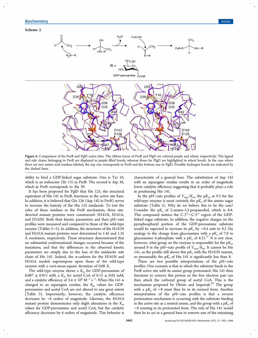
ability to bind a GDP-linked sugar substrate. One is Tyr 10,which is an isoleucine (Ile 13) in PerB. The second is Asp 36,which in PerB corresponds to Ala 39.It has been proposed for PglD that His 125, the structural
equivalent of His 141 in PerB, functions as the active site base.In addition, it is believed that Glu 126 (Asp 142 in PerB) servesto increase the basicity of the His 125 imidazole. To test theroles of these residues in the PerB mechanism, three site-directed mutant proteins were constructed: H141N, H141A,and D142N. Both their kinetic parameters and their pH−rateprofiles were measured and compared to those of the wild-typeenzyme (Tables 3−5). In addition, the structures of the H141Nand H141A mutant proteins were determined to 1.45 and 1.35Å resolution, respectively. These structures demonstrated thatno substantial conformational changes occurred because of themutations, and that the differences in the observed kineticparameters are simply due to the loss of the imidazole sidechain of His 141. Indeed, the α-carbons for the H141N andH141A models superimpose upon those of the wild-typeenzyme with a root-mean-square deviation of 0.08 Å.The wild-type enzyme shows a Km for GDP-perosamine of
0.087 ± 0.011 mM, a Km for acetyl CoA of 0.15 ± 0.02 mM,and a catalytic efficiency of 3.4 × 10⁶ M−1 s−1. When His 141 ischanged to an asparagine residue, the Km values for GDP-perosamine and acetyl CoA are not altered to any great extent(Table 3). Importantly, however, the catalytic efficiencydecreases by >4 orders of magnitude. Likewise, the H141Amutant protein demonstrates only slight alterations in the Km
values for GDP-perosamine and acetyl CoA, but the catalyticefficiency decreases by 6 orders of magnitude. This behavior is
characteristic of a general base. The substitution of Asp 142with an asparagine residue results in an order of magnitudelower catalytic efficiency suggesting that it probably plays a rolein positioning His 141.In the pH−rate profiles of Vmax/Km, the pKa2 at 9.3 for the
wild-type enzyme is most certainly the pKa of the amino sugarsubstrate (Table 5). Why do we believe this to be the case?Consider the pKa of 2-amino-1,3-propanediol, which is 8.8.This compound mimics the C-3‴−C-5‴ region of the GDP-linked sugar substrate. In addition, the negative charges on thepyrophosphoryl portion of the GDP-perosamine substratewould be expected to increase its pKa by ∼0.4 unit to 9.2 (byanalogy to the change from glucosamine with a pKa of 7.8 toglucosamine 6-phosphate with a pKa of 8.2).
37 It is not clear,however, what group on the enzyme is responsible for the pKa1
around 8 in the pH−rate profile of Vmax/Km. It cannot be His141, as the profile still shows this pKa with the H141N mutant,so presumably the pKa of His 141 is significantly less than 8.There are two possible interpretations of the pH−rate
profiles. One scenario is that in which the substrate binds in thePerB active site with its amino group protonated. His 141 thenfunctions to remove this proton so the free electron pair canthen attack the carbonyl group of acetyl CoA. This is themechanism proposed by Olivier and Imperiali.20 The groupwith a pKa of ∼8 must thus be in its ionized form. Anotherinterpretation of the pH−rate profiles is that a reverseprotonation mechanism is occurring with the substrate bindingin the active site as a neutral amine, and the group with a pKa of∼8 reacting in its protonated form. The role of His 141 wouldthen be to act as a general base to remove one of the remaining
Scheme 2
Figure 6. Comparison of the PerB and PglD active sites. The ribbon traces of PerB and PlgD are colored purple and wheat, respectively. The ligandand side chains belonging to PerB are displayed in purple filled bonds, whereas those for PlgD are highlighted in wheat bonds. In the case wherethere are two amino acid residues labeled, the top one corresponds to PerB and the bottom one to PglD. Possible hydrogen bonds are indicated bythe dashed lines.
Biochemistry Article
dx.doi.org/10.1021/bi300197h | Biochemistry 2012, 51, 3433−34443442

protons from the amino group of the substrate as it attacksacetyl CoA.Both of these mechanisms are consistent with the pH−rate
profiles, but the second allows His 141 to act in a truly catalyticmanner. In the first mechanism, His 141 deprotonates thesubstrate only to its neutral form, but there is then no catalysisof the transacylation reaction itself. In the second mechanism,His 141 acts as a general base to catalyze the formation of thetetrahedral transition state or intermediate, and thus, it isexpected that its mutation would have a major effect asobserved. We thus conclude that the pH−rate profiles arereverse protonation profiles, and that the substrate enters theactive site with a neutral amino group. The mechanism forPglD proposed by Rangarajan19 also assumes that the substratebinds to the enzyme in its neutral state. A possible reactionmechanism for PerB is shown in Scheme 3. The key players inthe mechanism include the backbone amide of Gly 159, whichprovides an “oxyanion” hole, and the imidazole of His 141,which serves as the general base.The catalytic mechanisms of PglD and PerB clearly involve a
general base provided by a histidine residue. This is in markedcontrast to the reaction mechanisms of WlbB, QdtC, and AntD,which are N-acetyl- or N-acyltransferases that have also beenstudied in our laboratory.21−23 Both WlbB and QdtC areinvolved in the biosynthesis of unusual acetylated sugars foundin the O-antigens of certain Gram-negative bacteria, and bothfunction on C-3‴ hexose amino groups. AntD is involved in theproduction of D-anthrose, an important carbohydrate found inthe endospores of Bacillus anthracis, the causative agent ofanthrax. Specifically, AntD catalyzes the transfer of an acylgroup from 3-hydroxy-3-methylbutyryl CoA to the C-4‴ aminogroup of dTDP-4-amino-4,6-dideoxy-α-D-glucose. In all threeenzymes, there is a decided lack of a catalytic base in their activesites. Likewise, in all three enzymes, the nucleotide-linkedsugars are bound similarly, but in an orientation completelydifferent from that observed for PerB and PglD. Clearly, the N-acetyltransferases (or N-acyltransferases) that function onnucleotide-linked sugars have evolved into two separate proteinclasses that differ with respect to both substrate bindingorientations and reaction mechanisms.
■ ASSOCIATED CONTENT
Accession Codes
X-ray coordinates have been deposited in the ResearchCollaboratory for Structural Bioinformatics, Rutgers University,New Brunswick, NJ (entries 4EA7, 4EA8, 4EA9, 4EAA, and4EAB).
■ AUTHOR INFORMATION
Corresponding Author*E-mail: [email protected] or [email protected]. Fax: (608) 262-1319. Phone: (608) 262-4988.
FundingThis research was supported in part by National Institutes ofHealth Grants DK47814 (to H.M.H.) and GM18938 (toW.W.C.).
NotesThe authors declare no competing financial interest.
■ ACKNOWLEDGMENTSA portion of the research described in this paper was performedat Argonne National Laboratory, Structural Biology Center atthe Advanced Source (U.S. Department of Energy, Office ofBiological and Environmental Research, under Contract DE-AC02-06CH11357). We gratefully acknowledge Dr. Norma E.C. Duke and Dr. Stephan L. Ginell for assistance during the X-ray data collection at Argonne.
■ ABBREVIATIONSCAPS, 3-(cyclohexylamino)propanesulfonic acid; CHES, 2-(cyclohexylamino)ethanesulfonic acid; CoA, coenzyme A; ESI,electrospray ionization; GDP, guanosine diphosphate; HEPPS,N-(2-hydroxyethyl)piperazine-N′-3-propanesulfonic acid;HPLC, high-performance liquid chromatography; MES, 2-(N-morpholino)ethanesulfonic acid; MOPS, 3-(N-morpholino)-propanesulfonic acid; NMR, nuclear magnetic resonance; PCR,polymerase chain reaction; TEV, tobacco etch virus; Tris,tris(hydroxymethyl)aminomethane.
■ REFERENCES(1) Wells, J. G., Davis, B. R., Wachsmuth, I. K., Riley, L. W., Remis, R.S., Sokolow, R., and Morris, G. K. (1983) Laboratory investigation ofhemorrhagic colitis outbreaks associated with a rare Escherichia coliserotype. J. Clin. Microbiol. 18, 512−520.(2) Mead, P. S., and Griffin, P. M. (1998) Escherichia coli O157:H7.Lancet 352, 1207−1212.(3) Perry, M. B., MacLean, L., and Griffith, D. W. (1986) Structure ofthe O-chain polysaccharide of the phenol-phase soluble lipopolysac-charide of Escherichia coli 0:157:H7. Biochem. Cell Biol. 64, 21−28.(4) Haishima, Y., Kondo, S., and Hisatsune, K. (1990) Theoccurrence of α(1----2) linked N-acetylperosamine-homopolymer inlipopolysaccharides of non-O1 Vibrio cholerae possessing an antigenicfactor in common with O1 V. cholerae. Microbiol. Immunol. 34, 1049−1054.(5) Ovchinnikova, O. G., Kocharova, N. A., Katzenellenbogen, E.,Zatonsky, G. V., Shashkov, A. S., Knirel, Y. A., Lipinski, T., andGamian, A. (2004) Structures of two O-polysaccharides of thelipopolysaccharide of Citrobacter youngae PCM 1538 (serogroup O9).Carbohydr. Res. 339, 881−884.
Scheme 3
Biochemistry Article
dx.doi.org/10.1021/bi300197h | Biochemistry 2012, 51, 3433−34443443
(6) Awram, P., and Smit, J. (2001) Identification of lipopolysacchar-ide O antigen synthesis genes required for attachment of the S-layer ofCaulobacter crescentus. Microbiology 147, 1451−1460.(7) Albermann, C., and Beuttler, H. (2008) Identification of theGDP-N-acetyl-D-perosamine producing enzymes from Escherichia coliO157:H7. FEBS Lett. 582, 479−484.(8) Somoza, J. R., Menon, S., Schmidt, H., Joseph-McCarthy, D.,Dessen, A., Stahl, M. L., Somers, W. S., and Sullivan, F. X. (2000)Structural and kinetic analysis of Escherichia coli GDP-mannose 4,6dehydratase provides insights into the enzyme’s catalytic mechanismand regulation by GDP-fucose. Struct. Folding Des. 8, 123−135.(9) Mulichak, A. M., Bonin, C. P., Reiter, W. D., and Garavito, R. M.(2002) Structure of the MUR1 GDP-mannose 4,6-dehydratase fromArabidopsis thaliana: Implications for ligand binding and specificity.Biochemistry 41, 15578−15589.(10) Webb, N. A., Mulichak, A. M., Lam, J. S., Rocchetta, H. L., andGaravito, R. M. (2004) Crystal structure of a tetrameric GDP-D-mannose 4,6-dehydratase from a bacterial GDP-D-rhamnose bio-synthetic pathway. Protein Sci. 13, 529−539.(11) Albermann, C., and Piepersberg, W. (2001) Expression andidentification of the RfbE protein from Vibrio cholerae O1 and its usefor the enzymatic synthesis of GDP-D-perosamine. Glycobiology 11,655−661.(12) Zhao, G., Liu, J., Liu, X., Chen, M., Zhang, H., and Wang, P. G.(2007) Cloning and characterization of GDP-perosamine synthetase(Per) from Escherichia coli O157:H7 and synthesis of GDP-perosamine in vitro. Biochem. Biophys. Res. Commun. 363, 525−530.(13) Cook, P. D., and Holden, H. M. (2008) GDP-perosaminesynthase: Structural analysis and production of a novel trideoxysugar.Biochemistry 47, 2833−2840.(14) Raetz, C. R., and Roderick, S. L. (1995) A left-handed parallel βhelix in the structure of UDP-N-acetylglucosamine acyltransferase.Science 270, 997−1000.(15) Brown, K., Pompeo, F., Dixon, S., Mengin-Lecreulx, D.,Cambillau, C., and Bourne, Y. (1999) Crystal structure of thebifunctional N-acetylglucosamine 1-phosphate uridyltransferase fromEscherichia coli: A paradigm for the related pyrophosphorylasesuperfamily. EMBO J. 18, 4096−4107.(16) Olsen, L. R., and Roderick, S. L. (2001) Structure of theEscherichia coli GlmU pyrophosphorylase and acetyltransferase activesites. Biochemistry 40, 1913−1921.(17) Kostrewa, D., D’Arcy, A., Takacs, B., and Kamber, M. (2001)Crystal structures of Streptococcus pneumoniae N-acetylglucosamine-1-phosphate uridyltransferase, GlmU, in apo form at 2.33 Å resolutionand in complex with UDP-N-acetylglucosamine and Mg2+ at 1.96 Åresolution. J. Mol. Biol. 305, 279−289.(18) Sulzenbacher, G., Gal, L., Peneff, C., Fassy, F., and Bourne, Y.(2001) Crystal structure of Streptococcus pneumoniae N-acetylglucos-amine-1-phosphate uridyltransferase bound to acetyl-coenzyme Areveals a novel active site architecture. J. Biol. Chem. 276, 11844−11851.(19) Rangarajan, E. S., Ruane, K. M., Sulea, T., Watson, D. C.,Proteau, A., Leclerc, S., Cygler, M., Matte, A., and Young, N. M.(2008) Structure and active site residues of PglD, an N-acetyltransferase from the bacillosamine synthetic pathway requiredfor N-glycan synthesis in Campylobacter jejuni. Biochemistry 47, 1827−1836.(20) Olivier, N. B., and Imperiali, B. (2008) Crystal structure andcatalytic mechanism of PglD from Campylobacter jejuni. J. Biol. Chem.283, 27937−27946.(21) Thoden, J. B., Cook, P. D., Schaffer, C., Messner, P., andHolden, H. M. (2009) Structural and functional studies of QdtC: AnN-acetyltransferase required for the biosynthesis of dTDP-3-acetamido-3,6-dideoxy-α-D-glucose. Biochemistry 48, 2699−2709.(22) Thoden, J. B., and Holden, H. M. (2010) Molecular structure ofWlbB, a bacterial N-acetyltransferase involved in the biosynthesis of2,3-diacetamido-2,3-dideoxy-D-mannuronic acid. Biochemistry 49,4644−4653.
(23) Kubiak, R. L., and Holden, H. M. (2012) Structural Studies onAntD: An N-Acyltransferase Involved in the Biosynthesis of D-Anthrose. Biochemistry 51, 867−878.(24) Holden, H. M., Cook, P. D., and Thoden, J. B. (2010)Biosynthetic enzymes of unusual microbial sugars. Curr. Opin. Struct.Biol. 20, 543−550.(25) Terwilliger, T. C., and Berendzen, J. (1999) Automated MADand MIR structure solution. Acta Crystallogr. D55 (Part 4), 849−861.(26) Terwilliger, T. C. (2000) Maximum-likelihood densitymodification. Acta Crystallogr. D56 (Part 8), 965−972.(27) Terwilliger, T. C. (2003) Automated main-chain model buildingby template matching and iterative fragment extension. ActaCrystallogr. D59, 38−44.(28) Emsley, P., and Cowtan, K. (2004) Coot: Model-building toolsfor molecular graphics. Acta Crystallogr. D60, 2126−2132.(29) Murshudov, G. N., Vagin, A. A., and Dodson, E. J. (1997)Refinement of macromolecular structures by the maximum-likelihoodmethod. Acta Crystallogr. D53, 240−255.(30) McCoy, A. J., Grosse-Kunstleve, R. W., Adams, P. D., Winn, M.D., Storoni, L. C., and Read, R. J. (2007) Phaser crystallographicsoftware. J. Appl. Crystallogr. 40, 658−674.(31) Minor, W., Cymborowski, M., Otwinowski, Z., and Chruszcz, M.(2006) HKL-3000: The integration of data reduction and structuresolution-from diffraction images to an initial model in minutes. ActaCrystallogr. D62, 859−866.(32) Sheldrick, G. M., and Schneider, T. R. (1997) SHELXL: High-resolution refinement. Methods Enzymol. 277, 319−343.(33) Cleland, W. W. (1979) Statistical analysis of enzyme kineticdata. Methods Enzymol. 63, 103−138.(34) Laskowski, R. A., MacArthur, M. W., Moss, D. S., and Thornton,J. M. (1993) PROCHECK: A program to check the stereochemicalquality of protein structures. J. Appl. Crystallogr. 26, 283−291.(35) Vuorio, R., Harkonen, T., Tolvanen, M., and Vaara, M. (1994)The novel hexapeptide motif found in the acyltransferases LpxA andLpxD of lipid A biosynthesis is conserved in various bacteria. FEBSLett. 337, 289−292.(36) Ray, W. J., Jr., and Puvathingal, J. M. (1985) A simple procedurefor removing contaminating aldehydes and peroxides from aqueoussolutions of polyethylene glycols and of nonionic detergents that arebased on the polyoxyethylene linkage. Anal. Biochem. 146, 307−312.(37) McCarthy, T. J., Plog, M. A., Floy, S. A., Jansen, J. A., Soukup, J.K., and Soukup, G. A. (2005) Ligand requirements for glmS ribozymeself-cleavage. Chem. Biol. 12, 1221−1226.(38) DeLano, W. L. (2002) The PyMOL Molecular Graphics System,DeLano Scientific, San Carlos, CA.
Biochemistry Article
dx.doi.org/10.1021/bi300197h | Biochemistry 2012, 51, 3433−34443444