Case Studies in Microscopy Resource Type: Publication Date ...
30
Case Studies in Microscopy Resource Type: Curriculum: Classroom Publication Date: 7/13/2005 Authors Susan Merkel Cornell University Ithaca, New York 14853 USA Email: [email protected] William Ghiorse Cornell University Ithaca, New York 14853 USA Email: [email protected] Marilyn Dispensa Academic Technologies Cornell University Ithaca, New York Abstract This website (http://instruct1.cit.cornell.edu/courses/biomi290/microscopycases/) has three auto-tutorial case studies that ask students to analyze and interpret microscopic images. One case study (Battle of the Biofilms) involves using fluorescent antibodies to determine how to best treat a Legionella biofilm on a cruise ship. In another (Cryptosporidium and the New York City watershed), the student must decide whether or not New York City should filter its drinking water. Another (The Fatal Flu) asks students to solve an outbreak mystery using information from Gram stains, fluorescent microscopy, and transmission electron micrographs. Worksheets are provided online, and each case requires the student to write a summary essay with a recommendation for action, based on the situation. Activity Invitation for User Feedback. If you have used the activity and would like to provide feedback, please send an e-mail to [email protected]. Feedback can include ideas which complement the activity and new approaches for implementing the activity. Your comments will be added to the activity under a separate section labeled "Feedback." Comments may be edited. INTRODUCTION Learning Objectives. For students to improve their understanding of: 1) how current microscopy techniques are applied, and 2) scientific reasoning and research approaches. Background. Students should have some introduction to microscopy and microscopic methods. PROCEDURE Materials. (if applicable) Students need access to the World Wide Web and a printer. All reading material and worksheets are provided online at: http://instruct1.cit.cornell.edu/courses/biomi290/microscopycases/. Student Version. See worksheet for each case study at: http://instruct1.cit.cornell.edu/courses/biomi290/microscopycases/. Instructor Version. We give out an instruction sheet before each case study that clearly explains where the site is, what they should do, and what their deadlines are (we give them a week to complete it). We state the essay question that they need to answer and give explicit instructions for the kinds of issues they should address. Otherwise, these are designed to be auto-tutorial, and students work independently (we do give them an e-mail address where they can send questions or problems). Note: During Spring 2005, students were not required to write a summary essay, instead we just had them answer the questions. Safety Issues. None. MicrobeLibrary http://archive.microbelibrary.org/edzine/details_print.asp?id=1972&lang= 1 of 2 3/13/2012 3:29 PM
Transcript of Case Studies in Microscopy Resource Type: Publication Date ...

Case Studies in Microscopy
Resource Type: Curriculum: Classroom
Publication Date: 7/13/2005
AuthorsSusan MerkelCornell UniversityIthaca, New York 14853USAEmail: [email protected]
William GhiorseCornell UniversityIthaca, New York 14853USAEmail: [email protected]
Marilyn DispensaAcademic TechnologiesCornell UniversityIthaca, New York
Abstract
This website (http://instruct1.cit.cornell.edu/courses/biomi290/microscopycases/) has three auto-tutorial case studies thatask students to analyze and interpret microscopic images. One case study (Battle of the Biofilms) involves using fluorescentantibodies to determine how to best treat a Legionella biofilm on a cruise ship. In another (Cryptosporidium and the NewYork City watershed), the student must decide whether or not New York City should filter its drinking water. Another (TheFatal Flu) asks students to solve an outbreak mystery using information from Gram stains, fluorescent microscopy, andtransmission electron micrographs. Worksheets are provided online, and each case requires the student to write a summaryessay with a recommendation for action, based on the situation.
Activity
Invitation for User Feedback. If you have used the activity and would like to provide feedback, please send an e-mail [email protected]. Feedback can include ideas which complement the activity and new approaches forimplementing the activity. Your comments will be added to the activity under a separate section labeled "Feedback."Comments may be edited.
INTRODUCTION
Learning Objectives.For students to improve their understanding of: 1) how current microscopy techniques are applied, and 2) scientific reasoningand research approaches.
Background.Students should have some introduction to microscopy and microscopic methods.
PROCEDURE
Materials.(if applicable) Students need access to the World Wide Web and a printer. All reading material and worksheets are providedonline at: http://instruct1.cit.cornell.edu/courses/biomi290/microscopycases/.
Student Version.See worksheet for each case study at: http://instruct1.cit.cornell.edu/courses/biomi290/microscopycases/.
Instructor Version.We give out an instruction sheet before each case study that clearly explains where the site is, what they should do, andwhat their deadlines are (we give them a week to complete it). We state the essay question that they need to answer andgive explicit instructions for the kinds of issues they should address. Otherwise, these are designed to be auto-tutorial, andstudents work independently (we do give them an e-mail address where they can send questions or problems).Note: During Spring 2005, students were not required to write a summary essay, instead we just had them answer thequestions.
Safety Issues.None.
MicrobeLibrary http://archive.microbelibrary.org/edzine/details_print.asp?id=1972&lang=
1 of 2 3/13/2012 3:29 PM

ASSESSMENT and OUTCOMES
Suggestions for Assessment.
During Fall 2003 we asked students general questions before and after giving the Cryptosporidium case study. Before thecase study, only 58% could define what the direct fluorescent-antibody assay (DFA) technique was; after the activity 81%could (n = 76). Before the activity, only 49% could discuss how a molecular microscopic method is applied to detectingmicrobes in the environment; after the activity 82% could (n = 71).
During 2004, we gave more structured pre- and post tests. There were three questions: a multiple-choice type questionabout the DFA technique and two open-ended questions about how to use DFA and electron microscopy to investigatespecific problems. In all cases, the students answered siginificantly better (P < 0.02) after doing the case study. In addition,students were asked what they felt they learned. 94% thought they learned about how microscopic methods were used in“real-life”; and 92% thought the case studies helped them better understand how scientists approach a problem.
Student grades are based on the essay they turn in. It is typically worth 10 points, and we develop a grading rubric for eachessay based on what we ask of students.
Field Testing.The Cryptosporidium case study has been used three times, the Fatal Flu case has been used two times, the Biofilm casehas been used once; all with 90 to 200 students in General Microbiology classes for Biology majors.
Student Data.
Below are examples of student work to provide faculty with a fuller sense of outcomes for the activity.
Sample Essays
Sample 1: Cryptosporidium case study
Sample 2: Cryptosporidium case study
Sample 1: Fatal flu case study
Sample 2: Fatal flu case study
Sample 3: Fatal flu case study
Sample 4: Fatal flu case study
Sample Short Answers
Sample 1: Biofilms case study
Sample 2: Biofilms case study
Sample 1: Fatal flu case study
Sample 2: Fatal flu case study
SUPPLEMENTAL MATERIALS
Biofilms student worksheet
Cryptosporidium student worksheet
Fatal Flu student worksheet Short answers
Fatal Flu student worksheet Essay
MicrobeLibrary http://archive.microbelibrary.org/edzine/details_print.asp?id=1972&lang=
2 of 2 3/13/2012 3:29 PM

Sample I Cryptosporidium Essay Page 1
BioMi 290
Cryptosporidium and the NYC Watershed
The protection of the NYC population from Cryptosporidium is the central
concern of this study. The question of whether or not an expensive filtration system
should be implemented must be based on the effectiveness of watershed protection.
Given the containment of the pathogen to its source (nearby fanns with livestock), this
study validates the effectiveness of watershed protection, and that filtration is not
necessary to prevent the spread of Cryptosporidium.
Data was gathered using microscopy-analysis with fluorescent antibodies at five
sites expected of exposure. These sites included an aqueduct, a reservoir, a stream, and a
nearby farm where cow manure and pasture soil were analyzed. As expected, a high
incidence of Cryptosporidium was found in the cow manure, with additional oocytes
present in the nearby pasture soil. However, the pathogen's presence was limited to the
farm area and did not spread into the neighboring stream. The reservoir also tested
negative for Cryptosporidium, as did an aqueduct on the opposite side of the reservoir.
Although fecal matter from cows and other livestock acts as the major source of
watershed contamination, other sources exists (such as geese, sewage, and runoff). It is
important to note that these areas were similarly negative for Cryptosporidium.
It is assumed that the lack of oocytes in the five fields sampled was a significant
negative, although further studies sampling a greater number of fields in a greater variety
of locations would provide greater statistical significance. Also, as outlined by the EP A
constant vigilance is essential to watershed protection. This study focused on one
reservoir from the Catskills watershed, but New York is required to provide constant,

Sample 1 Cryptosporidium EssayPage 2
-
5/9/2005
repeated testing of the entire watershed area. This study uses this reservoir as a sample
for developing conclusions on the whole'watershed, but in reality, one sample is
insufficient given the extent of the watershed and the variability in different locations.
While for the purposes of this case study, the results produced are assumed an accurate
depiction of the containment of Crytosporidium, NYC would require much more data
than this study provides.
Given the effectiveness of watershed protection, filtration is unnecessary .As a
replacement for watershed protection though, filtration would be a poor substitute.
Cryptosporidium is not killed by chlorination, and water treatment does not effectively
remove all the oocytes that spread infection. However, filtration processes cannot remove
a]1 oocytes either. The only way to tru]y secure the water supply is to ensure the
containment of the pathogen at its source- agricultural runoff. Farms along the
watershed are the major source of infection, with fecal matter from runoff containing a
huge amount of the oocytes that travel in runoff into reservoirs. Whether or not filtration
is implemented, watershed protection is absolutely essential
My final recommendation, given the cost of filtration and the observed
effectiveness of watershed protection, is that NYC need not filter the entire water supply
to meet EP A standards for water safety in terms of Cryptosporidium parvum.
Sample 2 Cryptosporidium Essay Page 1
~
Lab T/R 12:20
28 April 2004
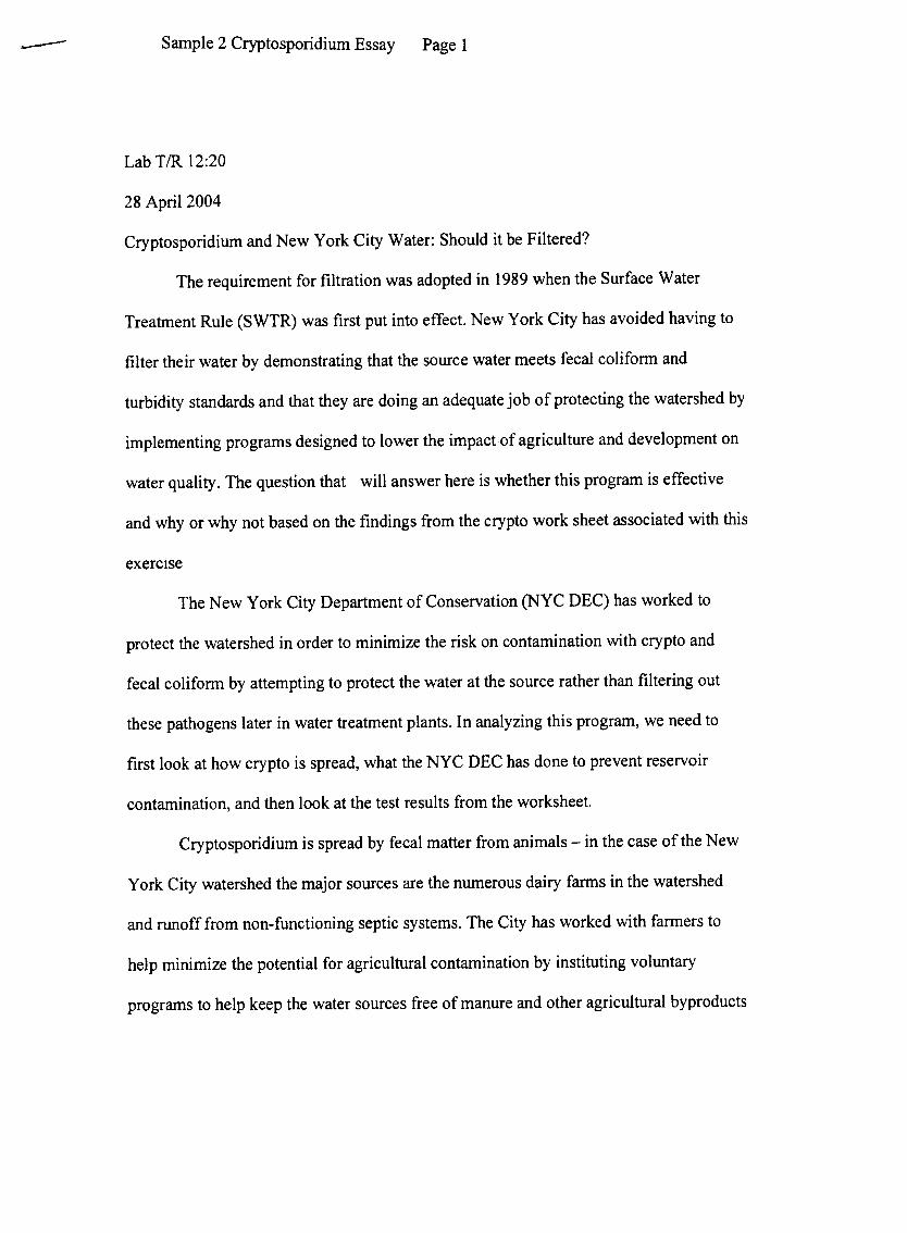
Cryptosporidium and New York City Water: Should it be Filtered?
The requirement for filtration was adopted in 1989 when the Surface Water
Treatment Rule (SWTR) was first put into effect. New York City has avoided having to
filter their water by demonstrating that the source water meets fecal coliform and
turbidity standards and that they are doing an adequate job of protecting the watershed by
implementing programs designed to lower the impactof agriculture and development on
water quality .The question that will answer here is whether this program is effective
and why or why not based on the findings from the crypto work sheet associated with this
exercIse
The New York City Department of Conservation (NYC DEC) has worked to
protect the watershed in order to minimize the risk on contamination with crypto and
fecal coliform by attempting to protect the water at the source rather than filtering out
these pathogens later in water treatment plants. In analyzing this program, we need to
first look at how crypto is spread, what the NYC DEC has done to prevent reservoir
contamination, and then look at the test results from the worksheet.
Cryptosporidium is spread by fecal matter from animals -in the case of the New
York City watershed the major sources are the numerous dairy farms in the watershed
and runoff from non-functioning septic systems. The City has worked with farmers to
help minimize the potential for agricultural contamination by instituting voluntary
programs to help keep the water sources free of manure and other agricultural by products

Page 2Sample 2 Cryptosporidium Essay--
such as fertilizer. This has involved the construction of manure management systems and
the purchase of land in the watershed area by the City .The City has also funded projects
to fix the non-functioning septic systems and stonn run off, and so far has spent $288
million on these programs according to the Executive Summary
The cryptosporidiurn worksheet associated with this exercise shows the relative
number ofoocytes in five different sources: manure, pasture soil, a stream, the reservoir,
and the aqueduct that feeds the water treatment plant. As expected, there were plenty of
oocytes in the manure -after all, this is the source for contamination in the first place.
The pasture soil also had measurable oocyte concentrations, although they tended to be
much lower than the manure concentration. In the three water sources tested, there was
no detectable oocyte contamination. This suggests that the crypto, which is present in the
manure, does not move into the water sources.
New York City had limited the potential for cryptosporidium to enter the
watershed through the implementation of watershed protection programs designed to
keep storm water, agricultural runoff and failing septic systems from contributing
contamination to the water supply. The tests in the worksheet show that crypto is not
entering the water supply under normal conditions. In addition, the storm water
management systems as well as agricultural protection will keep this contamination
potential low even when there is a heavy load on the system (as in storms which
contribute lots of extra runoff). This will prevent a disaster like that which occurred in
Milwaukee in 1993 when the extra load from heavy rains allowed crypto to enter the
water system. As Carol Browner of the US EP A said 'tBy putting in place the
mechanisms for protecting New York City's drinking water at the source, by keeping

Page 3Sample 2 Cryptosporidium Essay
contamination out of the water supply in the first place, we offer the promise of
," I think that theprotecting public health while saving billions of dollars for rate payers.
results of these tests show that the system is safe, and the programs to protect the
watershed will keep it that way under circumstances which place stress on it
Page 1Sample 1 Fatal Flu Essay
Lab T/R 12:2010 May 2004
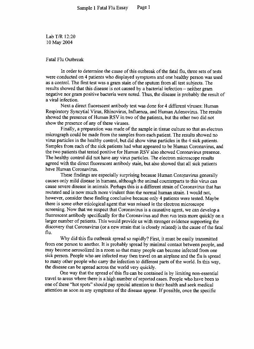
Fatal Flu Outbreak
In order to determine -the cause of this outbreak of the fatal flu, three sets of testswere conducted on 4 patients who displayed symptoms and one healthy person was usedas a control. The first test was a gram stain of the sputum from all test subjects. Theresults showed that this disease is not caused by a bacterial infection -neither gramQegative nor gram positive bacteria were noted. Thus, the disease is probably the result ofa viral infection.
Next a direct fluorescent antibody test was done for 4 different viruses: HumanRespiratory Syncytial Virus, Rhinovirus, Influenza, and Human Adenovirus. The resultsshowed the presence of Human RSV in two of the patients, but the other two did notshow the presence of any of these viruses.
Finally, a preparation was made of the sample in tissue culture so that an electronmicrograph could be made from the samples from each patient. The results showed novirus particles in the healthy control, but did show virus particles in the 4 sick patients.Samples from each of the sick patients had what appeared to be Human Coronavirus, andthe two patients that tested positive for Human RSV also showed Coronavirus presence.The healthy control did not have any virus particles. The electron microscope resultsagreed with the direct fluorescent antibody stain, but also showed that all sick patientshave Human Coronavirus.
These findings are especially surpris~ng because Human Coro.navirus generallycauses only mild disease in humans, although the animal counterparts to this virus cancause severe disease in animals. Perhaps this is a different strain of Coronavirus that hasmutated and is now much more virulent than the normal human strain. I would not,however, consider these finding conclusive because only 4 patients were tested. Maybethere is some other etiological agent that was missed in the electron microscopescreening. Now that we suspect that Coronavirus is a causative agent, we can develop afluorescent antibody specifically for the Coronavirus and then run tests more quickly on alarger number of patients. This would provide us with stronger evidence supporting thediscovery that Coronavirus ( or a new strain that is closely related) is the cause of the fatalflu.
Why did this flu outbreak spread so rapidly? First, it must be easily transmittedfrom one person to another. It is probably spread by minimal contact between people, andmay become aerosolized in a room so that many people can become infected from onesick person. People who are infected may then travel on an airplane and the flu is spreadto many other people who carry the infection to different parts of the world. In this way,the disease can be spread across the world very quickly.
One way that the spread of this flu can be contained is by limiting non-essentialtravel to areas where there is a high number of reported cases. People who have been toone of these "hot spots" should pay special attention to their health and seek medicalattention as soon as any symptoms of the disease appear. If possible, once the specific

Sample 1 Fatal Flu Essay Page 2
nature of the pathogen has been determined, a vaccine against it could be developed.Although this may take time, ultimately it will be the best way of controlling the spreadof this viral infection.

Page 1Sample 2 Fatal Flu Essay
10BIOMI 290 ~
Fatal Flu Case Study12/03/04
The electron micrograph images show a consistency through all four patients of
Human Coronavirus, This leads me to believe that Human Coronavirus is the cause of the
community-acquired pneumonia outbreak. I took the patients' sputum samples and grew the
virus in tissue culture cells, inoculating three mammalian cell lines. Cell culture line C
showed the Human Coronavirus in all patients, while Cell culture line A showed Human
Respiratory Syncytial 'irus only in patients 2 and 3. These results are indicative of
complications in patients 2 and 3, but given that cell culture C tested positive in a..!lfow~es
for Human Coronavirus, I find it difficult to blame RSV for the outbreaks, and I beli~at
Human Coronavirus is a much more probable candidate
The electron micrographs are the helpful in analyzing samples, because they are
able to easily resolve images only nanometers in length, and show good contrast.' I also
tested the patients using the direct fluorescent antibody test, which detects the presence of a
particular antigen, generating a very sensitive protein tag. However, for this test I
unfortunately did not include a positive control of Human Coronavirus, instead sampling for
:>nly 3 ),
Rhinovirus, Influenza and Human Adenovirus. In retrospect, I should have also used the
/direct fluorescent antibody test for Human Coronavirus, in order to verify the results of the
electron micrograph
used the gram stain test, a typical test for community acquired pneumonia.Finally,
The gram stain test uses microscopy to differentiate between bacteria based on the
composition of their cell walls. I tested Legionella, Staphylococcus, Streptococcus,
Pseudomonas, and Heamophilis. None of the patients had evidence of any of these

Page 2Sample 2 Fatal Flu Essay
bacterium in their sputum s I was able to rule out bacterial athogens as
the cause of the outbreak. This result was unexpected because bacterial pneumonias tend to
be the most serious especially Streptococcus pneumoniae. I was surprised to find that
mild,
I feel my results are conclusive, but would definitely recommend further testing of
RSV to detennine if serious complications arise in patients infected with both viruses
simultaneously. In the future, would also have included Human Coronavirus in my direct
would also like to test a greater number of patients to obtainfluorescent antibody testing. .,1
legitimate statistical results, and also be provided with documentation of symptoms.
Finally, I beJieve this pathogen spread so quickly because the virus particles are able to
travel through the air, contaminating anyone within a certain diameter and under specific
conditions. Physical contact, of course, also greatly increases the risk of contamination. To
0/control further spread of this virus, I recommend that all medical professionals treating
patients with symptoms indicative of the disease (rapid onset of high fever, chills, sore throat,
shortness of breath, cough, and evidence ofpneumonia) should use standard hygiene
precautions, together with airborne and contact precautions (respirator, gowns and gloves,
Suspected carriers of the virus should be diagnosed using' chest x -ray, grameyewear , etc
stain and sputum cultures to confinn the presense of Human Coronavirus. Family members
or anyone who has come in near proximity to the patient should be immediately quarantined
for a matter of at least one week. Furthennore, travel should be restricted in areas of severe
outbreak, and passengers needing to travel should be examined by security for any sign of
illness.

Page 1Sample 3 Fatal Flu Essay
\0
BioMi 290Dec. 3,04
The Fatal Flu: Case Study
At first, SARS or severe acute respiratory syndrome, looked like pneumonia with very
similar symptoms. However, the rapid spread of the illness coupled with numerous deaths caught
the attention ofhealth officials. The pathogen responsible for these outbreaks was unknown and
testing was necessary to try to identify this deadly agent.
The symptoms of SARS include a rapid onset of high fever, myalgia (muscle pain),
chills, rigor (stiffness), sore throat, and development of cough, shortness of breath, and other
pneumonia-like symptoms. After growing cultures from patients, a pathogen still wasn't
identified, however, the most reliable pathogens for diagnosis of SARS were identified as
influenza A or B, or human respiratory syncytial virus. The methods used for diagnosis included
chest radiograph, pulse oximetry , blood cultures, sputum Gram stains and cultures, and other
tests for viral respiratory pathogens.
Results from various tests weren't helpful in making a definite identification but did lead
to a general idea of the culprit. The first test, a Gram stain, was done to ~etect potential bacterial
pathogens. Four patients were each tested for Legionella, Streptococcus, Staphylococcus,
Pseudomonas, Heamophilis. The Gram stain~~~t~~ling o~ial
pathogens that could cause respiratory illness. There were no visible bacteria in any of the
p~tients' sampJes. The second test, a direct fluorescent antibody (DF A) test, was more helpful in
identifying a potential pathogen. This test uses antibodies which are specific to surface proteins
of the target and labeled with fluorescent chemicals so the target becomes labeled and visible.
This tested the same four patients for the viruses Human Respiratory Syncytial Virus (RSV),
Rhinovirus, Influenza, and Human Adenovirus. Only the seEQ!!!!J!p.dlhir~Q~tient had any ~~sults
and they were both positive for Human RSV. This is the same virus that was initially used to
diagnose SARS in patients. However, the dilemma is-~h~~lLtJ1e patients were posi~or
the virus, so there must be another factor .~ The third test was to isolate viruses from the sputum samples and this was done by
growing cultures of the viruses in tissues and then inoculating other tissues with the samples to
test for damage. Examination follows with an electron microscope to get an accurate picture of
"

Sample 3 Fatal Flu Essay Page 2
the virus in the culture. This test was interesting because the s~e virus, t!1e Hum~~SV, was
present in the second and third atient as be re, as well a~nother virus present in~!l £a~s.
Th~~ th~-Human ~r~~~-~s! which wasn't tested for in the DF A test. Since it was present
in all the patients, it makes more sense as a culprit; however, it doesn't usually cause serious
illn~ss ~~. It is also responsible for enteric diseases. This makes its presence slightly
unexpected as it isn't commonly considered as a cause of serious illness in humans.
The two pathogens detected are both viruses and so this tells us something about the
spread of the disease. Both the Human RSV and Coronavirus are enveloped, which increases
virulence, and there are fifteen types of Coronavirus which ~lso cause disease in animals. This
could mean that another type, previously unknown and originating in animals, could be the cause
of SARS .The Human RSV is the most identifiable as a cause of respiratory illness as it also
causes the cold, flu, bronchitis, and pneumonia. Thereforej for the second and third patients who
had both viruses, the Coronavirus may have initially initiated the illness and the RSV may have
enhanced the pneumonia-like symptoms. For those that didn't have the RSV present, the
Coronavirus was the sole cause of the illness.
From these results, the most probable cause of SARS is the Human Coronavirus. The-~---
only likely explanation for this unlikely .cause is the emergence of a new strain in animals thatthen spread to humans in more rural regions of China when the outbreaks first started. Again, v'
this pathogen is highly unusual since it usually prevails in animals and only causes mild illness in
humans. Because the illness spread so quickly, the virus is obviously prevalent in secretions from
coughing, sneezing, mucus, etc. This means that masks, hand washing, and avoiding crowded
areas and other sick people would be the most effective means of protecting oneself from the
illness

Sample 4 Fatal Flu Essay ...Page 1
-1
Case Studies in Microscopy: The Fatal Flu
Controlling the outbreak of disease is a gaunting enough task, but doing so with a disease of
unknown etiology presents an even more extensive list of challenges.
Bacterial infection was ruled out for all Patients using Gram stains. However, small, dark,
separate objects in Patient 4 were initially labeled cocci, but are smaller than the chained cocci found in
the positive control of Streptococcus, with which they are most similar. The Staphylococcus I had initially
identified in Patient 3 was even less convincing. The requisite cluster of cocci was very faint, hardly
noticeable at fIrst glance, and could just be pieces from the damaged cells. In either case, Staphylococcus
is usually only present in pneumonia cases as a secondary infection, so the question of its presence does
not overly concern me.
The fluorescent antibody tests were much more conclusive. Human Adenovirus, Influenza, and
Rhinovirus were ruled out in all four cases. However, antigens belonging to Human Respiratory Syncytial
Virus were identified in Patients 2 and 3. Morphology (size and shape) characteristic of Human RSV was
also observed in TEM analysis of their sputum samples. Human RSV is the most common cause of lower
respiratory infections, such as pneumonia. It is not surprisingly that Patients exhibiting symptoms of
pneumonia test positive for it. However, these Patients may be infected, not by Human RSV, but by the
SARS virus, because both Human RSV and (according to some scientists) the SARS viruses are members
of the Paramyxoviridae family. This could explain their similar morphology (EM Cell Line A results) and
surface proteins (F AB results). Both Human RSV and SARS are spread through close contact and
manifest themselves as respiratory infections. However, these Patients are severely ill, whereas victims of
Human RSV usually recover in 8-10 days.
Cell Line C of the EM test in all four cases showed the presence of Human Coronavirus. This
virus has been linked neither to SARS, nor to severe cases of pneumonia. However, since the etiology of
SARS is still unknown, our results cannot be ruled out. Nevertheless, the virus is known to cause severe
disease in animals, from which the tissue samples for these cell cultures was drawn. It is unlikely that the
cell line was contaminated, since the sample inoculated using the healthy patient was negative. It may be
that Human Coronavirus has infected the sick patients opportunistically, causing infection only when the
immune system is weakened by their current infection. However, the same could be said of the Human
RSV identified if a variant of Human Coronavirus is the cause of SARS. This is entirely possible since,
according to the WHO Update, a virus entirely different from Human RSV and the Paramyxoviridae
family could be the cause.
In conclusion, my fmdings show that the severe cases of pneumonia in these four Patients are
caused by a Human Coronavirus-like pathogen. Although Human RSV was also identified in two of the
cases, this could be the result of a secondary infection of an impaired immune system, as Human RSV
Sample 4 Fatal Flu Essay Page 2
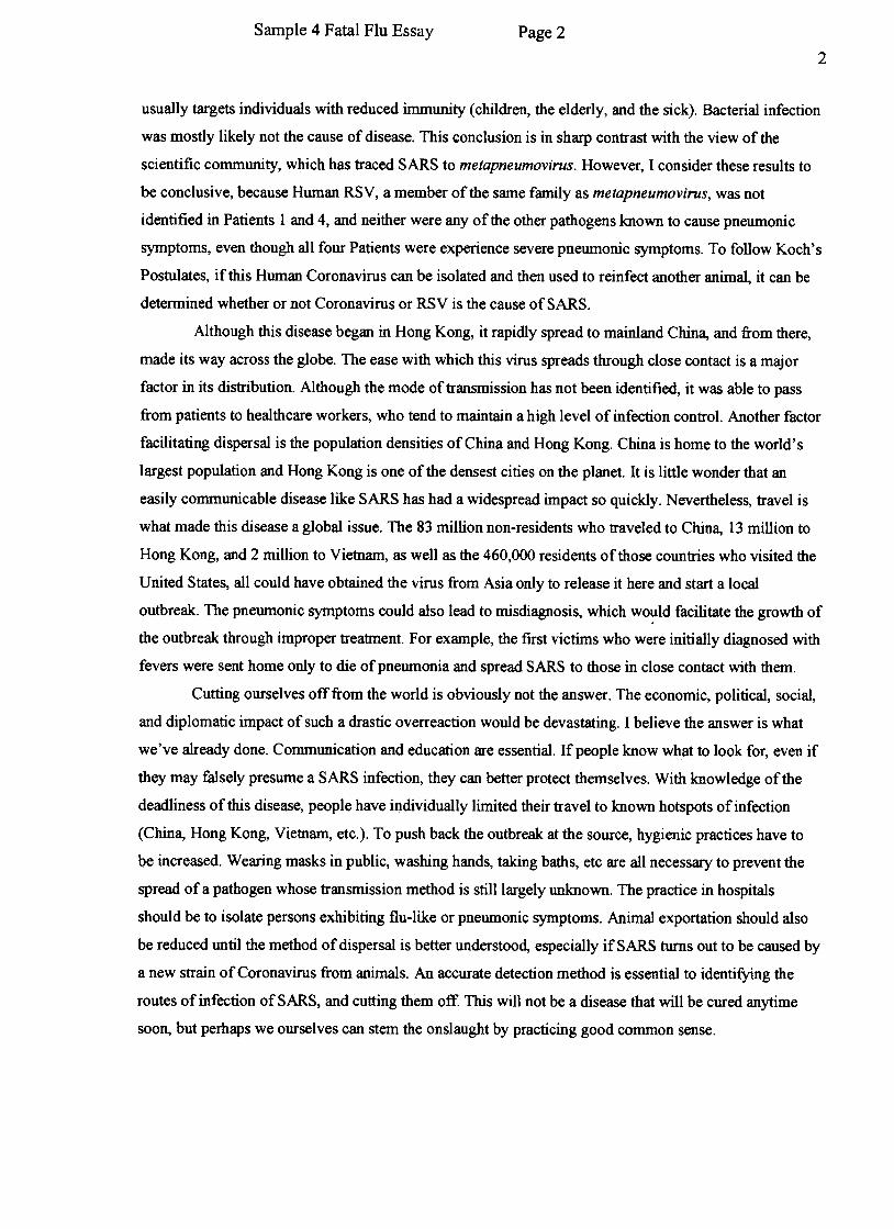
2
usually targets individuals with reduced immunity (children, the elderly, and the sick). Bacterial infection
was mostly likely not the cause of disease. This conclusion is in sharp contrast with the view of the
scientific community, which has traced SARS to metapneumovirus. However, I consider these results to
be conclusive, because Human RSV, a member of the same family as metapneumovirus, was not
identified in Patients I and 4, and neither were any of the other pathogens known to cause pneumonic
symptoms, even though all four Patients were experience severe pneumonic symptoms. To follow Koch's
Postulates, if this Human Coronavirus can be isolated and then used to reinfect another animal, it can be
determined whether or not Coronavirus or RSV is the cause of SARS.
Although this disease began in Hong Kong, it rapidly spread to mainland China, and from there,
made its way across the globe. The ease with which this virus spreads through close contact is a major
factor in its distribution. Although the mode of transmission has not been identified, it was able to pass
from patients to healthcare workers, who tend to maintain a high level of infection control. Another factor
facilitating dispersal is the population densities of China and Hong Kong. China is home to the world's
largest population and Hong Kong is one of the densest cities on the planet. It is little wonder that an
easily communicable disease like SARS has had a widespread impact so quickly. Nevertheless, travel is
what made this disease a global issue. The 83 million non-residents who traveled to China, 13 million to
Hong Kong, and 2 million to Vietnam, as well as the 460,000 residents of those countries who visited the
United States, all could have obtained the virus from Asia only to release it here and start a local
outbreak. The pneumonic symptoms could also lead to misdiagnosis, which wo~ld facilitate the growth of
the outbreak through improper treatment. For example, the first victims who were initially diagnosed with
fevers were sent home only to die of pneumonia and spread SARS to those in close contact with them.
Cutting ourselves off from the world is obviously not the answer. The economic, political, social,
and diplomatic impact of such a drastic overreaction would be devastating. I believe the answer is what
we've already done. Communication and education are essential. If people know what to look for, even if
they may falsely presume a SARS infection, they can better protect themselves. With knowledge of the
deadliness of this disease, people have iI)dividually limited their travel to known hotspots of infection
(China, Hong Kong, Vietnam, etc.). To push back the outbreak at the source, hygienic practices have to
be increased. Wearing masks in public, washing hands, taking baths, etc are all necessary to prevent the
spread of a pathogen whose transmission method is still largely unknown. The practice in hospitals
should be to isolate persons exhibiting flu-like or pneumonic symptoms. Animal exportation should also
be reduced until the method of dispersal is better understood, especially if SARS turns out to be caused by
a new strain of Coronavirus from animals. An accurate detection method is essential to identifying the
routes of infection of SARS, and cutting them off. This will not be a disease that will be cured anytime
soon, but perhaps we ourselves can stem the onslaught by practicing good common sense.

~/Sample Biofilms Short answer Page 1
19y
IQ
BIOMI 290
Case Studies 1
March 14,200:
Bioj
Part I: The Disease .1(: t.""'
Legionelliosis, also known as Legionnaires' disease, is caused by the bacterium c
the Legionella species growing intracellularly within protozoa in biofilms. Lo
pneumophilia is a specific species found naturally in aquatic environments. The
pneumonia associated with Legionelliosis is caused by the invasion of alveolar
macl-ophages in the lung tissue of mammals by the Legionella bacterium. The protozoa
inhaled serve as cofactors for pulmonary disease induced by inhalation of respiratory
pathogens, causing pneumonia in the affected Transmission occurs via aerosols from
environmental conditions, such as those found in hot tubs, water coolers, spas, vegetable
misters, and showerheads. Lo pneumophilia, often associated with pneumatic symptoms
in Legionelliosis, is commonly found in potable water plumbing systems, such as that on
a cruise ship. The protozoa that harbor L. pneumophilia have been found to be tolerant o
temperatures above 50°C (122°F), which is why it was found in the hot tub pipes. In
addition, L. pneumophilia populations were shown to be most abundant in plastic water
plumbing systems at 40°C (104°F). In one study, amoebas were also found to be hosting
L. pneumophilia in 42% of whirlpools and hot tubs. 0 The passengers got Legionelliosis
from,inhaling the steam coming from the hot tub water which contained the L.
pneumophilia pathogen picked up from the pipes.
Part II: The Environment-\-' \
Biofilms are, simply put, surfaces covered with a layer of bacteria. In reality, t~e
are much more complex. The surfaces most commonly occur in aqueous environments,
and there can be more than just bacteria living on them. Biofilms can consist of fungi,
algae, protozoa, corrosion products, and other debris. They form when a planktonic
bacterial cell anchors itself to the surface, sometimes using fimbriae to aid in anchoring
itself. oth~ receive chemical signals in a system called "quorum"
sensing, where special genes are activated and the cells all begin to secrete a
polysaccharide-rich substance. This will eventually form a glycocalyx, a protective,
slimy layer that helps the ~stay ~nch9r~g,GPmmunicate, share nutrients, and protect
themselves from the environment. They can excrete other metabolic byproducts that can
be supportive to other bacteria but be harmful to humans and even the surface of the
biofilm itself. In the case of Legionelliosis, biofiJms are iinportant because the pathogen,
a Legionella bacterium, grows on the hot tub pipe surfaces on the cruise ship. This is the
passengers' source of exposure to the L. pnell1110philia on the cruise ship and what cause(
them to become ill. ~\*,vf\f\~ ~ ~\.f:(-\Wv\ \0 ~~
Part Ill: Experimental Results J(?
The experiment used included a
Endemic. There was a system of 24 refpiece could be removed for sampling. ,
would grow. The flow rate of the wateJ
model of a water system like that on the ship t
novable square PVC pieces on one side; each
rhese were the surfaces on which the biofilms
r, its temperature, and its contents through the

Page 2Sample 1 Biofilms Short answer
PVC piping could be controlled with this setup. The water came from the cruise shipEndemic and was pumped through the pipe for 2 weeks. The samples all contained asmall sub-sample of the Legionella biofilrn. Different treatments were applied to thewater affecting flow rate and additives. These were added on day 0. Samples of thebiofilms were taken 10 and 25 days after the treatments began. Analysis of the biofilmswas done using a Legionella- specific tagged fluorescence antibody assay, viewableunder fluorescence microscopy. This was done to view the cells in their natural habitatsand to assure specificity of the organism of interest. In addition, biofilms samples wereviewed using differential intelierence contrast microscopy. This allowed live samples to
be viewed with a more 3-dimensional aspect.The treatments included high pulse flow (2 m/s as opposed to regular at 0.2 m/s),
free chlorine (0.5mg/mL), monochloramine (0.5mgimL), ~rythromycin antibiotic(5mg/mL), or some combination thereof. In all, there were 7 different treatments used.Temperature was held constant at 38°C (101°F) for all treatments.
The first ofjust high pulse flow, applied on day 9 for 2 hours, showed asignificant reduction the day after treatment on day 10. Day 0 showed about 30 micronsdiameter of growth, while day 10 showed only 10 microns. However, afterdiscontinuation of the treatment, day 25 showed significant growth filling the microscopy
field.Treatment ofjust free chlorine showed no reduction in growth. Day 0 showed 25
microns diameter of growth; day 10 showed 50 microns, and day 25 showed significant
growth filling the microscopy field.A treatment of free chlorine and high pulse flow for 2 hours on day 9 reduced
growth from 25 microns on day 0 to almost no growth (less than 5 microns) on day 10.However, day 25 showed enough growth to almost fill the microscopy field.
Treatment with monochloramine reduced growth from 25 microns on day O to athin, almost absent growth on day 10. Day 25 showed select growth spots of 10 and 20
mIcrons across.Monochloramine combined with high pulse flow on day 9 for 2 hours seemed
successful. Day 0 showed 25 microns of growth, while there were only a few spots onday 10 following high pulse flow treatment. On day 25, there was very thin growth of
barely 10 microns across present.Treatment with antibiotics (erythromycin) did not affect growth. Biofilm growth
was more than 50 microns across on day 10, double from 25 microns on day 0. By day25, there was enough growth to fill completely the microscopy field.
Antibiotics combined with high pulse flow reduced growth from 25 microns onday 0 to a few small spots up to 10 microns in diameter on day 10. However, by day 25,growth was significant enough to fill the entire microscopy field.
Part IV: My Conclusion ~ \
Based on the treatments, I recommend combining 0.5 mg/mL monochloramine
with high pulse flow of 2m/s for 2 hours every 9 or 10 days. This is better than the other
methods used because of the combination of physical and ~b"~!!!-!9~!-~-t!a~.k. The
monochloramine is a more reactive speciesOTChiorlne and therefore can better attack the
tough surface of the biofilm. The high pulse flow physically disrupts the integrity of the
gly~?calyx and erodes some of the bact~!i~lgrowth, exposing the more susceptible inner
~
'"

Page 3Sample 1 Biofilms Short answer
Prolonged exposure to the monochloramine helps keep bacterial growth to a
and eliminates the rest of the f:olony that was originally present. With repeated
}SeSSIOns of high pulse flow and exposure to monochloramine, t~e biofilm can help be
-~~p~lean and f~f disease-causing bacterial pathogens, specifiCa1fyTii[Oiiena-
pneumophilia.

-~'" c "
:;
~,,:
3! 14mS
Legionelliosis is a disease that is a form of pneumonia. It is caused by thebacteria Legi()nella pneum()phila. This bacterium is found in water which is of anenviroJ11nentaJ source. The bacteria were found in the hot tubs of the cruise ship.
A biofilm is a colony of microbial cells t~1t lu1ve attached themse1ves to a surfacetmd have secreted a poJysaccharide which encases the coJon)r. BjofiJms OCJ;;Uf in aqueousen\1ronments. The bacteria L. pnt'umophilt.l grows using bjofi Ims" gro\~ing insjde ofprotozoa whjch make up the bjofilms. This makes the bacteria more complicated todispose of, as it is doubly protected by the protozoa and the biofilm itself
Part 3
In order to detennine which method wouJd be best in ridding the cruise ship of thebacteria, we set up a model of the ship' s water system. This model consists of a PVCtube \vith removable pieces through which ,"-ater flows. The controllable variables arewater contents, flow rote, and water temperature. Water from the ship's storage tank \vithsamples of the bacteria was allowed to flow through the system for 2 weeks in order togive the bacteria a chance to develop. Then on day 0, the system \\'as tested tor biofilms,Each of the 7 treatment conditions were started on day 0, and the biofilnlS were S..1mpledat day 10 and day 25 after the treatments began. .
The seven treatments are listed in table I. The control had no treatment added toIt HIgh flow pulse Involves increasing the flow of \~'ater (from 0.2 wls to 2 w's) throughthe pipe for two hours on day 9. The chlorine tre.1tment involved adding free chlorine tothe \\'ater on d.1.y O at a nonnal flow (0.2 mJs). The mono-chloramine treatment involvedadding the mono-chloramine on day O at nomlal flo\\,-. The antibiotics treatment involvedaddin{! 5 m{!!ml ef'l1hromvcin to the water on dav 0...., ---~
Samples of the biofilms were al1alyzed in t\vo ways. They were looked at \\-ithDifferential Interference Contrast Microscope and again using a Direct FluorescentAntibody T est
I table 1 i resultsI treatment! dayO \ da 10 !, da" 25
control I one-third .half / full
hj 71 flo"' uJse haJf a1rnost none I fuJJ
Lc.hlori~e-- --! half) two-tl!irds I fl111 -j\ high flo\v ulse and chlorine \ one-third [ almost one: t'vo-t~
t mono-chloramine [ one-third !. almost none I one-third I,'-= ~ -~
I mono-chloramine and hi ulse i one-third I almost none! almost none 1I1 antibiotics i one-tl-!ird : t\vo-thirds ~ t\~o-thjrds -J
antibiotics and hi ulse \ one-third \ almost none \ full
~
Page 2Sample 2 Biofilms Short answer
The results of this experiment tell us that chlorine, mono-chloramine, and theantibiotic erythromycin are as effective on their ~~ at removing biofilms as they are
\vhen used in conjunction with a high flow pulse. The combination of mono-chlommineand the hjgh flow pulse was the most effective at removing biofilms and keep1ng them
gone till day 1.5.
Sample 1 Fatal Flu Short answer Page 1
I~'- / ~ D
~
-r~~::,-:;",,- .~ -BioMI 290 -
Case Study 2 -Flu
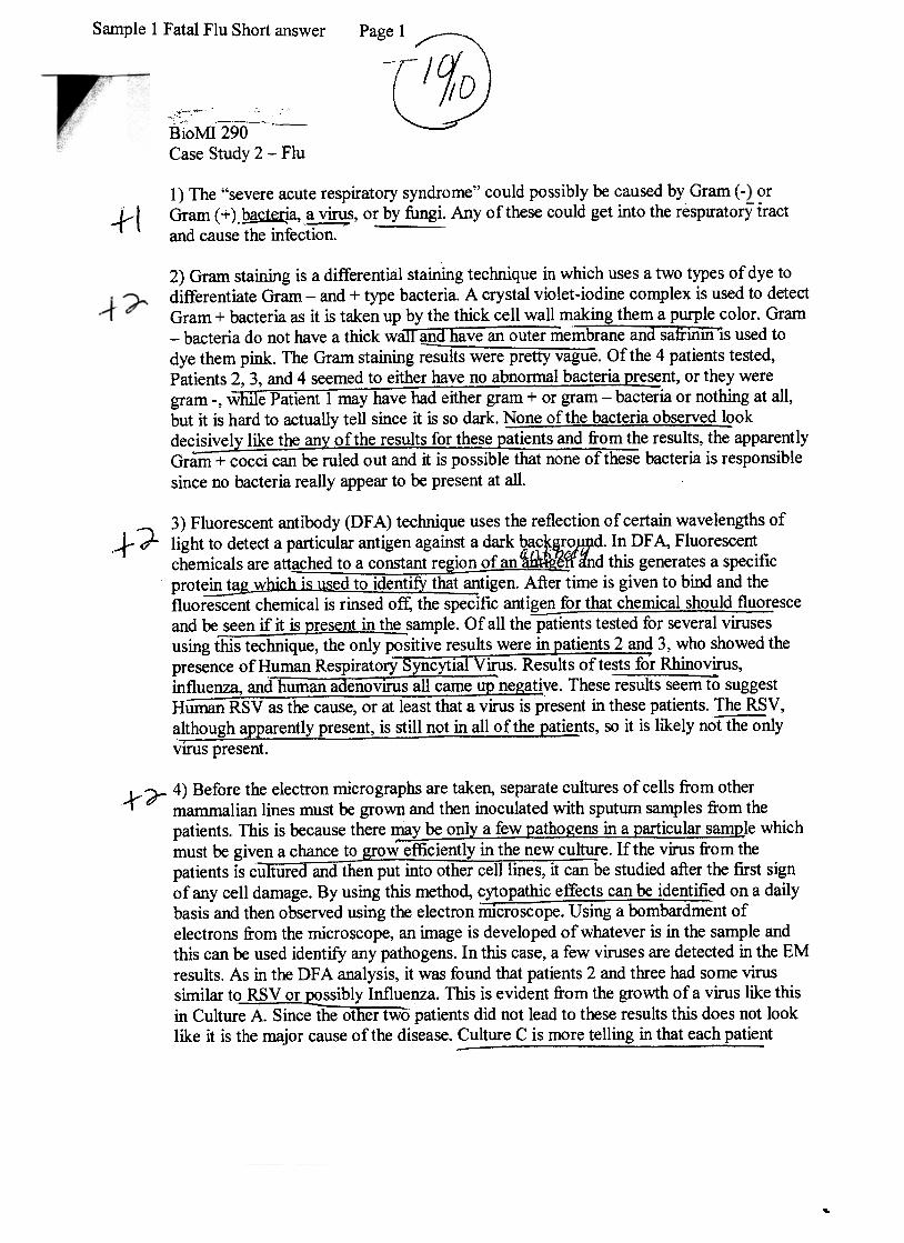
1) The "severe acute respiratory syndrome" could possibly be caused by Gram ( -orGram ( + ) .~a, ,~s, or by fungi. Any o f these could get into the resp1IatorY tractand cause the infection. -
.-r-l
2) Gram staining is a differential staining technique in which uses a two types of dye todifferentiate Gram -and + type bacteria. A crystal violet-iodine complex is used to detectGram + bacteria as it is taken up by the thick cell wall making them a purple color. Gram-bacteria do not have a thick wan-and have an outer membrane and saftin~s used todye them pink. The Gram staining ~~ts were pretty vague. Of the 4 patients tested,Patients 2, 3, and 4 seemed to either have no abnormal bacteria resent, or they weregram -, we Patient 1 may have had either gram + or gram- bacteria or nothing at all,but it is hard to actually tell since it is so dark. ~one of the bacteria observed l~okdec~ly l~e theanv ~f~he resu~t~ ~or thes~ ~at~ents and ~o~ t results, the apparentlyGram + cocci can be ruled out and it is possible that none of these bacteria is responsiblesince no bacteria really appear to be present at all.
" 3) Fluorescent antibody (DF A) technique uses the reflection of certain wavelengths ofct d"'" light to detect a particular antigen against a dark bac cQ.r.o)Jl}d. In DF A, Fluorescent
chemicals are attached to a constant region of an {~Wand this generates a specificprot~hich is used to iQent!f}: tha~tigen. After time is given to bind and thefluorescent chemical is rinsed off, the specific antigen for that chemical should fluoresceand 00. s~n if it is Qresent in t~ sample. Of all thepai1ents tested for several vifUsesusing this technique, the only positive results were in atients 2 and 3, who showed thepresence ofHuman Respirat~ry ~yncytlal V~. Results of tests for Rhinovirus,in~nza,~dh~man adenoviru-s all c.ameu~ne~ve. These results seem 10 suggestHuman RSV as the cause, or at least that a virus is present in these patients. The RSV,a~?ugh apEarently present, is still not in all of the pat~nts, so it is likely no1"liie""Q"nly
VIrUS present.
Jr r 4) Before the electron micrographs are taken, separate cultures of cells from othermammalian lines must be grown and then inoculated with sputum samples from thepatients. This is because there ~be only a few pathog~ns in~ ~arti;ular -samt?-le whichmust be given a chance to row efficiently in the new culture. If the virus from thepatients is cu ture and then put into other cell lines, it can be studied after the first signof any cell damage. By using this method, cytopathic effects can be identified on a dailybasis and then observed using the electron rnIcioscope. Using a bombardment ofelectrons from the microscope, an image is developed of whatever is in the sample andthis can be used identify any pathogens. In this case, a few viruses are detected in the EMresults. As in the DF A analysis, it was found that patients 2 and three had some virussimilar tQR§V or ~ibly Influenza. This is evident from the growth of a virus like thisin Culture A. Since the other two patients did not lead to these results this does not looklike it is the major cause of the disease. Culture C is more telling in that each patient
-
..
Page 2Sample 1 Fatal Flu Short answer
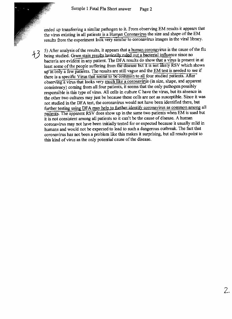
ended up transferring a similar pathogen to it. From observing EM results it appears thatthe virus existing in all patients is a Human Coronavirus the size and shape of the EMresults from the experiment look-very sirilar to coronavirus images in the viral library.
+35) After analysis of the results, it appears that ~Euman coro~irus is the cause of the flubeing studied. Gr~~.i('~ ~~ ~lled~ut ~ ba,cteri~~ ~u~nc since nobacteria are evident in any patient. The DF A results do show that a virus is present in atleast some of the people suffering from the disease but it IS not likelyRSV which showsu!f1iiOiiry a few patients. The results are still vague and the EM test is needed to see ifthere is a specific Virus that seems to be common to all four-studied patients. After~observ~s that looks very much I~e a coronav~s (in size, shape, and apparentconsistency) coming from all four patients, it seems that the only pathogen possiblyrespQnsible is this type of virus. All cells in culture C have the virus, but its absence inthe other two cultures may just be because these cells are not as susceptible. Since it wasnot studied in the DF A test, the coronavirus would not have been identified there, butfurther testing usin DF A ma he} t ..coronavirus as common amon allp~. The apparent RSV does show up in the same two patients when EM is used butit is not consistent among all patients so it can't be the cause of disease. A humancoronavirus may not have been initially tested for or expected because it usually mild inhumans and would not be expected to lead to such a dangerous outbreak. The fact thatcoronavirus has not been a problem like this makes it surprising, but all results point tothis kind of virus as the only potential cause of the disease.
2.

Sarnple2 Fatal Flu Short answerPage 1
Biol\-:n 290
"~ioMI 290
.
Case Studv in MicroscoDv (The Fatal Flu}
The Disease
)Types of pathogens could be viral respiratory pathogens such as influenza types A & B
measles, humanmumps,Syncytial paramxyovlrus,Virus,+1
Human Respiratory
Human Coronavirus,Adenovirus, Rhinovirus:Humanparainfluenza viruses,
can beAlso, theychlamydophilametapneumovirus, mycoplasma,heni paviruses,
bacteria, such as 1~g!2nella, ~se\ldom2na~, and He~mQQhilus.
The Gram Stain Results
2) Gram stain is one of the important differential stains. Gram stain differentiates
~I
thicker cell walls and no outer me~brane (Gram+ type).
'.The crystal violet-iodine complex gets trapped inside bacteria with a thick cell wall, and
those cell (Gram +) remains purple. In bacteria with a thin cell wall, the crystal violet-
: d .1 1 k .-ujh hel1 ?11 b 1 1 f .. 10 me comp ex ea s out, anu t e ce s ecome co or ess: Because o countenng stam
The end result .is purple-colored'with safrinin, the Gram- cells will be stained pink.
not bacterialThe... Viral' pathogens,Gram( + ) cells and pink -color~d Gram( -) cells
pathogens, will be ruled out.
is possibly affected by Gr~( + ) bacte!ia,In this case, the results indicate that patient
and patient 2,3, 4 are hard to tell whether they are affected by bacteria or not, the pink
~
stained cells are hard to be distinguished whether they are human cells or bacter~a cells. -»,,"")
5tJ u~LJZ- AI1::f:I.~d .,)Patle~nt 3 .~ay posslbly be affe~ted by Gram(+) bactena. fY .~ IJ..,t ~
J ~~ : k~X:;:'?K-f1~
-5l;]tJ w(..(!... ~~ t/1~ ~~~ ~..J fl..-:VI L1 ~
::::;tjt.t, ~ C!J A " A , -..tf A1 {jJ.4-') tL."'".l.Mt..-'I ./ --1-
.~~"-7~ -1-- fi "' A jJ IJ A I ,,:7'

The DFA Results
,.}~ 3) The direct fluorescent antibody (DF A) technique detects the presence of a particular
antigen, typically a specific protein on the surface of a virus, bacterium or other microbe.
Fluorescent chemicals are attached to the constant regio~ ~ ~ a~ .The antibody ,,-L. A#f)
~ ' c;:t .J::>() ~
binds to generate a very specific, very sensitive protein tag if the antigen is present. ~ A ~
In this case, the DFA results showed that patient 1 and patient 4 WIJ.S not affected b~~J4!JI.
four viruses, Human adenovirus, Influenza, Human Respiratory Syncytial Virus (RSV),
Rhinovirus, as the DF A results showed similar pattern as the healthy person, these
samples do not emit light, indicating there are no presence of the viral antigen.~tient~
and Patient 3 are possibly affec~ Human RSV, but not the other three viruses. The
two samples only emit light because of the presence ofHuman RSV antigen
..

Student Worksheet
Battle of the Biofilms
Spring 2005 Short Answers
The purpose of this activity is for you to think about the results and to use the information to make a recommendation regarding how to rid a Cruise ship of a source of disease. Think about the information, and answer these questions as you go through the case.
The case study can be found by going to http://instruct1.cit.cornell.edu/courses/biomi290/microscopycases/
The Disease 1What is Legionelliosis? What causes it and why did they find it here?
The Environment 2What are biofilms? Why are they important in this case?
Experimental Results 3Explain your experiments, the methods you used, and what information these methods gave you (including your results).
The table below may help you organize the results. For each result, indicate how much biofilm growth you obtained from each different treatment.
Results Day 0 Day 10 Day 25 Treatment
Treatment
Treatment
Treatment
Treatment
Treatment
Treatment
Your Conclusion 4Based on your results, what treatment are you going to recommend? Explain why it is better than the other methods used.

Cryptosporidium and the NYC Watershed
Student Worksheet
Page 1 of 2
These questions are meant to help you research the background information you will need to evaluate some of the water quality issues in New York City (NYC), with respect to Cryptosporidium. You should answer the following questions as you go through the Cryptosporidium Case Study. We will not collect and grade this work sheet.
The case study can be found by going to: http://instruct1.cit.cornell.edu/courses/biomi290/microscopycases/
Part 1. The NYC Water Supply
1From where does NYC get its drinking water?
2List two ways that NYC is currently trying to protect the CatskillDelaware watershed.
Part 2. Cryptosporidium
3How is Cryptosporidium transmitted? What are the disease symptoms? Is it highly lethal?
4What is the most likely source of Cryptosporidium in the NYC watershed?
5What is the current EPAaccepted method for detecting Cryptosporidium?
6Why do you think the other methods (FISH, Gram stain, phase contrast) on the list are not as useful as the EPA method? What problems do you think they have with respect to detecting Cryptosporidium?

Cryptosporidium and the NYC Watershed
Student Worksheet
Page 2 of 2
Part 3. Record your results below:
8 What do the results tell you about where in the watershed Cryptosporidium is found?
9 What do the results tell you about whether or not Cryptosporidium moves through the watershed?
Final Question: Do you think it is necessary for NYC to filter its water supply to control the spread of Cryptosporidium? Explain why or why not.
The purpose of this activity is for you to think about the results and to use the information to make a recommendation to the EPA regarding Cryptosporidium and the effectiveness of watershed protection versus the cost of filtration.
Your argument for or against filtration should include information about the nature of Cryptosporidium and the disease it causes what NYC is currently doing to protect its watershed whether you think this is good enough protection or not
You must refer to your results and include information from the references as needed. Your answer should be no more than 2 pages long and handed in on a separate piece of paper.
Sample site # oocytes /field
# oocytes /field
# oocytes /field
# oocytes /field
# oocytes /field
Student worksheet
Spring 2005 Short Answers
The Fatal Flu The purpose of this activity is for you to use the information presented in the results and the references to determine the cause of an outbreak. Think about the information, and answer these questions as you go through case.
The case study can be found by going to http://instruct1.cit.cornell.edu/courses/biomi290/microscopycases/
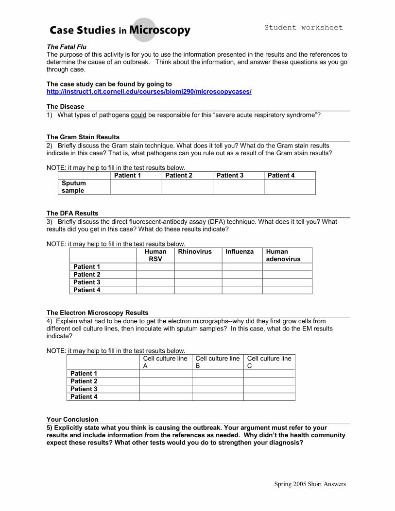
The Disease 1) What types of pathogens could be responsible for this “severe acute respiratory syndrome”?
The Gram Stain Results 2) Briefly discuss the Gram stain technique. What does it tell you? What do the Gram stain results indicate in this case? That is, what pathogens can you rule out as a result of the Gram stain results?
NOTE: it may help to fill in the test results below. Patient 1 Patient 2 Patient 3 Patient 4
Sputum sample
The DFA Results 3) Briefly discuss the direct fluorescentantibody assay (DFA) technique. What does it tell you? What results did you get in this case? What do these results indicate?
NOTE: it may help to fill in the test results below. Human RSV
Rhinovirus Influenza Human adenovirus
Patient 1 Patient 2 Patient 3 Patient 4
The Electron Microscopy Results 4) Explain what had to be done to get the electron micrographswhy did they first grow cells from different cell culture lines, then inoculate with sputum samples? In this case, what do the EM results indicate?
NOTE: it may help to fill in the test results below. Cell culture line A
Cell culture line B
Cell culture line C
Patient 1 Patient 2 Patient 3 Patient 4
Your Conclusion 5) Explicitly state what you think is causing the outbreak. Your argument must refer to your results and include information from the references as needed. Why didn’t the health community expect these results? What other tests would you do to strengthen your diagnosis?

Student worksheet
2004 Essay Page 1 of 2
The Fatal Flu These questions are meant to help you research the background information you will need to evaluate this current outbreak of severe pneumonia. Answer the following questions as you go through the Fatal Flu Case Study. We will not collect and grade this work sheet.
The case study can be found by going to: http://instruct1.cit.cornell.edu/courses/biomi290/microscopycases/
Part 1 Questions 1.1 What are the symptoms of this “severe acute respiratory syndrome” ?
1.2 What types of pathogens could be responsible for community acquired pneumonia?
1.3 What tests are recommended for the diagnosis of this severe acute respiratory syndrome?
Part 2 Question: 2.1 In general, what information does a Gram stain give you?
2.2 Fill in the Gram stain results below. Patient 1 Patient 2 Patient 3 Patient 4
Sputum sample
2.3 These results should help you narrow the list of potential pathogens. What pathogens can you rule out as a result of the Gram stain results?
Part 3 Questions 3.1 In general, what information does a direct fluorescentantibody assay (DFA) test give you?
3.2 Fill in the direct fluorescentantibody assay test results below. Human
RSV Rhinovirus Influenza Human
adenovirus Patient 1
Patient 2
Patient 3
Patient 4

Student worksheet
2004 Essay Page 2 of 2
3.3 What do these results indicate?
Part 4 Questions 4.1 In general, what information do Electron Micrographs give you?
4.2 Fill in the Electron Microscope results below. Cell culture line A
Cell culture line B
Cell culture line C
Patient 1
Patient 2
Patient 3
Patient 4
4.3 What do these results indicate?
Your Conclusion: What do you think is causing the outbreak? What is the evidence for your conclusion? Why are these results somewhat unexpected?
Time is running out the World Heath Organization (WHO) needs your recommendation now! As an epidemiologist, you must now write a report to the WHO on your research.
Your report should include the following: • What did each set of results tell you? Do all the results agree? • What do you think is causing the outbreak? Why is the culprit somewhat unexpected? • Do you think these are conclusive results? If yes, explain why. If not, what would you recommend next to conclusively prove the etiological agent of this outbreak?
• Why did this pathogen spread so quickly? What do you recommend to control further spread?
Your answer should be thoughtful and well organized, and it must incorporate the results you obtained in this case study. That is, use the information from your results and in the references to support your final conclusion.