Case Report Totally Ossified Metaplastic Spinal … · ic resonance imaging revealed a large T9-10...
4
257 his half lower body below the umbilicus. She also noticed pro- gressive urinary incontinence without weakness in lower ex- tremities. Neurological examination revealed a hypoesthesia to touch and pain from T10 to S2 level. Also, she had positive sign in Romberg test. Both patella and ankle reflexes were hypoac- tive and the Barbinski signs were bilaterally negative. Comput- ed tomographic images without enhancement show a large hy- perdense intradural round mass occupying the whole spinal canal at T9-10 level (Fig. 1). Magnetic resonance imaging re- vealed a large T9-10 intradural extramedullary mass that was hypointense to spinal cord on T1- and T2-weighted sequences, partial enhancement was apparent aſter Gd administration (Fig. 2). e spinal cord was severely compressed and displaced to- ward the right. Under general anesthesia, the patient was positioned prone aſter placement of transcranial muscle motor evoked potentials (Tc-MEPs) and somatosensory evoked potentials (SSEPs) mon- itoring devices. A T9-10 laminectomy was performed after midline splitting of T9-10 spinous process. e dura mater was exposed and the tumor location has been confirmed at the T9- 10 level by ultrasonography. An operative microscope was used during opening the dura, the dura was incised at the midline, exposing a hard and totally ossified, round dural-based extra- INTRODUCTION Spinal meningiomas are the second most common tumors after schwannoma and they represent about represent 25 to 46% of spinal tumors 18) . ey most frequently occur in the tho- racic region in middle-aged women 9,12,14,16,24) . Although the most common calcified intradural spinal tumor is a meningioma, to- tally ossified cases are uncommon and account for only 1-5% of all spinal meningiomas 2,6) . A ossified spinal meningioma is more difficult to resect than the usual type of meningioma. Hy- pointensity in T1 as well as T2-weighted images of MRI should make the surgeon suspicious of calcified condition, which may in some cases complicate tumor resection. We describe a case of symptomatic totally ossified large spinal metaplastic meningioma at the level of T9-10, and discuss the differential diagnosis of ossified spinal intradural tumor based on neuroimaging and the surgical strategy for the totally calci- fied and ossified spinal meningioma. CASE REPORT A 61-year-old woman had a 1-year history of numbness in right lower extremities that progress bilaterally and ascend to Totally Ossified Metaplastic Spinal Meningioma Chang Il Ju, M.D., Ph.D., 1,2 Kazutoshi Hida, M.D., 1 Tomohiro Yamauchi, M.D., 1 Kiyohiro Houkin, M.D. 1 Department of Neurosurgery, 1 Hokkaido University, Sapporo, Japan Department of Neurosurgery, 2 School of Medicine, Chosun University, Gwangju, Korea A 61-year-old woman with a very rare case of totally ossified large thoracic spinal metaplastic meningioma, showing progressing myelopathy is presented. Computed tomographic images showed a large totally ossfied intradural round mass occupying the spinal canal on T9-10 level. Magnet- ic resonance imaging revealed a large T9-10 intradural extramedullary mass that was hypointense to spinal cord on T1- and T2-weighted sequenc- es, partial enhancement was apparent after Gadolinium administration. The spinal cord was severely compressed and displaced toward the right at the level of T9-10. Surgical removal of the tumor was successfully accomplished via the posterior midline approach and the histological diagnosis verified an ossified metaplastic meningioma. The clinical neurological symptoms of patient were improved postoperatively. In this article we discuss the surgical and pathological aspects of rare case of spinal totally ossified metaplastic meningioma. Key Words : Ossified spinal tumor · Metaplastic meningioma · Grossly total resection. Case Report • Received : April 4, 2013 • Revised : July 14, 2013 • Accepted : September 8, 2013 • Address for reprints : Chang Il Ju, M.D., Ph.D. Department of Neurosurgery, School of Medicine, Chosun University, 365 Pilmun-daero, Dong-gu, Gwangju 501-717, Korea Tel : +82-62-220-3126, Fax : +82-62-227-4575, E-mail : [email protected] • This is an Open Access article distributed under the terms of the Creative Commons Attribution Non-Commercial License (http://creativecommons.org/licenses/by-nc/3.0) which permits unrestricted non-commercial use, distribution, and reproduction in any medium, provided the original work is properly cited. J Korean Neurosurg Soc 54 : 257-260, 2013 http://dx.doi.org/10.3340/jkns.2013.54.3.257 Copyright © 2013 The Korean Neurosurgical Society Print ISSN 2005-3711 On-line ISSN 1598-7876 www.jkns.or.kr
-
Upload
truongdien -
Category
Documents
-
view
219 -
download
0
Transcript of Case Report Totally Ossified Metaplastic Spinal … · ic resonance imaging revealed a large T9-10...
257
his half lower body below the umbilicus. She also noticed pro-gressive urinary incontinence without weakness in lower ex-tremities. Neurological examination revealed a hypoesthesia to touch and pain from T10 to S2 level. Also, she had positive sign in Romberg test. Both patella and ankle reflexes were hypoac-tive and the Barbinski signs were bilaterally negative. Comput-ed tomographic images without enhancement show a large hy-perdense intradural round mass occupying the whole spinal canal at T9-10 level (Fig. 1). Magnetic resonance imaging re-vealed a large T9-10 intradural extramedullary mass that was hypointense to spinal cord on T1- and T2-weighted sequences, partial enhancement was apparent after Gd administration (Fig. 2). The spinal cord was severely compressed and displaced to-ward the right.
Under general anesthesia, the patient was positioned prone after placement of transcranial muscle motor evoked potentials (Tc-MEPs) and somatosensory evoked potentials (SSEPs) mon-itoring devices. A T9-10 laminectomy was performed after midline splitting of T9-10 spinous process. The dura mater was exposed and the tumor location has been confirmed at the T9-10 level by ultrasonography. An operative microscope was used during opening the dura, the dura was incised at the midline, exposing a hard and totally ossified, round dural-based extra-
INTRODUCTION
Spinal meningiomas are the second most common tumors after schwannoma and they represent about represent 25 to 46% of spinal tumors18). They most frequently occur in the tho-racic region in middle-aged women9,12,14,16,24). Although the most common calcified intradural spinal tumor is a meningioma, to-tally ossified cases are uncommon and account for only 1-5% of all spinal meningiomas2,6). A ossified spinal meningioma is more difficult to resect than the usual type of meningioma. Hy-pointensity in T1 as well as T2-weighted images of MRI should make the surgeon suspicious of calcified condition, which may in some cases complicate tumor resection.
We describe a case of symptomatic totally ossified large spinal metaplastic meningioma at the level of T9-10, and discuss the differential diagnosis of ossified spinal intradural tumor based on neuroimaging and the surgical strategy for the totally calci-fied and ossified spinal meningioma.
CASE REPORT
A 61-year-old woman had a 1-year history of numbness in right lower extremities that progress bilaterally and ascend to
Totally Ossified Metaplastic Spinal Meningioma
Chang Il Ju, M.D., Ph.D., 1,2 Kazutoshi Hida, M.D.,1 Tomohiro Yamauchi, M.D.,1 Kiyohiro Houkin, M.D.1
Department of Neurosurgery,1 Hokkaido University, Sapporo, JapanDepartment of Neurosurgery,2 School of Medicine, Chosun University, Gwangju, Korea
A 61-year-old woman with a very rare case of totally ossified large thoracic spinal metaplastic meningioma, showing progressing myelopathy is presented. Computed tomographic images showed a large totally ossfied intradural round mass occupying the spinal canal on T9-10 level. Magnet-ic resonance imaging revealed a large T9-10 intradural extramedullary mass that was hypointense to spinal cord on T1- and T2-weighted sequenc-es, partial enhancement was apparent after Gadolinium administration. The spinal cord was severely compressed and displaced toward the right at the level of T9-10. Surgical removal of the tumor was successfully accomplished via the posterior midline approach and the histological diagnosis verified an ossified metaplastic meningioma. The clinical neurological symptoms of patient were improved postoperatively. In this article we discuss the surgical and pathological aspects of rare case of spinal totally ossified metaplastic meningioma.
Key Words : Ossified spinal tumor · Metaplastic meningioma · Grossly total resection.
Case Report
• Received : April 4, 2013 • Revised : July 14, 2013 • Accepted : September 8, 2013• Address for reprints : Chang Il Ju, M.D., Ph.D. Department of Neurosurgery, School of Medicine, Chosun University, 365 Pilmun-daero, Dong-gu, Gwangju 501-717, Korea Tel : +82-62-220-3126, Fax : +82-62-227-4575, E-mail : [email protected]• This is an Open Access article distributed under the terms of the Creative Commons Attribution Non-Commercial License (http://creativecommons.org/licenses/by-nc/3.0) which permits unrestricted non-commercial use, distribution, and reproduction in any medium, provided the original work is properly cited.
J Korean Neurosurg Soc 54 : 257-260, 2013
http://dx.doi.org/10.3340/jkns.2013.54.3.257
Copyright © 2013 The Korean Neurosurgical Society
Print ISSN 2005-3711 On-line ISSN 1598-7876www.jkns.or.kr

258
J Korean Neurosurg Soc 54 | September 2013
up the stairs for herself. She benefited from a short stay in the inpatient reha-bilitation unit.
DISCUSSION
The incidence of intradural tumors in the spine is estimated to be from 3 to 10 per 100000 persons per year, intradural extramedullary tumors account for 2/3 of all intraspinal neoplasms and are mainly represented by meningiomas and schwannomas. Among these spinal intradural tumors, spinal meningiomas are the second common and account for approximately 25-46% of all spinal cord tumors. These tumors occur more frequent in females and peak in the fourth to sixth decade of life10,16).
Spinal meningiomas usually arise in-tradurally from the arachnoid villi closely related to an emerging nerve root. However, approximately 3-15% of all cases of spinal meningiomas is ex-tradural4,23).
Although spinal meningiomas are not uncommon, calcified cases are rare. A totally calcified spinal me-ningioma is very rare in spinal lesion1,3,5). As compared to a crani-al lesion, the incidence of calcification visible by plain radiogra-phy has been reported to be 1-5% of cases1,3,5,7,8,11,17,20,22,23). Because intraspinal spaces are much smaller than intracranial spaces, symptoms may appear more rapidly with intraspinal tumors than with intracranial tumors.
In the present case, on CT scans, intradural extramedullary large masses occupying the whole spinal canal were totally re-placed with calcifications with a higher density compared with the adjacent bone marrow density (Fig. 1). The masses showed mainly low intensity compared to spinal cord on T1-weighted image and very low signal intensity on T2-weighted image. T1-weighted imaging gadolinium showed a round homogenously enhanced tumor and the lower signal intensity of central tumor portions were less enhanced compared with the other portions of the masses (Fig. 2).
Calcified or ossified meningiomas usually have adhesion of the tumor to the surrounding nerve tissue. This adhesion makes the surgery more difficult to dissect in surgical resection and may bring more worse surgical outcome than usual meningio-ma14,20,25). It appears that the functional outcome depends on the location and extent of the tumor. Levy et al.16) reported the different long-term outcomes after calcified or ossified menin-gioma surgery in calcified 4 cases of 97 spinal meningiomas. Three out of these four patients had a “disastrous surgical out-come”. Freidberg7) reported on an ossified meningioma on the
medullary mass compressing and displacing the spinal cord to the right. We performed tumor debulking with an ultrasonic surgical aspirators. Adhesions between the tumor and the spinal cord were meticulously separated after adequate internal de-compression of the tumor (Fig. 3). Surgery proceeded slowly and en block resection of the tumor was performed without sig-nificant changes in MEP monitoring. Using the microscissors and microdissector, the tumor and its dural attachment at the left side were removed from the spinal cord. The dural attach-ment was thoroughly coagulated using bipolar cauterization. A gross-total resection could be achieved with en block manner. Finally, the dura was closed with a Gore-Tex membrane (W. L. Gore & Associates, Inc., Flagstaff, AZ, USA) as an intradural sheet to prevent arachnoiditis.
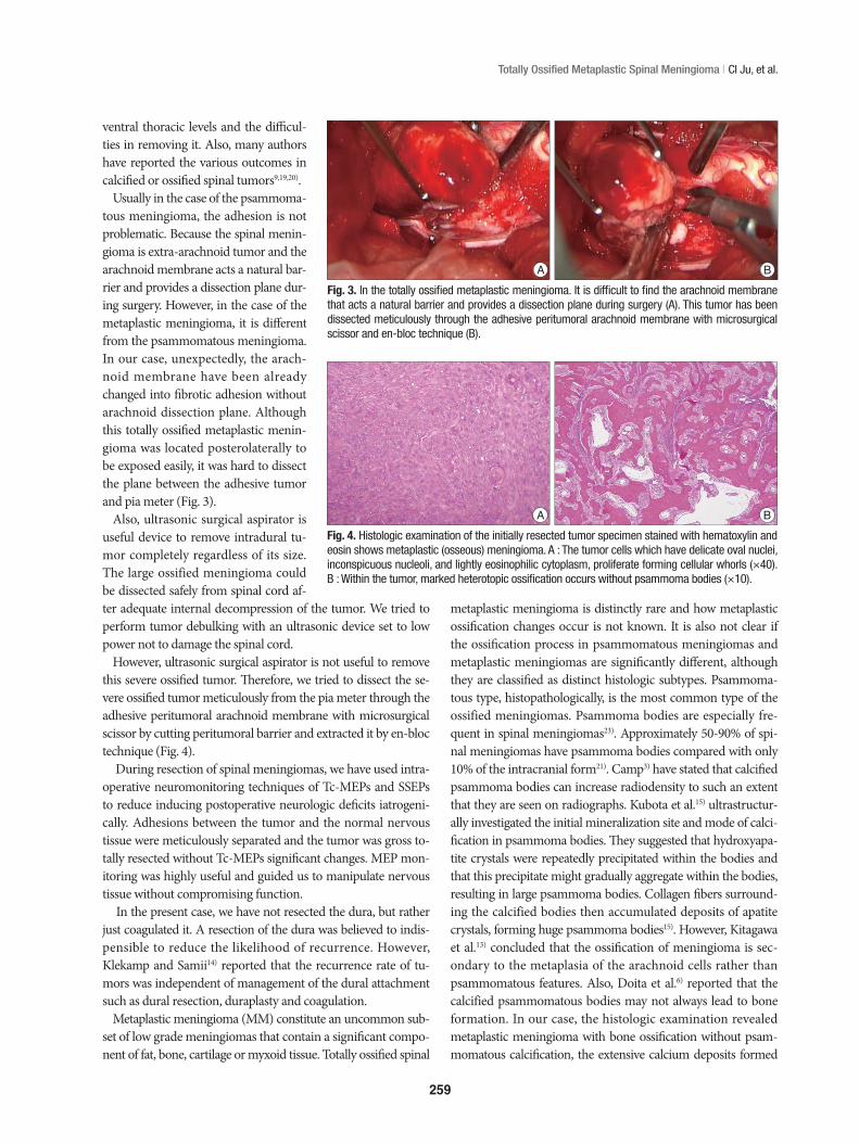
Histopathological findings demonstrated delicate oval nuclei and inconspicuous nucleoli, lightly eosinophilic cytoplasm, pro-liferate forming cellular whorls. Within the tumor, marked het-erotopic ossification occurs without psammoma bodies (Fig. 4). The findings were consistent with spinal metaplastic meningio-ma. Postoperative MR imaging revealed no evidence of residual tumor. Three days after surgery, the patient had regained full urinary continence, ambulated without an assistive device, and exhibited normal motor strength with residual numbness in her both feet. Prior to discharge from the hospital on postoperative day 31, she could ambulate without a cane and both leg numb-ness and sensation of pain as well as touch had improved. How-ever, left foot numbness persisted and it caused her hard to go
Fig. 1. Computed tomographic scan without enhancement show a large totally ossified intradural round mass occupying the whole spinal canal on T9-T10 level. Axial view on T9 (A),10 level (B) and sagittal view (C).
Fig. 2. Magnetic resonance image showing the tumor was very low signal intensity on T2-weighted image and mainly low intensity compared to spinal cord on T1-weighted image (A and B). T1-weighted imaging gadolinium showed a round homogenously enhanced tumor and the lower signal intensity of central tumor portions were less enhanced compared with the other portions of the masses (C).
A
A
B
B
C
C
259
Totally Ossified Metaplastic Spinal Meningioma | CI Ju, et al.
metaplastic meningioma is distinctly rare and how metaplastic ossification changes occur is not known. It is also not clear if the ossification process in psammomatous meningiomas and metaplastic meningiomas are significantly different, although they are classified as distinct histologic subtypes. Psammoma-tous type, histopathologically, is the most common type of the ossified meningiomas. Psammoma bodies are especially fre-quent in spinal meningiomas23). Approximately 50-90% of spi-nal meningiomas have psammoma bodies compared with only 10% of the intracranial form21). Camp3) have stated that calcified psammoma bodies can increase radiodensity to such an extent that they are seen on radiographs. Kubota et al.15) ultrastructur-ally investigated the initial mineralization site and mode of calci-fication in psammoma bodies. They suggested that hydroxyapa-tite crystals were repeatedly precipitated within the bodies and that this precipitate might gradually aggregate within the bodies, resulting in large psammoma bodies. Collagen fibers surround-ing the calcified bodies then accumulated deposits of apatite crystals, forming huge psammoma bodies15). However, Kitagawa et al.13) concluded that the ossification of meningioma is sec-ondary to the metaplasia of the arachnoid cells rather than psammomatous features. Also, Doita et al.6) reported that the calcified psammomatous bodies may not always lead to bone formation. In our case, the histologic examination revealed metaplastic meningioma with bone ossification without psam-momatous calcification, the extensive calcium deposits formed
ventral thoracic levels and the difficul-ties in removing it. Also, many authors have reported the various outcomes in calcified or ossified spinal tumors9,19,20).
Usually in the case of the psammoma-tous meningioma, the adhesion is not problematic. Because the spinal menin-gioma is extra-arachnoid tumor and the arachnoid membrane acts a natural bar-rier and provides a dissection plane dur-ing surgery. However, in the case of the metaplastic meningioma, it is different from the psammomatous meningioma. In our case, unexpectedly, the arach-noid membrane have been already changed into fibrotic adhesion without arachnoid dissection plane. Although this totally ossified metaplastic menin-gioma was located posterolaterally to be exposed easily, it was hard to dissect the plane between the adhesive tumor and pia meter (Fig. 3).
Also, ultrasonic surgical aspirator is useful device to remove intradural tu-mor completely regardless of its size. The large ossified meningioma could be dissected safely from spinal cord af-ter adequate internal decompression of the tumor. We tried to perform tumor debulking with an ultrasonic device set to low power not to damage the spinal cord.
However, ultrasonic surgical aspirator is not useful to remove this severe ossified tumor. Therefore, we tried to dissect the se-vere ossified tumor meticulously from the pia meter through the adhesive peritumoral arachnoid membrane with microsurgical scissor by cutting peritumoral barrier and extracted it by en-bloc technique (Fig. 4).
During resection of spinal meningiomas, we have used intra-operative neuromonitoring techniques of Tc-MEPs and SSEPs to reduce inducing postoperative neurologic deficits iatrogeni-cally. Adhesions between the tumor and the normal nervous tissue were meticulously separated and the tumor was gross to-tally resected without Tc-MEPs significant changes. MEP mon-itoring was highly useful and guided us to manipulate nervous tissue without compromising function.
In the present case, we have not resected the dura, but rather just coagulated it. A resection of the dura was believed to indis-pensible to reduce the likelihood of recurrence. However, Klekamp and Samii14) reported that the recurrence rate of tu-mors was independent of management of the dural attachment such as dural resection, duraplasty and coagulation.
Metaplastic meningioma (MM) constitute an uncommon sub-set of low grade meningiomas that contain a significant compo-nent of fat, bone, cartilage or myxoid tissue. Totally ossified spinal
Fig. 3. In the totally ossified metaplastic meningioma. It is difficult to find the arachnoid membrane that acts a natural barrier and provides a dissection plane during surgery (A). This tumor has been dissected meticulously through the adhesive peritumoral arachnoid membrane with microsurgical scissor and en-bloc technique (B).
A B
Fig. 4. Histologic examination of the initially resected tumor specimen stained with hematoxylin and eosin shows metaplastic (osseous) meningioma. A : The tumor cells which have delicate oval nuclei, inconspicuous nucleoli, and lightly eosinophilic cytoplasm, proliferate forming cellular whorls (×40). B : Within the tumor, marked heterotopic ossification occurs without psammoma bodies (×10).
A B

260
J Korean Neurosurg Soc 54 | September 2013
8. Garfinkle W, Yudd AP : Calcified intraspinal meningioma detected by computed tomography. Comput Radiol 6 : 305-307, 1982
9. Gezen F, Kahraman S, Canakci Z, Bedük A : Review of 36 cases of spi-nal cord meningioma. Spine (Phila Pa 1976) 25 : 727-731, 2000
10. Huang TY, Kochi M, Kuratsu J, Ushio Y : Intraspinal osteogenic menin-gioma : report of a case. J Formos Med Assoc 98 : 218-221, 1999
11. Kaufman AB, Dunsmore RH : Clinicopathological considerations in spinal meningeal calcification and ossification. Neurology 21 : 1243-1248, 1971
12. King AT, Sharr MM, Gullan RW, Bartlett JR : Spinal meningiomas : a 20-year review. Br J Neurosurg 12 : 521-526, 1998
13. Kitagawa M, Nakamura T, Aida T, Iwasaki Y, Abe H, Nagashima K : [Clinicopathologic analysis of ossification in spinal meningioma]. Noshuyo Byori 11 : 115-119, 1994
14. Klekamp J, Samii M : Surgical results for spinal meningiomas. Surg Neurol 52 : 552-562, 1999
15. Kubota T, Sato K, Yamamoto S, Hirano A : Ultrastructural study of the formation of psammoma bodies in fibroblastic meningioma. J Neuro-surg 60 : 512-517, 1984
16. Levy WJ Jr, Bay J, Dohn D : Spinal cord meningioma. J Neurosurg 57 : 804-812, 1982
17. Memon MY, Schneck L : Ventral spinal tumor : the value of computed tomography in its localization. Neurosurgery 8 : 108-111, 1981
18. Mirimanoff RO, Dosoretz DE, Linggood RM, Ojemann RG, Martuza RL : Meningioma : analysis of recurrence and progression following neurosurgical resection. J Neurosurg 62 : 18-24, 1985
19. Naderi S, Yilmaz M, Canda T, Acar U : Ossified thoracic spinal menin-gioma in childhood : a case report and review of the literature. Clin Neurol Neurosurg 103 : 247-249, 2001
20. Niijima K, Huang YP, Malis LI, Sachdev VP : Ossified spinal meningio-ma en plaque. Spine (Phila Pa 1976) 18 : 2340-2343, 1993
21. Nyström SH, Nyholm M : The origin of the calcium deposits in psam-moma bodies of human spinal meningiomas. Naturwissenschaften 53 : 703-704, 1966
22. Parizel PM, Balériaux D, Rodesch G, Segebarth C, Lalmand B, Chris-tophe C, et al. : Gd-DTPA-enhanced MR imaging of spinal tumors. AJR Am J Roentgenol 152 : 1087-1096, 1989
23. Pear BL, Boyd HR : Roentgenographically visible calcifications in spinal meningioma. Am J Roentgenol Radium Ther Nucl Med 120 : 32-45, 1974
24. Roux FX, Nataf F, Pinaudeau M, Borne G, Devaux B, Meder JF : Intra-spinal meningiomas : review of 54 cases with discussion of poor prog-nosis factors and modern therapeutic management. Surg Neurol 46 : 458-463; discussion 463-464, 1996
crystalline structures rather than the characteristic concentric laminations of psammoma bodies that support this hyposthesis.
Many authors reported that recurrence of calcification of me-ningioma is rare and variable depending on incomplete remov-al of the tumor6,7,16). In this case of a extremely rare totally ossi-fied metaplastic meningioma with grossly total resection, the postoperative recurrence and outcomes should be followed up carefully in outpatient clinic to compare with other type of me-ningioma.
CONCLUSION
Spinal MM is rare and a calcified or ossified meningioma usual-ly have adhesion of the tumor to the surrounding nerve tissue. Adhesions between the tumor and the normal nervous tissue could be meticulously separated and the ossified tumor could be totally resected without complication under Tc-MEPs monitoring. Tc-MEP monitoring is very useful method to guide us to manipu-late nervous tissue without compromising function. However, how metaplastic ossification changes occur is not known.
• Acknowledgements This study was supported by research funds from Chosun University 2011.
References 1. Antons K : Calcified spinal meningioma visible on the roentgen film.
Acta Psychiat Scand 19 : 5-9, 19442. Bonstelle CT, Vines FS : Calcification in a cervical intraspinal neurilem-
moma. Neuroradiology 10 : 231-233, 19763. Camp JD : The roentgenologic localization of tumors affecting the spi-
nal cord. Am J Roentgenol 40 : 540-544, 19384. Calogero JA, Moossy J : Extradural spinal meningiomas. Report of four
cases. J Neurosurg 37 : 442-447, 19725. Culver GJ, Concannon JP, Koenig EC : Calcification in intraspinal me-
ningiomas. Am J Roentgenol Radium Ther 62 : 237-246, 19496. Doita M, Harada T, Nishida K, Marui T, Kurosaka M, Yoshiya S : Recur-
rent calcified spinal meningioma detected by plain radiograph. Spine (Phila Pa 1976) 26 : E249-E252, 2001
7. Freidberg SR : Removal of an ossified ventral thoracic meningioma. Case report. J Neurosurg 37 : 728-730, 1972