Case Report Systemic Capillary Leak Syndrome: Is Methylene...
7
Case Report Systemic Capillary Leak Syndrome: Is Methylene Blue the Silver Bullet? Michele Umbrello, 1 Marco Gardinali, 2 Davide Ottolina, 1 Giancarlo Zanforlin, 1 and Gaetano Iapichino 1,3 1 Unit` a Operativa di Anestesia e Rianimazione, Azienda Ospedaliera San Paolo, Polo Universitario, Via A. Di Rudin` ı 8, 20142 Milano, Italy 2 Unit` a Operativa di Medicina IV, Azienda Ospedaliera San Paolo, Polo Universitario, Via A. Di Rudin` ı 8, 20142 Milano, Italy 3 Dipartimento di Fisiopatologia Medico-Chirurgica e dei Trapianti, Universit` a degli Studi di Milano, Via F. Sforza 35, 20122 Milano, Italy Correspondence should be addressed to Michele Umbrello; [email protected] Received 20 October 2014; Accepted 17 November 2014; Published 7 December 2014 Academic Editor: Zsolt Moln´ ar Copyright © 2014 Michele Umbrello et al. is is an open access article distributed under the Creative Commons Attribution License, which permits unrestricted use, distribution, and reproduction in any medium, provided the original work is properly cited. Background. Systemic capillary leak syndrome (SCLS) is a rare disorder characterized by unexplained, recurrent episodes of transient, abrupt increase in endothelial permeability, leading to severe hypotension, generalized edema, and hemoconcentration. Case Report. We report the case of a patient suffering from systemic capillary leak syndrome and present a possible interpretation of the pathophysiology of this condition. Besides the classical triad of hypotension, edema, and hemoconcentration, we recorded increased levels of methemoglobin, an index of NO overproduction. We present a possible interpretation of the pathophysiology of this condition based on the fast and complete reversal of symptoms aſter methylene blue administration (which opposes NO- induced effects) and speculate that increased NO levels could be implicated in the pathophysiology of the capillary leak phase. Why should an emergency physician be aware of this? e safety of this treatment and its fluid- and cathecolamine-sparing effect deserve consideration and further research. 1. Introduction Systemic capillary leak syndrome (SCLS) is a rare disorder characterized by unexplained, recurrent episodes of tran- sient, abrupt increase in endothelial permeability, leading to severe hypotension, generalized edema, and hemocon- centration [1]. Both etiology and pathogenesis are currently unknown, and systematic research is clearly limited by the rarity of the disease. Several hypotheses have been formu- lated, but clear evidence is lacking to support any. We describe the improvement of one patient with SCLS in two occasions with acute IV administration of methylene blue aſter failure of the medications commonly used in this setting. We present a possible interpretation of the pathophysiology of this response to therapy. 2. Case Presentation In March 2013, a 56-year-old man, otherwise healthy, suf- fered from a cold and low-grade fever and gradually devel- oped oliguria and weight gain. e general practitioner prescribed some biochemical tests, which resulted normal except for serum albumin 3 g/dL and a monoclonal IgG/ peak. An empirical course of broad-spectrum antibiotic and furosemide was started. Ten days later, he developed massive peripheral edema, bilateral pleural and pericardial effusion, and ascites. Body temperature was normal, arterial blood pressure (ABP) was 90/60, and heart rate was (HR) 120/min. Blood tests showed albumin 2.3 g/dL, creatinine 1.4 mg/dL, sodium 129 mEq/L, hemoglobin (Hb) 22 g/dL, and hematocrit (Ht) Hindawi Publishing Corporation Case Reports in Critical Care Volume 2014, Article ID 141670, 6 pages http://dx.doi.org/10.1155/2014/141670
Transcript of Case Report Systemic Capillary Leak Syndrome: Is Methylene...

Case ReportSystemic Capillary Leak Syndrome: Is MethyleneBlue the Silver Bullet?
Michele Umbrello,1 Marco Gardinali,2 Davide Ottolina,1
Giancarlo Zanforlin,1 and Gaetano Iapichino1,3
1Unita Operativa di Anestesia e Rianimazione, Azienda Ospedaliera San Paolo, Polo Universitario,Via A. Di Rudinı 8, 20142 Milano, Italy2Unita Operativa di Medicina IV, Azienda Ospedaliera San Paolo, Polo Universitario, Via A. Di Rudinı 8, 20142 Milano, Italy3Dipartimento di Fisiopatologia Medico-Chirurgica e dei Trapianti, Universita degli Studi di Milano,Via F. Sforza 35, 20122 Milano, Italy
Correspondence should be addressed to Michele Umbrello; [email protected]
Received 20 October 2014; Accepted 17 November 2014; Published 7 December 2014
Academic Editor: Zsolt Molnar
Copyright © 2014 Michele Umbrello et al. This is an open access article distributed under the Creative Commons AttributionLicense, which permits unrestricted use, distribution, and reproduction in any medium, provided the original work is properlycited.
Background. Systemic capillary leak syndrome (SCLS) is a rare disorder characterized by unexplained, recurrent episodes oftransient, abrupt increase in endothelial permeability, leading to severe hypotension, generalized edema, and hemoconcentration.Case Report. We report the case of a patient suffering from systemic capillary leak syndrome and present a possible interpretationof the pathophysiology of this condition. Besides the classical triad of hypotension, edema, and hemoconcentration, we recordedincreased levels of methemoglobin, an index of NO overproduction. We present a possible interpretation of the pathophysiologyof this condition based on the fast and complete reversal of symptoms after methylene blue administration (which opposes NO-induced effects) and speculate that increased NO levels could be implicated in the pathophysiology of the capillary leak phase.Whyshould an emergency physician be aware of this?The safety of this treatment and its fluid- and cathecolamine-sparing effect deserveconsideration and further research.
1. Introduction
Systemic capillary leak syndrome (SCLS) is a rare disordercharacterized by unexplained, recurrent episodes of tran-sient, abrupt increase in endothelial permeability, leadingto severe hypotension, generalized edema, and hemocon-centration [1]. Both etiology and pathogenesis are currentlyunknown, and systematic research is clearly limited by therarity of the disease. Several hypotheses have been formu-lated, but clear evidence is lacking to support any.
We describe the improvement of one patient with SCLSin two occasions with acute IV administration of methyleneblue after failure of the medications commonly used inthis setting. We present a possible interpretation of thepathophysiology of this response to therapy.
2. Case Presentation
In March 2013, a 56-year-old man, otherwise healthy, suf-fered from a cold and low-grade fever and gradually devel-oped oliguria and weight gain. The general practitionerprescribed some biochemical tests, which resulted normalexcept for serum albumin 3 g/dL and a monoclonal IgG/𝜅peak. An empirical course of broad-spectrum antibiotic andfurosemide was started.
Ten days later, he developed massive peripheral edema,bilateral pleural and pericardial effusion, and ascites. Bodytemperature was normal, arterial blood pressure (ABP)was 90/60, and heart rate was (HR) 120/min. Blood testsshowed albumin 2.3 g/dL, creatinine 1.4mg/dL, sodium129mEq/L, hemoglobin (Hb) 22 g/dL, and hematocrit (Ht)
Hindawi Publishing CorporationCase Reports in Critical CareVolume 2014, Article ID 141670, 6 pageshttp://dx.doi.org/10.1155/2014/141670
2 Case Reports in Critical Care
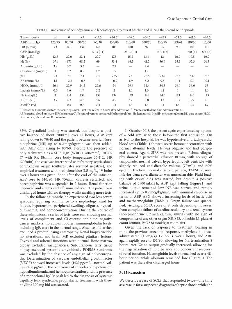
Table 1: Time course of hemodynamic and laboratory parameters at baseline and during the second acute episode.
Time (hours) BL 0 +5 +13.5 +24.5∗ +36.5 +39.5 +47.5 +54.5 +61.5 +65.5ABP (mmHg) 125/75 80/50 90/60 65/30 135/80 110/60 100/70 110/50 129/61 110/50 115/60HR (1/min) 73 140 134 120 105 100 97 112 98 102 101CVP (mmHg) — — — 2/−3 (−1) — 2/−3 (−1) — 16/7 (12) — 7/0 (4) 8/4 (6)Hb (g/dL) 12.5 22.0 22.4 22.7 17.5 15.2 13.4 12 10.9 10.5 10.2Ht (%) 37.1 67.1 68.2 69 53.4 46.5 41.2 36.9 33.5 32.3 31.5Albumin (g/dL) 3.9 3.7 3.3 — 2.7 — 2.4 — — — —Creatinine (mg/dL) 1 1.2 0.9 1.2 1.5 1.2pH 7.4 7.4 7.4 7.4 7.35 7.4 7.46 7.46 7.46 7.47 7.43BE (mmol/L) 1.1 −2.8 −0.8 −4 −0.9 4.9 8.2 9.8 11.4 12.1 10.1HCO3 (mmol/L) 26.4 22.9 24.2 22.6 24 29.6 32.4 34.3 36.1 36.6 35Lactate (mmol/L) 0.6 1.6 1.7 2.2 2 1.3 1.6 1.2 1 1.1 1.3Na (mEq/L) 141 134 134 131 137 139 141 142 143 143 143K (mEq/L) 3.7 4.3 4.6 5.6 4.2 3.7 3.8 3.4 3.3 3.5 4.1MetHb (%) — 0.5 0.6 0.4 1.3 1.4 1.5 1.4 1.5 1.3 1.7BL: baseline (3 months before the acute episode). First line refers to hours after admission. ∗Denotes methylene blue administration.ABP: arterial blood pressure; HR: heart rate; CVP: central venous pressure; Hb: haemoglobin; Ht: hematocrit;MetHb:methaemoglobin; BE: base excess; HCO3:bicarbonate; Na: sodium; K: potassium.
62%. Crystalloid loading was started, but despite a posi-tive balance of about 7000mL over 12 hours, ABP keptfalling down to 70/40 and the patient became anuric. Nore-pinephrine (NE) up to 0.2mcg/kg/min was then added,with ABP only rising to 80/60. Despite the presence ofonly tachycardia as a SIRS sign (WBC 11780/mm3, PaCO237 with RR 18/min, core body temperature 36.4∘C, HR120/min), the case was interpreted as refractory septic shockof unknown origin (cultures later resulted negative), andempirical treatment with methylene blue (1.5mg/kg IV bolusover 1 hour) was given. Soon after the end of the infusion,ABP rose to 140/80, HR 75/min, diuresis restarted, andnorepinephrine was suspended in 2 hours. Renal functionimproved and edema and effusions reduced. The patient wasdischarged home with no therapy, whilst awaiting more tests.
In the following months he experienced two less severeepisodes, requiring admittance to a nephrology ward forfatigue, hypotension, peripheral swelling, oliguria, hypoal-buminemia, and hemoconcentration. During the course ofthese admissions, a series of tests were run, showing normallevels of complement and C1-esterase inhibitor, negativecancer markers, no autoantibodies; immunoglobulin levels,including IgE, were in the normal range. Absence of diarrheaexcluded a protein losing enteropathy. Renal biopsy yieldedno alterations, and brain MR excluded pituitary lesions.Thyroid and adrenal functions were normal. Bone marrowbiopsy excluded malignancies. Subcutaneous fatty tissuebiopsy excluded systemic amyloidosis. POEMS syndromewas excluded by the absence of any sign of polyneuropa-thy. Determination of vascular endothelial growth factor(VEGF) showed increased levels (1429 pg/mL—normal val-ues <450 pg/mL).The recurrence of episodes of hypotension,hypoalbuminemia, and hemoconcentration and the presenceof a monoclonal IgG/𝜅 peak led to the diagnosis of systemiccapillary leak syndrome: prophylactic treatment with theo-phylline 300mg bid was started.
In October 2013, the patient again experienced symptomsof a cold similar to those before the first admission. Onarrival to the hospital, he was hypotensive and tachycardic;blood tests (Table 1) showed severe hemoconcentration withnormal albumin levels. He was oliguric and had periph-eral edema. Again, SIRS was not present. Echocardiogra-phy showed a pericardial effusion 18mm, with no sign oftamponade, normal valves, hypertrophic left ventricle withslightly reduced end-diastolic volume (70mL), and 60%ejection fraction, normal diastolic pattern, TAPSE 20mm.Inferior vena cava diameter was unmeasurable. Fluid load-ing with crystalloids was started, but despite a positivebalance of 5500mL/12 h, ABP kept falling (Figure 1) andurine output remained low. NE was started and rapidlyincreased up to 0.2mcg/kg/min, with minimal response interms of ABP. ABG showed increasing levels of lactic acidand methaemoglobin (Table 1). Organ failure was quanti-fied, yielding a SOFA score of 8, only depending, however,from complete failure of cardiocirculatory and renal system(norepinephrine 0.2mcg/kg/min, anuria) with no sign ofcompromise of any other organ (GCS 15, bilirubin 1.1, plateletcount 180000, PaO2 81mmHg at room air).
Given the lack of response to treatment, bearing inmind the previous anecdotal response, methylene blue wasadministered (1.5mg/kg IV bolus over 1 hour), and ABPagain rapidly rose to 135/90, allowing for NE termination 8hours later. Urine output gradually increased, allowing forthe negativization of fluid balance and concurrent recoveryof renal function. Haemoglobin levels normalized over a 48-hour period, while albumin remained low (Figure 1). Thepatient was thereafter discharged home.
3. Discussion
We describe a case of SCLS that responded twice—one timeas a rescue for a suspected diagnosis of septic shock, while the
Case Reports in Critical Care 3
Fluid balanceHb
ABPAlbFluid therapy
Time from admission (hours)
0 4 8
12
16
20
24
28
32
36
40
44
48
52
56
60
64
68
72
(mL)
0
2000
4000
6000
8000
10000
Varia
tion
from
bas
elin
e (%
)
(mm
Hg)
30
40
50
60
70
80
90
100
110
120
130
140
Methylene blue
BL
Norepinephrine (mcg/kg/min)
0.2
0.11
0.04
80
70
60
50
40
30
20
10
0
−10
−20
−30
−40
−50
−2000
−4000
(a)
Time from admission (hours)
0 4 8
12
16
20
24
28
32
36
40
44
48
52
56
60
64
68
72
Varia
tion
from
bas
elin
e (%
)
0
20
40
60
80
100
120
140
160
180
200
220
240
260
BL
−20
−40
MetHb
(b)
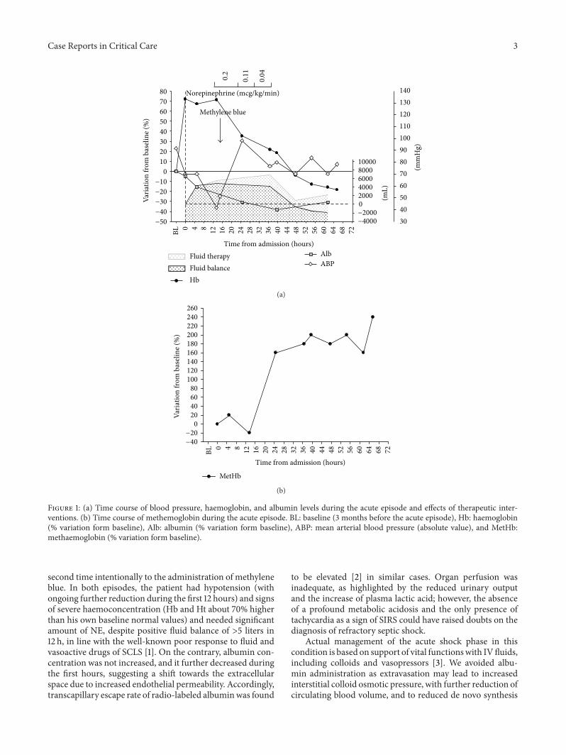
Figure 1: (a) Time course of blood pressure, haemoglobin, and albumin levels during the acute episode and effects of therapeutic inter-ventions. (b) Time course of methemoglobin during the acute episode. BL: baseline (3 months before the acute episode), Hb: haemoglobin(% variation form baseline), Alb: albumin (% variation form baseline), ABP: mean arterial blood pressure (absolute value), and MetHb:methaemoglobin (% variation form baseline).
second time intentionally to the administration of methyleneblue. In both episodes, the patient had hypotension (withongoing further reduction during the first 12 hours) and signsof severe haemoconcentration (Hb and Ht about 70% higherthan his own baseline normal values) and needed significantamount of NE, despite positive fluid balance of >5 liters in12 h, in line with the well-known poor response to fluid andvasoactive drugs of SCLS [1]. On the contrary, albumin con-centration was not increased, and it further decreased duringthe first hours, suggesting a shift towards the extracellularspace due to increased endothelial permeability. Accordingly,transcapillary escape rate of radio-labeled albuminwas found
to be elevated [2] in similar cases. Organ perfusion wasinadequate, as highlighted by the reduced urinary outputand the increase of plasma lactic acid; however, the absenceof a profound metabolic acidosis and the only presence oftachycardia as a sign of SIRS could have raised doubts on thediagnosis of refractory septic shock.
Actual management of the acute shock phase in thiscondition is based on support of vital functionswith IVfluids,including colloids and vasopressors [3]. We avoided albu-min administration as extravasation may lead to increasedinterstitial colloid osmotic pressure, with further reduction ofcirculating blood volume, and to reduced de novo synthesis
4 Case Reports in Critical Care
[4]. Infusion of pentastarch or dextrans [5] which couldseal the negatively charged endothelial fenestrations [4],were unavailable for clinical use in Italy. Echocardiographicassessment showed signs of both hypovolemia (completecollapse of the inferior vena cava) and loss of systemicvascular resistance (hypotension despite preserved systolicfunction, as indicated by the normal values of ejectionfraction and TAPSE). After about 13 hours of standardtreatment, we decided to administer methylene blue andconsequently observed the rapid reversal of hypotension, aconcomitant gradual reduction of Hb andHt back to baselinelevels, an increase in urine output, a negative fluid balance,and resolution of edema. All these signs are compatible withnormalization of endothelial permeability. Albumin levels,however, remained low, likely because the escaped moleculesonly partially flow back into vessels and are degraded intissues [6], while the turnover rate for de novo synthesis cantake up to 20 days to restore normal values [7]. Meanwhile,during the second episode, we observed a significant increaseof methaemoglobin levels.
Despite severe hypotension, the patient was awake andcooperative; moreover, hypoxemia was not present ad admis-sion, and the lung CT scan excluded pulmonary edema. Usu-ally, during SCLS attacks, musculature and connective tissueare the principal target sites of the extravasation of plasma,while the lungs, brain, and kidneys seem to be infrequentlyinvolved [8]; as such, we think that themeasurement vascularpermeability, such as the pulmonary vascular permeabilityindex derived by transpulmonary thermodilution [9], is oflimited help in this phase. However, it is quite common thatpulmonary edema develops during the recovery phase, as aniatrogenic side effect of the huge amount of intravenous fluidoften administered. In this phase, we can speculate that theavailability of such an index might potentially be of help forthe evaluation of the effects of therapy.
This patient, as the majority of those described in seriesand case reports, presented a monoclonal gammopathy [10]and elevated VEGF levels [2]; many Clarkson’s disease casespredominantly have IgG-𝜅 or IgA-𝜅 monoclonal gammopa-thy [8]: both multiple myeloma and systemic amyloidosiswere excluded via abdominal wall fat pad and bone mar-row biopsy. POEMS syndrome (polyneuropathy, organo-megaly, endocrinopathy, monoclonal gammopathy, and skinchanges), which is almost always associated with IgG-𝜆 orIgA-𝜆, was excluded by the absence of any sign of polyneu-ropathy.
The patient was also in long-term treatment with theo-phylline [11], a drug that increases intracellular cAMP levels.Any possible pathophysiologic interpretation of the responseto methylene blue should take these findings into account.In addition to its mitogenic effect, VEGF was shown toincrease endothelial permeability, in a process mediated bya NO-dependent increase in cGMP levels [12], the mainmodulator of increased vascular permeability. Moreover, IL-2 might contribute to the pathogenesis of SCLS [1], based onthe fact that IL-2 therapy can develop a leakage syndromeundistinguishable from SCLS [13]. Indeed, increased IL-2expression was demonstrated on perivascular cells of symp-tomatic cases of SCLS [14]. Nitric oxide (NO) was suggested
as themediator of IL-2-induced endothelial permeabilization[15], and inhibition of NO synthesis resulted in reversal ofIL-2-induced leakage [16]. Hence, the NO system seems tobe a common pathway for both IL-2- and VEGF-mediatedendothelial permeabilization. The vascular effects of NOare mainly mediated by the activation of soluble guanylatecyclase (sGC), which leads to the synthesis of cGMP; the lat-ter, in turn, acts on several targets eventually causing smoothmuscle relaxation [17] and increased endothelial permeability[18]. cGMP-mediated signalling also contributes to vasopres-sor hyporesponsiveness [19]. Instead, cAMP (increased bytheophylline) counteracts with this pathway, protecting thebasal barrier function [18].
The response to methylene blue supports the hypoth-esis that increased NO levels could be implicated in thepathophysiology of the acute capillary leak phase. Methyleneblue (3,7-bis(dimethylamino)-phenothiazin-5-ium chloride)is generally used in the setting of methaemoglobinemia [20]and proved effective in reversing hypotension and restoringthe response to vasoactive drugs inmany different conditionscharacterized by increased levels of NO (sepsis, anaphylaxis,severe burns, ischaemia-reperfusion injury and liver fail-ure) [21–25]. Methylene blue opposes NO-induced effectsmainly by inhibition of sGC [26–30], ultimately reducing thegeneration of cGMP. Acute methylene blue administration,unlike drugs that increase intracellular cAMP levels (such astheophylline), aims at decreasing cGMP levels to reduce theseverity of supposedly NO-mediated attacks.
Although multiple therapies were administered duringthe clinical management of the patient, both times werecorded a clear temporal relation between methylene blueadministration and reversal of the symptoms, leading us toidentify this treatment as the main cause for the change inthe clinical course. We did not report direct measures ofNO levels in the case we described. Indeed, NO has a veryshort half-life [31].However, in septic populations, circulatinglevels of methaemoglobin proved effective indicators of NOoverproduction [32]. Methaemoglobin is generated by thereaction of haemoglobin with NO [33]. In the present case,methaemoglobin levels increased up to 250% their baselinevalue during the course of the episode. We observed adelay from symptoms appearance and methaemoglobin rise:methaemoglobin generation in the presence of elevated NOmay require up to several hours to occur in vitro [34]; underin vivo conditions, this timemay even increase, depending onplasma redox state.
At pharmacologic doses methylene blue is a reducingagent via the NADPH-methemoglobin reductase pathway[20]. Methylene blue, when injected intravenously as an anti-dote, is reduced to leucomethylene blue, which then reducesthe heme group from methemoglobin to hemoglobin. How-ever, when given in higher doses, methylene blue mayoxidize the ferrous iron of hemoglobin to ferric iron, thuspotentially resulting in methemoglobin production. How-ever, the dose we administered in the present case is wellwithin the doses used in the setting of methaemoglobinemia(1-2mg/kg, repeated up to twice if symptoms of hypoxiafail to subside) [35], thus reducing the possibility that theelevated methaemoglobin levels we recorded were generated

Case Reports in Critical Care 5
by the methylene blue we administered. Elevated levels ofmethaemoglobin persisted up to the last blood sample takenin the ICU, likely after the effects of methylene blue ended;however, this is not surprising as the elimination half-life ofmethaemoglobin has been reported to be as high as 15–20hours [36].
The main limit of the present investigation is its case-report nature. However, the rarity of this condition makesexperimental studies difficult to plan, and the incompleteunderstanding of the underlying mechanisms does notallow for preclinical modeling. Moreover, the hemodynamicresponse to methylene blue might have simply faced bychance the natural course of the syndrome, which sponta-neously reversed. However, the response we observed wassimilar in two different occasions, and both times it occurredsoon after the administration of the drug.
SCLS is a rare yet potentially severe condition, oftendifficult to recognize and diagnose upon initial presentation.Current therapies are mainly supportive, and the condition isstill associated with a 10-year mortality rate of about 30% [1].We report our experience with a treatment whose safety [37],alongwith its fluid- and catecholamine-sparing effect deserveconsideration and further research.
Abbreviation List
ABP: Arterial blood pressurecAMP: Cyclic adenosine monophosphatecGMP: Cyclic guanosine monophosphateHb: HemoglobinHR: Heart rateHt: HematocritNE: NorepinephrineNO: Nitric oxideSCLS: Systemic capillary leak syndromesGC: Soluble guanilate cyclaseVEGF: Vascular endothelial growth factor.
Conflict of Interests
The authors declare that there is no conflict of interestsregarding the publication of this paper.
Authors’ Contribution
Michele Umbrello performed the literature search anddrafted the first version of the paper, Marco Gardinali andGaetano Iapichino revised the text for important intellectualcontent, and Davide Ottolina and Giancarlo Zanforlin col-lected clinical data and helped to draft the paper.
Funding
This study was carried out by departmental funding only.
References
[1] K. M. Druey and P. R. Greipp, “Narrative review: the systemiccapillary leak syndrome,” Annals of Internal Medicine, vol. 153,no. 2, pp. 90–98, 2010.
[2] W. J. Lesterhuis, A. J. Rennings, W. P. Leenders et al., “Vascularendothelial growth factor in systemic capillary leak syndrome,”The American Journal of Medicine, vol. 122, no. 6, pp. e5–e7,2009.
[3] R. P. Dellinger, M. M. Levy, A. Rhodes et al., “Surviving sepsiscampaign: international guidelines for management of severesepsis and septic shock, 2012,” Intensive Care Medicine, vol. 39,no. 2, pp. 165–228, 2013.
[4] T. E. Woodcock and T. M. Woodcock, “Revised Starling equa-tion and the glycocalyx model of transvascular fluid exchange:an improved paradigm for prescribing intravenous fluid ther-apy,” British Journal of Anaesthesia, vol. 108, no. 3, pp. 384–394,2012.
[5] S. L. Young, Y. K. Sun, W. K. Chin et al., “Two cases of systemiccapillary leak syndrome that were treated with pentastarch,”Korean Journal of Internal Medicine, vol. 22, no. 2, pp. 130–132,2007.
[6] S. Yedgar, T. E. Carew, R. C. Pittman, W. F. Beltz, and D.Steinberg, “Tissue sites of catabolism of albumin in rabbits,”American Journal of Physiology, vol. 244, no. 1, pp. E101–E107,1983.
[7] J. P. Nicholson, M. R. Wolmarans, and G. R. Park, “The role ofalbumin in critical illness,” British Journal of Anaesthesia, vol.85, no. 4, pp. 599–610, 2000.
[8] P. Kapoor, P. T. Greipp, E.W. Schaefer et al., “Idiopathic systemiccapillary leak syndrome (Clarkson’s disease): the Mayo Clinicexperience original article,”Mayo Clinic Proceedings, vol. 85, no.10, pp. 905–912, 2010.
[9] X. Monnet, N. Anguel, D. Osman, O. Hamzaoui, C. Richard,and J.-L. Teboul, “Assessing pulmonary permeability by trans-pulmonary thermodilution allows differentiation of hydrostaticpulmonary edema from ALI/ARDS,” Intensive Care Medicine,vol. 33, no. 3, pp. 448–453, 2007.
[10] W. Zhang, P. W. Ewan, and P. J. Lachmann, “The paraproteinsin systemic capillary leak syndrome,” Clinical and ExperimentalImmunology, vol. 93, no. 3, pp. 424–429, 1993.
[11] R. M. Droder, R. A. Kyle, and P. R. Greipp, “Control of systemiccapillary leak syndrome with aminophylline and terbutaline,”The American Journal of Medicine, vol. 92, no. 5, pp. 523–526,1992.
[12] W. G. Mayhan, “VEGF increases permeability of the blood-brain barrier via a nitric oxide synthase/cGMP-dependentpathway,” The American Journal of Physiology, vol. 276, no. 5,pp. C1148–C1153, 1999.
[13] S. A. Rosenberg, M. T. Lotze, L. M. Muul et al., “Observationson the systemic administration of autologous lymphokine-activated killer cells and recombinant interleukin-2 to patientswith metastatic cancer,” The New England Journal of Medicine,vol. 313, no. 23, pp. 1485–1492, 1985.
[14] M. Cicardi, M. Gardinali, G. Bisiani, A. Rosti, P. Allavena, andA.Agostoni, “The systemic capillary leak syndrome: appearanceof interleukin-2-receptor-positive cells during attacks,” Annalsof Internal Medicine, vol. 113, no. 6, pp. 475–477, 1990.
[15] J. B. Hibbs Jr., C. Westenfelder, R. Taintor et al., “Evidence forcytokine-inducible nitric oxide synthesis from L-arginine inpatients receiving interleukin-2 therapy,”The Journal of ClinicalInvestigation, vol. 89, no. 3, pp. 867–877, 1992.
[16] A. Orucevic and P. K. Lala, “NG-nitro-L-arginine methyl ester,an inhibitor of nitric oxide synthesis, ameliorates interleukin2-induced capillary leakage and reduces tumour growth inadenocarcinoma-bearing mice,” British Journal of Cancer, vol.73, no. 2, pp. 189–196, 1996.
6 Case Reports in Critical Care
[17] M. Umbrello, A. Dyson, M. Feelisch, and M. Singer, “The keyrole of nitric oxide in hypoxia: hypoxic vasodilation and energysupply-demandmatching,”Antioxidants &Redox Signaling, vol.19, no. 14, pp. 1690–1710, 2013.
[18] S. Y. Yuan, “Protein kinase signaling in the modulation ofmicrovascular permeability,” Vascular Pharmacology, vol. 39,no. 4-5, pp. 213–223, 2002.
[19] J. E. da Silva-Santos, M. R. Terluk, and J. Assreuy, “Differentialinvolvement of guanylate cyclase and potassium channels innitric oxide-induced hyporesponsiveness to phenylephrine inendotoxemic rats,” Shock, vol. 17, no. 1, pp. 70–76, 2002.
[20] W. R. Layne and R. P. Smith, “Methylene blue uptake and thereversal of chemically induced methemoglobinemias in humanerythrocytes,” Journal of Pharmacology and Experimental Ther-apeutics, vol. 165, no. 1, pp. 36–44, 1969.
[21] C. R. G. H. Daemen-Gubbels, P. H. P. Groeneveld, A. B. J.Groeneveld, G. J. Van Kamp, W. Bronsveld, and L. G. Thijs,“Methylene blue increasesmyocardial function in septic shock,”Critical Care Medicine, vol. 23, no. 8, pp. 1363–1370, 1995.
[22] J.-C. Preiser, P. Lejeune, A. Roman et al., “Methylene blueadministration in septic shock: a clinical trial,” Critical CareMedicine, vol. 23, no. 2, pp. 259–264, 1995.
[23] A. D. Jaskille, J. C. Jeng, and M. H. Jordan, “Methylene blue inthe treatment of vasoplegia following severe burns,” Journal ofBurn Care and Research, vol. 29, no. 2, pp. 408–410, 2008.
[24] H. Koelzow, J. A. Gedney, J. Baumann, N. J. Snook, and M. C.Bellamy, “The effect of methylene blue on the hemodynamicchanges during ischemia reperfusion injury in orthotopic livertransplantation,” Anesthesia and Analgesia, vol. 94, no. 4, pp.824–829, 2002.
[25] M. Y. Kirov, O. V. Evgenov, N. V. Evgenov et al., “Infusion ofmethylene blue in human septic shock: a pilot, randomized,controlled study,” Critical Care Medicine, vol. 29, no. 10, pp.1860–1867, 2001.
[26] W. Martin, G. M. Villani, D. Jothianandan, and R. F. Furchgott,“Selective blockade of endothelium-dependent and glyceryltrinitrate-induced relaxation by hemoglobin and by methyleneblue in the rabbit aorta,” The Journal of Pharmacology andExperimental Therapeutics, vol. 232, no. 3, pp. 708–716, 1985.
[27] T.M. Griffith, D.H. Edwards,M. J. Lewis, andA.H.Henderson,“Evidence that cyclic guanosinemonophosphate (cGMP)medi-ates endothelium-dependent relaxation,” European Journal ofPharmacology, vol. 112, no. 2, pp. 195–202, 1985.
[28] L. J. Ignarro, R. G. Harbison, K. S. Wood, and P. J. Kad-owitz, “Dissimilarities between methylene blue and cyanide onrelaxation and cyclic GMP formation in endothelium-intactintrapulmonary artery caused by nitrogen oxide-containingvasodilators and acetylcholine,” Journal of Pharmacology andExperimental Therapeutics, vol. 236, no. 1, pp. 30–36, 1986.
[29] C. A. Gruetter, P. J. Kadowitz, and L. J. Ignarro, “Methyleneblue inhibits coronary arterial relaxation and guanylate cyclaseactivation by nitroglycerin, sodium nitrite, and amyl nitrite,”Canadian Journal of Physiology and Pharmacology, vol. 59, no.2, pp. 150–156, 1981.
[30] R. J. Gryglewski, A. Zembowicz, D. Salvemini, G. W. Taylor,and J. R. Vane, “Modulation of the pharmacological actionsof nitrovasodilators by methylene blue and pyocyanin,” BritishJournal of Pharmacology, vol. 106, no. 4, pp. 838–845, 1992.
[31] X. Liu, Q. Yan, K. L. Baskerville, and J. L. Zweier, “Estimationof nitric oxide concentration in blood for different rates ofgeneration: evidence that intravascular nitric oxide levels are
too low to exert physiological effects,” The Journal of BiologicalChemistry, vol. 282, no. 12, pp. 8831–8836, 2007.
[32] K. Ohashi, H. Yukioka, M. Hayashi, and A. Asada, “Elevatedmethemoglobin in patients with sepsis,” Acta AnaesthesiologicaScandinavica, vol. 42, no. 6, pp. 713–716, 1998.
[33] V. S. Sharma, R. A. Isaacson, M. E. John, M. R. Waterman,and M. Chevion, “Reaction of nitric oxide with heme pro-teins: studies on metmyoglobin, opossummethemoglobin, andmicroperoxidase,” Biochemistry, vol. 22, no. 16, pp. 3897–3902,1983.
[34] N. Maeda, K. Imaizumi, K. Kon, and T. Shiga, “A kineticstudy on functional impairment of nitric oxide-exposed raterythrocytes,” Environmental Health Perspectives, vol. 73, pp.171–177, 1987.
[35] A. Skold, D. L. Cosco, and R. Klein, “Methemoglobinemia:pathogenesis, diagnosis, and management,” Southern MedicalJournal, vol. 104, no. 11, pp. 757–761, 2011.
[36] S. M. Raso, J. B. Fernandez, E. A. Beobide, and A. F. Landaluce,“Methemoglobinemia andCNS toxicity after topical applicationof EMLA to a 4-year-old girl with molluscum contagiosum,”Pediatric Dermatology, vol. 23, no. 6, pp. 592–593, 2006.
[37] L. Pasin, M. Umbrello, T. Greco et al., “Methylene blue as avasopressor: a meta-analysis of randomised trials,”Critical Careand Resuscitation, vol. 15, no. 1, pp. 42–48, 2013.

Submit your manuscripts athttp://www.hindawi.com
Stem CellsInternational
Hindawi Publishing Corporationhttp://www.hindawi.com Volume 2014
Hindawi Publishing Corporationhttp://www.hindawi.com Volume 2014
MEDIATORSINFLAMMATION
of
Hindawi Publishing Corporationhttp://www.hindawi.com Volume 2014
Behavioural Neurology
EndocrinologyInternational Journal of
Hindawi Publishing Corporationhttp://www.hindawi.com Volume 2014
Hindawi Publishing Corporationhttp://www.hindawi.com Volume 2014
Disease Markers
Hindawi Publishing Corporationhttp://www.hindawi.com Volume 2014
BioMed Research International
OncologyJournal of
Hindawi Publishing Corporationhttp://www.hindawi.com Volume 2014
Hindawi Publishing Corporationhttp://www.hindawi.com Volume 2014
Oxidative Medicine and Cellular Longevity
Hindawi Publishing Corporationhttp://www.hindawi.com Volume 2014
PPAR Research
The Scientific World JournalHindawi Publishing Corporation http://www.hindawi.com Volume 2014
Immunology ResearchHindawi Publishing Corporationhttp://www.hindawi.com Volume 2014
Journal of
ObesityJournal of
Hindawi Publishing Corporationhttp://www.hindawi.com Volume 2014
Hindawi Publishing Corporationhttp://www.hindawi.com Volume 2014
Computational and Mathematical Methods in Medicine
OphthalmologyJournal of
Hindawi Publishing Corporationhttp://www.hindawi.com Volume 2014
Diabetes ResearchJournal of
Hindawi Publishing Corporationhttp://www.hindawi.com Volume 2014
Hindawi Publishing Corporationhttp://www.hindawi.com Volume 2014
Research and TreatmentAIDS
Hindawi Publishing Corporationhttp://www.hindawi.com Volume 2014
Gastroenterology Research and Practice
Hindawi Publishing Corporationhttp://www.hindawi.com Volume 2014
Parkinson’s Disease
Evidence-Based Complementary and Alternative Medicine
Volume 2014Hindawi Publishing Corporationhttp://www.hindawi.com