Case Report Pseudomembranous trigonitis in a male with ... · Case Report Pseudomembranous...
5
Int J Clin Exp Pathol 2014;7(6):3375-3379 www.ijcep.com /ISSN:1936-2625/IJCEP0000435 Case Report Pseudomembranous trigonitis in a male with Klinefelter syndrome: a case report and evidence of a hormonal etiology Derrick WQ Lian 1 , Fay X Li 2 , Caroline CP Ong 2 , CH Kuick 1 , Kenneth TE Chang 1 Departments of 1 Pathology and Laboratory Medicine, 2 Paediatric Surgery, KK Women’s and Children’s Hospital, Singapore Received April 6, 2014; Accepted May 26, 2014; Epub May 15, 2014; Published June 1, 2014 Abstract: Klinefelter syndrome is a clinical syndrome with a distinct 47, XXY karyotype. Patients are characterized by a tall eunuchoid stature, small testes, hypergonotrophic hypogonadism, gynecomastia, learning difficulties and infertility. These patients have also been found to have raised estrogen levels. We report a 16 year old boy with Kline- felter syndrome presenting to our institution with gross hematuria. Cystoscopy and biopsy revealed the diagnosis of pseudomembranous trigonitis. Immunohistochemical stains showed an increase in estrogen and progesterone receptors in the trigone area but not in the rest of the bladder. In view of the patient’s mildly raised estrogen levels and the histological findings, we postulate that estrogen is the driver of the development of pseudomembranous trigonitis. This is the first reported case of pseudomembranous trigonitis seen in association with Klinefelter syn- drome, and also the first case of pseudomembranous trigonitis occurring within the male adolescent age group. Keywords: Klinefelter syndrome, pseudomembranous trigonitis, pediatric Introduction Klinefelter syndrome is the most common chro- mosomal aberration in males and the most common cause of male hypogonadism [1]. The genotype of Klinefelter syndrome is the result of meiotic nondisjunction, resulting in a 47, XXY karyotype. Patients have a tall eunuchoid stat- ure, small testes, hypergonotrophic hypogonad- ism, gynecomastia, learning difficulties and infertility. In addition, Klinefelter syndrome is associated with raised estrogen levels, and cor- respondingly, an increased risk of breast can- cer, mediastinal germ cell cancer, endocrine complications and osteoporosis [1, 2]. Case report A 16 year old boy with Klinefelter syndrome, presented to the surgical outpatient clinic with the complaint of recurrent intermittent gross hematuria over the past 1 year. The gross hematuria affected mainly the terminal urine stream and occasionally, fresh blood clots would also be passed. This was associated with a sense of incomplete voiding. These epi- sodes were initially treated with a course of oral antibiotics by a primary care physician with no resolution of symptoms. Initial investigations consisting of a plain radio- graph and an ultrasound scan of the urinary system were unrevealing of a diagnosis. Uri- nalysis did not demonstrate any casts or crys- tals, and urine phase contrast could not be car- ried out due to inadequate red blood cell counts in the sample of urine provided. Urine culture was negative for bacterial growth. The full blood count and coagulation profile were also normal. His serum estradiol level was marginally high at 165 pmol/L (upper limit of normal estradiol for males 161 pmol/L). During rigid cystoscopy, the bladder mucosa appeared inflamed with prominent superficial vessels. Mucosa over the trigone area had a raised, whitish-grey appearance, suspicious for pseudomembranous trigonitis (Figure 1). Biop- sies from the trigone and the dome of the blad- der were taken for histological confirmation.
Transcript of Case Report Pseudomembranous trigonitis in a male with ... · Case Report Pseudomembranous...
Int J Clin Exp Pathol 2014;7(6):3375-3379www.ijcep.com /ISSN:1936-2625/IJCEP0000435
Case Report Pseudomembranous trigonitis in a male with Klinefelter syndrome: a case report and evidence of a hormonal etiology
Derrick WQ Lian1, Fay X Li2, Caroline CP Ong2, CH Kuick1, Kenneth TE Chang1
Departments of 1Pathology and Laboratory Medicine, 2Paediatric Surgery, KK Women’s and Children’s Hospital, Singapore
Received April 6, 2014; Accepted May 26, 2014; Epub May 15, 2014; Published June 1, 2014
Abstract: Klinefelter syndrome is a clinical syndrome with a distinct 47, XXY karyotype. Patients are characterized by a tall eunuchoid stature, small testes, hypergonotrophic hypogonadism, gynecomastia, learning difficulties and infertility. These patients have also been found to have raised estrogen levels. We report a 16 year old boy with Kline-felter syndrome presenting to our institution with gross hematuria. Cystoscopy and biopsy revealed the diagnosis of pseudomembranous trigonitis. Immunohistochemical stains showed an increase in estrogen and progesterone receptors in the trigone area but not in the rest of the bladder. In view of the patient’s mildly raised estrogen levels and the histological findings, we postulate that estrogen is the driver of the development of pseudomembranous trigonitis. This is the first reported case of pseudomembranous trigonitis seen in association with Klinefelter syn-drome, and also the first case of pseudomembranous trigonitis occurring within the male adolescent age group.
Keywords: Klinefelter syndrome, pseudomembranous trigonitis, pediatric
Introduction
Klinefelter syndrome is the most common chro-mosomal aberration in males and the most common cause of male hypogonadism [1]. The genotype of Klinefelter syndrome is the result of meiotic nondisjunction, resulting in a 47, XXY karyotype. Patients have a tall eunuchoid stat-ure, small testes, hypergonotrophic hypogonad-ism, gynecomastia, learning difficulties and infertility. In addition, Klinefelter syndrome is associated with raised estrogen levels, and cor-respondingly, an increased risk of breast can-cer, mediastinal germ cell cancer, endocrine complications and osteoporosis [1, 2].
Case report
A 16 year old boy with Klinefelter syndrome, presented to the surgical outpatient clinic with the complaint of recurrent intermittent gross hematuria over the past 1 year. The gross hematuria affected mainly the terminal urine stream and occasionally, fresh blood clots would also be passed. This was associated
with a sense of incomplete voiding. These epi-sodes were initially treated with a course of oral antibiotics by a primary care physician with no resolution of symptoms.
Initial investigations consisting of a plain radio-graph and an ultrasound scan of the urinary system were unrevealing of a diagnosis. Uri- nalysis did not demonstrate any casts or crys-tals, and urine phase contrast could not be car-ried out due to inadequate red blood cell counts in the sample of urine provided. Urine culture was negative for bacterial growth. The full blood count and coagulation profile were also normal. His serum estradiol level was marginally high at 165 pmol/L (upper limit of normal estradiol for males 161 pmol/L).
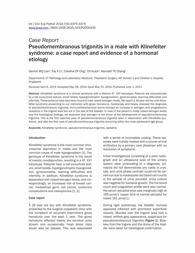
During rigid cystoscopy, the bladder mucosa appeared inflamed with prominent superficial vessels. Mucosa over the trigone area had a raised, whitish-grey appearance, suspicious for pseudomembranous trigonitis (Figure 1). Biop- sies from the trigone and the dome of the blad-der were taken for histological confirmation.

Pseudomembranous trigonitis in a male with Klinefelter syndrome
3376 Int J Clin Exp Pathol 2014;7(6):3375-3379
Figure 1. Cystoscopic images of the bladder. The trigone area showed the characteristic gross appearance of pseu-domembranous trigonitis, with a raised, whitish grey appearance (A). The bladder neck (B) and verumontanum (C) were inflamed. The urethra (D) was unremarkable.
Figure 2. Histological examination of the trigone area. There was focal non-keratinizing squamous metaplasia (A). Elsewhere in the trigone, unremarkable urothelium is seen (B).

Pseudomembranous trigonitis in a male with Klinefelter syndrome
3377 Int J Clin Exp Pathol 2014;7(6):3375-3379
Histological examination revealed non-keratin-izing squamous metaplasia isolated to the tri-gone region (Figure 2). The squamous cells were vacuolated and contained glycogen, giv-ing an appearance similar to that of the vaginal
epithelium. The rest of the bladder biopsies were unremarkable. Immunohistochemical st- udies showed expression of estrogen and pro-gesterone receptors in the epithelial cells of the trigone area, with increased density of expres-
Figure 3. Immunohistochemical study of the trigone area. The region of squamous metaplasis within the trigone was positive for estrogen receptors (A) and progesterone receptors (B). The urothelium in the trigone adjacent to the area of squamous metaplasia also showed patchy positivity for estrogen receptors (C) and progesterone receptors (D).

Pseudomembranous trigonitis in a male with Klinefelter syndrome
3378 Int J Clin Exp Pathol 2014;7(6):3375-3379
sion at the site of non-keratinizing squamous metaplasia (Figure 3). In contrast, neither estr- ogen receptors nor progesterone receptors were expressed in the transitional epithelium of the other bladder sites of the same patient (Figure 4).
Discussion
This is the first known case report of an asso-ciation between Klinefelter syndrome and pse- udomembranous trigonitis.
The term pseudomembranous trigonitis is a misnomer as the condition does not have a pseudomembrane histologically, and is a meta-plastic rather than inflammatory entity. It is seen almost exclusively in women [3], who present with urgency and frequency of micturi-tion, termed as the ‘urethral syndrome’. The uri-nalysis is normal with a sterile urine culture. On cystoscopy, an abnormal, white to greyish, heaped up mucosa with a serpiginous and hyperemic margin is seen, confined to the tri-gone region of the bladder. The rest of the blad-der is normal. On histology, the epithelium at the trigone area shows a non-keratinizing squa-mous metaplasia, thicker than normal transi-tional epithelium [3]. Studies have shown the expression of estrogen receptors in the trigonal transitional epithelium of female bladders, and not at other bladder sites, supporting a hor-monal etiology for the development of pseudo-membranous trigonitis [4, 5].
The findings of raised estrogen levels in con-junction with increased expression of estrogen
receptors in the trigone area of the biopsy, sug-gest that estrogen is the etiological driver for the development of pseudomembranous cysti-tis in our patient. One of the rare male patients reported to have pseudomembranous cystitis in the literature had a similar hormonal etiolo-gy; he was receiving estrogen for carcinoma of the prostate [3].
With raised estrogen levels, it is entirely plausi-ble for patients with Klinefelter syndrome to be at risk for female-predominated diseases such as pseudomembranous trigonitis. In support of this is the association of Klinefelter syndrome with estrogen-driven cancers such as breast cancer.
Pseudomembranous trigonitis is exceedingly rare in men. To date, only 6 men with pseudo-membranous trigonitis have been reported in the literature [3, 6]. Although the presence of estrogen receptors in the trigone area of female bladders has been well documented [4], this is the first report demonstrating a similar immu-nohistochemical hormonal profile in the male bladder. This suggests an identical embryologi-cal development of the trigone region in both genders.
Bacterial cystitis, especially recurrent infec-tions, has been cited to be associated with the development of pseudomembranous trigonitis [6]. However, our patient did not have any docu-mentation of urinary tract infections prior to cystoscopy, and no histological features of chronic inflammation such as cystitis cystica, cystitis glandularis or a chronic inflammatory
Figure 4. Immunohistochemical study of urothelium away from the trigone area. In contrast, both estrogen receptors (A) and progesterone receptors (B) are negative in the urothelium away from the trigone area. Some stromal cells show positive staining.

Pseudomembranous trigonitis in a male with Klinefelter syndrome
3379 Int J Clin Exp Pathol 2014;7(6):3375-3379
infiltrate within the lamina propria was seen. Thus, in our patient, it is likely that urinary tract infection played any major role in the develop-ment of pseudomembranous trigonitis.
Cytological examination of the urine in pseudo-membranous cystitis is likely to yield squamous cells. This case report raises the possibility of using clean-catch urine cytology to make a pre-sumptive diagnosis of pseudomembranous tri-gonitis in a patient with Klinefelter syndrome, with known raised serum estrogen levels and presenting with gross hematuria and/or lower urinary tract symptoms. In this select group of patients with otherwise normal radiological and biochemical investigations, and who are at low risk of urologic malignancies, the use of clean-catch urine cytology may potentially avoid the need for an invasive procedure such as cystoscopy.
Disclosure of conflict of interest
None.
Address correspondence to: Dr. Derrick WQ Lian, Department of Pathology and Laboratory Medicine, KK Women’s and Children’s Hospital, Basement 1 Children’s Tower, 100 Bukit Timah Road, Singapore 229899. Tel: (65) 6394 8748; Fax: (65) 6394 1372; E-mail: [email protected]
References
[1] Wosnitzer MS, Paduch DA. Endocrinological is-sues and hormonal manipulation in children and men with Klinefelter syndrome. Am J Med Genet C Semin Med Genet 2013; 163C: 16-26.
[2] Groth KA, Skakkebæk A, Høst C, Gravholt CH, Bojesen A. Clinical review: Klinefelter syn-drome--a clinical update. J Clin Endocrinol Metab 2013; 98: 20-30.
[3] Henry L, Fox M. Histological findings in pseudo-membranous trigonitis. J Clin Pathol 1971; 24: 605-8.
[4] Stephenson TJ, Henry L, Harris SC, Giri DD, Fox M, Underwood JC. Pseudomembranous trigo-nitis of the bladder: hormonal aetiology. J Clin Pathol 1989; 42: 922-6.
[5] Pacchioni D, Revelli A, Casetta G, Cassoni P, Piana P, Tizzani A, Bussolati G, Massobrio M. Immunohistochemical detection of estrogen and progesterone receptors in the normal uri-nary bladder and in pseudomembranous trigo-nitis. J Endocrinol Invest 1992; 15: 719-25.
[6] Ozbey I, Aksoy Y, Polat O, Biçgi O, Demirel A. Squamous metaplasia of the bladder: findings in 14 patients and review of the literature. Int Urol Nephrol 1999; 31: 457-61.