Case Report OrbitalApexSyndromeinHerpesZosterOphthalmicus · 4 Case Reports in Ophthalmological...
5
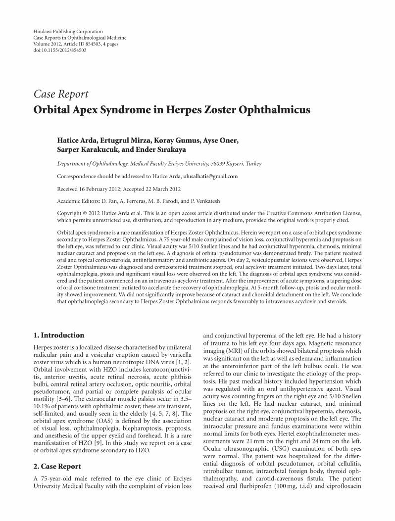
Hindawi Publishing Corporation Case Reports in Ophthalmological Medicine Volume 2012, Article ID 854503, 4 pages doi:10.1155/2012/854503 Case Report Orbital Apex Syndrome in Herpes Zoster Ophthalmicus Hatice Arda, Ertugrul Mirza, Koray Gumus, Ayse Oner, Sarper Karakucuk, and Ender Sırakaya Department of Ophthalmology, Medical Faculty Erciyes University, 38039 Kayseri, Turkey Correspondence should be addressed to Hatice Arda, [email protected] Received 16 February 2012; Accepted 22 March 2012 Academic Editors: D. Fan, A. Ferreras, M. B. Parodi, and P. Venkatesh Copyright © 2012 Hatice Arda et al. This is an open access article distributed under the Creative Commons Attribution License, which permits unrestricted use, distribution, and reproduction in any medium, provided the original work is properly cited. Orbital apex syndrome is a rare manifestation of Herpes Zoster Ophthalmicus. Herein we report on a case of orbital apex syndrome secondary to Herpes Zoster Ophthalmicus. A 75 year-old male complained of vision loss, conjunctival hyperemia and proptosis on the left eye, was referred to our clinic. Visual acuity was 5/10 Snellen lines and he had conjunctival hyperemia, chemosis, minimal nuclear cataract and proptosis on the left eye. A diagnosis of orbital pseudotumor was demonstrated firstly. The patient received oral and topical corticosteroids, antiinflammatory and antibiotic agents. On day 2, vesiculopustular lesions were observed, Herpes Zoster Ophthalmicus was diagnosed and corticosteroid treatment stopped, oral acyclovir treatment initiated. Two days later, total ophthalmoplegia, ptosis and significant visual loss were observed on the left. The diagnosis of orbital apex syndrome was consid- ered and the patient commenced on an intravenous acyclovir treatment. After the improvement of acute symptoms, a tapering dose of oral cortisone treatment initiated to accelarate the recovery of ophthalmoplegia. At 5-month follow-up, ptosis and ocular motil- ity showed improvement. VA did not significantly improve because of cataract and choroidal detachment on the left. We conclude that ophthalmoplegia secondary to Herpes Zoster Ophthalmicus responds favourably to intravenous acyclovir and steroids. 1. Introduction Herpes zoster is a localized disease characterised by unilateral radicular pain and a vesicular eruption caused by varicella zoster virus which is a human neurotropic DNA virus [1, 2]. Orbital involvement with HZO includes keratoconjunctivi- tis, anterior uveitis, acute retinal necrosis, acute phthisis bulbi, central retinal artery occlusion, optic neuritis, orbital pseudotumor, and partial or complete paralysis of ocular motility [3–6]. The extraocular muscle palsies occur in 3.5– 10.1% of patients with ophthalmic zoster; these are transient, self-limited, and usually seen in the elderly [4, 5, 7, 8]. The orbital apex syndrome (OAS) is defined by the association of visual loss, ophthalmoplegia, blepharoptosis, proptosis, and anesthesia of the upper eyelid and forehead. It is a rare manifestation of HZO [9]. In this study we report on a case of orbital apex syndrome secondary to HZO. 2. Case Report A 75-year-old male referred to the eye clinic of Erciyes University Medical Faculty with the complaint of vision loss and conjunctival hyperemia of the left eye. He had a history of trauma to his left eye four days ago. Magnetic resonance imaging (MRI) of the orbits showed bilateral proptosis which was significant on the left as well as edema and inflammation at the anteroinferior part of the left bulbus oculi. He was referred to our clinic to investigate the etiology of the prop- tosis. His past medical history included hypertension which was regulated with an oral antihypertensive agent. Visual acuity was counting fingers on the right eye and 5/10 Snellen lines on the left. He had nuclear cataract, and minimal proptosis on the right eye, conjunctival hyperemia, chemosis, nuclear cataract and moderate proptosis on the left eye. The intraocular pressure and fundus examinations were within normal limits for both eyes. Hertel exophthalmometer mea- surements were 21 mm on the right and 24 mm on the left. Ocular ultrasonographic (USG) examination of both eyes were normal. The patient was hospitalized for the differ- ential diagnosis of orbital pseudotumor, orbital cellulitis, retrobulbar tumor, intraorbital foreign body, thyroid oph- thalmopathy, and carotid-cavernous fistula. The patient received oral flurbiprofen (100 mg, t.i.d) and ciprofloxacin
Transcript of Case Report OrbitalApexSyndromeinHerpesZosterOphthalmicus · 4 Case Reports in Ophthalmological...
Hindawi Publishing CorporationCase Reports in Ophthalmological MedicineVolume 2012, Article ID 854503, 4 pagesdoi:10.1155/2012/854503
Case Report
Orbital Apex Syndrome in Herpes Zoster Ophthalmicus
Hatice Arda, Ertugrul Mirza, Koray Gumus, Ayse Oner,Sarper Karakucuk, and Ender Sırakaya
Department of Ophthalmology, Medical Faculty Erciyes University, 38039 Kayseri, Turkey
Correspondence should be addressed to Hatice Arda, [email protected]
Received 16 February 2012; Accepted 22 March 2012
Academic Editors: D. Fan, A. Ferreras, M. B. Parodi, and P. Venkatesh
Copyright © 2012 Hatice Arda et al. This is an open access article distributed under the Creative Commons Attribution License,which permits unrestricted use, distribution, and reproduction in any medium, provided the original work is properly cited.
Orbital apex syndrome is a rare manifestation of Herpes Zoster Ophthalmicus. Herein we report on a case of orbital apex syndromesecondary to Herpes Zoster Ophthalmicus. A 75 year-old male complained of vision loss, conjunctival hyperemia and proptosis onthe left eye, was referred to our clinic. Visual acuity was 5/10 Snellen lines and he had conjunctival hyperemia, chemosis, minimalnuclear cataract and proptosis on the left eye. A diagnosis of orbital pseudotumor was demonstrated firstly. The patient receivedoral and topical corticosteroids, antiinflammatory and antibiotic agents. On day 2, vesiculopustular lesions were observed, HerpesZoster Ophthalmicus was diagnosed and corticosteroid treatment stopped, oral acyclovir treatment initiated. Two days later, totalophthalmoplegia, ptosis and significant visual loss were observed on the left. The diagnosis of orbital apex syndrome was consid-ered and the patient commenced on an intravenous acyclovir treatment. After the improvement of acute symptoms, a tapering doseof oral cortisone treatment initiated to accelarate the recovery of ophthalmoplegia. At 5-month follow-up, ptosis and ocular motil-ity showed improvement. VA did not significantly improve because of cataract and choroidal detachment on the left. We concludethat ophthalmoplegia secondary to Herpes Zoster Ophthalmicus responds favourably to intravenous acyclovir and steroids.
1. Introduction
Herpes zoster is a localized disease characterised by unilateralradicular pain and a vesicular eruption caused by varicellazoster virus which is a human neurotropic DNA virus [1, 2].Orbital involvement with HZO includes keratoconjunctivi-tis, anterior uveitis, acute retinal necrosis, acute phthisisbulbi, central retinal artery occlusion, optic neuritis, orbitalpseudotumor, and partial or complete paralysis of ocularmotility [3–6]. The extraocular muscle palsies occur in 3.5–10.1% of patients with ophthalmic zoster; these are transient,self-limited, and usually seen in the elderly [4, 5, 7, 8]. Theorbital apex syndrome (OAS) is defined by the associationof visual loss, ophthalmoplegia, blepharoptosis, proptosis,and anesthesia of the upper eyelid and forehead. It is a raremanifestation of HZO [9]. In this study we report on a caseof orbital apex syndrome secondary to HZO.
2. Case Report
A 75-year-old male referred to the eye clinic of ErciyesUniversity Medical Faculty with the complaint of vision loss
and conjunctival hyperemia of the left eye. He had a historyof trauma to his left eye four days ago. Magnetic resonanceimaging (MRI) of the orbits showed bilateral proptosis whichwas significant on the left as well as edema and inflammationat the anteroinferior part of the left bulbus oculi. He wasreferred to our clinic to investigate the etiology of the prop-tosis. His past medical history included hypertension whichwas regulated with an oral antihypertensive agent. Visualacuity was counting fingers on the right eye and 5/10 Snellenlines on the left. He had nuclear cataract, and minimalproptosis on the right eye, conjunctival hyperemia, chemosis,nuclear cataract and moderate proptosis on the left eye. Theintraocular pressure and fundus examinations were withinnormal limits for both eyes. Hertel exophthalmometer mea-surements were 21 mm on the right and 24 mm on the left.Ocular ultrasonographic (USG) examination of both eyeswere normal. The patient was hospitalized for the differ-ential diagnosis of orbital pseudotumor, orbital cellulitis,retrobulbar tumor, intraorbital foreign body, thyroid oph-thalmopathy, and carotid-cavernous fistula. The patientreceived oral flurbiprofen (100 mg, t.i.d) and ciprofloxacin

2 Case Reports in Ophthalmological Medicine
Figure 1: Vesicular lesions on the left upper eyelid.
(750 mg, b.i.d), topical moxifloxacine and dexamethasone6 times daily and cold compresses as an initial treatment.Orbital computerized tomography showed no foreign body,and there was no murmur on auscultation of the glob. Theproptosis was not pulsatile. The visual acuity on the left eyedecreased to 4/10 Snellen lines one day after the hospitaliza-tion although the other ocular signs remained the same. Afterthe consultations with the departments of infectious diseasesand endocrinology, the diagnosis of orbital cellulitis and/orthyroid ophthalmopathy was not considered. An oral steroid(fluocortolone 1 mg/kg,) and preventive treatment for thegastric mucosa (ranitidine HCL, b.i.d) were added to thetreatment because of the possible diagnosis of orbitalpseudotumor.
On the second day of the hospitalization, vesicular andpustular lesions occurred around the left eyelid and forehead,and the diagnosis of HZO was made (Figure 1). The visualacuity of the left eye rapidly decreased to counting fingersfrom 3 meters, and a punctate keratopathy was detectedon slit-lamp examination. After the consultation with thedepartment of dermatology, oral corticosteroid treatmentwas stopped, and the patient was commenced on a treatmentof oral valacyclovir (1 g, t.i.d. for 7 days), vitamin B1 (250 mg,b.i.d), vitamin B6 (250 mg, b.i.d), and dressing with ethacri-dine lactate for the skin lesions. The visual acuity of the lefteye decreased to hand motions one day after the initializationof this treatment. Total ophthalmoplegia and ptosis ofthe left eye were detected (Figure 2). There were dendriticlesions on the cornea (Figure 3). Since a brief period ofdisorientation and a syncope was developed, a diagnosis ofOAS and a probable cranial involvement was consideredand the patient was referred to the department of infectiousdiseases. The patient was commenced on intravenous ampi-cillin sulbactam (2 gr, t.i.d.) and acyclovir (750 mg, t.i.d.) for14 days, topical acyclovir ointment 5 times, moxifloxacinand polyacrylic acid 6 times, and a fixed combination ofdorzolamide HCL and timolol maleate 2 times per day. Twodays later uveitis with cyclitic membrane and hemorrhagein the anterior chamber were developed (Figure 4). Topicalcycloplegic drops and hot eye compresses were added to thetreatment 5 times per day. Topical steroids were not consid-ered because of the diffuse corneal epithelial defects. MRI
examination showed an enlargement of the extraocularmuscles and an increase in the amount of the soft tissuesaround the preseptal area; both of the cavernous sinuses werenormal. Both of the superior orbital veins were dilated, whichwere more significant on the left. There was a choroidaldetachment and an intraocular hemorrhage on the left eye.Diffusion MRI showed a deficiency of the diffusion on theright frontal and frontoparietal regions.
When the acute clinical signs of HZO were regressed,the patient was commenced on a tapering dose of oral pred-nisolone (1 mg/kg per day) to accelerate the recovery of thetotal ophthalmoplegia, proptosis, and ptosis. Topical corti-sone treatment (dexamethasone) 8 times was added after therecovery of the corneal epithelial defects.
At the end of 5-month follow-up period, the visual acuityof the left eye was counting fingers from 1.5 meters. Anteriorsegment examination showed an epithelial defect at theinferior half of the cornea, posterior synechiae at thepupillary area, fibrinoid reaction in the anterior chamber andmature cataract. The intraocular pressure of the left eye was4 mmHg (applanation). The USG examination of the lefteye revealed a choroidal detachment. The corneal sensation,ocular motility, and the ptosis showed partial improvementat the end of five months (Figure 5).
3. Discussion
HZO is a disease in which the ophthalmic division of thetrigeminal nerve is affected by the varicella virus [10]. Thecommon ocular manifestations of HZO include blepharo-conjunctivitis, keratitis, and uveitis [11]. OAS is a rare andsevere manifestation of HZO which was to the best of ourknowledge reported in only five cases previously [9, 12–14].We reported the sixth case of HZO which progressed to OASin the literature. OAS is characterized by the involvement ofthe 2nd, 3rd, 4th, and 6th cranial nerves with the paralysis ofophthalmic branch of the 5th cranial nerve due to inflamma-tory, infectious, neoplastic, traumatic, vascular, and some-times iatrogenic reasons along the ophthalmic canal region[15]. In our case there was a history of trauma and a viralinfection but not any malignancy. The two of the previouslyreported cases had immunodeficiency; one had Hodgkin’sdisease and in the other case, human immunodeficiencyvirus was positive [12, 14]. All of the cases which had orbitalapex syndrome secondary to HZO in the literature weretreated with systemic acyclovir and steroids. The treatmentwith intravenous acyclovir and steroids usually carries a goodprognosis with the exception of the presence of immuno-suppression. Superior orbital fissure syndrome (SOFS) hasthe same clinical findings with OAS. The clinical signs ofSOFS include proptosis, ptosis, and total ophthalmoplegiasimilar to orbital apex syndrome. SOFS can be distinguishedfrom the orbital apex syndrome by the absence of opticnerve involvement. The visual acuity of the left eye rapidlydecreased in our patient. The appearance of the optic discwas normal during the initial fundus examination. After thedevelopment of anterior uveitis and secondary cataract, we

Case Reports in Ophthalmological Medicine 3
Figure 2: Total ophthalmoplegia and ptosis of the left eye.
Figure 3: Dendritic lesions on the left cornea.
were unable to evaluate the optic disc and retina to disclosean optic nerve atrophy.
The time interval for resolution of ophthalmoplegiasecondary to HZO was found between 2 weeks to 1.5 yearsand a mean of 4.4 months in a previous review report [16]. Atthe end of the 5-month follow-up period, partial resolutionof the ocular movements and ptosis was observed in thepresented case. The follow-up period could have been longerto observe the complete resolution; however, our patientwent abroad and we were unable to follow him up any longer.The frequency of complete or near complete resolution ofopthalmoplegia was reported to be 76.5% and the resolutionin optic neuropathy was reported to be 75% in the literature[16]. Many pathogenic mechanisms are proposed as thecause of total ophthalmoplegia in zoster infection. Theseinclude direct viral cytopathic effect and a reactive immuno-logic response to the virus [17, 18]. Naumann et al. reportedchronic inflammatory cell infiltration in the long posteriorciliary vessels and nerves of 21 enucleated eyes which wereaffected by HZO [18]. These findings can be suggested by
Figure 4: Cyclitic membrane and hemorrhage in the left anteriorchamber.
microinfarction of the cranial nerves which was reportedpreviously by Garg et al. [19]. On the other hand, orbital softtissue edema may affect the third, fourth, and sixth cranialnerves by direct compression [8]. Direct spread of the HZVvirus from the fifth cranial nerve to the third, fourth, andsixth in the region of the orbital apex may be one of the otherpossible mechanisms of total ophthalmoplegia [20]. In ourcase, we hypnotize that both the compressive effect of theorbital soft tissue edema and the direct spread of the viruscaused the total ophthalmoplegia. Also the history of disori-entation and syncope in our patient may be due to micro-infarction of the brain.
Orbital apex syndrome is a very rare and severe compli-cation of HZO. Although the effect of systemic steroids andantiviral therapy on HZO-associated ophthalmoplegia hasnot been studied with a randomised controlled clinical trial,the combination treatment of intravenous acyclovir andsteroids usually carries good prognosis especially with nor-mal immunity.

4 Case Reports in Ophthalmological Medicine
Figure 5: Partial improvement of the ocular motility and ptosis of the left eye after 5-month followup.
Disclosure
A part of the paper was published as a poster at 44. TODNational Congress, 2010, Antalya, Turkey.
References
[1] R. P. Kirwan, M. Abdalla, A. Hogan, N. Tubridy, P. Barry, andW. Power, “Superior orbital fissure syndrome in herpes zosterophthalmicus,” Irish Journal of Medical Science, vol. 178, no. 3,pp. 355–358, 2009.
[2] M. K. Shin, C. P. Choi, and M. H. Lee, “A case of herpes zosterwith abducens palsy,” Journal of Korean Medical Science, vol.22, no. 5, pp. 905–907, 2007.
[3] P. Schoenlaub, F. Grange, X. Nasica, and J. C. Guillaume,“Oculomotor nerve paralysis with complete ptosis in herpeszoster ophthalmicus: 2 cases,” Ann Dermatol Venereol, vol. 124,pp. 401–403, 1997.
[4] R. J. Marsh and M. Cooper, “Ophthalmic herpes zoster,” Eye,vol. 7, no. 3, pp. 350–370, 1993.
[5] L. W. Womack and T. J. Liesegang, “Complications of herpeszoster ophthalmicus,” Archives of Ophthalmology, vol. 101, no.1, pp. 42–45, 1983.
[6] D. Pavan-Langston, “Herpes zoster ophthalmicus,” Neurology,vol. 45, no. 12, pp. 50–51, 1995.
[7] J. S. Glaser, “Infranuclear disorders of eye movement,” inDuane’s Clinical Ophthalmology, W. Tasman and E. A. Jaeger,Eds., vol. 2, pp. 17–18, JB Lippincott, Philadelphia, Pa, USA,1994.
[8] A. Chang-Godinich, A. G. Lee, P. W. Brazis, T. J. Liesegang, andD. B. Jones, “Complete ophthalmoplegia after zoster oph-thalmicus,” Journal of Neuro-Ophthalmology, vol. 17, no. 4, pp.262–265, 1997.
[9] T. Baha Ali, A. Moutaouakil, B. Ouaggag et al., “Orbital apexsyndrome secondary to herpes zoster infection. A case report,”Bulletin of the Belgian Society of Ophthalmology, no. 307, pp.39–43, 2008.
[10] S. E. Straus, K. E. Schmader, and M. N Oxman, “Varicella andherpes zoster,” in Dermatology in General Medicine, I. M.
Freedberg, A. Z. Eisen, K. Wolff, K. F. Austen, L. A. Goldsmith,and S. I. Katz, Eds., Fitzpatrick’s Dermatology in General Med-icine, pp. 2070–2079, McGraw-Hill, New York, NY, USA, 6thedition, 2003.
[11] V. K. Yong, C. C. Yip, and V. S. Yong, “Herpes zoster oph-thalmicus and the superior orbital fissure syndrome,” Singa-pore Medical Journal, vol. 42, no. 10, pp. 485–486, 2001.
[12] J. C. Kattah and J. S. Kennerdell, “Orbital apex syndrome sec-ondary to herpes zoster ophthalmicus,” American Journal ofOphthalmology, vol. 85, no. 3, pp. 378–382, 1978.
[13] R. D. Bourke and J. Pyle, “Herpes zoster ophthalmicus and theorbital apex syndrome,” Australian and New Zealand Journalof Ophthalmology, vol. 22, no. 1, pp. 77–80, 1994.
[14] R. Saxena, S. Phuljhele, L. Aalok et al., “A rare case of orbitalapex syndrome with herpes zoster ophthalmicus in a humanimmunodeficiency virus-positive patient,” Indian Journal ofOphthalmology, vol. 58, no. 6, pp. 527–530, 2010.
[15] S. Yeh and R. Foroozan, “Orbital apex syndrome,” CurrentOpinion in Ophthalmology, vol. 15, pp. 490–498, 2004.
[16] S. Sanjay, E. W. Chan, L. Gopal, S. R. Hegde, and B. C.M. Chang, “Complete unilateral ophthalmoplegia in herpeszoster ophthalmicus,” Journal of Neuro-Ophthalmology, vol.29, no. 4, pp. 325–337, 2009.
[17] F. J. Lexa, S. L. Galetta, D. M. Yousem, M. Farber, J. C. Ober-holtzer, and S. W. Atlas, “Herpes zoster ophthalmicus withorbital pseudotumor syndrome complicated by optic nerveinfarction and cerebral granulomatous angiitis: MR-patho-logic correlation,” American Journal of Neuroradiology, vol. 14,no. 1, pp. 185–190, 1993.
[18] G. Naumann, J. D. Gass, and R. L. Font, “Histopathology ofherpes zoster ophthalmicus,” American Journal of Ophthalmol-ogy, vol. 65, no. 4, pp. 533–541, 1968.
[19] R. K. Garg, A. M. Kar, and A. K. Jain, “Herpes zoster opthalmi-cus with complete external ophthalmoplegia,” Journal of theAssociation of Physicians of India, vol. 40, no. 7, pp. 486–497,1992.
[20] H. M. Shin, H. Lew, and Y. S. Yun, “A case of completeophthalmoplegia in herpes zoster ophthalmicus,” Korean Jour-nal of Ophthalmology, vol. 19, no. 4, pp. 302–304, 2005.

Submit your manuscripts athttp://www.hindawi.com
Stem CellsInternational
Hindawi Publishing Corporationhttp://www.hindawi.com Volume 2014
Hindawi Publishing Corporationhttp://www.hindawi.com Volume 2014
MEDIATORSINFLAMMATION
of
Hindawi Publishing Corporationhttp://www.hindawi.com Volume 2014
Behavioural Neurology
EndocrinologyInternational Journal of
Hindawi Publishing Corporationhttp://www.hindawi.com Volume 2014
Hindawi Publishing Corporationhttp://www.hindawi.com Volume 2014
Disease Markers
Hindawi Publishing Corporationhttp://www.hindawi.com Volume 2014
BioMed Research International
OncologyJournal of
Hindawi Publishing Corporationhttp://www.hindawi.com Volume 2014
Hindawi Publishing Corporationhttp://www.hindawi.com Volume 2014
Oxidative Medicine and Cellular Longevity
Hindawi Publishing Corporationhttp://www.hindawi.com Volume 2014
PPAR Research
The Scientific World JournalHindawi Publishing Corporation http://www.hindawi.com Volume 2014
Immunology ResearchHindawi Publishing Corporationhttp://www.hindawi.com Volume 2014
Journal of
ObesityJournal of
Hindawi Publishing Corporationhttp://www.hindawi.com Volume 2014
Hindawi Publishing Corporationhttp://www.hindawi.com Volume 2014
Computational and Mathematical Methods in Medicine
OphthalmologyJournal of
Hindawi Publishing Corporationhttp://www.hindawi.com Volume 2014
Diabetes ResearchJournal of
Hindawi Publishing Corporationhttp://www.hindawi.com Volume 2014
Hindawi Publishing Corporationhttp://www.hindawi.com Volume 2014
Research and TreatmentAIDS
Hindawi Publishing Corporationhttp://www.hindawi.com Volume 2014
Gastroenterology Research and Practice
Hindawi Publishing Corporationhttp://www.hindawi.com Volume 2014
Parkinson’s Disease
Evidence-Based Complementary and Alternative Medicine
Volume 2014Hindawi Publishing Corporationhttp://www.hindawi.com