CASE REPORT Open Access Biclonal myelodysplastic syndrome ... · CASE REPORT Open Access Biclonal...
6
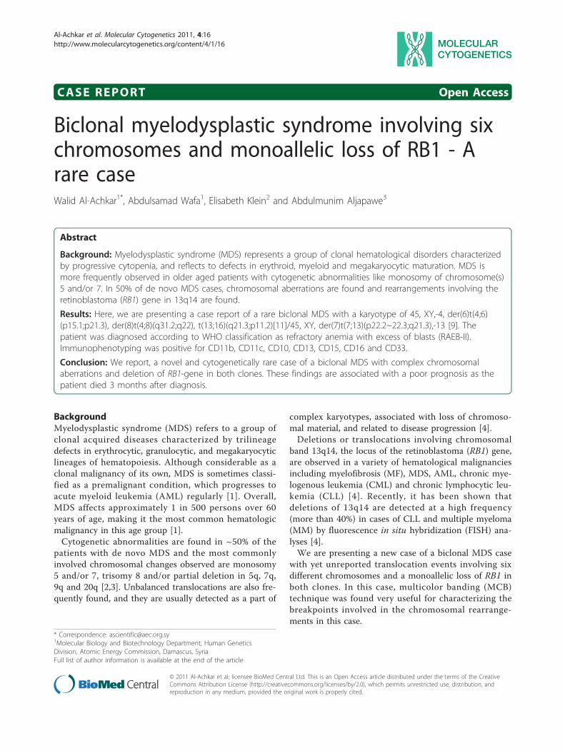
CASE REPORT Open Access Biclonal myelodysplastic syndrome involving six chromosomes and monoallelic loss of RB1 - A rare case Walid Al-Achkar 1* , Abdulsamad Wafa 1 , Elisabeth Klein 2 and Abdulmunim Aljapawe 3 Abstract Background: Myelodysplastic syndrome (MDS) represents a group of clonal hematological disorders characterized by progressive cytopenia, and reflects to defects in erythroid, myeloid and megakaryocytic maturation. MDS is more frequently observed in older aged patients with cytogenetic abnormalities like monosomy of chromosome(s) 5 and/or 7. In 50% of de novo MDS cases, chromosomal aberrations are found and rearrangements involving the retinoblastoma (RB1) gene in 13q14 are found. Results: Here, we are presenting a case report of a rare biclonal MDS with a karyotype of 45, XY,-4, der(6)t(4;6) (p15.1;p21.3), der(8)t(4;8)(q31.2;q22), t(13;16)(q21.3;p11.2)[11]/45, XY, der(7)t(7;13)(p22.2~22.3;q21.3),-13 [9]. The patient was diagnosed according to WHO classification as refractory anemia with excess of blasts (RAEB-II). Immunophenotyping was positive for CD11b, CD11c, CD10, CD13, CD15, CD16 and CD33. Conclusion: We report, a novel and cytogenetically rare case of a biclonal MDS with complex chromosomal aberrations and deletion of RB1-gene in both clones. These findings are associated with a poor prognosis as the patient died 3 months after diagnosis. Background Myelodysplastic syndrome (MDS) refers to a group of clonal acquired diseases characterized by trilineage defects in erythrocytic, granulocytic, and megakaryocytic lineages of hematopoiesis. Although considerable as a clonal malignancy of its own, MDS is sometimes classi- fied as a premalignant condition, which progresses to acute myeloid leukemia (AML) regularly [1]. Overall, MDS affects approximately 1 in 500 persons over 60 years of age, making it the most common hematologic malignancy in this age group [1]. Cytogenetic abnormalities are found in ~50% of the patients with de novo MDS and the most commonly involved chromosomal changes observed are monosomy 5 and/or 7, trisomy 8 and/or partial deletion in 5q, 7q, 9q and 20q [2,3]. Unbalanced translocations are also fre- quently found, and they are usually detected as a part of complex karyotypes, associated with loss of chromoso- mal material, and related to disease progression [4]. Deletions or translocations involving chromosomal band 13q14, the locus of the retinoblastoma (RB1) gene, are observed in a variety of hematological malignancies including myelofibrosis (MF), MDS, AML, chronic mye- logenous leukemia (CML) and chronic lymphocytic leu- kemia (CLL) [4]. Recently, it has been shown that deletions of 13q14 are detected at a high frequency (more than 40%) in cases of CLL and multiple myeloma (MM) by fluorescence in situ hybridization (FISH) ana- lyses [4]. We are presenting a new case of a biclonal MDS case with yet unreported translocation events involving six different chromosomes and a monoallelic loss of RB1 in both clones. In this case, multicolor banding (MCB) technique was found very useful for characterizing the breakpoints involved in the chromosomal rearrange- ments in this case. * Correspondence: [email protected] 1 Molecular Biology and Biotechnology Department, Human Genetics Division, Atomic Energy Commission, Damascus, Syria Full list of author information is available at the end of the article Al-Achkar et al. Molecular Cytogenetics 2011, 4:16 http://www.molecularcytogenetics.org/content/4/1/16 © 2011 Al-Achkar et al; licensee BioMed Central Ltd. This is an Open Access article distributed under the terms of the Creative Commons Attribution License (http://creativecommons.org/licenses/by/2.0), which permits unrestricted use, distribution, and reproduction in any medium, provided the original work is properly cited.
Transcript of CASE REPORT Open Access Biclonal myelodysplastic syndrome ... · CASE REPORT Open Access Biclonal...
CASE REPORT Open Access
Biclonal myelodysplastic syndrome involving sixchromosomes and monoallelic loss of RB1 - Arare caseWalid Al-Achkar1*, Abdulsamad Wafa1, Elisabeth Klein2 and Abdulmunim Aljapawe3
Abstract
Background: Myelodysplastic syndrome (MDS) represents a group of clonal hematological disorders characterizedby progressive cytopenia, and reflects to defects in erythroid, myeloid and megakaryocytic maturation. MDS ismore frequently observed in older aged patients with cytogenetic abnormalities like monosomy of chromosome(s)5 and/or 7. In 50% of de novo MDS cases, chromosomal aberrations are found and rearrangements involving theretinoblastoma (RB1) gene in 13q14 are found.
Results: Here, we are presenting a case report of a rare biclonal MDS with a karyotype of 45, XY,-4, der(6)t(4;6)(p15.1;p21.3), der(8)t(4;8)(q31.2;q22), t(13;16)(q21.3;p11.2)[11]/45, XY, der(7)t(7;13)(p22.2~22.3;q21.3),-13 [9]. Thepatient was diagnosed according to WHO classification as refractory anemia with excess of blasts (RAEB-II).Immunophenotyping was positive for CD11b, CD11c, CD10, CD13, CD15, CD16 and CD33.
Conclusion: We report, a novel and cytogenetically rare case of a biclonal MDS with complex chromosomalaberrations and deletion of RB1-gene in both clones. These findings are associated with a poor prognosis as thepatient died 3 months after diagnosis.
BackgroundMyelodysplastic syndrome (MDS) refers to a group ofclonal acquired diseases characterized by trilineagedefects in erythrocytic, granulocytic, and megakaryocyticlineages of hematopoiesis. Although considerable as aclonal malignancy of its own, MDS is sometimes classi-fied as a premalignant condition, which progresses toacute myeloid leukemia (AML) regularly [1]. Overall,MDS affects approximately 1 in 500 persons over 60years of age, making it the most common hematologicmalignancy in this age group [1].Cytogenetic abnormalities are found in ~50% of the
patients with de novo MDS and the most commonlyinvolved chromosomal changes observed are monosomy5 and/or 7, trisomy 8 and/or partial deletion in 5q, 7q,9q and 20q [2,3]. Unbalanced translocations are also fre-quently found, and they are usually detected as a part of
complex karyotypes, associated with loss of chromoso-mal material, and related to disease progression [4].Deletions or translocations involving chromosomal
band 13q14, the locus of the retinoblastoma (RB1) gene,are observed in a variety of hematological malignanciesincluding myelofibrosis (MF), MDS, AML, chronic mye-logenous leukemia (CML) and chronic lymphocytic leu-kemia (CLL) [4]. Recently, it has been shown thatdeletions of 13q14 are detected at a high frequency(more than 40%) in cases of CLL and multiple myeloma(MM) by fluorescence in situ hybridization (FISH) ana-lyses [4].We are presenting a new case of a biclonal MDS case
with yet unreported translocation events involving sixdifferent chromosomes and a monoallelic loss of RB1 inboth clones. In this case, multicolor banding (MCB)technique was found very useful for characterizing thebreakpoints involved in the chromosomal rearrange-ments in this case.
* Correspondence: [email protected] Biology and Biotechnology Department, Human GeneticsDivision, Atomic Energy Commission, Damascus, SyriaFull list of author information is available at the end of the article
Al-Achkar et al. Molecular Cytogenetics 2011, 4:16http://www.molecularcytogenetics.org/content/4/1/16
© 2011 Al-Achkar et al; licensee BioMed Central Ltd. This is an Open Access article distributed under the terms of the CreativeCommons Attribution License (http://creativecommons.org/licenses/by/2.0), which permits unrestricted use, distribution, andreproduction in any medium, provided the original work is properly cited.

Case reportIn June 2009, a 60 year old male patient was referred withanemia, thrombocytopenia, loss of weight and fever. Hiswhite blood cell count was 7 × 10⁹/l, with 64.5% neutro-phils, 24.6% lymphocytes, 4.2% monocytes, 1% eosino-philes, 1% basophils and 4.6% blast. Bone marrow washypercellular with 19% blast cells. Dysplastic changes inbone marrow included cytoplasmic hypogranulation ofneutrophils, erythroblasts and micromegakaryocytes. Thered blood cell count was 3.38 × 10⁶/cmm with 8.3 g/dlhemoglobin level along with platelet count of 49 × 109/l,and LDH value of 571 U/l. Physical examination of thepatient showed splenomegaly. The patient was treatedwith Zyloric (300 mg as a daily dose) and Hydroxyurea(500 mg as a daily dose) for 1 month and later continuedon Hydroxyurea (500 mg as a daily dose) for 3 month.The patient died 3 months after diagnosis.Karyotyping was done after the initiation of the treat-
ment which showed a mosaic and biclonal karyotypewith 45, XY, -4, der(6)t(4;6)(?;?), der(8)t(4;8)(?;?), t(13;16)(?;?)/45, XY, der(7)t(7;13)(?;?),-13 (Figure 1), which wasfurther studied by molecular cytogenetics (Figure 2, 3,4). Dual-color-FISH using WCP and CEP probes specificfor chromosomes 4, 6, 7, 8, 13 and 16 confirmed thetranslocation seen in GTG-banding. Application of sub-telomeric probes for 7pter and 7qter revealed two sig-nals of subtelomeric 7qter on both homologouschromosomes 7 and one signal of subtelomeric 7pter onintact chromosome 7. Thus, subtelomeric region 7pterwas deleted on the derivative chromosome 7 (Figure 2).Applying an RB1-specific probe showed one signal onlyon the normal chromosome 13 in both clones. The ana-lysis using MCB probes specific for individual chromo-some involved in translocation, determined thebreakpoint location and the final karyotype was foundto be with 45, XY, -4, der(6)t(4;6)(p15.1;p21.3), der(8)t(4;8)(q31.2;q22), t(13;16)(q21.3;p11.2)[11]/45, XY, der(7)t(7;13)(p22.2~22.3;q21.3),-13 [9].Immunophenotyping of peripheral blood characterized
the neutrophiles which showed abnormal side scatterpattern, as well as abnormal intensity staining patternsfor CD11b(63%), CD11c(59.3%), CD10(17.4%), CD13(44%), CD15(59.3%), CD16(46%) and CD33(20.4%). Themajority of monocytes were HLADr+ (4.2%). Lympho-cyte subsets percentages were low. The patient was diag-nosed as having common MDS, refractory anemia withexcess of blasts (RAEB) in the French-American-British(FAB) classification, or RAEB-II in the World HealthOrganization (WHO) classification [5].
DiscussionWe described a biclonal MDS case with cytogeneticrearrangements involving six different chromosomes
together with a monoallelic loss of the RB1 gene in bothclones.The Cancer Genome Anatomy Project databases
(http://cgap.nci.nih.gov/Chromosomes/AbnCytSearch-Form) and atlas of genetics and cytogenetics in oncologyand hematology (http://atlasgeneticsoncology.org/)showed not a single case of MDS with a der(6)t(4;6)(p15.1;p21.3), a der(7)t(7;13)(p22.2~22.3;q21.3), a der(8)t(4;8)(q31.2;q22), or a t(13;16)(q21.3;p11.2). Contrarilythe involvement of RB1 gene in MDS is known andappears to be a rare event [4]. The RB1 protein acts as acell cycle regulator which blocks the transition of nor-mal cells from G0/G1 into S phase of the cycle. Micewith homozygous disruption of the RB1 alleles resultedin an overall normal development but had lethal anemia,suggesting a critical role of the RB1 gene in erythropoi-esis [4]. In the present case, anemia and thrombocytope-nia were predominantly observed during the clinicalcourse, while white blood cells count was relatively pre-served. This impaired erythropoiesis might be related tomonoallelic loss of the RB1 gene [4].Besides imbalances of chromosomes 13, the observed
rearrangements lead to a partial monosomy 4p15.1 to4q31.2 in the slightly larger of both clones. In accor-dance with the international prognostic scoring system(IPSS) classification of chromosomes 4 and 13 loss in aRAEB-II stage patient supports an intermediate-2 prog-nosis group and/or poor prognosis group [6,7].The decreased heterogeneous expression of those anti-
gens was consistent with myelodysplastic disease intransition.Concerning the additional observed acquired imbal-
ances, up to present somehow similar minor terminaldeletions of 6p21.3 in malignant hematological disorderswas observed, and an involvement of the breakpoint8q22 in an oncogene induced solid tumor [8,9]. Dele-tions of 7p confer an inferior outcome in children withALL, regardless of the presence of other poor prognosticfeatures. Monosomy 7 is also associated with inferiorevent-free survival (EFS) in children with ALL [10].13q21.3 region was involved in CLL cases and 16p11.2region was found in classical Hodgkin lymphoma but toour knowledge our breakpoints have not been reportedin MDS, yet [11,12].According to a recent study, the LDH level was nearly
as powerful as a prognostic parameter as karyotypingand an elevated LDH was associated with poor prog-nosis in MDS [13]. The LDH level for the presentedpatient was 571 U/l, which compared to the normalvalue (up to 480 U/l) is enhanced. Thus, also the LDHlevel as well as cytogenetics supported an adverse prog-nosis, which unfortunately was confirmed by the clinicaloutcome.
Al-Achkar et al. Molecular Cytogenetics 2011, 4:16http://www.molecularcytogenetics.org/content/4/1/16
Page 2 of 6

In conclusion, here we reported a novel translocationinvolving six chromosomes distributed in two clonesand monoallelic loss of RB1 in both clones. Our findingand according to WHO classification, IPSS and LDH isconsidered to a poor prognostic factor in MDS patients,as no response was observed after the application ofchemotherapy.
Materials and methodsChromosome analysisChromosome analysis using GTG-banding was per-formed according to standard procedures [14]. Twenty
metaphases derived from unstimulated bone marrow ofthe patient were analyzed. Karyotypes were describedaccording to the International System for Human Cyto-genetic Nomenclature [15].
Molecular cytogeneticsFluorescence in situ hybridization (FISH) using wholechromosome painting (WCP) probe for chromosomes 4,6, 7, 13 and 16 (MetaSystems/Germany) and subtelo-meric probes for 7pter and 7qter (Abbott Molecular/Vysis, USA) were applied according to manufacturer’sinstructions together with a chromosome enumeration
Figure 1 GTG-banding revealed a biclonal condition in the present case. All derivative chromosomes are marked by an arrow head. (A) Atranslocation between chromosomes 7, 13 and loss of one chromosome 13 was observed. (B) A complex karyotype involving the chromosomes4, 6, 8, 13 and 16.
Al-Achkar et al. Molecular Cytogenetics 2011, 4:16http://www.molecularcytogenetics.org/content/4/1/16
Page 3 of 6

probe (CEP) for chromosome 8 and specific probe forRB1 (LSI 13 (RB1) Abbott Molecular/Vysis, USA) [14].Multicolor banding probe (MCB) sets based on micro-dissection derived region-specific libraries for chromo-some 4, 7, 13 and 16 were applied as previouslydescribed [16]. Twenty metaphase spreads were ana-lyzed, each using a fluorescence microscope (AxioIma-ger.Z1 mot, Zeiss) equipped with appropriate filter sets.Image capturing and processing were carried out usingan Isis mFISH imaging system (MetaSystems, Altlus-sheim, Germany).
ImmunophenotypingImmunophenotyping of leukemic blasts was done usinggeneral panel of fluorescent antibodies against the fol-lowing antigens typical for different cell lineages andcell types: CD1a, CD2, CD3, CD4, CD5, CD8, CD10,CD11b, CD11c, CD13, CD14, CD15, CD16, CD19,CD20, CD22, CD23, CD32, CD33, CD34, CD38, CD41a,CD45, CD56, CD57, CD64, CD103, CD117, CD123,CD209, CD235a and CD243; In addition to antibodiesto Kappa and Lambda light Chains, sIgD, sIgM, andHLADr. All antibodies were product from BD
Figure 2 Karyotype and chromosomal aberrations were confirmed using molecular cytogenetic approach. (A) The translocation amongchromosomes 4, 6 and 8 were identified using WCP for chromosomes 4 and 6 (MetaSystems, Altlussheim, Germany) mixed with CEP 8 (AbbottMolecular/Vysis, USA). (B) The translocation between chromosomes 7 and 13 was identified using WCP for chromosomes 7 and 13 (MetaSystems,Altlussheim, Germany). (C) The translocation between chromosomes 13 and 16 was identified using WCP for chromosomes 13 and 16(MetaSystems, Altlussheim, Germany). (D) The subtelomeric deletion of chromosome 7 was identified using subtelomeric 7pter and subtelomeric7qter (Abbott Molecular/Vysis, USA). Abbreviations: #, chromosome; der, derivative chromosome.
Figure 3 Fluorescence in situ hybridization (FISH) using probefor RB1 gene on metaphase spread showed one RB1 signal onnormal chromosome 13 in the second clone. Abbreviations: #,chromosome.
Al-Achkar et al. Molecular Cytogenetics 2011, 4:16http://www.molecularcytogenetics.org/content/4/1/16
Page 4 of 6
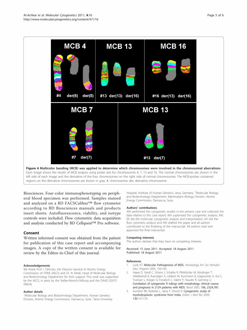
Biosciences. Four-color immunophenotyping on periph-eral blood specimen was performed. Samples stainedand analyzed on a BD FACSCalibur™ flow cytometeraccording to BD Biosciences manuals and productsinsert sheets. Autofluorescence, viability, and isotypecontrols were included. Flow cytometric data acquisitionand analysis conducted by BD Cellquest™ Pro software.
ConsentWritten informed consent was obtained from the patientfor publication of this case report and accompanyingimages. A copy of the written consent is available forreview by the Editor-in-Chief of this journal.
AcknowledgementsWe thank Prof. I. Othman, the Director General of Atomic EnergyCommission of SYRIA (AECS) and Dr. N. Mirali, Head of Molecular Biologyand Biotechnology Department for their support. This work was supportedby the AECS, in parts by the Stefan-Morsch-Stiftung and the DAAD (D/07/09624).
Author details1Molecular Biology and Biotechnology Department, Human GeneticsDivision, Atomic Energy Commission, Damascus, Syria. 2Jena University
Hospital, Institute of Human Genetics, Jena, Germany. 3Molecular Biologyand Biotechnology Department, Mammalians Biology Division, AtomicEnergy Commission, Damascus, Syria.
Authors’ contributionsAW performed the cytogenetic studies in the present case and collected thedata relative to this case report; WA supervised the cytogenetic analysis; AW,EK did the molecular cytogenetic analysis and interpretation; AA did theflow cytometry analysis and AW drafted the paper and all authorscontributed to the finalizing of the manuscript. All authors read andapproved the final manuscript.
Competing interestsThe authors declare that they have no competing interests.
Received: 15 June 2011 Accepted: 18 August 2011Published: 18 August 2011
References1. Look AT: Molecular Pathogenesis of MDS. Hematology Am Soc Hematol
Educ Program 2005, 156-160.2. Haase D, Steidl C, Schanz J, Schabla R, Pfeilstöcker M, Nösslinger T,
Hildebrandt B, Kuendgen A, Lübbert M, Kunzmann B, Giagounidis A, Aul C,Trumper L, Krieger O, Fonatsch C, Valent P, Stauder R, Germing U:Correlation of cytogenetic fi ndings with morphology, clinical courseand prognosis in 2124 patients with MDS. Blood 2005, 106, 232A,787.
3. Vundinti BR, Kerketta L, Jijina F, Ghosh K: Cytogenetic study ofmyelodysplastic syndrome from India. Indian J Med Res 2009,130:155-159.
Figure 4 Multicolor banding (MCB) was applied to determine which chromosomes were involved in the chromosomal aberrations.Each image shows the results of MCB analysis using probe sets for chromosomes 4, 7, 13 and 16. The normal chromosomes are shown in theleft side of each image and the derivative of the four chromosomes on the right side of normal chromosomes. The MCB-probes unstainedregions on the derivative chromosomes are shown in gray. #, chromosome; der, derivative chromosome.
Al-Achkar et al. Molecular Cytogenetics 2011, 4:16http://www.molecularcytogenetics.org/content/4/1/16
Page 5 of 6

4. Yamamoto K, Ito M, Minagawa K, Urahama N, Sada A, Okamura A, Matsui T:A der(13)t(7;13)(p13;q14) with monoallelic loss of RB1 and D13S319 inmyelodysplastic syndrome. Cancer Genet Cytogenet 2005, 162:160-165.
5. Invernizzi R, Filocco A: Myelodysplastic syndrome: classification andprognostic systems. Oncol Rev 2010, 4:25-33.
6. Mitelman F, Johansson B, Mertens F, (Eds.): Mitelman Database ofChromosome Aberrations in Cancer. 2009 [http://cgap.nci.nih.gov/Chromosomes/Mitelman”].
7. Fenaux P: Chromosome and molecular abnormalities in myelodysplasticsyndromes. Int J Hematol 2001, 73:429-437.
8. Chen Z, Issa B, Brothman LJ, Hendricksen M, Button D, Brothman AR:Nonrandom rearrangements of 6p in malignant hematological disorders.Cancer Genet Cytogenet 2000, 121:22-25.
9. Kao C, Wu SQ, DeVries S, Reznikoff WS, Waldman FM, Reznikoff CA:Carcinogen-induced amplification of SV40 DNA inserted at 9q12-21.1associated with chromosome breakage, deletions, and translocations inhuman uroepithelial cell transformation in vitro. Genes ChromosomesCancer 1993, 8:155-166.
10. Heerema NA, Nachman JB, Sather HN, La MK, Hutchinson R, Lange BJ,Bostrom B, Steinherz PG, Gaynon PS, Uckun FM: Deletion of 7p ormonosomy 7 in pediatric acute lymphoblastic leukemia is an adverseprognostic factor: a report from the Children’s Cancer Group. Leukemia2004, 18:939-947.
11. David Ng, Ousmane Toure, Ming-Hui Wei, Arthur CDiane, Fatima Abbasi,Laura Fontaine, Marti EGerald, Fraumeni FJoseph Jr, Goldin RLynn,Neil Caporaso, Toro RJorge: Identification of a novel chromosome region,13q21.33-q22.2, for susceptibility genes in familial chronic lymphocyticleukemia. Blood 2007, 109:916-925.
12. Chui DT, Hammond D, Baird M, Shield L, Jackson R, Jarrett RF: ClassicalHodgkin lymphoma is associated with frequent gains of 17q. GenesChromosomes Cancer 2003, 38:126-136.
13. Germing U, Hildebrandt B, Pfeilstöcker M, Nösslinger T, Valent P,Fonatsch C, Lübbert M, Haase D, Steidl C, Krieger O, Stauder R,Giagounidis AA, Strupp C, Kündgen A, Mueller T, Haas R, Gattermann N,Aul C: Refinement of the international prognostic scoring system (IPSS)by including LDH as an additional prognostic variable to improve riskassessment in patients with primary myelodysplastic syndromes (MDS).Leukemia 2005, 19:2223-2231.
14. AL-achkar W, Wafa A Nweder MS: A complex translocation t(5;9;22) inPhiladelphia cells involving the short arm of chromosome 5 in a case ofchronic myelogenous leukemia. Journal of Experimental and Clinical CancerResearch 2007, 26:411-415.
15. Shaffer L, Slovak M, Cambell L, (eds): ISCN (2009): An International Systemfor Human Cytogenetic Nomenclature. 2009, S. Karger, Basel.
16. Liehr T, Heller A, Starke H, Rubtsov N, Trifonov V, Mrasek K, Weise A,Kuechler A, Claussen U: Microdissection based high resolution multicolorbanding for all 24 human chromosomes. Int J Mol Med 2002, 9:335-339.
doi:10.1186/1755-8166-4-16Cite this article as: Al-Achkar et al.: Biclonal myelodysplastic syndromeinvolving six chromosomes and monoallelic loss of RB1 - A rare case.Molecular Cytogenetics 2011 4:16.
Submit your next manuscript to BioMed Centraland take full advantage of:
• Convenient online submission
• Thorough peer review
• No space constraints or color figure charges
• Immediate publication on acceptance
• Inclusion in PubMed, CAS, Scopus and Google Scholar
• Research which is freely available for redistribution
Submit your manuscript at www.biomedcentral.com/submit
Al-Achkar et al. Molecular Cytogenetics 2011, 4:16http://www.molecularcytogenetics.org/content/4/1/16
Page 6 of 6