Case report Metastatic well-differentiated neuroendocrine ...
6
Case report Open Access Metastatic well-differentiated neuroendocrine carcinoma of the pancreas: case report and review of literature Mersadies R Martin 1 , Umer F Malik 2 *, Deepak Mohan 3 and Ahmed Mahmoud 4 Addresses: 1 Saint George’s University School of Medicine, 7405 Greenback Lane #109 Citrus Heights, CA 95610, USA 2 Department of Internal Medicine San Joaquin General Hospital, 500 West Hospital Road French Camp, CA 95231, USA 3 Department of Pathology San Joaquin General Hospital, 500 West Hospital Road French Camp, CA 95231, USA 4 Department of Surgery San Joaquin General Hospital, 500 West Hospital Road French Camp, California, CA 95231, USA Email: MRM - [email protected]; UFM* - [email protected]; DM - [email protected]; AM - [email protected] * Corresponding author Received: 27 January 2009 Accepted: 19 June 2009 Published: 1 September 2009 Cases Journal 2009, 2:8973 doi: 10.4076/1757-1626-2-8973 This article is available from: http://casesjournal.com/casesjournal/article/view/8973 © 2009 Martin et al.; licensee Cases Network Ltd. This is an Open Access article distributed under the terms of the Creative Commons Attribution License ( http://creativecommons.org/licenses/by/3.0), which permits unrestricted use, distribution, and reproduction in any medium, provided the original work is properly cited. Introduction Neuroendocrine carcinoma accounts for less than 5% of cancers of unknown primary site. When primary pancrea- tic cancer is diagnosed, 95% of cases are classified as aggressive adenocarcinoma and the remaining 5% of cases are caused by indolent behaving neuroendocrine carci- noma. We present a rare case of well-differentiated neuroendocrine carcinoma of the pancreas extending to the transverse colon, small bowel, stomach, and lymph nodes with metastasis to the caudate lobe of the liver. The patient had a two-year history of midepigastric abdominal pain that eventually sent him to the Emergency Depart- ment and after extensive tests we removed the entire tumor with margins free of growth. Included in this case report are many illustrations to show the severity and we further emphasize the importance of examining patients thor- oughly especially when vague symptoms are chronic. Case presentation A 58-year-old African American male presented to the Emergency Department by ambulance complaining of a constant, sharp, and worsening midepigastric pain that radiated posterior for approximately 30 minutes. The pain was perceived to be 10 out of 10 and was described as an inflated balloon that was squeezing from the inside out. The paramedics found the patient in his house hypoten- sive and lying in a right lateral decubitus position. The patient was given a bolus of normal saline and his blood pressure responded appropriately. Upon examination, the patient claimed to of had a two-year history of worsening mild midepigastric and left upper quadrant abdominal pain and was seen by his primary care provider whom prescribed him a proton pump inhibitor (PPI) which partially relieved some symptoms. In addition, the patient complained of being nauseated, feeling bloated after meals, having a history of bloody stools one week prior to admission, and increased pain after eating. He denied vomiting or early satiety. Vital signs on admission were stable. Past medical history included gastroesophageal reflux disease (GERD), hyperlipidemia and impaired fasting glucose. His medications consisted of a PPI, H2-blocker and simvastatin. The only past surgical history was an appendectomy. The patient was a truck driver for several years. He denied alcohol, drug use, and smoked approxi- mately ½ packs of cigarettes a day for the past 40 years. He reported his two nieces having an unspecified type of cancer but no other known family history of cancer or heart diseases. Review of systems was negative except for Page 1 of 6 (page number not for citation purposes)
Transcript of Case report Metastatic well-differentiated neuroendocrine ...

Case report
Open Access
Metastatic well-differentiated neuroendocrine carcinoma of thepancreas: case report and review of literatureMersadies R Martin1, Umer F Malik2*, Deepak Mohan3 andAhmed Mahmoud4
Addresses: 1Saint George’s University School of Medicine, 7405 Greenback Lane #109 Citrus Heights, CA 95610, USA2Department of Internal Medicine San Joaquin General Hospital, 500 West Hospital Road French Camp, CA 95231, USA3Department of Pathology San Joaquin General Hospital, 500 West Hospital Road French Camp, CA 95231, USA4Department of Surgery San Joaquin General Hospital, 500 West Hospital Road French Camp, California, CA 95231, USA
Email: MRM - [email protected]; UFM* - [email protected]; DM - [email protected]; AM - [email protected]
*Corresponding author
Received: 27 January 2009 Accepted: 19 June 2009 Published: 1 September 2009
Cases Journal 2009, 2:8973 doi: 10.4076/1757-1626-2-8973
This article is available from: http://casesjournal.com/casesjournal/article/view/8973
© 2009 Martin et al.; licensee Cases Network Ltd.This is an Open Access article distributed under the terms of the Creative Commons Attribution License (http://creativecommons.org/licenses/by/3.0),which permits unrestricted use, distribution, and reproduction in any medium, provided the original work is properly cited.
IntroductionNeuroendocrine carcinoma accounts for less than 5% ofcancers of unknown primary site. When primary pancrea-tic cancer is diagnosed, 95% of cases are classified asaggressive adenocarcinoma and the remaining 5% of casesare caused by indolent behaving neuroendocrine carci-noma. We present a rare case of well-differentiatedneuroendocrine carcinoma of the pancreas extending tothe transverse colon, small bowel, stomach, and lymphnodes with metastasis to the caudate lobe of the liver. Thepatient had a two-year history of midepigastric abdominalpain that eventually sent him to the Emergency Depart-ment and after extensive tests we removed the entire tumorwith margins free of growth. Included in this case reportare many illustrations to show the severity and we furtheremphasize the importance of examining patients thor-oughly especially when vague symptoms are chronic.
Case presentationA 58-year-old African American male presented to theEmergency Department by ambulance complaining of aconstant, sharp, and worsening midepigastric pain thatradiated posterior for approximately 30 minutes. The painwas perceived to be 10 out of 10 and was described as aninflated balloon that was squeezing from the inside out.
The paramedics found the patient in his house hypoten-sive and lying in a right lateral decubitus position. Thepatient was given a bolus of normal saline and his bloodpressure responded appropriately. Upon examination, thepatient claimed to of had a two-year history of worseningmild midepigastric and left upper quadrant abdominalpain and was seen by his primary care provider whomprescribed him a proton pump inhibitor (PPI) whichpartially relieved some symptoms. In addition, the patientcomplained of being nauseated, feeling bloated aftermeals, having a history of bloody stools one week priorto admission, and increased pain after eating. He deniedvomiting or early satiety. Vital signs on admission werestable.
Past medical history included gastroesophageal refluxdisease (GERD), hyperlipidemia and impaired fastingglucose. His medications consisted of a PPI, H2-blockerand simvastatin. The only past surgical history was anappendectomy. The patient was a truck driver for severalyears. He denied alcohol, drug use, and smoked approxi-mately ½ packs of cigarettes a day for the past 40 years. Hereported his two nieces having an unspecified type ofcancer but no other known family history of cancer orheart diseases. Review of systems was negative except for
Page 1 of 6(page number not for citation purposes)

gastrointestinal complaints as described above. Thepatient denied weight loss, fatigue, fever, shortness ofbreath, and/or chest pain. On physical examination, theabdomen was hard, guarding was noted, and there wasdirect and rebound tenderness noted in all four quadrantsbut worse in the epigastric area.
On admission, the patient had a complete laboratoryworkup involving all systems. The abnormalities foundmildly decreased were hemoglobin of 12.9 g/dl, hematocritof 39.6%, albumin of 3.1 g/dl, and sodium of 132 mEq/l.The only abnormalities found mildly increased was thered blood distribution width of 16%, white blood cellcount of 15,100 ul, corrected calcium of 11.32 mg/dl,glucose of 129 mg/dl, prothrombin time of 12.2 seconds,and the international ratio of 1.2 seconds. Stool guaiacwas negative.
Computed tomography (CT) of the abdomen and pelvison admission revealed a large 4.7 centimeter (cm)lobulated hypodense mass in the region of the portahepatis probably arising from the liver with the possibilityof adjacent morphologic tubular lymph nodes or satellitelesions (Figure 1). Extending from the lobulated mass ofthe liver was a broad lobulated 10 cm band of density justdeep to the rectus muscle extending from the upperabdomen to the level below the iliac crest, which possiblyrepresented extraperitoneal infiltration. Additionally, anenhancing lesion was seen in the caudate lobe of the liverthat was most likely metastasis. Blood was noted in bothparacolic gutters, and there was a pancake-type densityalong the anterior abdominal wall, which was most likely
blood that extended into the right pelvis representingomental caking. After review of the CT, serum measure-ments of Cancer antigen (CA) 19-9, Carcino embryonicantigen (CEA), and Alpha-fetoprotein (AFP) were found tobe within normal limits. Serum Helicobacter pylori waspositive. Colonoscopy and esophagastroduodenoscopywas recommended after review of the CT and findingswere mild antral gastritis and a nearly obstructing mass inthe descending colon. Recommendations were to rule outother malignancies and surgical intervention. An acuteabdomen series with chest radiograph was consistent withthe CT with additional findings of patchy and streakyinfiltrate of the right and left lung bases and question ofthe mass in the left upper quadrant displacing the stomachmedially. There was no evidence of acute obstruction or airunder the diaphragm. Positron emission tomography scanwas performed to evaluate for metastases and resultsshowed increase uptake in the left upper quadrant, andabout 4 cm of caudate lobe of the liver.
Utilizing CT guidance, biopsy of the large mass in the leftupper quadrant was performed by Interventional Radi-ology on the same day of admission. Pathology results twodays later showed a well-differentiated neuroendocrinecarcinoma. The architecture revealed insular and trabecu-lar morphologies, and the Ki-67 index was approximately30% positive (Figure 2). Microscopic sections showednested epithelial cells with moderately increased mitoticactivity, nuclear atypia, irregular nuclear contours andhyperchromasia. Immunohistological stains were
Figure 1. Computed tomography (CT) of the abdomenand pelvis. A large 4.7 centimeter (cm) lobulated hypodensemass is shown in the region of the porta hepatis likelysecondary to the tumor mass.
Figure 2. Histological demonstration of the well-differentiated neuroendocrine carcinoma. Pathology slidesfrom the tumourus mass showing a well-differentiatedneuroendocrine carcinoma. The architecture is consistentwith the insular and trabecular morphologies, and theKi-67 index is approximately 30% positive.
Page 2 of 6(page number not for citation purposes)
Cases Journal 2009, 2:8973 http://casesjournal.com/casesjournal/article/view/8973

performed with the following results: the epithelialcomponent was positive for cytokeratin AE1/AE3, neu-roendocrine marker synaptophysin was positive, neuroen-docrine marker chromogranin-A was positive,neuroendocrine marker neurone specific enolase waspositive, and CA 19-9 was negative.
On hospital day number six the surgery service performeda diagnostic laparoscopy, exploratory laparotomy withdistal pancreatectomy, splenectomy, partial gastrectomy,left colectomy, and resection of the caudate lobe of theliver (Figure 3). Pre and postoperative diagnoses werewell-differentiated neuroendocrine tumor of the pancrea-tic tail with metastasis to the caudate lobe of the liver. Afterdiagnostic laparoscopy revealed a moderate amount ofblood around the peritoneal cavity and metastasis to theliver it was necessary to convert to exploratory laparotomy.Blood was aspirated from the abdominal cavity and sent tocytology, which later revealed numerous neutrophilsadmixed with red blood cells yet no malignant cells.General inspection of the peritoneal cavity revealed atumor in the caudate lobe as well as a large 10 cm massthat was fixed in the left upper quadrant. The tumor itselfwas quite vascular and was surrounded by varices, whichwas most likely the cause for the blood throughout theabdomen as well as within the lesser sac. Additionally, aperiaortic lymph node appeared grossly positive and wasthen dissected and sent for analysis. There was no evidenceof metastases in the pelvis, small bowel, or omentum. Thediaphragm was not involved or infiltrated by the tumorand neither was the left kidney or adrenal glands.
Overall, the surgery was successful. At the end of theprocedure two 10 ml Jackson-Pratt drains were insertedinto the lesser sac and left upper quadrant.
Gross specimens were sent to pathology in three parts:frozen section of the periaortic lymph node measured 1.0by 0.8 by 0.4 cm, the caudate lobe of the liver measured4.0 by 3.5 by 2.5 cm, and the massive left upper quadranttumor that consisted of an en bloc resection of the spleen,partial transverse colon, partial small bowel, pancreatictail, partial distal stomach, adipose tissue, and lymphnodes. The spleenmeasured 12 by 7.5 by 2.5 cm, the colonmeasured 33 cm in length and 4 cm in diameter, thepancreatic tail measured 12 by 8 by 6 cm, a portion of thesmall bowel measured 15 cm in length and about 3 cm indiameter, and a small portion of the distal stomachmeasured 9 by 6.5 cm (Figure 4). The massive tumorinvaded into the serosa of the transverse colon, distalstomach, and small bowel. The tumor appeared tocompress the capsule of the spleen, however, no directinvasion was identified (Figure 5). After sectioning of allthe involved organs it was noted that the tumor originated
Figure 3. Exploratory laparotomy. Distal pancreatectomy,splenectomy, partial gastrectomy, left colectomy, andresection of the caudate lobe of the liver wereperformed during the surgical procedure.
Figure 4. Gross specimen of the liver. One of the specimensremoved from the body are shown here which presents thecaudate lobe of the liver measured 4.0 by 3.5 by 2.5 cm.
Page 3 of 6(page number not for citation purposes)
Cases Journal 2009, 2:8973 http://casesjournal.com/casesjournal/article/view/8973

from the pancreas. Within the adjacent adipose tissue,multiple lymph nodes ranging in size from 0.3 to 1.0 cmwere analyzed.
Microscopic sections were analyzed and found theperioarotic lymph node to be negative for metastaticcarcinoma. Microscopic sections of the caudate lobe of theliver showed metastatic well-differentiated neuroendo-crine carcinoma (Figures 6 and 7). Sections of the massiveleft upper quadrant mass showed invasive well-differen-tiated neuroendocrine carcinoma characterized by cellswith high nuclear to cytoplasmic ratio, irregular nuclearcontours and hyperchromasia arranged in trabecular andinsular patterns. There were 21 of 21 lymph nodes positivefor metastatic carcinoma. Margins of resection were freeof tumor. Tumor node metastasis (TNM) staging wasT4, N1, M1.
Overall, the patient tolerated the procedure and hospitalstay well. During the course of hospitalization his pain waswell controlled with morphine, and his glucose was wellcontrolled after being weaned off of an insulin drip andplaced on a sliding scale. Vaccinations for encapsulatedorganisms including pneumococcal, Streptococcus pneumo-niae, Haemophilus influenza and Neisseria meningitides wereadministered. Twenty days post-admission the patient wasdischarged from the hospital with instructions not to liftmore than 15 pounds, and to follow-up with surgery inone week.
DiscussionWhen the initial work-up, including physical examination,laboratory and radiographic studies fails to identify the
primary site of a tumor with symptoms of referable to ametastatic site, light microscopic evaluation of biopsymaterial is necessary. Cancer of unknown primary site is acommon entity and based on five histological categories:adenocarcinoma, poorly-differentiated carcinoma, poorly-differentiated neoplasm, squamous cell carcinoma, andneuroendocrine carcinoma [1]. Neuroendocrine carci-noma accounts for less than 5% of cancers of unknownprimary site [2]. With that in mind, we present the clinical,pathological, histological and diagnostic features of well-differentiated neuroendocrine carcinoma which emphasisplaced on pancreatic neuroendocrine tumors as seen inthis case report.
Neuroendocrine carcinomas falls into three broad cate-gories: poorly, moderately and well-differentiated
Figure 5. Splenic tissue. This histological picture is showingthe tumor compressing the capsule of the spleen withoutany direct invasion.
Figures 6 & 7. Microscopic sections of the caudate lobe ofthe liver. These pictures are demonstrating metastaticwell-differentiated neuroendocrine carcinoma in differentmagnifications (exact numbers are mentioned in the figures).
Page 4 of 6(page number not for citation purposes)
Cases Journal 2009, 2:8973 http://casesjournal.com/casesjournal/article/view/8973

neuroendocrine carcinomas. Poorly-differentiated neuro-endocrine carcinomas usually have a high grade malig-nancy and are characteristically small cell undifferentiatedor morphologically anaplastic by light microscopy [3].Moderately-differentiated neuroendocrine carcinomas,such as atypical carcinoid tumors, manifest cellular pleo-morphism, nuclear atypia, frequent mitosis, and necrosis[4]. In contrast, well-differentiated neuroendocrine carci-nomas have variable and most often indolent biologicbehaviors [5]. Histologically, well-differentiated neuro-endocrine carcinomas show uniform and small cellsorganized in organoid or trabecular architecture [6].Subtypes of well-differentiated neuroendocrine carcino-mas include typical carcinoid tumors, pancreatic islet cell(neuroendocrine) tumors, paragangliomas, pheochromo-cytomas, and medullary thyroid carcinomas [5].
With well-differentiated neuroendocrine carcinomasencompassing such a broad spectrum of neoplasms it ispertinent to further classify them depending on whetherthey have a neural or an epithelial origin. Paragangliomasare of neural origin and the epithelial group can besubdivided into the remaining types of neuroendocrinecarcinomas [7]. In our case, the epithelial component wasillustrated microscopically by areas of nested and trabe-cular (neuroendocrine) growth, large cell size, irregularnuclear contours, and coarse nuclear chromatin. Inaddition, the immunohistochemical documentationshowed immunoreactivity for markers of epithelial andneuroendocrine differentiation with positivity for cytoker-atin AE1/AE3, synaptophysin, chromogranin, and neuronespecific enolase [8]. Recognition of this unusual morpho-logic appearance is of importance to avoid mistaking theselesions for other types of malignant neoplasms.
The majority of well-differentiated neuroendocrine carci-nomas involve the gastrointestinal tract, and therefore arereferred to as gastroenteropancreatic neuroendocrinecarcinomas, however neuroendocrine carcinomas havebeen found to occur in almost every organ in the body [9].Gastroenteropancreatic neuroendocrine tumors are rela-tively rare, and pancreatic endocrine tumors are even morerare [10]. In our case the tumor originated in the pancreasand further infiltrated the transverse colon, small bowl,stomach, and lymph nodes within the adipose tissue.Literature review reported that low-grade neuroendocrinecarcinomas, such as metastatic carcinoid or pancreaticendocrine tumors, are occasionally found at metastaticsites without an obvious primary site but almost alwaysinvolve the liver [11]. Following the literature, in this caseof pancreatic neuroendocrine origin there was metastasisspecific to the caudate lobe of the liver.
Pancreatic endocrine tumors or islet cell tumors may benonfunctional or functional depending on whether or not
the tumor was secreting excessive amounts bioactivesubstances such as insulin, gastrin, somatostatin, vasoac-tive intestinal peptide, or glucagon and further causingassociated symptoms [12]. Initially the patient presentedwith hypercalcemia, impaired fasting glucose, and GERDbut due to urgency to undergo surgery and expected delayof the pathology it was not evident to look into theseconditions until after the surgery. Therefore, we recom-mend looking into abnormal laboratory values or diseasesbefore surgery to evaluate for associations related toneuroendocrine syndromes. In this case we cannot becertain if the patient was suffering from functional ornonfunctional well-differentiated neuroendocrine carci-noma of the pancreas. Since part of the stomach wasremoved it was difficult to determine if the GERD that thepatient was suffering from preoperatively was due to highgastrin levels and/or if the tumor infiltration pushingupwards on the stomach was causing the acid refluxsymptoms. In addition, it was difficult to determinepostoperatively if the hypercalcemia, renal stone and/orthe impaired glucose fasting levels as seen in our patientwere attributed to a functional versus nonfunctionalpancreatic endocrine tumor.
We looked into reasons of why metastasis occurred in thecaudate lobe of the liver and not the other three lobes.Interesting, the caudate lobe is the only part of the liverthat has hepatic venous drainage encompassing a fewsizeable and several smaller branches that flow directlyinto the inferior vena cava [13]. Whether or not that isrelevant it was interesting because even though manystudies have reported tumors frequently infiltrating theparenchyma of the caudate lobe and/or its bile ducts it isstill a rare entity [14,15].
Although there is abundant literature on neuroendocrinecarcinomas [5,7,9], to our knowledge, there has not been acase presentation of neuroendocrine carcinoma evolvingfrom the pancreas with metastasis to the caudate lobe ofliver of such massive size. We have included manyillustrations to show the severity of this case and toemphasize the importance of examining patients thor-oughly especially when vague symptoms are chronic.When primary pancreatic cancer is diagnosed, 95% ofcases are adenocarcinoma in origin and the remaining 5%are due to neuroendocrine carcinoma [16]. In contrast toneuroendocrine carcinomas, adenopancreatic cancers aregenerally very aggressive [17]. A study assessed the clinicalrelevance of the World Health Organization and TNMclassifications in patients with pancreatic neuroendocrinecarcinomas and found that the five-year survival rate to be44% [18]. Another study found the five-year survival rateto be 58.3% and concluded that patients with well tomoderately differentiated tumors had a better (63.9%)five-year prognosis than those with poorly differentiated
Page 5 of 6(page number not for citation purposes)
Cases Journal 2009, 2:8973 http://casesjournal.com/casesjournal/article/view/8973
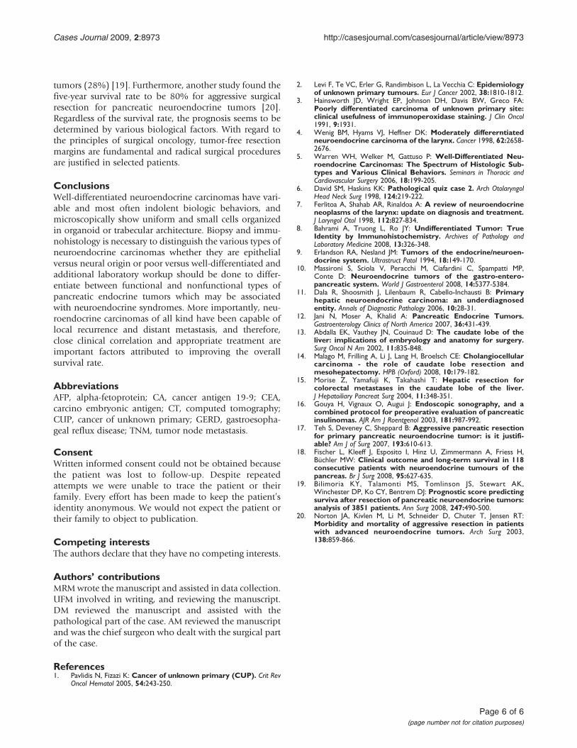
tumors (28%) [19]. Furthermore, another study found thefive-year survival rate to be 80% for aggressive surgicalresection for pancreatic neuroendocrine tumors [20].Regardless of the survival rate, the prognosis seems to bedetermined by various biological factors. With regard tothe principles of surgical oncology, tumor-free resectionmargins are fundamental and radical surgical proceduresare justified in selected patients.
ConclusionsWell-differentiated neuroendocrine carcinomas have vari-able and most often indolent biologic behaviors, andmicroscopically show uniform and small cells organizedin organoid or trabecular architecture. Biopsy and immu-nohistology is necessary to distinguish the various types ofneuroendocrine carcinomas whether they are epithelialversus neural origin or poor versus well-differentiated andadditional laboratory workup should be done to differ-entiate between functional and nonfunctional types ofpancreatic endocrine tumors which may be associatedwith neuroendocrine syndromes. More importantly, neu-roendocrine carcinomas of all kind have been capable oflocal recurrence and distant metastasis, and therefore,close clinical correlation and appropriate treatment areimportant factors attributed to improving the overallsurvival rate.
AbbreviationsAFP, alpha-fetoprotein; CA, cancer antigen 19-9; CEA,carcino embryonic antigen; CT, computed tomography;CUP, cancer of unknown primary; GERD, gastroesopha-geal reflux disease; TNM, tumor node metastasis.
ConsentWritten informed consent could not be obtained becausethe patient was lost to follow-up. Despite repeatedattempts we were unable to trace the patient or theirfamily. Every effort has been made to keep the patient’sidentity anonymous. We would not expect the patient ortheir family to object to publication.
Competing interestsThe authors declare that they have no competing interests.
Authors’ contributionsMRMwrote the manuscript and assisted in data collection.UFM involved in writing, and reviewing the manuscript.DM reviewed the manuscript and assisted with thepathological part of the case. AM reviewed the manuscriptand was the chief surgeon who dealt with the surgical partof the case.
References1. Pavlidis N, Fizazi K: Cancer of unknown primary (CUP). Crit Rev
Oncol Hematol 2005, 54:243-250.
2. Levi F, Te VC, Erler G, Randimbison L, La Vecchia C: Epidemiologyof unknown primary tumours. Eur J Cancer 2002, 38:1810-1812.
3. Hainsworth JD, Wright EP, Johnson DH, Davis BW, Greco FA:Poorly differentiated carcinoma of unknown primary site:clinical usefulness of immunoperoxidase staining. J Clin Oncol1991, 9:1931.
4. Wenig BM, Hyams VJ, Heffner DK: Moderately differerntiatedneuroendocrine carcinoma of the larynx. Cancer 1998, 62:2658-2676.
5. Warren WH, Welker M, Gattuso P: Well-Differentiated Neu-roendocrine Carcinomas: The Spectrum of Histologic Sub-types and Various Clinical Behaviors. Seminars in Thoracic andCardiovascular Surgery 2006, 18:199-205.
6. David SM, Haskins KK: Pathological quiz case 2. Arch OtolaryngolHead Neck Surg 1998, 124:219-222.
7. Ferlitoa A, Shahab AR, Rinaldoa A: A review of neuroendocrineneoplasms of the larynx: update on diagnosis and treatment.J Laryngol Otol 1998, 112:827-834.
8. Bahrami A, Truong L, Ro JY: Undifferentiated Tumor: TrueIdentity by Immunohistochemistry. Archives of Pathology andLaboratory Medicine 2008, 13:326-348.
9. Erlandson RA, Nesland JM: Tumors of the endocrine/neuroen-docrine system. Ultrastruct Patol 1994, 18:149-170.
10. Massironi S, Sciola V, Peracchi M, Ciafardini C, Spampatti MP,Conte D: Neuroendocrine tumors of the gastro-entero-pancreatic system. World J Gastroenterol 2008, 14:5377-5384.
11. Dala R, Shoosmith J, Lilenbaum R, Cabello-Inchausti B: Primaryhepatic neuroendocrine carcinoma: an underdiagnosedentity. Annals of Diagnostic Pathology 2006, 10:28-31.
12. Jani N, Moser A, Khalid A: Pancreatic Endocrine Tumors.Gastroenterology Clinics of North America 2007, 36:431-439.
13. Abdalla EK, Vauthey JN, Couinaud D: The caudate lobe of theliver: implications of embryology and anatomy for surgery.Surg Oncol N Am 2002, 11:835-848.
14. Malago M, Frilling A, Li J, Lang H, Broelsch CE: Cholangiocellularcarcinoma - the role of caudate lobe resection andmesohepatectomy. HPB (Oxford) 2008, 10:179-182.
15. Morise Z, Yamafuji K, Takahashi T: Hepatic resection forcolorectal metastases in the caudate lobe of the liver.J Hepatoiliary Pancreat Surg 2004, 11:348-351.
16. Gouya H, Vignaux O, Augui J: Endoscopic sonography, and acombined protocol for preoperative evaluation of pancreaticinsulinomas. AJR Am J Roentgenol 2003, 181:987-992.
17. Teh S, Deveney C, Sheppard B: Aggressive pancreatic resectionfor primary pancreatic neuroendocrine tumor: is it justifi-able? Am J of Surg 2007, 193:610-613.
18. Fischer L, Kleeff J, Esposito I, Hinz U, Zimmermann A, Friess H,Büchler MW: Clinical outcome and long-term survival in 118consecutive patients with neuroendocrine tumours of thepancreas. Br J Surg 2008, 95:627-635.
19. Bilimoria KY, Talamonti MS, Tomlinson JS, Stewart AK,Winchester DP, Ko CY, Bentrem DJ: Prognostic score predictingsurviva after resection of pancreatic neuroendocrine tumors:analysis of 3851 patients. Ann Surg 2008, 247:490-500.
20. Norton JA, Kivlen M, Li M, Schneider D, Chuter T, Jensen RT:Morbidity and mortality of aggressive resection in patientswith advanced neuroendocrine tumors. Arch Surg 2003,138:859-866.
Page 6 of 6(page number not for citation purposes)
Cases Journal 2009, 2:8973 http://casesjournal.com/casesjournal/article/view/8973