Case Report JLA - KoreaMed...atrioventricular block, or vascular events. There was no significant...
6
85 Received: Revised: Accepted: August 4, 2013 October 10, 2013 November 8, 2013 Corresponding Author: Myeong-Ki Hong, Division of Cardiology, Severance Cardiovascular Hospital, Severance Biomedical Science Institute, Yonsei University College of Medicine, 50 Yonsei-ro, Seodaemun-gu, Seoul 120-752, Kore a Tel: +82-2-2228-8460, Fax: +82-2-2227-7732, E-mail: [email protected] This is an Open Access article distributed under the terms of the creative Commons Attribution Non-Commercial License (http://creativecommons.org/licenses/by-nc/3.0) which permits unrestricted non-commercial use, distribution, and reproduction in any medium, provided the original work is properly cited. Case Report http://dx.doi.org/10.12997/jla.2013.2.2.85 pISSN 2287-2892 • eISSN 2288-2561 JL A Transcatheter Aortic Valve Implantation Using CoreValve by Transaortic Approach Kyeong-Hyeon Chun 1 , Young-Guk Ko 1 , Ji-Young Shim 1 , Sak Lee 2 , Byung-Chul Chang 2 , Jae-Kwang Shim 3 , Young-Ran Kwak 3 , Myeong-Ki Hong 1 1 Division of Cardiology, Severance Cardiovascular Hospital, Yonsei University Health System, Seoul, 2 Department of Thoracic and Cardiovascular Surgery, Severance Cardiovascular Hospital, Yonsei University Health System, Seoul, 3 Department of Anesthesiology and Pain Medicine, Severance Cardiovascular Hospital, Yonsei University Health System, Seoul, Korea Introduction: Transcatheter aortic valve implantation (TAVI) is now considered as an alternative treatment option for severe aortic stenosis (AS) patients who cannot undergo surgical aortic valve replacement (AVR). Case report: We describe the first Korean case of transaortic TAVI with mini-sternotomy using CoreValve. A 83-year-old woman with severe AS and recent history of non-ST elevation myocardial infarction was referred to our institution for TAVI intervention. There was no amenable peripheral vascular access for transfemoral or trans-subclavian approach. Considering the relatively high procedural risk of transapical approach in this patient, we performed transaortic TAVI with mini-sternotomy. Conclusion: The present case suggests transaortic approach may be an effective and safe strategy for TAVI in high risk severe AS patients without eligible femoral or subclavian access routes. Key Words: Heart valve prosthesis implantation, Aortic stenosis, Vascular access devices INTRODUCTION Surgical aortic valve replacement (AVR) is the treatment of choice of symptomatic severe aortic stenosis (AS). With technological advancements, an alternative to surgical AVR, transcatheter aortic valve implantation (TAVI) has recently emerged and has been performed widely after the first case a decade ago. 1,2 The most common route used to deliver transcatheter aortic valve is femoral artery. For patients with significant peripheral arterial diseases, especially common femoral or iliac artery stenosis, other access approach such as transapical or trans-subclavian approach can be considered. For whom with unsuitable conditions for previously mentioned routes, direct trans- aortic access with a mini-sternotomy could be an alternative option. In this case report, we describe the first Korean case of trans-aortic TAVI using CoreValve (Medtronic) for an 83-year-old woman. CASE REPORT An 83-year-old woman (height, 1.43 m; weight, 40 Copyright ⓒ 2013 The Korean Society of Lipidology and Atherosclerosis
Transcript of Case Report JLA - KoreaMed...atrioventricular block, or vascular events. There was no significant...

www.lipid.or.kr 85
Received:Revised:Accepted:
August 4, 2013 October 10, 2013November 8, 2013
Corresponding Author: Myeong-Ki Hong, Division of Cardiology, Severance Cardiovascular Hospital, Severance Biomedical Science Institute, Yonsei University College of Medicine, 50 Yonsei-ro, Seodaemun-gu, Seoul 120-752, KoreaTel: +82-2-2228-8460, Fax: +82-2-2227-7732, E-mail: [email protected]
This is an Open Access article distributed under the terms of the creative Commons Attribution Non-Commercial License (http://creativecommons.org/licenses/by-nc/3.0) which permits unrestricted non-commercial use, distribution, and reproduction in any medium, provided the original work is properly cited.
Case Report
http://dx.doi.org/10.12997/jla.2013.2.2.85pISSN 2287-2892 • eISSN 2288-2561 JLATranscatheter Aortic Valve Implantation Using CoreValve by Transaortic ApproachKyeong-Hyeon Chun1, Young-Guk Ko1, Ji-Young Shim1, Sak Lee2, Byung-Chul Chang2, Jae-Kwang Shim3, Young-Ran Kwak3, Myeong-Ki Hong1
1Division of Cardiology, Severance Cardiovascular Hospital, Yonsei University Health System, Seoul, 2Department of Thoracic and Cardiovascular Surgery, Severance Cardiovascular Hospital, Yonsei University Health System, Seoul, 3Department of Anesthesiology and Pain Medicine, Severance Cardiovascular Hospital, Yonsei University Health System, Seoul, Korea
Introduction: Transcatheter aortic valve implantation (TAVI) is now considered as an alternative treatment option for severe aortic stenosis (AS) patients who cannot undergo surgical aortic valve replacement (AVR). Case report: We describe the first Korean case of transaortic TAVI with mini-sternotomy using CoreValve. A 83-year-old woman with severe AS and recent history of non-ST elevation myocardial infarction was referred to our institution for TAVI intervention. There was no amenable peripheral vascular access for transfemoral or trans-subclavian approach. Considering the relatively high procedural risk of transapical approach in this patient, we performed transaortic TAVI with mini-sternotomy. Conclusion: The present case suggests transaortic approach may be an effective and safe strategy for TAVI in high risk severe AS patients without eligible femoral or subclavian access routes.
Key Words: Heart valve prosthesis implantation, Aortic stenosis, Vascular access devices
INTRODUCTION
Surgical aortic valve replacement (AVR) is the treatment
of choice of symptomatic severe aortic stenosis (AS). With
technological advancements, an alternative to surgical
AVR, transcatheter aortic valve implantation (TAVI) has
recently emerged and has been performed widely after
the first case a decade ago.1,2 The most common route
used to deliver transcatheter aortic valve is femoral artery.
For patients with significant peripheral arterial diseases,
especially common femoral or iliac artery stenosis, other
access approach such as transapical or trans-subclavian
approach can be considered. For whom with unsuitable
conditions for previously mentioned routes, direct trans-
aortic access with a mini-sternotomy could be an
alternative option. In this case report, we describe the
first Korean case of trans-aortic TAVI using CoreValve
(Medtronic) for an 83-year-old woman.
CASE REPORT
An 83-year-old woman (height, 1.43 m; weight, 40
Copyright ⓒ 2013 The Korean Society of Lipidology and Atherosclerosis
J Lipid Atheroscler 2013;2(2):85-90 JOURNAL OF LIPID AND ATHEROSCLEROSIS
86 www.lipid.or.kr
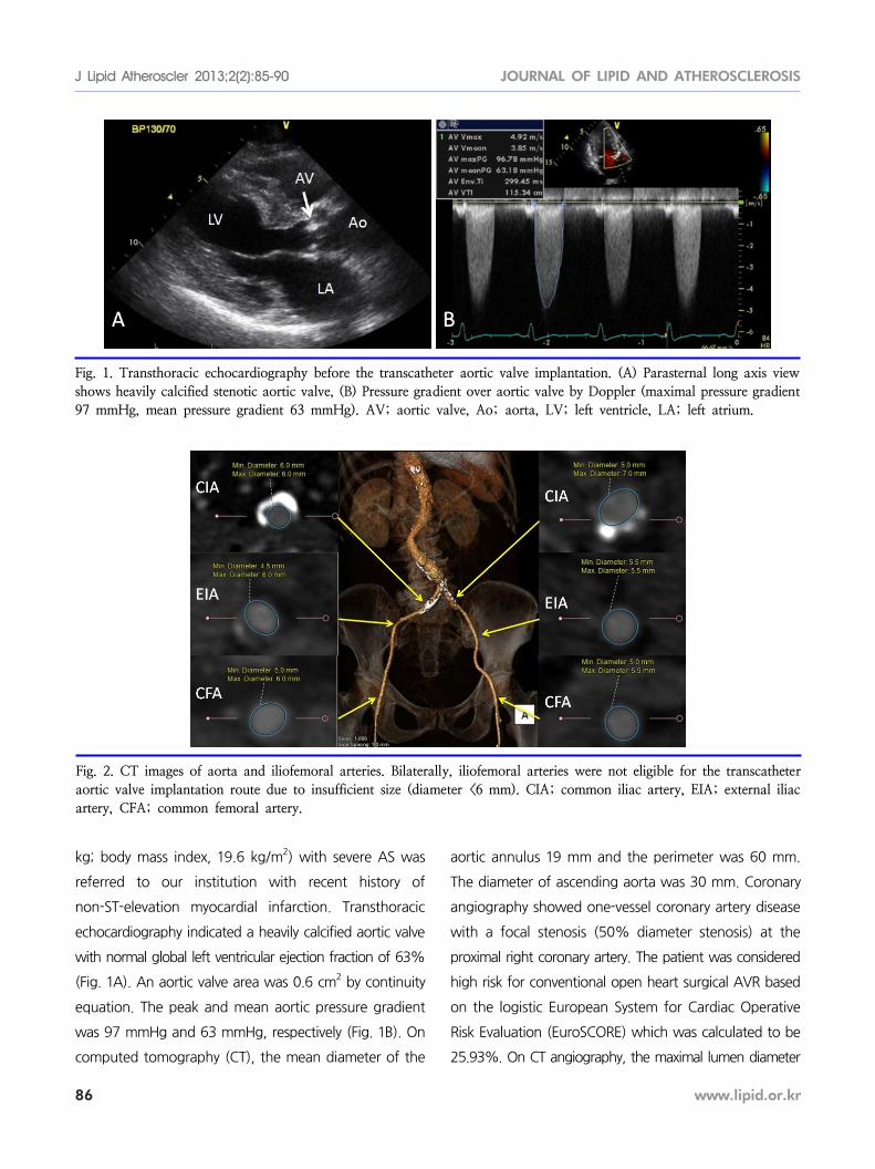
Fig. 1. Transthoracic echocardiography before the transcatheter aortic valve implantation. (A) Parasternal long axis view shows heavily calcified stenotic aortic valve, (B) Pressure gradient over aortic valve by Doppler (maximal pressure gradient97 mmHg, mean pressure gradient 63 mmHg). AV; aortic valve, Ao; aorta, LV; left ventricle, LA; left atrium.
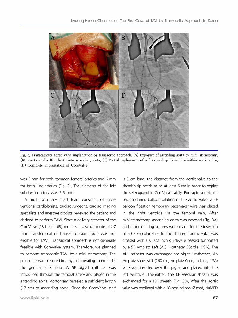
Fig. 2. CT images of aorta and iliofemoral arteries. Bilaterally, iliofemoral arteries were not eligible for the transcatheteraortic valve implantation route due to insufficient size (diameter <6 mm). CIA; common iliac artery, EIA; external iliacartery, CFA; common femoral artery.
kg; body mass index, 19.6 kg/m2) with severe AS was
referred to our institution with recent history of
non-ST-elevation myocardial infarction. Transthoracic
echocardiography indicated a heavily calcified aortic valve
with normal global left ventricular ejection fraction of 63%
(Fig. 1A). An aortic valve area was 0.6 cm2 by continuity
equation. The peak and mean aortic pressure gradient
was 97 mmHg and 63 mmHg, respectively (Fig. 1B). On
computed tomography (CT), the mean diameter of the
aortic annulus 19 mm and the perimeter was 60 mm.
The diameter of ascending aorta was 30 mm. Coronary
angiography showed one-vessel coronary artery disease
with a focal stenosis (50% diameter stenosis) at the
proximal right coronary artery. The patient was considered
high risk for conventional open heart surgical AVR based
on the logistic European System for Cardiac Operative
Risk Evaluation (EuroSCORE) which was calculated to be
25.93%. On CT angiography, the maximal lumen diameter
Kyeong-Hyeon Chun, et al: The First Case of TAVI by Transaortic Approach in Korea
www.lipid.or.kr 87
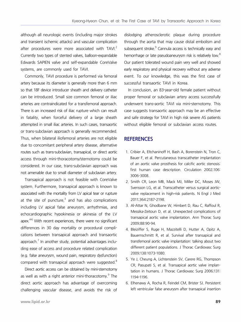
Fig. 3. Transcatheter aortic valve implantation by transaortic approach. (A) Exposure of ascending aorta by mini-sternotomy,(B) Insertion of a 18F sheath into ascending aorta, (C) Partial deployment of self-expanding CoreValve within aortic valve,(D) Complete implantation of CoreValve.
was 5 mm for both common femoral arteries and 6 mm
for both iliac arteries (Fig. 2). The diameter of the left
subclavian artery was 5.5 mm.
A multidisciplinary heart team consisted of inter-
ventional cardiologists, cardiac surgeons, cardiac imaging
specialists and anesthesiologists reviewed the patient and
decided to perform TAVI. Since a delivery catheter of the
CoreValve (18 french (F)) requires a vascular route of ≥7
mm, transfemoral or trans-subclavian route was not
eligible for TAVI. Transapical approach is not generally
feasible with CoreValve system. Therefore, we planned
to perform transaortic TAVI by a mini-sternotomy. The
procedure was prepared in a hybrid operating room under
the general anesthesia. A 5F pigtail catheter was
introduced through the femoral artery and placed in the
ascending aorta. Aortogram revealed a sufficient length
(>7 cm) of ascending aorta. Since the CoreValve itself
is 5 cm long, the distance from the aortic valve to the
sheath’s tip needs to be at least 6 cm in order to deploy
the self-expandble CoreValve safely. For rapid ventricular
pacing during balloon dilation of the aortic valve, a 4F
balloon flotation temporary pacemaker wire was placed
in the right ventricle via the femoral vein. After
mini-sternotomy, ascending aorta was exposed (Fig. 3A)
and a purse string sutures were made for the insertion
of a 6F vascular sheath. The stenosed aortic valve was
crossed with a 0.032 inch guidewire passed supported
by a 5F Amplatz Left (AL) 1 catheter (Cordis, USA). The
AL1 catheter was exchanged for pig-tail cathether. An
Amplatz super stiff (260 cm, Amplatz Cook, Indiana, USA)
wire was inserted over the pigtail and placed into the
left ventricle. Thereafter, the 6F vascular sheath was
exchanged for a 18F sheath (Fig. 3B). After the aortic
valve was predilated with a 18 mm balloon (Z-med, NuMED

J Lipid Atheroscler 2013;2(2):85-90 JOURNAL OF LIPID AND ATHEROSCLEROSIS
88 www.lipid.or.kr
Fig. 4. Transthoracic echocardiography after the transcatheter aortic valve implantation. (A) Parasternal long axis view showsimplanted CoreValve, (B) Pressure gradient over aortic valve by Doppler (maximal pressure gradient 17 mmHg, mean pressuregradient 8 mmHg). AV; aortic valve, Ao; aorta, LV; left ventricle, LA; left atrium.
Fig. 5. Electrocardiogram (ECG) findings. (A) ECG before the procedure, (B) ECG after the procedure.
Inc., Hopkinton, NY, USA) under rapid pacing, a 26 mm
CoreValve was successfully deployed under fluoroscopic
guidance (Fig. 3C, D). Aortography immediately after the
CoreValve deployment demonstrated mild aortic regurgi-
tation (grade II). Transesophageal echocardiography
showed proper position of the prosthetic valve and
confirmed mild paravalvular leak. The peak and mean
transaortic pressure gradient was lowered to 17 mmHg
and 8 mmHg, respectively (Fig. 4). The patient was
monitored in a coronary care unit during 3 subsequent
days for potential complications such as stroke, complete
atrioventricular block, or vascular events. There was no
significant interval change in electrocardiogram (ECG)
findings between before the procedure and after that
(Fig. 5). She recovered fully without any adverse event
and was discharged on the 9th day after the procedure.
The patient is currently being followed for 18 months
without significant adverse cardiovascular event.
DISCUSSION
TAVI is accepted as an alternative treatment modality
for patient with severe AS at high surgical risk. Patients
with an estimated morality risk >20% by logistic
EuroSCORE or >10% by Society of Thoracic Surgeons (STS)
score system are generally considered candidates for the
TAVI procedure. In the literature, TAVI and surgical AVR
were associated with similar rates of survival at 1 year,
Kyeong-Hyeon Chun, et al: The First Case of TAVI by Transaortic Approach in Korea
www.lipid.or.kr 89
although all neurologic events (including major strokes
and transient ischemic attacks) and vascular complication
after procedures were more associated with TAVI.2
Currently two types of stented valves, balloon-expandable
Edwards SAPIEN valve and self-expandable CoreValve
systems, are commonly used for TAVI.
Commonly, TAVI procedure is performed via femoral
artery because its diameter is generally more than 6 mm
so that 18F device introducer sheath and delivery catheter
can be introduced. Small size common femoral or iliac
arteries are contraindicated for a transfemoral approach.
There is an increased risk of iliac rupture which can result
in fatality, when forceful delivery of a large sheath
attempted in small iliac arteries. In such cases, transaortic
or trans-subclavian approach is generally recommended.
Thus, when bilateral iliofemoral arteries are not eligible
due to concomitant peripheral artery disease, alternative
routes such as trans-subclavian, transapical, or direct aortic
access through mini-thoracotomy/sternotomy could be
considered. In our case, trans-subclavian approach was
not amenable due to small diameter of subclavian artery.
Transapical approach is not feasible with CoreValve
system. Furthermore, transapical approach is known to
associated with the mortality from LV apical tear or rupture
at the site of puncture,3 and has also complications
including LV apical false aneurysm, arrhythmias, and
echocardiographic hypokinesia or akinesia of the LV
apex.4-6 With recent experiences, there were no significant
differences in 30 day mortality or procedural compli-
cations between transapical approach and transaortic
approach.7 In another study, potential advantages inclu-
ding ease of access and procedure related complication
(e.g. false aneurysm, wound pain, respiratory dysfunction)
compared with transapical approach were suggested.8
Direct aortic access can be obtained by mini-sternotomy
as well as with a right anterior mini-thoracotomy.9 The
direct aortic approach has advantage of overcoming
challenging vascular disease, and avoids the risk of
dislodging atherosclerotic plaque during procedure
through the aorta that may cause distal embolism and
subsequent stroke.9 Cannula access is technically easy and
hemorrhage or late pseudoaneurysm risk is relatively low.8
Our patient tolerated wound pain very well and showed
early respiratory and physical recovery without any adverse
event. To our knowledge, this was the first case of
successful transaortic TAVI in Korea.
In conclusion, an 83-year-old female patient without
proper femoral or subclavian artery access successfully
underwent trans-aortic TAVI via mini-sternotomy. This
case suggests transaortic approach may be an effective
and safe strategy for TAVI in high risk severe AS patients
without eligible femoral or subclavian access routes.
REFERENCES
1. Cribier A, Eltchaninoff H, Bash A, Borenstein N, Tron C,
Bauer F, et al. Percutaneous transcatheter implantation
of an aortic valve prosthesis for calcific aortic stenosis:
first human case description. Circulation 2002;106:
3006-3008.
2. Smith CR, Leon MB, Mack MJ, Miller DC, Moses JW,
Svensson LG, et al. Transcatheter versus surgical aortic-
valve replacement in high-risk patients. N Engl J Med
2011;364:2187-2198.
3. Al-Attar N, Ghodbane W, Himbert D, Rau C, Raffoul R,
Messika-Zeitoun D, et al. Unexpected complications of
transapical aortic valve implantation. Ann Thorac Surg
2009;88:90-94.
4. Bleiziffer S, Ruge H, Mazzitelli D, Hutter A, Opitz A,
Bauernschmitt R, et al. Survival after transapical and
transfemoral aortic valve implantation: talking about two
different patient populations. J Thorac Cardiovasc Surg
2009;138:1073-1080.
5. Ye J, Cheung A, Lichtenstein SV, Carere RG, Thompson
CR, Pasupati S, et al. Transapical aortic valve implan-
tation in humans. J Thorac Cardiovasc Surg 2006;131:
1194-1196.
6. Elhenawy A, Rocha R, Feindel CM, Brister SJ. Persistent
left ventricular false aneurysm after transapical insertion

J Lipid Atheroscler 2013;2(2):85-90 JOURNAL OF LIPID AND ATHEROSCLEROSIS
90 www.lipid.or.kr
of an aortic valve. J Card Surg 2011;26:51-53.
7. Bapat V, Khawaja MZ, Attia R, Narayana A, Wilson K,
Macgillivray K, et al. Transaortic Transcatheter Aortic
valve implantation using Edwards Sapien valve: a novel
approach. Catheter Cardiovasc Interv 2012;79:733-740.
8. Clarke A, Wiemers P, Poon KK, Aroney CN, Scalia G,
Burstow D, et al. Early experience of transaortic
TAVI--the future of surgical TAVI? Heart Lung Circ
2013;22:265-269.
9. Bruschi G, de Marco F, Botta L, Cannata A, Oreglia J,
Colombo P, et al. Direct aortic access for transcatheter
self-expanding aortic bioprosthetic valves implantation.
Ann Thorac Surg 2012;94:497-503.