Case Report High Grade Glioma Mimicking Voltage Gated ...
5
Case Report High Grade Glioma Mimicking Voltage Gated Potassium Channel Complex Associated Antibody Limbic Encephalitis Dilan Athauda, 1 R. S. Delamont, 2 and E. De Pablo-Fernandez 1 1 Neurology Department, Darent Valley Hospital, Darenth Wood Road, Dartford & Gravesham NHS Trust, Dartford DA2 8DA, UK 2 Neurology Department, Kings College Hospital, London SE5 3RS, UK Correspondence should be addressed to Dilan Athauda; [email protected] Received 30 October 2013; Accepted 19 December 2013; Published 12 January 2014 Academic Editors: K. Arnautov´ ıc, R. Koide, and M. Kurz Copyright © 2014 Dilan Athauda et al. is is an open access article distributed under the Creative Commons Attribution License, which permits unrestricted use, distribution, and reproduction in any medium, provided the original work is properly cited. ough raised titres of voltage gated potassium channel (VGKC) complex antibodies have been occasionally associated with extracranial tumours, mainly presenting as Morvan’s Syndrome or neuromyotonia, they have not yet been reported to be associated with an intracranial malignancy. is is especially important as misdiagnosis of these conditions and delay of the appropriate treatment can have important prognostic implications. We describe a patient with a high grade glioma presenting with clinical, radiological, and serological features consistent with the diagnosis of VGKC antibody associated limbic encephalitis (LE). is is the first association between a primary brain tumour and high titre of VGKC complex antibodies. Clinicoradiological progression despite effective immunosuppressive treatment should prompt clinicians to look for alternative diagnoses. Further studies to elucidate a possible association between VGKC complex and other surface antigen antibodies with primary brain tumours should be carried out. 1. Introduction VGKC complex antibody associated LE is considered a nonparaneoplastic, immunoresponsive condition typically presenting subacutely with memory loss, confusion, seizures, and behavioural changes (though visual hallucinations and psychosis have also been reported) [1]. Approximately 60% of patients have high signal changes unilaterally or bilaterally in the medial temporal lobes on FLAIR and T2 weighted MR imaging [1]. Serum hyponatremia, another diagnosti- cally useful feature, is present in around 60% of patients [2]. e diagnosis is usually made with a combination of clinicoradiological findings and the presence of high titre VGKC complex antibodies (normal reference range is <100 picomolar (pM)). ough it is uncertain at what level a titre of antibody becomes significant, previous studies have suggested titres greater than 400 pM as “high” and levels above this seem to be more associated with CNS disease [1, 2]. ough the phenotype of VGKC complex antibody associated LE is continually expanding, other important conditions can oſten mimic the initial presentation and alternative diagnoses should be considered in atypical cases. Intracranial malignancy is an important differential diagnosis and can present with similar symptoms; however, there have been no reports of elevated levels of VGKC complex antibodies being found in association with an intracranial malignancy. We report a case of a patient presenting initially with characteristic features of VGKC complex associated LE with elevated levels of VGKC antibodies in his serum who, despite treatment with immunosuppression, clinically deteriorated and was subsequently discovered to have a high grade glioma. 2. Case Presentation An 86-year-old man complaining of 1 week of lethargy and irritability was found unconscious at home and taken to the emergency department whereupon he had two generalised tonic clonic (GTC) seizures. He had no past medical history and was on no medication. ere was no history of recent illness, cognitive decline or travel. On examination he had a temperature of 38 ∘ C and had a GCS of 10. ough his consciousness improved over the next Hindawi Publishing Corporation Case Reports in Neurological Medicine Volume 2014, Article ID 458790, 4 pages http://dx.doi.org/10.1155/2014/458790
Transcript of Case Report High Grade Glioma Mimicking Voltage Gated ...
Case ReportHigh Grade Glioma Mimicking Voltage Gated PotassiumChannel Complex Associated Antibody Limbic Encephalitis
Dilan Athauda,1 R. S. Delamont,2 and E. De Pablo-Fernandez1
1 Neurology Department, Darent Valley Hospital, Darenth Wood Road, Dartford & Gravesham NHS Trust, Dartford DA2 8DA, UK2Neurology Department, Kings College Hospital, London SE5 3RS, UK
Correspondence should be addressed to Dilan Athauda; [email protected]
Received 30 October 2013; Accepted 19 December 2013; Published 12 January 2014
Academic Editors: K. Arnautovı́c, R. Koide, and M. Kurz
Copyright © 2014 Dilan Athauda et al. This is an open access article distributed under the Creative Commons Attribution License,which permits unrestricted use, distribution, and reproduction in any medium, provided the original work is properly cited.
Though raised titres of voltage gated potassium channel (VGKC) complex antibodies have been occasionally associated withextracranial tumours, mainly presenting asMorvan’s Syndrome or neuromyotonia, they have not yet been reported to be associatedwith an intracranial malignancy. This is especially important as misdiagnosis of these conditions and delay of the appropriatetreatment can have important prognostic implications. We describe a patient with a high grade glioma presenting with clinical,radiological, and serological features consistent with the diagnosis of VGKC antibody associated limbic encephalitis (LE). This isthe first association between a primary brain tumour and high titre of VGKC complex antibodies. Clinicoradiological progressiondespite effective immunosuppressive treatment should prompt clinicians to look for alternative diagnoses. Further studies toelucidate a possible association between VGKC complex and other surface antigen antibodies with primary brain tumours shouldbe carried out.
1. Introduction
VGKC complex antibody associated LE is considered anonparaneoplastic, immunoresponsive condition typicallypresenting subacutely with memory loss, confusion, seizures,and behavioural changes (though visual hallucinations andpsychosis have also been reported) [1]. Approximately 60%of patients have high signal changes unilaterally or bilaterallyin the medial temporal lobes on FLAIR and T2 weightedMR imaging [1]. Serum hyponatremia, another diagnosti-cally useful feature, is present in around 60% of patients[2]. The diagnosis is usually made with a combination ofclinicoradiological findings and the presence of high titreVGKC complex antibodies (normal reference range is <100picomolar (pM)). Though it is uncertain at what level atitre of antibody becomes significant, previous studies havesuggested titres greater than 400 pM as “high” and levelsabove this seem to be more associated with CNS disease[1, 2]. Though the phenotype of VGKC complex antibodyassociated LE is continually expanding, other importantconditions can often mimic the initial presentation andalternative diagnoses should be considered in atypical cases.
Intracranialmalignancy is an important differential diagnosisand can present with similar symptoms; however, therehave been no reports of elevated levels of VGKC complexantibodies being found in association with an intracranialmalignancy. We report a case of a patient presenting initiallywith characteristic features of VGKC complex associatedLE with elevated levels of VGKC antibodies in his serumwho, despite treatment with immunosuppression, clinicallydeteriorated and was subsequently discovered to have a highgrade glioma.
2. Case Presentation
An 86-year-old man complaining of 1 week of lethargy andirritability was found unconscious at home and taken to theemergency department whereupon he had two generalisedtonic clonic (GTC) seizures. He had no past medical historyand was on no medication. There was no history of recentillness, cognitive decline or travel.
On examination he had a temperature of 38∘C and had aGCS of 10. Though his consciousness improved over the next
Hindawi Publishing CorporationCase Reports in Neurological MedicineVolume 2014, Article ID 458790, 4 pageshttp://dx.doi.org/10.1155/2014/458790

2 Case Reports in Neurological Medicine
few hours he remained confused and verbally and physicallyaggressive. Repeated neurological examination was normal.He was started on ceftazidime, amoxicillin, and acyclovir fora possible CNS infection.
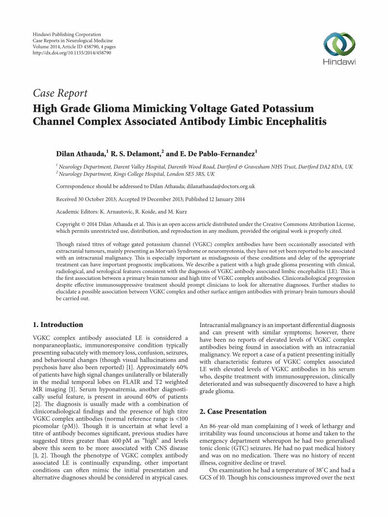
Extensive blood tests including full blood count, elec-trolytes, liver and thyroid function tests, CRP, ESR, ANA,ANCA, anticardiolipin antibodies, vitamin B12, folate, ACE,protein electrophoresis, Borrelia, and HIV serologies werenegative or normal. His initial brain CT scanwithout contrastwas normal and CSF analysis revealed a white cell count of 35(30% lymphocytes), with normal protein and glucose, cultureand viral PCR. MRI brain showed high signal changes on T2weighted images involving the right insula, parahippocampalgyrus, hippocampus, and splenium of the corpus callosumsuggestive of limbic encephalitis (Figures 1(a) and 1(b)).A paraneoplastic screen (for anti-Yo, anti-Hu, and anti-Ri antibodies) was negative and CT chest, abdomen, andpelvis did not identify any occult malignancy. His confusionand behavioural abnormalities resolved a few days afteradmission and he was discharged after completing 14 days oftreatment with intravenous acyclovir. An extended autoim-mune screen was sent including voltage gated potassiumchannel (VGKC) complex antibodies, N-methyl-D-aspartatereceptor (NMDAr), and glutamic acid decarboxylase (GAD)antibodies.
Unfortunately, he was readmitted 15 days later withfluctuating disorientation, aggressiveness, and complex visualhallucinations. Neurological examination was unremarkableincluding cognitive assessment (Mini-Mental State Examina-tion score was 30/30). His extended immune screen returnedwhich was negative for NMDAr and GAD antibodies, buthe had a raised VGKC complex antibody titre of 437 pico-molar (pM). Repeat MRI brain scan 5 weeks after initialpresentation showed persistent high T2 signal changes of theright medial temporal lobe structures previously describedwith new involvement of the right precuneus (Figures 1(c)and 1(d)). A diagnosis of VGKC complex antibody limbicencephalitis (LE) was made. The patient declined treatmentwith plasmapheresis or intravenous immunoglobulins andwas started on oral prednisolone 40mg a day. He improvedand was discharged. Repeat serum testing sent for VGKCantibodies 3 weeks after starting the steroids was nowundetectable.
He presented 25 days later with 3 generalised tonicclonic seizures and gradual progressive cognitive decline.On examination, he was disorientated, had mild left sidedhemiparesis, and was neglecting the left side. He underwenta course of plasmapheresis for 5 days, but a repeat MRIbrain scan following the plasmapheresis showed aggressiveprogression of the previous lesions, with new nodular fociwithin the medial aspect of the right temporoparietal lobeswith mass effect, heterogeneous contrast enhancement, andareas of necrosis more in keeping with a high grade gliomathan LE (Figures 1(e) and 1(f)).
Following review by a neurooncology multidisciplinaryteam he was offered supportive care only. He subsequentlyhad a gradual deterioration of his condition with progressivecognitive decline and general physical decline and occasionalgeneralized tonic seizures despite antiepileptic treatment.
3. Discussion
Our patient initially illustrated many of the classic clinical,radiological, and serological features of VGKC antibodycomplex LE: he had well-recognised symptoms of seizures,amnesia, and personality change. He had unilateral highsignal changes in his medial temporal lobe on MRI T2weighted imaging which is usually observed in such cases.We also detected elevated levels of VGKC antibody in hisserum of 407 pM—usually undetectable. The suggestedtreatment for VGKC complex associated LE is earlyaggressive immunotherapies with a combination of high dosesteroids, plasma exchange, or intravenous immunoglobulins,with a concurrent rapid fall of the antibody level witheffective therapy [3]. Typically prognosis is generally goodthough radiological changes may take longer to improve,with subsequent persistent temporomesial atrophy asa common finding [1].
Despite the subsequent immunosuppressive therapies,the patient developed progressive cognitive decline, visualhallucinations, and left sided hemiparesis, with spreadingof the MRI changes involving new areas including themedial parietal lobe and nodular enhancement—which isatypical for VGKC associated LE. Though no histologicalconfirmation is available, repeat brainMRI 8months after theinitial presentation showed radiological findings in keepingwith an aggressive, high grade glioma.
The spectrum of conditions associated with VGKC com-plex antibodies is in continuous expansion and raised titresof VGKC complex antibodies have also been reported inother clinical syndromes includingMorvan’s Syndrome, neu-romyotonia, and faciobrachial dystonic seizures [1]. Thoughneuromyotonia and Morvan’s Syndrome can be associatedwith tumours, particularly thymomas, they are uncommonin VGKC complex antibody LE and it is thought to be anonparaneoplastic condition.
A study to identify possible neurological and oncologicalassociations in patients with raised VGKC antibodies foundthat 33% of patients identified with raised VGKC antibodieshad active tumours, mostly lung carcinomas, but also 3haematological malignancies and 1 thymoma [4], though nodistinction between different clinical syndromes was made.
However, a more recent study that identified the clinicalphenotype and tumour association of 96 patients with VGKCantibody titres >400 pM found that the majority—66%—had LE, and of these, none were associated with underlyingtumours. Of the six patients identified in the studywith activetumours all had either Morvan’s Syndrome or neuromy-otonia, 5 had thymomas and one patient had endometrialadenocarcinoma [5].
To our knowledge, no intracranial malignancy, eitherprimary or metastases, has been associated with raisedVGKC antibodies and this is the first case reporting thisassociation. This is extremely important as misdiagnosis ofthese conditions and delay of the appropriate treatment couldhave important prognostic implications.
It is impossible to ascertain that if an early diagnosisin our case could have made an impact on his prognosisthough, as once the diagnosis was made, the patient was
Case Reports in Neurological Medicine 3
(a) (b)
(c) (d)
(e) (f)
Figure 1: (a, b) Coronal T2 weighted FLAIR brainMRI showing high signal changes involvingmedial temporal areas. (c, d) T1 weightedMRI10 weeks later showing enhancement of the previous lesions after gadolinium administration with extension posterior to the occipital lobe.(e, f) T1 weighted brain MRI showing rapid progression of the initial lesion with marked mass effect, areas of necrosis, and heterogeneouscontrast enhancement consistent with a high grade glioma.
only suitable for supportive care. This case illustrates thatclinicians treating patients with a presumed diagnosis ofVGKC complex antibody LE must maintain a high index ofsuspicion, even in the presence of high levels of antibodies,and should consider appropriate neuroimaging and alter-native diagnoses if the patient fails to respond to effectiveimmunotherapy. It also highlights the difficulty of diagnosinggliomas in the early stages despite MR imaging. Furtherstudies to investigate a possible association between VGKC
complex and other surface antigen antibodies with primarybrain tumours should be carried out in order to betterelucidate their role in these conditions.
Conflict of Interests
The authors declare that there is no conflict of interestsregarding the publication of this paper.

4 Case Reports in Neurological Medicine
Acknowledgments
Many thanks are due to Angela Vincent who provided apaper review and to the Nuffield Department of ClinicalSciences, John Radcliffe Hospital, Oxford, for analysis of theantibodies.
References
[1] L. Zuliani, F. Graus, B. Giometto, C. Bien, and A. Vincent,“Central nervous system neuronal surface antibody associatedsyndromes: Review and guidelines for recognition,” Journal ofNeurology, Neurosurgery and Psychiatry, vol. 83, no. 6, pp. 638–645, 2012.
[2] A. Vincent, C. Buckley, J. M. Schott et al., “Potassiumchannel antibody-associated encephalopathy: a potentiallyimmunotherapy-responsive form of limbic encephalitis,” Brain,vol. 127, no. 3, pp. 701–712, 2004.
[3] S. A. B. Ahmad, H. A. Archer, C. M. Rice, S. Gerhand, M.Bradley, and A.Wilkins, “Seronegative limbic encephalitis: casereport, literature review and proposed treatment algorithm,”Practical Neurology, vol. 11, no. 6, pp. 355–361, 2011.
[4] K. M. Tan, V. A. Lennon, C. J. Klein, B. F. Boeve, and S. J.Pittock, “Clinical spectrum of voltage-gated potassium channelautoimmunity,” Neurology, vol. 70, no. 20, pp. 1883–1890, 2008.
[5] S. R. Irani, S. Alexander, P. Waters et al., “Antibodies to Kv1potassium channel-complex proteins leucine-rich, glioma inac-tivated 1 protein and contactin-associated protein-2 in limbicencephalitis,Morvan’s syndrome and acquired neuromyotonia,”Brain, vol. 133, no. 9, pp. 2734–2748, 2010.

Submit your manuscripts athttp://www.hindawi.com
Stem CellsInternational
Hindawi Publishing Corporationhttp://www.hindawi.com Volume 2014
Hindawi Publishing Corporationhttp://www.hindawi.com Volume 2014
MEDIATORSINFLAMMATION
of
Hindawi Publishing Corporationhttp://www.hindawi.com Volume 2014
Behavioural Neurology
EndocrinologyInternational Journal of
Hindawi Publishing Corporationhttp://www.hindawi.com Volume 2014
Hindawi Publishing Corporationhttp://www.hindawi.com Volume 2014
Disease Markers
Hindawi Publishing Corporationhttp://www.hindawi.com Volume 2014
BioMed Research International
OncologyJournal of
Hindawi Publishing Corporationhttp://www.hindawi.com Volume 2014
Hindawi Publishing Corporationhttp://www.hindawi.com Volume 2014
Oxidative Medicine and Cellular Longevity
Hindawi Publishing Corporationhttp://www.hindawi.com Volume 2014
PPAR Research
The Scientific World JournalHindawi Publishing Corporation http://www.hindawi.com Volume 2014
Immunology ResearchHindawi Publishing Corporationhttp://www.hindawi.com Volume 2014
Journal of
ObesityJournal of
Hindawi Publishing Corporationhttp://www.hindawi.com Volume 2014
Hindawi Publishing Corporationhttp://www.hindawi.com Volume 2014
Computational and Mathematical Methods in Medicine
OphthalmologyJournal of
Hindawi Publishing Corporationhttp://www.hindawi.com Volume 2014
Diabetes ResearchJournal of
Hindawi Publishing Corporationhttp://www.hindawi.com Volume 2014
Hindawi Publishing Corporationhttp://www.hindawi.com Volume 2014
Research and TreatmentAIDS
Hindawi Publishing Corporationhttp://www.hindawi.com Volume 2014
Gastroenterology Research and Practice
Hindawi Publishing Corporationhttp://www.hindawi.com Volume 2014
Parkinson’s Disease
Evidence-Based Complementary and Alternative Medicine
Volume 2014Hindawi Publishing Corporationhttp://www.hindawi.com