
Case Report Haihong Liao, Pengtao Song, Quan Qi, Yizhen ...
7
Case Report Haihong Liao, Pengtao Song, Quan Qi, Yizhen Jiang, Shuwen Han, Yuefen Pan*, Yulong Liu* Synchronous primary malignancy of colon cancer and mantle cell lymphoma: A case report https://doi.org/10.1515/med-2020-0239 received July 8, 2020; accepted September 23, 2020 Abstract: Multiple primary malignancies in a single patient are relatively rare; however, the frequency of this has increased significantly in recent decades. Here, we retrospectively reported an unusual case of a 70-year-old man who was admitted to the hospital with mantle cell lymphoma (MCL) and colon cancer and aimed to explore measures to reduce missed diagnosis. Based on the data, the investigation of the related literatures, colonoscopy examination, and abdom- inal computed tomography (CT) scan were conducted for the detection of colon cancer. Following this, a precise diagnosis of MCL was confirmed by immunohistochemistry and bone marrow biopsy, which were performed to analyze the clinical characteristics and essentials for the diagnosis and differential of the disease. The results of colonoscopy showed that the patient had colon cancer, while the abdom- inal CT scan demonstrated colon cancer accompanied by multiple lymphadenopathy throughout the entire body. Besides, the results of immunohistochemistry confirmed that the patient suffered from MCL. The bone marrow biopsy revealed the active hematopoietic tissue hyperplasia but no tumor issue involvement in the bone marrow. In conclu- sion, our study combined the analysis and summary of the diagnosis and treatment of the colon cancer with MCL to provide clinical guidance for the rare multiple primary malignancy. Keywords: multiple primary cancer, colon cancer, mantle cell lymphoma, colonoscopy, computed tomography 1 Introduction Multiple primary malignancies are defined as the occur- rence of two or more malignancies without subordinate relationship in different organs of an individual patient, while the underlying mechanism remains unclear [1]. The incidence of multiple primary malignancy is rare although the prevalence rate is increasing recently [2]. Mantle cell lymphoma (MCL) is a unique B-cell lymphoma that accounts for 3–10% of non-Hodgkin’s lymphoma (NHL) with a median survival of 3–4 years [3]. It is more common in the elderly, which is often diagnosed at an advanced stage [4]. Lymph node metastasis in colon cancer and extra- lateral lymphoma in gastrointestinal tract are common, whereas conventional lymph node biopsy during the opera- tion of colon cancer and partial enlarged lymph nodes that are found in MCL are rare [5]. Only one case reported that a patient developed NHL after curative resection of colon cancer in recent decades, but no report was found about synchronous primary malignancy, including the colon cancer and lymphoma [6]. Herein, we present such an unu- sual case of colon cancer and MCL. To our knowledge, this is the first article that has been reported on double primary malignancies associated with MCL in a patient with colon cancer. We report the following case according to the CARE reporting checklist. 2 Case presentation A 70-year-old man was admitted to our hospital in March 2017 due to upper abdominal pain for more than 2 weeks. He had no obvious nausea, vomiting, chills, fever, black Haihong Liao: Department of Medical Oncology, Affiliated Central Hospital Huzhou University, Huzhou 313000, Zhejiang, China Pengtao Song, Quan Qi, Yizhen Jiang, Shuwen Han, Haihong Liao: Department of Medical Oncology, Huzhou Central Hospital, Huzhou 313000, Zhejiang, China * Corresponding author: Yuefen Pan, Department of Medical Oncology, Huzhou Central Hospital, Huzhou 313000, Zhejiang, China, e-mail: [email protected] * Corresponding author: Yulong Liu, Department of Oncology, The Second Affiliated Hospital of Soochow University, Suzhou 215004, Jiangsu, China; State Key Laboratory of Radiation Medicine and Protection, School of Radiation Medicine and Protection, Soochow University, Suzhou 215123, Jiangsu, China; Collaborative Innovation Center of Radiation Medicine of Jiangsu Higher Education Institutions, Suzhou 215123, Jiangsu, China, e-mail: [email protected] Open Medicine 2021; 16: 696–702 Open Access. © 2021 Haihong Liao et al., published by De Gruyter. This work is licensed under the Creative Commons Attribution 4.0 International License.
Transcript of Case Report Haihong Liao, Pengtao Song, Quan Qi, Yizhen ...

Case Report
Haihong Liao, Pengtao Song, Quan Qi, Yizhen Jiang, Shuwen Han, Yuefen Pan*, Yulong Liu*
Synchronous primary malignancy of colon cancerand mantle cell lymphoma: A case report
https://doi.org/10.1515/med-2020-0239received July 8, 2020; accepted September 23, 2020
Abstract: Multiple primary malignancies in a single patientare relatively rare;however, the frequencyof thishas increasedsignificantly in recent decades. Here, we retrospectivelyreported an unusual case of a 70-year-old man who wasadmitted to the hospital with mantle cell lymphoma (MCL)and colon cancer and aimed to explore measures to reducemissed diagnosis. Based on the data, the investigation of therelated literatures, colonoscopy examination, and abdom-inal computed tomography (CT) scan were conducted forthe detection of colon cancer. Following this, a precisediagnosis of MCL was confirmed by immunohistochemistryand bone marrow biopsy, which were performed to analyzethe clinical characteristics and essentials for the diagnosisand differential of the disease. The results of colonoscopyshowed that the patient had colon cancer, while the abdom-inal CT scan demonstrated colon cancer accompaniedby multiple lymphadenopathy throughout the entire body.Besides, the results of immunohistochemistry confirmedthat the patient suffered fromMCL. The bonemarrow biopsyrevealed the active hematopoietic tissue hyperplasia butno tumor issue involvement in the bone marrow. In conclu-sion, our study combined the analysis and summary of the
diagnosis and treatment of the colon cancer with MCL toprovide clinical guidance for the rare multiple primarymalignancy.
Keywords:multiple primary cancer, colon cancer, mantlecell lymphoma, colonoscopy, computed tomography
1 Introduction
Multiple primary malignancies are defined as the occur-rence of two or more malignancies without subordinaterelationship in different organs of an individual patient,while the underlying mechanism remains unclear [1]. Theincidence of multiple primary malignancy is rare althoughthe prevalence rate is increasing recently [2]. Mantle celllymphoma (MCL) is a unique B-cell lymphoma thataccounts for 3–10% of non-Hodgkin’s lymphoma (NHL)with a median survival of 3–4 years [3]. It is more commonin the elderly, which is often diagnosed at an advancedstage [4]. Lymph nodemetastasis in colon cancer and extra-lateral lymphoma in gastrointestinal tract are common,whereas conventional lymph node biopsy during the opera-tion of colon cancer and partial enlarged lymph nodesthat are found in MCL are rare [5]. Only one case reportedthat a patient developed NHL after curative resection ofcolon cancer in recent decades, but no report was foundabout synchronous primarymalignancy, including the coloncancer and lymphoma [6]. Herein, we present such an unu-sual case of colon cancer andMCL. To our knowledge, this isthe first article that has been reported on double primarymalignancies associated with MCL in a patient with coloncancer. We report the following case according to the CAREreporting checklist.
2 Case presentation
A 70-year-old man was admitted to our hospital in March2017 due to upper abdominal pain for more than 2 weeks.He had no obvious nausea, vomiting, chills, fever, black
Haihong Liao: Department of Medical Oncology, Affiliated CentralHospital Huzhou University, Huzhou 313000, Zhejiang, ChinaPengtao Song, Quan Qi, Yizhen Jiang, Shuwen Han, Haihong Liao:Department of Medical Oncology, Huzhou Central Hospital, Huzhou313000, Zhejiang, China
* Corresponding author: Yuefen Pan, Department of MedicalOncology, Huzhou Central Hospital, Huzhou 313000, Zhejiang,China, e-mail: [email protected]* Corresponding author: Yulong Liu, Department of Oncology, TheSecond Affiliated Hospital of Soochow University, Suzhou 215004,Jiangsu, China; State Key Laboratory of Radiation Medicine andProtection, School of Radiation Medicine and Protection, SoochowUniversity, Suzhou 215123, Jiangsu, China; Collaborative InnovationCenter of Radiation Medicine of Jiangsu Higher EducationInstitutions, Suzhou 215123, Jiangsu, China,e-mail: [email protected]
Open Medicine 2021; 16: 696–702
Open Access. © 2021 Haihong Liao et al., published by De Gruyter. This work is licensed under the Creative Commons Attribution 4.0International License.

stools, and other discomforts. The patient was previouslyhealthy and had no family history of colon cancer or malig-nant lymphoma, and social history was noncontributory.
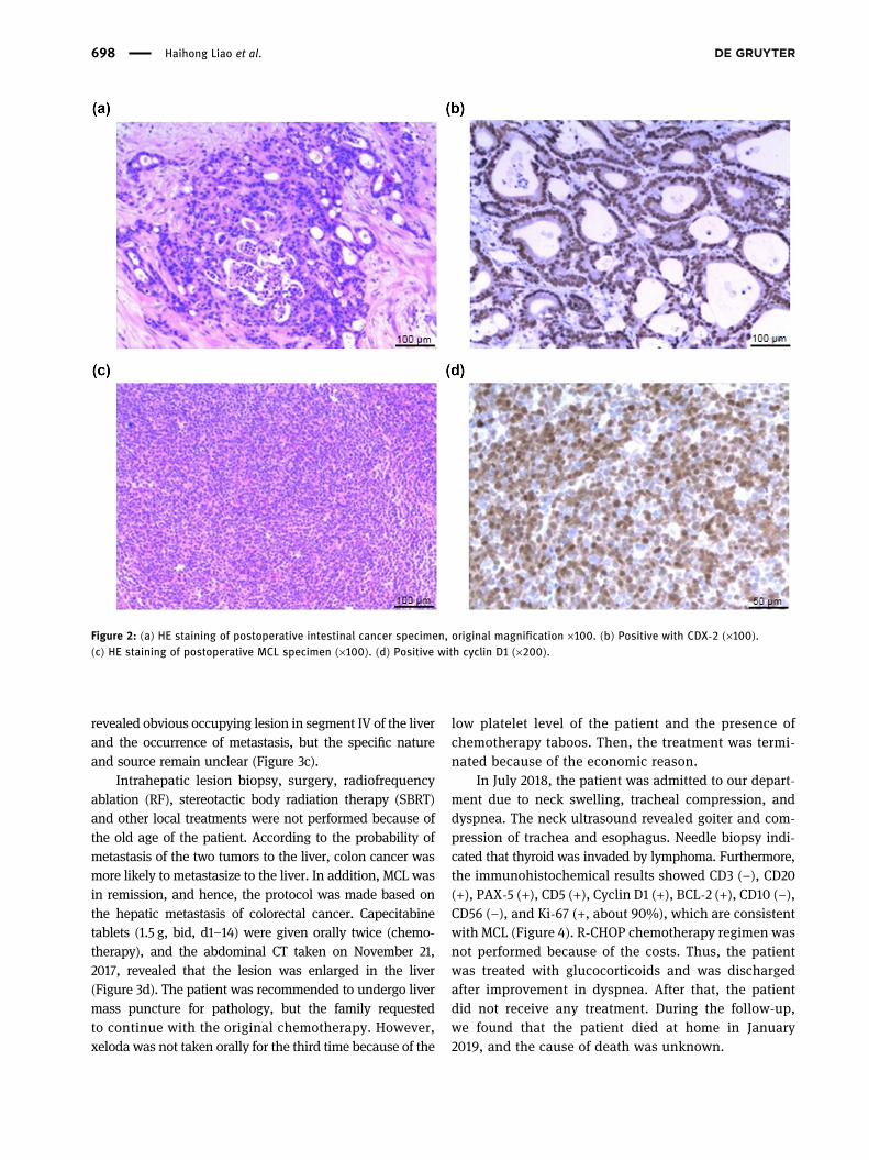
The colonoscopy revealed colon cancer, and theabdominal computed tomography (CT) indicated coloncancer and systemic multiple lymphadenopathy that mightbe malignant lymphoma (Figure 1). On March 17, 2017, thepathological diagnoses were as follows: (descending colon)uplifted moderately differentiated adenocarcinoma with amaximum mass diameter of 4.5 cm infiltrating into the ser-osal layer; the peripheral lymph node metastases were 3/10perienteric group, 2 cancer nodules, and 0/5 mesentericgroup; the upper and lower cutting edges were negative(Figure 2a). The immunohistochemical results showed P53(3+), Ki-67 (+, about 70%), CEA (+), E-cadherin (+), Topo-II(2+), and CDX-2 (+) (Figure 2b). Fourteen retroperitoneallymph nodes with a maximum diameter of 0.3–6 cm werefound: NHL (Figure 2c). Immunohistochemical resultsshowed LCA (+), CD3 (−), CD20 (+), PAX-5 (+), CD10 (−),Bcl-6 (−), Mum-1 (+), Bcl-2 (+), Ki-67 (+, about 50%), andcyclin D1 (+) (Figure 2d). Finally, the patient was diagnosedas NHL [MCL at stage IIIA, CD20+ with International Prog-nostic Index (IPI) score of 4 points, postoperative coloncancer (pT3N1M0, stage IIIB)]. During the course of thedisease, the patient had no fever, night sweats, or signifi-cant weight loss. The bone marrow biopsy on April 10, 2017,revealed that bone marrow hematopoietic tissue hyper-plasia was active, and the hematopoietic tissue accountedfor about 40% with the degree of hyperplasia (++). Besides,granulocytes proliferate actively, protocells were observed,and the proportion of mature granulocytes was normal.
Furthermore, red blood cell lines were hyperproliferative,and clusters of juvenile red blood cells were visible, whichwere distributed in the trabecular region, and the propor-tion of the megakaryocytes was normal. The bone marrowbiopsy revealed that bone marrow hematopoietic hyper-plasia was active and no tumor involving bone marrowwas found. Bone marrow routine examination showedthat the smear had an active proliferation of nuclear cellswithout obvious abnormal cells on April 12, 2017. Afteradmission to our department, the patient went to a hos-pital in Shanghai for pathological consultation based onthe data and pathological sections to confirm the diag-nosis of the double primary tumor. The result of consul-tation was the same as that of our hospital. The patientpresented significant abdominal pain while waitingfor the result of pathological consultation after surgery.CT scan showed obvious abdominal lymphadenopathy,indicating the progression of lymphoma (no CT imagesbecause of many enlarged lymph nodes).
According to the principle of dual primary tumortreatment, this patient underwent a radical resection ofa tumor in the descending colon cancer in our hospital onMarch 7, 2017, and postoperative adjuvant chemotherapywas performed to reduce the risk of recurrence and meta-stasis. Patients with NHL at stage III weremainly treatedwithinternal medicine, and chemotherapy was required becauseof obvious clinical symptoms. However, there is no chemo-therapy scheme for both tumors. In addition, the sideeffects of two chemotherapy regimens on bilateral tumorsare too severe to be tolerated by elderly patients. After dis-cussing themultidisciplinary diagnosis and treatmentmodel(MDT), and actively communicating with the patient’sfamily, MCL with clinical symptoms was treated. R-CHOPchemotherapy regimenwas performed for five times, includ-ing metrology (600mg, day 1), dexamethasone (10mg, days2–6), pharmorubicin (80mg, day 2), cyclophosphamide(0.8 g, day 2), and vindesine (4mg, day 2 q3w). IV degreeof the bone marrowwas suppressed, and granulocytes werelacking after the first chemotherapy. Following the subse-quent chemotherapy, PEGylated recombinant human gran-ulocyte colony-stimulating factor needle was used to pre-vent leukopenia, and no obvious bone marrow suppressionoccurred. Abdominal pain was relieved, and abdominallymph nodes became smaller upon the first chemotherapy.On June 6, 2017, abdominal CT indicated no obvious occu-pying lesion in hepatic segment IV (Figure 3a). After thefifth course of chemotherapy, abdominal CT was performedon August 30, 2017, and the result showed the hepatic seg-ment IV occupation and metastasis may occur (Figure 3b).Enhanced MRI was performed on September 5, 2017,because the abdominal CT was not clear and the results
Figure 1: Abdominal CT indicating a mass lesion of the colon beforesurgery.
Diagnosis of synchronous primary malignancy 697
revealed obvious occupying lesion in segment IV of the liverand the occurrence of metastasis, but the specific natureand source remain unclear (Figure 3c).
Intrahepatic lesion biopsy, surgery, radiofrequencyablation (RF), stereotactic body radiation therapy (SBRT)and other local treatments were not performed because ofthe old age of the patient. According to the probability ofmetastasis of the two tumors to the liver, colon cancer wasmore likely to metastasize to the liver. In addition, MCL wasin remission, and hence, the protocol was made based onthe hepatic metastasis of colorectal cancer. Capecitabinetablets (1.5 g, bid, d1–14) were given orally twice (chemo-therapy), and the abdominal CT taken on November 21,2017, revealed that the lesion was enlarged in the liver(Figure 3d). The patient was recommended to undergo livermass puncture for pathology, but the family requestedto continue with the original chemotherapy. However,xeloda was not taken orally for the third time because of the
low platelet level of the patient and the presence ofchemotherapy taboos. Then, the treatment was termi-nated because of the economic reason.
In July 2018, the patient was admitted to our depart-ment due to neck swelling, tracheal compression, anddyspnea. The neck ultrasound revealed goiter and com-pression of trachea and esophagus. Needle biopsy indi-cated that thyroid was invaded by lymphoma. Furthermore,the immunohistochemical results showed CD3 (−), CD20(+), PAX-5 (+), CD5 (+), Cyclin D1 (+), BCL-2 (+), CD10 (−),CD56 (−), and Ki-67 (+, about 90%), which are consistentwith MCL (Figure 4). R-CHOP chemotherapy regimen wasnot performed because of the costs. Thus, the patientwas treated with glucocorticoids and was dischargedafter improvement in dyspnea. After that, the patientdid not receive any treatment. During the follow-up,we found that the patient died at home in January2019, and the cause of death was unknown.
Figure 2: (a) HE staining of postoperative intestinal cancer specimen, original magnification ×100. (b) Positive with CDX-2 (×100).(c) HE staining of postoperative MCL specimen (×100). (d) Positive with cyclin D1 (×200).
698 Haihong Liao et al.

Figure 3: (a) Abdominal enhanced CT revealing no obvious occupying lesion in the hepatic segment IV on June 6, 2017. (b) Abdominalenhanced CT revealing hepatic segment IV occupation and metastasis may occur on August 30, 2017. (c) Abdominal enhanced MRI showingobvious occupying lesion in the hepatic segment IV, and metastasis may occur on September 5, 2017. (d) Abdominal enhanced CTsuggesting enlarged hepatic segment IV lesions.
Figure 4: (a) HE staining of fine needle aspiration sample of thyroid (×100). (b) Positive with PAX-5 (×100).
Diagnosis of synchronous primary malignancy 699

Ethical statement: The research related to human usehas been complied with all the relevant national regula-tions, institutional policies, and in accordance the tenetsof the Helsinki Declaration, and has been approvedby the Ethics Committee of Huzhou Central Hospital(20191210-01).
Informed consent: Written informed consent was obtainedfrom the patient for publication of this study and anyaccompanying images.
3 Discussion
The diagnosis of multiple primary malignancies in thestudy was performed according to the criteria previouslydescribed: each tumor is malignant and has its ownunique pathological features; two or more tumors needto be spaced at a normal interval or occur at differentsites, but the two are not continuous; and recurrenceand metastasis must be excluded [7]. This case showeda combination of colon cancer and MCL, which are twodifferent types occurred simultaneously, thus meetingthe criteria for multiple primary malignancy.
In terms of treatment, there is no chemotherapyscheme for both tumors. Currently, the treatment forcolon cancer is mostly surgical resection followed byadjuvant chemotherapy [8]. Previous studies have reportedthat although the first-line treatment for MCL patientsis generally considered too toxic for elderly patients,moderate intensive chemotherapy, such as R-CHOP, stillremained the best induction regimen for elderly patientswith MCL [9]. In this case, according to the principleof dual primary tumor treatment, radical resection ofcolon cancer and postoperative adjuvant chemotherapywere performed, whereas patients with MCL were mainlytreated with internal medicine, and R-CHOP chemotherapywas required because of obvious clinical symptoms. More-over, oral chemotherapy was also adopted in the patient forthe treatment of hepatic and thyroid metastasis.
The pathogenesis of multiple primary malignanciesremains unclear. Several researches have reported thatrisk factors, including low immunity, the existence oftumor-susceptibility genes, mutual signal channels andcontinuous exposure to toxic and harmful carcinogens,are related with multiple primary malignancy [10–12].Colon cancer is often associated with endometrial, gas-tric, and ovarian cancers, but little is reported to itscorrelations with blood malignancies [13]. It has beenreported that the patients underwent secondary breast,
ovarian, and uterine cancers after colorectal cancer inKorea [14]. Liu et al. reported a case with male breastcancer and coexisting MCL in 2017 [7]. Besides, Hu et al.reported that a 74-year-old woman had a history of coloncancer and developed multiple primary malignanciesincluding stomach, lung, breast, and liver cancers [15].In our study, two types of malignant tumor components(colon cancer and MCL) were noticed simultaneously inthe patient, followed by invasion of thyroid by lym-phoma. Multiple primary tumor is usually found in thesame system or in different parts of the same organ [16].Colon cancer and coexisting MCL in this case are rarebecause colon belongs to the parenchymal organ, whereasMCL is a disease of the lymphatic hematopoietic system.The patient was admitted to the hospital because of diges-tive symptoms, and the colonoscopy revealed colon cancer.Furthermore, abdominal CT showed lymphadenectasis.Lymph node metastasis of colon malignant tumor generallyoccurs through lymphatic circulation path [17]. Therefore,colon cancer with lymph node metastasis rather than thesecond primary tumor would be considered when theswollen lymph nodes were observed in imagological diag-nosis, resulting in missed diagnosis. In addition, the lymphnodes submitted for examination in the case were colorectalcancer metastasis, and retroperitoneal lymph nodes wereMCL. Thus, pathological diagnosis should be performedfor the specimens of the lymph node to avoid missed diag-nosis. Moreover, auxiliary examination including regionallymph nodes and puncture of mass should be completedbefore treatment because the liver lesions were not exam-ined for pathology with the unknown nature. As for thera-peutic effect, the prognosis of dual primary tumors is worsethan that of the corresponding single primary tumors,which may be related to the low immunity of patients andthe failure of treatment of both tumors [18–21].
The synchronous of MCL and colonic carcinoma inone patient is rare. Liao et al. [22] previously reported acase of advanced sigmoid adenomcarcinoma and duo-denal MCL with the involvement of lymph nodes. Thiscase was considered as extremely rare because the loca-tion of MCL was identified 20 months after the chemor-adiotherapy of adenocarcinoma. In the case described bySztarkier et al. [23], there was also a case of adenocarci-noma; however, the MCL was present in tubular adenomawith polyp formation. One polyp was infiltered by thelymphoid cell, whereas two regional lymph nodes werepartially replaced by MCL. Handra-Luca [24] has describeda patient with intestinal tumors of adenoma-lymphomawithout compromising the lymph nodes. All three patientsdescribed earlier received R-CHOP regimen after the diag-nosis of MCL, which is consistent with our case. Yet, the
700 Haihong Liao et al.

used drugs and its dosage in R-CHOP regimen were dif-ferent, whichmay depend on the patients’ clinical situation.Besides, compared with other studies, our study is the firstone that has not only introduced a rare case of synchronousof colon cancer and MCL but also provided a clinical gui-dance of composite tumors to reduce the probability ofmisdiagnosis.
In conclusion, colon cancer with multiple lymphade-nopathy throughout the body (malignant lymphomawas considered) was detected through abdominal CT.Combined with the immunohistochemical results, thepatient was diagnosed as synchronous primary malig-nancy colon cancer and MCL, avoiding missed diagnosis.The diagnosis rate of dual primary tumors is high due tothe medical advancements. Doctors require detailed ana-lysis ability and rich clinical experience from multipledisciplines to discuss together before making best choiceto formulate the most effective treatment regimen, pro-long the survival time of the patients, and improve thequality of life needs to be solved in the future.
Funding information: This work was supported by theYoung Talents of Zhejiang Provincial Medical and HealthScience and Technology Project [grant number 2019RC285].
Conflict of interest: The authors have no conflicts ofinterest to declare.
Data availability statement: The datasets generated duringand/or analysed during the current study are availablefrom the corresponding author on reasonable request.
References
[1] Xu W, Liao WJ, Ge PL, Ren JJ, Xu HF, Yang HY, et al. Multipleprimary malignancies in patients with hepatocellular carci-noma: a largest series with 26-year follow-up. Medicine.2016;95(17):e3491. doi: 10.1097/MD.0000000000003491.
[2] Banimostafavi SE, Tayebi S, Tayebi M, Montazer F. CaseReport: Synchronous primary malignancy including the breastand endometrium. F1000Res. 2017;6:1502. doi: 10.12688/f1000research.11971.3.
[3] Cao LH, Jin J. Prognostic stratification in patients with mantlecell lymphoma. Chin J Pract Int Med. 2015;27(481–2):13.doi: 10.1629/2048-7754.111.
[4] Fernàndez V, Hartmann E, Ott G, Campo E, Rosenwald A.Pathogenesis of mantle-cell lymphoma: all oncogenic roadslead to dysregulation of cell cycle and DNA damage responsepathways. J Clin Oncol. 2005;23(26):6364–9. doi: 10.1200/JCO.2005.05.019.
[5] Lee SY, Yeom SS, Kim CH, Kim YJ, Kim HR. Distribution of lymphnode metastasis and the extent of lymph node dissection in
descending colon cancer patients. ANZ J Surg.2019;89(9):373–8. doi: 10.1111/ans.15400.
[6] Hirano K, Yamashita K, Yamashita N, Nakatsumi Y, Esumi H,Kawashima A, et al. Non-hodgkin’s lymphoma in a patient withprobable hereditary nonpolyposis colon cancer: report of acase and review of the literature. Dis Colon Rectum.2002;45(2):273–9. doi: 10.1007/s10350-004-6161-9.
[7] Liu J, Wei HQ, Zhu KQ, Lai LQ, Han XY, Yang Y. Male breastcancer and mantle cell lymphoma in a single patient: a casereport and literature review. Medicine. 2017;96(48):e8911.doi: 10.1097/MD.0000000000008911.
[8] Rajdev K, Siddiqui AH, Agarwal S, Ansari Y, Maroun R.Mediastinal recurrence after curative resection of coloncancer: a case report and review of literature. J Oncol PharmPract. 2019;25(5):1243–7. doi: 10.1177/1078155218784744.
[9] Kluin-Nelemans JC, Doorduijn JK. What is the optimal initialmanagement of the older MCL patient? Best Pract Res ClinHaematol. 2018;31(1):99–104. doi: 10.1016/j.beha.2017.07.006.
[10] Gokyer A, Kostek O, Hacioglu MB, Erdogan B, Kodaz H,Turkmen E, et al. Clinical features of the patient with multipleprimary tumors: Single center experience. North Clin Istanb.2017;4(1):43–51. doi: 10.14744/nci.2017.67044.
[11] Vogt A, Schmid S, Heinimann K, Frick H, Herrmann C, Cerny T,et al. Multiple primary tumours: challenges and approaches,a review. Esmo Open. 2017;2(2):e000172. doi: 10.1136/esmoopen-2017-000172.
[12] Hwang HS, Yoon DH, Suh C, Park CS, Huh J. Prognostic value ofimmunohistochemical algorithms in gastrointestinal diffuselarge B-cell lymphoma. Blood Res. 2013;48(4):266–73.doi: 10.5045/br.2013.48.4.266.
[13] He F, Gan XL, Hu L. Clinical analysis of 10 cases of synchronousprimary cancers of the endometrium and ovary. J Pract ObstetGynecol. 2012;15(1):159–64. doi: 10.1111/j.1525-1438.2007.00975.x.
[14] Shin DW, Choi YJ, Kim HS, Han KD, Yoon H, Park YS, et al.Secondary breast, ovarian, and uterine cancers after colorectalcancer: a nationwide population-based cohort study in Korea.Dis Colon Rectum. 2018;61(11):1250–7. doi: 10.1097/DCR.0000000000001203.
[15] Hu NC, Hsieh SC, Chen TJ, Chang JY. Multiple primary malig-nancies including colon, stomach, lung, breast, and livercancer: a case report and literature review. Chin Med J.2009;122(24):3091–3. doi: 10.3760/cma.j.issn.0366-6999.2009.24.034.
[16] Chai W, Gong FY, Zhang WL, Wen Y, Cui LF. Multiple primarycancer in the female genital system: two rare case reports anda literature review. Medicine. 2017;96(47):e8860.doi: 10.1097/MD.0000000000008860.
[17] Liu M, Xing LQ. Basic fibroblast growth factor as a potential bio-marker for diagnosing malignant tumor metastasis in women.Oncol Lett. 2017;14(2):1561–7. doi: 10.3892/ol.2017.6335.
[18] Gou HF, Zang J, Jiang M, Yang Y, Cao D, Chen XC. Clinicalprognostic analysis of 116 patients with primary intestinalnon-Hodgkin lymphoma. Med Oncol. 2012;29(1):227–34.doi: 10.1007/s12032-010-9783-x.
[19] Lv M, Zhang X, Shen YW, Wang F, Yang J, Wang BY, et al.Clinical analysis and prognosis of synchronous and meta-chronous multiple primary malignant tumors. Medicine.2017;96(17):e6799. doi: 10.1097/MD.0000000000006799.
Diagnosis of synchronous primary malignancy 701

[20] Park I, Ryu MH, Choi YH, Kang HJ, Yook JH, Park YS, et al.A phase II study of neoadjuvant docetaxel, oxaliplatin, and S-1(DOS) chemotherapy followed by surgery and adjuvant S-1chemotherapy in potentially resectable gastric or gastroeso-phageal junction adenocarcinoma. Cancer ChemotherPharmacol. 2013;72(4):815–23. doi: 10.1007/s00280-013-2257-z.
[21] Xiong BH, Cheng Y, Ma L, Zhang CQ. An updated meta-analysisof randomized controlled trial assessing the effect of neoad-juvant chemotherapy in advanced gastric cancer. CancerInvest. 2014;32(6):272–84. doi: 10.3109/07357907.2014.911877.
[22] Liao MT, Cheng MF, Chang WC, Wu YC, Lee HS, Tsai SH.Duodenal mantle cell lymphoma in a patient with advancedsigmoid adenocarcinoma. South Med J. 2009;102(4):429–31.doi: 10.1097/SMJ.0b013e31819bc4b0.
[23] Sztarkier I, Levy I, Walfisch S, Delgado J, Benharroch D. Mantlecell lymphoma in a tubular adenoma: unusual presentationwith synchronous colonic carcinoma. Ann Diagn Pathol.2009;13(1):47–9. doi: 10.1016/j.anndiagpath.2007.05.017.
[24] Handra-Luca A. Diagnostic challenges of composite colorectaltumors of adenoma–mantle cell lymphoma type. Ann DiagnPathol. 2013;17(6):544–6. doi: 10.1016/j.anndiagpath.2013.04.005.
702 Haihong Liao et al.


![arXiv:1911.11502v1 [cs.CV] 26 Nov 2019 · Hearing Lips: Improving Lip Reading by Distilling Speech Recognizers Ya Zhao,1 Rui Xu,1 Xinchao Wang,2 Peng Hou,3 Haihong Tang,3 Mingli Song1](https://static.fdocuments.us/doc/165x107/6014a8c2cec4c025af3225a0/arxiv191111502v1-cscv-26-nov-2019-hearing-lips-improving-lip-reading-by-distilling.jpg)


