Case Report: Gallstone Ileus - israeli journal of emergency medicine
4
Gallstone Ileus Israeli Journal of Emergency Medicine – Vol. 9, No.1 April 2009 - דחופה לרפואה הישראלי העת כתב9 Case Report: Gallstone Ileus Koc A MD a , Caglayan K MD b , Dogan H MD c , Akkus A MD b Departments of Radiology a , General Surgery b , and Emergency Medicine c , Kars State Hospital, Kars, Turkey Abstract Gallstone ileus is an infrequent complication of cholelithiasis. The formation of a fistula between the gallbladder and the duodenum may allow a gallstone to enter the intestinal tract. Gallstone ileus usually occurs in the elderly with a female predominance and is associated with a high mortality rate. The diagnosis is difficult; early diagnosis could reduce the mortality. We describe a 74-year-old woman who presented with a 3-day history of vomiting, distension, constipation, and abdominal pain. Laboratory tests showed elevated levels of blood urea nitrogen and creatinine and leukocytosis. Plain abdominal x-ray film showed a dilated small intestine in the left upper quadrant. The patient was hospitalized in the internal medicine ward with a diagnosis of acute renal failure with abdominal pain. Sonographic examination performed the next day indicated gallstone ileus. The diagnosis was confirmed by findings of pneumobilia, bowel obstruction, and an ectopic stone within the terminal ileum on emergency abdominal computed tomography. MeSH Words: abdominal computed tomography, gallstone, ileus Introduction Mechanical small bowel intestinal obstruction may be caused by gallstones, foreign bodies, bezoars, tumors, adhesions, congenital abnormalities, intussusception, and volvulus [1]. Gallstone-induced intestinal obstruction, also referred to as gallstone ileus, is an uncommon and potentially serious complication of cholelithiasis [2]. The formation of a fistula between the gallbladder and the duodenum may allow a gallstone to enter the intestinal tract. Cholecystoduodenal fistulas are the most frequent (75%), followed by cholecystocolic fistulas(10-20%), and a variety of other types (15%). Spontaneous enterobiliary fistulas occur secondary to biliary disease and disease of adjacent structures. They are usually associated with gallstones but have also been reported with peptic ulcer disease, abdominal trauma, Crohn’s disease, and malignancies of the biliary tract, bowel, and head of the pancreas [3]. The obstruction usually occurs at the level of the ileocecal valve [4]. Gallstone ileus accounts for 1-3% of all cases of surgery for intestinal obstruction [5-7]. However, in the over-65-year age group, the prevalence is much higher, with gallstone ileus accounting for 25% of all cases of nonstrangulated small bowel obstruction. The associated morbidity and mortality rates are high because of the age of the
Transcript of Case Report: Gallstone Ileus - israeli journal of emergency medicine

Gallstone Ileus
Israeli Journal of Emergency Medicine – Vol. 9, No.1 April 2009 - 9 כתב העת הישראלי לרפואה דחופה
Case Report: Gallstone Ileus Koc A MDa, Caglayan K MDb , Dogan H MDc, Akkus A MDb Departments of Radiologya, General Surgeryb, and Emergency Medicinec, Kars State Hospital, Kars, Turkey Abstract Gallstone ileus is an infrequent complication of cholelithiasis. The formation of a fistula between the gallbladder and the duodenum may allow a gallstone to enter the intestinal tract. Gallstone ileus usually occurs in the elderly with a female predominance and is associated with a high mortality rate. The diagnosis is difficult; early diagnosis could reduce the mortality. We describe a 74-year-old woman who presented with a 3-day history of vomiting, distension, constipation, and abdominal pain. Laboratory tests showed elevated levels of blood urea nitrogen and creatinine and leukocytosis. Plain abdominal x-ray film showed a dilated small intestine in the left upper quadrant. The patient was hospitalized in the internal medicine ward with a diagnosis of acute renal failure with abdominal pain. Sonographic examination performed the next day indicated gallstone ileus. The diagnosis was confirmed by findings of pneumobilia, bowel obstruction, and an ectopic stone within the terminal ileum on emergency abdominal computed tomography. MeSH Words: abdominal computed tomography, gallstone, ileus Introduction Mechanical small bowel intestinal obstruction may be caused by gallstones, foreign bodies, bezoars, tumors, adhesions, congenital abnormalities, intussusception, and volvulus [1]. Gallstone-induced intestinal obstruction, also referred to as gallstone ileus, is an uncommon and potentially serious complication of cholelithiasis [2]. The formation of a fistula between the gallbladder and the duodenum may allow a gallstone to enter the intestinal tract. Cholecystoduodenal fistulas are the most frequent (75%), followed by cholecystocolic fistulas(10-20%), and a variety of other types (15%). Spontaneous enterobiliary fistulas occur
secondary to biliary disease and disease of adjacent structures. They are usually associated with gallstones but have also been reported with peptic ulcer disease, abdominal trauma, Crohn’s disease, and malignancies of the biliary tract, bowel, and head of the pancreas [3]. The obstruction usually occurs at the level of the ileocecal valve [4]. Gallstone ileus accounts for 1-3% of all cases of surgery for intestinal obstruction [5-7]. However, in the over-65-year age group, the prevalence is much higher, with gallstone ileus accounting for 25% of all cases of nonstrangulated small bowel obstruction. The associated morbidity and mortality rates are high because of the age of the

Gallstone Ileus
Israeli Journal of Emergency Medicine – Vol. 9, No.1 April 2009 - 10 כתב העת הישראלי לרפואה דחופה
typical patient, the presence of concomitant medical conditions, and the difficulty in diagnosis [5]. The aim of this report was to describe a case of gallstone ileus presenting with acute renal failure and to review the literature on this rare disease. Case Report A 74-year-old woman presented to the emergency department with a three day history of vomiting, distension, constipation, and abdominal pain. Laboratory tests revealed elevated levels of blood urea nitrogen (182 mg/dl; normal, 15-43) and creatinine (1.90 mg/dl; normal 0.6-1.1). Plain abdominal x-ray film showed a dilated small intestine in the left upper quadrant (Fig. 1). The patient was admitted to the internal medicine ward with a diagnosis of acute renal failure with abdominal pain.
Figure 1: Plain abdominal radiograph showing dilated small intestine. Abdominal sonographic examination was performed one day after admission. Findings included increased gallbladder wall thickness with tiny mural gas shadowing, collapsed wall appearance, pericholecystic fluid collection, and increased echo in the gallbladder fossa. The small intestine was dilated and the valvula conniventes were visible. On follow-through of the dilated segments, an enterolith was noted in the right lower quadrant, with posterior acoustic
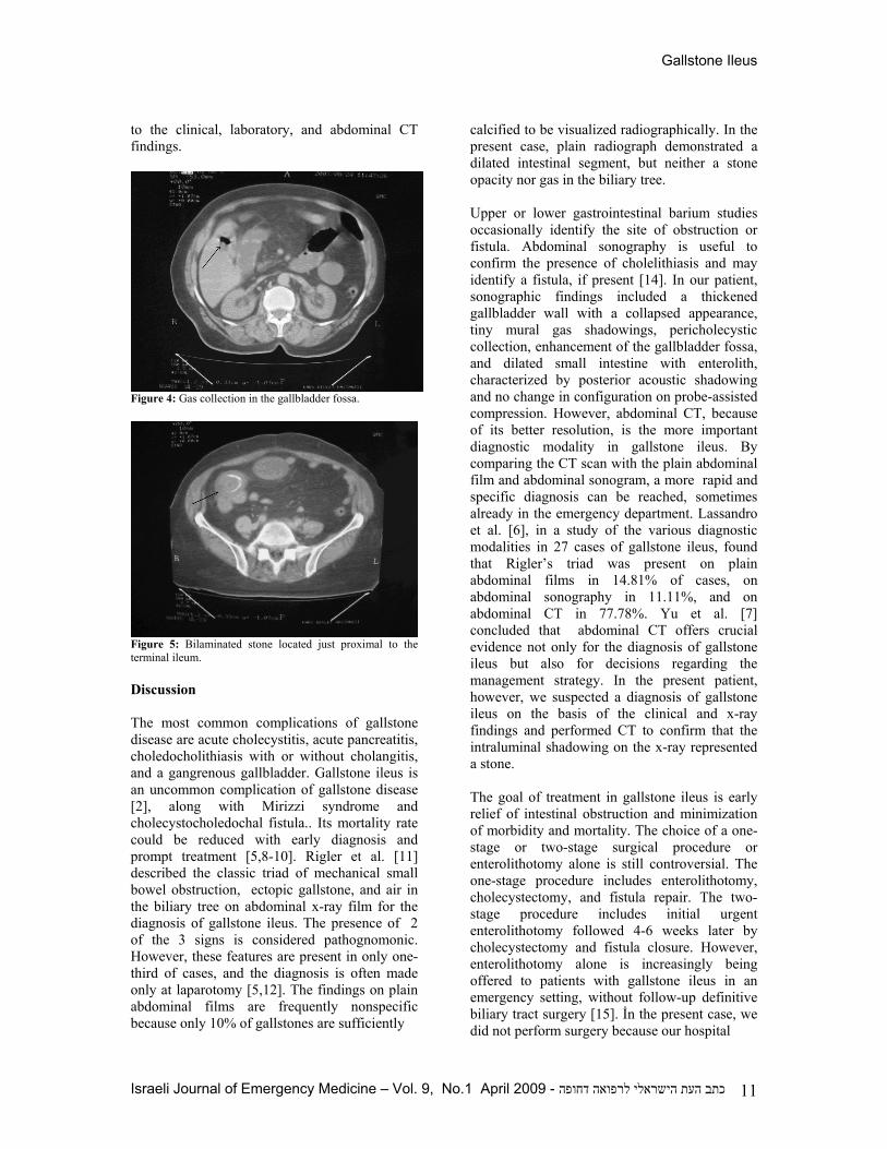
shadowing; compression yielded no change in configuration. For a detailed analysis of intraluminal shadowing, the patient was referred for nonspiral, noncontrast, abdominal computed tomography (CT). Axial images showed discontinuity of the duodenal wall at the area neighboring the gallbladder (Fig. 2), in addition to phlegmonous changes in the subhepatic fatty tissues and pericholecystic and periduodenal areas (Fig. 3), abnormal gas in the gallbladder fossa (Fig. 4), dilated small intestine, and a bilaminated stone, 30 mm in diameter, located just proximal to the terminal ileum (Fig. 5). These features confirmed the presence of gallstone ileus.
Figure 2: Axial CT image showing discontiunity of duodenal wall, suggesting a cholecystoduodenal fistula.
Figure 3: Phlegmonous changes in subhepatic fatty tissues and pericholecystic and periduodenal areas. The patient underwent primary treatment with intravenous fluids (0.9% sodium chloride) and nasogastric decompression and was transferred to an advanced hospital with a diagnosis of gallstone ileus with acute renal failure according
Gallstone Ileus
Israeli Journal of Emergency Medicine – Vol. 9, No.1 April 2009 - 11 כתב העת הישראלי לרפואה דחופה
to the clinical, laboratory, and abdominal CT findings.
Figure 4: Gas collection in the gallbladder fossa.
Figure 5: Bilaminated stone located just proximal to the terminal ileum. Discussion The most common complications of gallstone disease are acute cholecystitis, acute pancreatitis, choledocholithiasis with or without cholangitis, and a gangrenous gallbladder. Gallstone ileus is an uncommon complication of gallstone disease [2], along with Mirizzi syndrome and cholecystocholedochal fistula.. Its mortality rate could be reduced with early diagnosis and prompt treatment [5,8-10]. Rigler et al. [11] described the classic triad of mechanical small bowel obstruction, ectopic gallstone, and air in the biliary tree on abdominal x-ray film for the diagnosis of gallstone ileus. The presence of 2 of the 3 signs is considered pathognomonic. However, these features are present in only one-third of cases, and the diagnosis is often made only at laparotomy [5,12]. The findings on plain abdominal films are frequently nonspecific because only 10% of gallstones are sufficiently
calcified to be visualized radiographically. In the present case, plain radiograph demonstrated a dilated intestinal segment, but neither a stone opacity nor gas in the biliary tree. Upper or lower gastrointestinal barium studies occasionally identify the site of obstruction or fistula. Abdominal sonography is useful to confirm the presence of cholelithiasis and may identify a fistula, if present [14]. In our patient, sonographic findings included a thickened gallbladder wall with a collapsed appearance, tiny mural gas shadowings, pericholecystic collection, enhancement of the gallbladder fossa, and dilated small intestine with enterolith, characterized by posterior acoustic shadowing and no change in configuration on probe-assisted compression. However, abdominal CT, because of its better resolution, is the more important diagnostic modality in gallstone ileus. By comparing the CT scan with the plain abdominal film and abdominal sonogram, a more rapid and specific diagnosis can be reached, sometimes already in the emergency department. Lassandro et al. [6], in a study of the various diagnostic modalities in 27 cases of gallstone ileus, found that Rigler’s triad was present on plain abdominal films in 14.81% of cases, on abdominal sonography in 11.11%, and on abdominal CT in 77.78%. Yu et al. [7] concluded that abdominal CT offers crucial evidence not only for the diagnosis of gallstone ileus but also for decisions regarding the management strategy. In the present patient, however, we suspected a diagnosis of gallstone ileus on the basis of the clinical and x-ray findings and performed CT to confirm that the intraluminal shadowing on the x-ray represented a stone. The goal of treatment in gallstone ileus is early relief of intestinal obstruction and minimization of morbidity and mortality. The choice of a one-stage or two-stage surgical procedure or enterolithotomy alone is still controversial. The one-stage procedure includes enterolithotomy, cholecystectomy, and fistula repair. The two-stage procedure includes initial urgent enterolithotomy followed 4-6 weeks later by cholecystectomy and fistula closure. However, enterolithotomy alone is increasingly being offered to patients with gallstone ileus in an emergency setting, without follow-up definitive biliary tract surgery [15]. İn the present case, we did not perform surgery because our hospital

Gallstone Ileus
Israeli Journal of Emergency Medicine – Vol. 9, No.1 April 2009 - 12 כתב העת הישראלי לרפואה דחופה
lacked an intensive care unit. The patient was transferred to an advanced hospital after administration of intravenous fluids (0.9% sodium chloride) and nasogastric decompression. Conclusion In conclusion, gallstone ileus must be considered in the differential diagnosis of intestinal obstruction in patients with sonographic findings of inflammatory changes of gallbladder, especially elderly women. Abdominal CT is usually the preferred diagnostic modality because of its high resolution. References 1. Richards WO, Williams LF Jr. Obstruction
of the large and small intestine. Surg Clin North Am. 1988;68:355-376.
2. Abou-Saif A, Al-Kawas FH. Complications
of gallstone disease: Mirizzi syndrome, cholecystocholedochal fistula, and gallstone ileus. Am J Gastroenterol. 2002;97:249-254.
3. Hernandez C, Heuman D, Vlahcevid ZR.
Pathophysiology of disease associated with deficiency of bile acids. Principles and Practice of Gastroenterology and Hepatology. New York: Elsevier Science. 1988;384-395.
4. Shahat AH, Obaideen AM, Pandey UC.
Images: Gallstone ileus. Indian J Radiol Imaging. 2002;12:349-351.
5. Reisner RM, Cohen JR. Gallstone ileus: A
review of 1001 reported cases. Am Surg. 1994;60:441-446.
6. Lassandro F, Gagliardi N, Scuderi M, Pinto
A, Gatta G, Mazzeo R. Gallstone ileus analysis of radiological findings in 27 patients. Eur J Radiol. 2004;50:23-29.
7. Yu CY, Lin CC, Shyu RY, Hsieh CB, Wu
HS, Tyan YS, et al. Value of CT in the diagnosis and management of gallstone ileus.World J Gastroenterol. 2005;11:2142-2147.
8. Kurtz RJ, Heimann TM, Kurtz AB.
Gallstone ileus: A diagnostic problem. Am J Surg. 1983;146:314-317.
9. Clavien PA, Richon J, Burgan S, Rohner A.
Gallstone ileus. Br J Surg. 1990;77:737-774.
10. Rodriguez Hermosa JI, Codina Cazador A,
Girones Vila J, Roig Garcia J, Figa Francesch M, et al. Gallstone Ileus: Results of analysis of a series of 40 patients. Gastroenterol Hepatol. 2001;24:489-494.
11. Rigler LG, Borman CN, Noble JF.
Gallstone obstruction: Pathogenesis and roentgen manifestations. JAMA. 1941;117:1753-1760.
12. Delabrousse E, Bartholomot B, Sohm O,
Wallerand H, Kastler B. Gallstone ileus: CT findings. Eur Radiol. 2000;10:938-940.
13. Balthazar EJ, Schechter LS. Air in
gallbladder: A frequent finding in gallstone ileus. Am J Roentgenol. 1978;131:219-222.
14. Lasson A, Loren I, Nilsson A, Nirhov N,
Nilsson P. Ultrasonography in gallstone ileus: A diagnostic challenge. Eur J Surg. 1995;161:259-263.
15. Ayantunde AA, Agrawal A. Gallstone ileus:
Diagnosis and management. World J Surg. 2007;31:1292–1297.
Competing Interests: None declared. Funding: None declared. This manuscript has been peer reviewed Correspondence: Kasım Cağlayan MD Kars Devlet Hastanesi Genel cerrahi kliniği 036100 Kars, Turkey Phone: +90 474-2125012 Fax: +904742122367 e-mail: [email protected]


![Clinical and radiological diagnosis of gallstone ileus: a ... · order to cause obstruction at an anatomically wide part of the gastrointestinal tract [40–42]. This is estimated](https://static.fdocuments.us/doc/165x107/5d62e92788c993e9588b86bc/clinical-and-radiological-diagnosis-of-gallstone-ileus-a-order-to-cause.jpg)