Case report from Dr. Josheph. Shaffu, et al
131
Case report from Dr. Josheph. Shaffu, et al
Transcript of Case report from Dr. Josheph. Shaffu, et al

Case report from Dr Josheph Shaffu et al
Case report
A 37-year-old man was admitted after syncope at rest with facial trauma
He had been examined for bradycardia 6 years earlier had a known complete right bundle branch block (RBBB) without apparent structural heart
disease
Familial background His 2 brothers both had ECG patterns typical for Brugada syndrome his mother had drug-induced Brugada syndrome His
father was from East Asian descent
Electrocardiography (ECG) findings (next slide)
Echo The left ventricular ejection fraction was 57 the right ventricle had normal wall thickness without dilatation
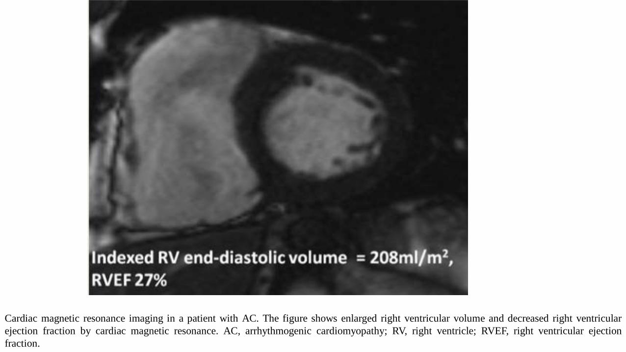
Cardiac magnetic resonance imaging (MRI) was entirely normal
EEP A monomorphic ventricular tachycardia was induced (second trace) Induction of symptomatic sustained wide complex monomorphic
tachycardia (cycle length 540ms) with one ventricular extra-stimulus on a drive train of 600ms The QRS width is 180ms with left axis and
positive complexes in V1 and V2 (ECG electrocardiography RBBB) The tachycardia was a left sided posterior fascicular tachycardia and was
easily ablated on a site with a Purkinje potential
A dual-chamber implantable cardioverter defibrillator was implanted given the bradycardia and conduction disease Genetic analysis showed 2
missense variants in the SCN5A gene and one TMEM43 variant all with unclear relation to his disease given his East Asian roots His gene
belongs to the TMEM43 family
Resting ECG Which is the diagnosis
Induction of symptomatic sustained wide complex monomorphic tachycardia (cycle length 540ms) with one ventricular extra-stimulus on a drive
train of 600ms The QRS width is 180ms with left axis and positive complexes in V1 and V2 (ECG electrocardiography RBBB right bundle
branch block)
1 How to guide us with the mutations found 2 missense variants in the SCN5A gene and one TMEM43 variant
2 Which is the clinical diagnosis
3 Which is the ECG diagnosis
4 Is this ECG compatible with Brugada syndrome
5 Are the anomalies in the right precordial leads a sign of another disease
6 Is the ventricular arrhythmia related to Brugada syndrome A sustained induced monomorphic ventricular tachycardia (second trace)
Induction of symptomatic sustained wide complex monomorphic tachycardia (cycle length 540ms) with one ventricular extra-stimulus on a
drive train of 600ms The QRS width is 180ms with left axis and positive complexes in V1 and V2 (ECG electrocardiography RBBB) The
tachycardia was a left sided posterior fascicular tachycardia and was easily ablated on a site with a Purkinje potential
Questions
Colleagues opinions
Thanks for the interesting case Baseline ECG shows Brugada pattern especially in V2in addition there are J waves in the inferior leads which point to a higher risk for arrhythmias In addition tall R in V1 and right axis deviation is noteworthy suggestive of additional fascicular disease ieleft septal block and Left posterior fascicular block The VT is certainly compatible with a left posterior fascicular focus
To try and put this all together a SCN5A mutation might explain the Brugada pattern as well as conduction delay in the fascicles leading to reentrant fascicular tachycardias We have described this in patients with bundle to bundle reentry and am aware of a case reported from Canada with SCN5A mutation and fascicular tachycardia
Professor Melvin M Sheinman
httpswwwyoutubecomwatchv=Fm3SwORdBS4
httpswwwyoutubecomwatchv=j9w86h7QvJwThe past present and future of ablation with Dr Melvin Scheinman
Dr Melvin A Scheinman is Professor of Medicine and holds the Walter HShorenstein Endowed Chair in Cardiology at the University of California SanFrancisco He has received awards including the Paul Dudley White Award forExcellence in Teaching by the American Heart Association and the DistinguishedScience Award of the American College of Cardiology He grew up in BrooklynNew York and took his undergraduate degree at Johns Hopkins Universitywhere he graduated first in his class Postgraduate medical education includedthe Albert Einstein College of Medicine residency training at the University ofNorth Carolina (Chapel Hill) and cardiology training at the University ofCalifornia San Francisco Medical Center
Dear Andreacutes Prof Melvin Scheinman brought very interesting comments
Actually his group (1) described SCN5A mutations in patients with idiopathic bundle branch reentry VT however none of the BBR-VT cases
was suggestive of the ldquoBelhassen VTrdquo type
In addition I do not find trace of the case from Canada with SCN5A mutation and fascicular VT Can you ask him to provide this reference
Thanks
Bernard Belahsen
1 Roberts JD Gollob MH Young C Connors SP Gray C Wilton SB Green MS Zhu DW Hodgkinson KA Poon A Li Q Orr N Tang
AS Klein GJ Wojciak J Campagna J Olgin JE Badhwar N Vedantham V Marcus GM Kwok PY Deo RC Scheinman
MM Bundle Branch Re-Entrant Ventricular Tachycardia Novel Genetic Mechanisms in a Life-Threatening ArrhythmiaJACC Clin
Electrophysiol 2017 Mar3(3)276-288 doi 101016jjacep201609019
Very interesting and infrequent case
With Brugada pattern in V2 early repolarization in inferior leads wide QRS complex very tall Racute wave in V1 and epsilon wave in V3In this case is important to rule out arrhythmogenic cardiomyopathy with important abnormalities in despolarization and repolarization ispossible to the MRI is currently negative because is an early stage of the myocardiopathy or it only presents with electrical abnormalities and notwith fibrofatty infiltration which is a rare presentation and it has been associated with PKP2 mutationThe genetic findings and the left ventricle VT are more associated with LV involvement of arrhythmogenic cardiomyopathy
Thanks for sharingHumberto Rodriguez -Reyes MD FACC AHA y HRS Member bull Instructor RCP Baacutesico Avanzado y Avanzado para Expertos (BLS ACLS ACLS-EP) de
la AHA bull Presidente Sociedad Cardiovascular y del Metabolismo Presidente SOMEEC 2013-2014 bull Coordinador del capiacutetulo de ReanimacioacutenCardio Pulmonar del Consejo de la Alianza contra la Muerte Suacutebita de la SIACinfocardiologicacommx
Aguas Calientes Meacutexico
Spanish Se trata de un paciente masculino joven con herencia y eacutetnica positiva para siacutendrome de Brugada que desarrolla siacutencope en reposo por
probable TV -FV Su ECG muestra ritmo sinusal bradicaacuterdico bloqueo AV de 1deg grado probable bloqueo interauricular parcial eje a la
derecha alrededor de 110 deg QRS ancho con imagen de BRD like (bifascicular BRD +BFPS) en V2 es caracteriacutestico de patroacuten de Brugada tipo
1 En V1 hay onda R alta y ondas S en precordiales izquierdas pero en las derechas en maacutes ancho El QTc es normal No veo desniveles del ST
en derivaciones inferiores ni ondas J si un ST raacutepidamente ascendente La onda R de aVR mide gt 03 mV Tampoco veo fQRS como otros
signos de peor pronoacutestico para TV-FV La TV inducida con un extra estiacutemulo es monomoacuterfica Habitualmente las TV del BrS son polimorfas
Aunque hay descriptas mono morfas (Shimada y col 1996) Me permito adjuntar un breve e improvisado diagrama sobre el ECG original
Esta descripto el gen autosoacutemico recesivo TRPM4 en el BrS ( Janin y col 2018) y el TRPM4 asociado al SCN5A en este siacutendrome (Gualandi y
col 2017) El siguiente comentario lo planteo como interrogante pensando en BRD like y habiendo hecho el estudio electrofisioloacutegico se podriacutea
haber intentado la Maniobra del Dr Pablo Chiale y asiacute desenmascarar al todo el BRD y el patroacuten de Brugada
Saludos respetuosos y muy cordiales estimado Profesor esperemos las opiniones de quienes de verdad saben mucho y desde luego su
acostumbrada resolucioacuten final
Dr Juan Carlos Manzzardo Mendoza-ArgentinaEnghish This is a male patient young with positive ethnicity and inheritance for BrS who develops syncope at rest due to probable TV-FV His
ECG shows sinus rhythm bradycardic 1st degree AV block probable partial interatrial block QRS axis around +110deg QRS width with RBBB
image like (bifascicular RBBB+ LSFB) in V2 is characteristic of Brugada type 1 pattern In V1 there is high R wave and S waves in left
precordial but in the right in more width The QTc is normal I do not see ST differences in inferior derivations or J waves if a rapidly rising ST
The R wave of aVR measuresgt 03 mV Nor do I see fQRS as other signs of worse prognosis for TV-FV TV induced with extra stimulation is
monomorphic Usually the TVs of the BrS are polymorphic Although there are described morphorphic (Shimada M1 Miyazaki T Miyoshi S
Soejima K Hori S Mitamura H Ogawa SSustained monomorphic ventricular tachycardia in a patient with Brugada syndromeJpn Circ
J 1996 Jun60(6)364-70) I allow myself to attach a brief and improvised diagram about the original ECG( see next slide) The autosomal
recessive TRPM4 gene in BrS (Janin et al 2018) and TRPM4 associated with SCN5A in this syndrome (Gualandi et al 2017) are described The
following comment I pose as a question thinking of RBBB like and having done the EPS could have tried the Chiale manouver and thus
unmask the whole RBBB and the Brugada pattern
Regards respectful and very cordial esteemed Professor lets hope the opinions of those who really know a lot and of course their usual final
resolution
Buenas noches estimado Andreacutes Good evening dear Andreacutes
Dear Juan Carlos Thank you for your lucid analysis As always denoting your intelligent mind Your analysis of the case seemed very complete to
me I will only make a few observations to see if you agree You commented that the electric axis of the QRS is around +110deg The QRS pattern in
aVR is of type QR with the Q discretely deeper than the height of the R that follows it however the latter is much wider than the Q in virtue of the
CRBBB that causes terminal conduction delay located forward and to the right just in the area of the RVOT faced by aVR therefore the area
of the final R is greater than the area of the initial Q consequently I think that the axis is minimally to the right of +120 deg Not discreetly to the left
(+ 110 deg) I observed with increase the voltage of the R of aVR and I had the impression that it does not exceed 2mm consequently I think it is
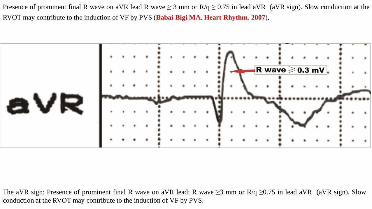
not present the aVR sign (Babai Bigi MA et al Heart Rhythm 2007) For those who do not know or do not remember these authors show the
existence of a significant correlation between a prominent R wave in aVR (aVR sign) and the risk of development of arrhythmic events in BrS In
the presence of BrS the prominent R wave in primary aVR may further reflect a delay in right ventricular conduction of the RVOT and
subsequently greater electrical heterogeneity which in turn is responsible for an increased of arrhythmiaacutes risk The aVR sign was defined as R
wave ge 03 mV or R qgt = 075 in the aVR lead In other words this sign is based on the depolarization theory of the BrS where there is a final
disturbance conduction on RVOT because of the minor expression of the connexin 43 contracted by Naademanee et al (Nademanee K et al
Journal of the American College of Cardiology 2015) You comment that the recessive TRPM4 gene in the BrS is described but you got
confused because the mutation found is TMEM43 and not TRPM4 Please look well in the description of the story
Regarding your question if it will not be interesting to perform ldquoChiale maneuverrdquo I answer that it is not necessary because in this case the
CRBBB does not eclipse the Type 1 Brugada pattern as seen in V2 The definition of the type 1 pattern currently does not require that it be present
in two or more right precordial leads as long as it has one for the diagnosis of the syndrome (Priori SG1 et al Heart Rhythm 2013)
An affectionate hug and thanks for your opinion Andreacutes
English
Querido Juan Carlos Gracias por tu lucido anaacutelisis Como siempre denotando tu mente inteligente Tu anaacutelisis del caso me parecioacute bien completo
Apenas hareacute miacutenimas observaciones para ver si concuerdas
1 Dices que el eje eleacutectrico del QRS estaacute por vuelta de los 110deg El patroacuten en aVR es de tipo QR con la Q discretamente maacutes profunda que la
altura de la R que le sigue no obstante esta uacuteltima es mucho maacutes ancha que la Q en virtud del BCRD que ocasiona retardo final de
conduccioacuten localizado adelante y a la derecha justamente en el aacuterea del RVOT enfrentada por aVR por lo tanto el aacuterea de la R final es
mayor de que el aacuterea de la Q inicial consecuentemente pienso que el eje estaacute miacutenimamente a la derecha de +120deg Y no discretamente a la
izquierda(+110deg)
2 Observeacute con aumento el voltaje de la R de aVR y tuve la impresioacuten que no pasa de 2mm consequetemte pienso que no estaacute presente o signo
de Babai Bigi MA (Babai Bigi MA et al Heart Rhythm 2007) Para los que no saben o no se recuerda estos autores muestran la existencia
de una correlacioacuten significativa entre una onda R prominente en aVR (signo aVR) y el riesgo de desarrollo de eventos arriacutetmicos en el
siacutendrome de Brugada En presencia de BrS la onda R prominente en aVR principal puede reflejar maacutes un retraso en la conduccioacuten ventricular
derecha del tracto de salida y posteriormente una mayor heterogeneidad eleacutectrica que a su vez es responsable de un mayor riesgo de arritmia
El signo aVR se definioacute como onda R ge 03 mV o R qgt = 075 en la derivacioacuten aVR En otras palabras este signo se basa en la teoriacutea de la
despolarizacioacuten del Brugada donde existe un disturbio final de conduccioacuten justamente por la menor expresioacuten de la conexiona 43 en contrada
por Naademee
3 Tu comentas que estaacute descripto el gen TRPM4 recesivo en el Brugada maacutes te confundiste porque la mutacioacuten encontrada es TMEM43 y no
TRPM4 fijate bien en la descripcioacuten de la historia
4 En relacioacuten a tu pregunta si no seraacute interesante tentar la maniobra de Chiale te respondo que no es necesario porque en este caso el BRD no
eclipsa el patroacuten Brugada como se ve en V2 La definicioacuten del patroacuten tipo 1 actualmente no exige que esteacute presente en dos o maacutes
derivaciones precordiales derechas basta que tenga una para el diagnoacutestico del siacutendrome (Priori SG1 et al Heart Rhythm 2013) Tu dices
ldquoQRS ancho con imagen de BRD like (bifascicular BRD +BFPS)rdquo Me parece que si tiene bloqueo AV de primer grado deberiacuteas decir
ldquoQRS ancho con imagen de BRD like (trifascicular BRD +BFPS)rdquo
Un abrazo afectuoso y gracias por opinar
PotroAndreacutes
Spanish
Answer to questions from Adail Paixatildeo Almeida MD
a Which is the clinical diagnosis A Unexplained syncope at rest We donrsquot know the subjacent cause Possible VT
b Which is the clinical diagnosis Syncope episode of unknow origin
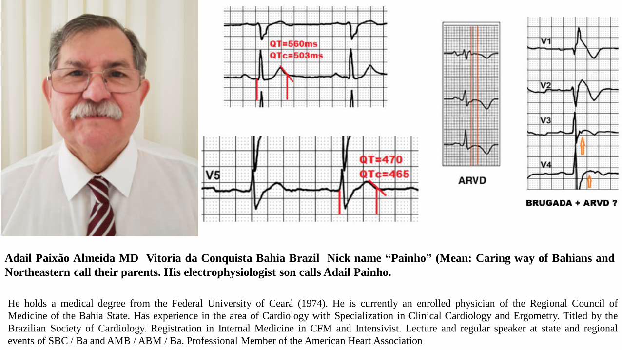
c Which is the ECG diagnosis Complete right bundle branch block spontaneous Type 1 Brugada pattern with ldquoLambda shaperdquo in V1
and typical in V2 parietal block presence of epsilon wave in V3 possible long QT interval (1-3)
d EPS Monomorphic sustained ventricular tachycardia type idiopathic fascicular ventricular tachycardia
e Are the anomalies compatible with Brugada syndrome Yes see next slide
f Are the anomalies in the right precordial leads compatible with Brugada syndrome Yes
g Are the abnomalities in the right precordial leads compatible with another entity Maybe ARVCD Gene TMEM43 mutation SCN5A
BrS
h Is the arrhythmia compatible with BrS Not frequently In BrS the typical VT is polymorphic VTVF The event of SMVT is observed in
ARVC but with LBBB pattern
1 Martini B Cannas S Nava A Brugada by any other name Eur Heart J 2001 Oct22(19)1835-6
2 Peacuterez Riera AR Antzelevitch C Schapacknik E Dubner S Ferreira CIs there an overlap between Brugada syndrome and arrhythmogenic
right ventricular cardiomyopathydysplasiaJ Electrocardiol 2005 Jul38(3)260-3
3 Hoogendijk MG1Diagnostic dilemmas overlapping features of brugada syndrome and arrhythmogenic right ventricular
cardiomyopathyFront Physiol 2012 May 233144 doi 103389fphys201200144
Adail Paixatildeo Almeida
He holds a medical degree from the Federal University of Cearaacute (1974) He is currently an enrolled physician of the Regional Council of
Medicine of the Bahia State Has experience in the area of Cardiology with Specialization in Clinical Cardiology and Ergometry Titled by the
Brazilian Society of Cardiology Registration in Internal Medicine in CFM and Intensivist Lecture and regular speaker at state and regional
events of SBC Ba and AMB ABM Ba Professional Member of the American Heart Association
Adail Paixatildeo Almeida MD Vitoria da Conquista Bahia Brazil Nick name ldquoPainhordquo (Mean Caring way of Bahians and
Northeastern call their parents His electrophysiologist son calls Adail Painho
Final comment by Andreacutes Ricardo Peacuterez-Riera MD PhD
Laboratoacuterio de Delineamento de Estudos e Escrita Cientiacutefica da Faculdade de Medicina do ABC Santo Andreacute SP Brazil
Laboratory of Study Design and Scientific Writing of the Faculty of Medicine of ABC Santo Andreacute SP Brazil
ECG VCG Peacuterez-Riera | my cardiology site of scientific interests
httpsekgvcgwordpresscom
(1 Gandjbakhch E et al J Am Coll Cardiol 2018 2 Merner ND et al Am J Hum Genet 2008 3 Hodgkinson et al Clin Genet 20134 4
Christensen et al Clin Genet 20115 5 Liang WC et al Ann Neurol 2011 6 Haywood AF et al Eur Heart J 2013)
This gene belongs to the TMEM43 family Defects in this gene are the cause of familial arrhythmogenic right ventricular dysplasia type 5
(ARVD5) also known as arrhythmogenic right ventricular cardiomyopathy type 5 (ARVC5) Arrhythmogenic right ventricular dysplasia is an
inherited disorder often involving both ventricles and is characterized by MVT with LBBB patternm heart failure SCD and fibrofatty
replacement of cardiomyocytes This gene contains a response element for PPAR gamma (an adipogenic transcription factor) which may explain
the fibrofatty replacement of the myocardium a characteristic pathological finding in AC May have an important role in maintaining nuclear
envelope structure by organizing protein complexes at the inner nuclear membrane Required for retaining emerin at the inner nuclear membrane
(By similarity) Emery-Dreifuss muscular dystrophy 7 autosomal ARVC type 5
From Emery-Dreifuss muscular dystrophy 7 autosomal dominant (EDMD7) A form of Emery-Dreifuss muscular dystrophy a degenerative
myopathy characterized by weakness and atrophy of muscle without involvement of the nervous system early contractures of the elbows achilles
tendons and spine and cardiomyopathy associated with cardiac conduction defects [MIM614302] (Liang WC et al Ann Neurol 2011)
Emery-Dreifuss
muscular
dystrophy-
related
myopathy 7 (5)
TMEM43
ARVD familial 5 (ARVD5) A congenital heart disease characterized by infiltration of adipose and fibrous tissue into the RV and loss of
myocardial cells resulting in ventricular and supraventricular arrhythmias [MIM604400] Cytogenetic Location 3p251 which is the short (p)
arm of chromosome 3 at position 251
Other names for this gene
ARVC5 In 83 affected individuals with ARCD-5 (604400) from 15 unrelated Newfoundland families (Merner et al Am J Hum Genet 2008)
identified heterozygosity for a missense mutation (S358L 6120480001) in the TMEM43 gene that was not found in 47 spouses or 161 controls In
an analysis of the TMEM43 gene in 55 Danish probands who fulfilled the criteria for ARVD and 10 patients with only some features of ARVD
Christensen et al (Christensen AH et al J Med Genet 2010) identified 1 woman fulfilling the criteria who carried the S358L variant In DNA
samples from 195 unrelated individuals with suspected ARVD Baskin et al (Baskin B et alHum Genet 2013) identified 6 patients who carried
the S358L Newfoundland mutation in TMEM43 including a 43-year-old New Zealand man who was not of Newfoundland descent In addition 5
patients carried 5 different rare sequence variants in the TMEM43 (see eg 6120480004) 2 of whom also carried a variant in the PKP2 and DSP
genes respectively EDMD7 Based on the putative role for TMEM43 in the nuclear envelope Liang et al (Liang WCC et al Ann Neurol
2011) analyzed the TMEM43 gene in 41 patients with Emery-Dreifuss muscular dystrophy (EDMD) who were negative for mutations in known
EDMD-related genes and identified different heterozygous missense mutations in 2 unrelated individuals LUMA
Gene Function (Bengtsson and Otto (2008) found that TMEM43 binds A- (LMNA 150330) and B- (LMNB1 150340) type lamins and depends
on A-type lamins for its inner nuclear membrane localization The TMEM43 protein was also shown to interact with emerin (EMD 300384)
Downregulation of TMEM43 and overexpression of dominant-negative acting TMEM43 caused redistribution of emerin The findings suggested
that TMEM43 functions as a nuclear membrane organizer (Liang WCC et al Ann Neurol 2011) demonstrated that TMEM43 interacts with
SUN2 (613569) another inner nuclear membrane protein
Main Alternative Diagnosis andor Potential ACM Mimicking Diseases
Brugada syndrome
Sarcoidosis
Dilated cardiomyopathy
Athletersquos heart
Idiopathic infundibular PVCVT
Myocarditis
Uhlrsquos disease
Other causes of RV dilatation andor dysfunction
Ebsteinrsquos anomaly
Left to right shunt interatrial septal defect anomalous pulmonary venous return
Tricuspid regurgitation
Inferior infarct with RV extension
1) Arrhythmogenic right ventricular cardiomyopathy (ARVC)
2) Arrhythmogenic right ventricular dysplasiacardiomyopathy (ARVDC)
3) Arrhythmogenic cardiomyopathy (AC) Important In this presentation we will use AC nomenclature
Main nomenclatures used in the literature
Arrhythmogenic cardiomyopathy (AC) is a hereditary disease characterized by ventricular arrhythmias right ventricular andor
left ventricular dysfunction and fibrofatty replacement of cardiomyocytes Patients with AC typically present between the second
and the fourth decade of life with ventricular tachycardias However sudden cardiac death (SCD) may be the first manifestation
often at young age in the concealed stage of disease AC is diagnosed by a set of clinically applicable criteria defined by an
international
Task Force The current Task Force Criteria are the essential standard for a correct diagnosis in individuals suspected of AC The
genetic substrate for AC is predominantly identified in genes encoding desmosomal proteins
In a minority of patients a non-desmosomal mutation predisposes to the phenotype
Risk stratification in AC is imperfect at present
Genotype-phenotype correlation analysis may provide more insight into risk profiles of index patients and family members
In addition to symptomatic treatment prevention of SCD is the most important therapeutic goal in AC Therapeutic options in
symptomatic patients include antiarrhythmic drugs catheter ablation and ICD implantation Furthermore patients with AC and
also all pathogenic mutation carriers should be advised against practising competitive and endurance sports
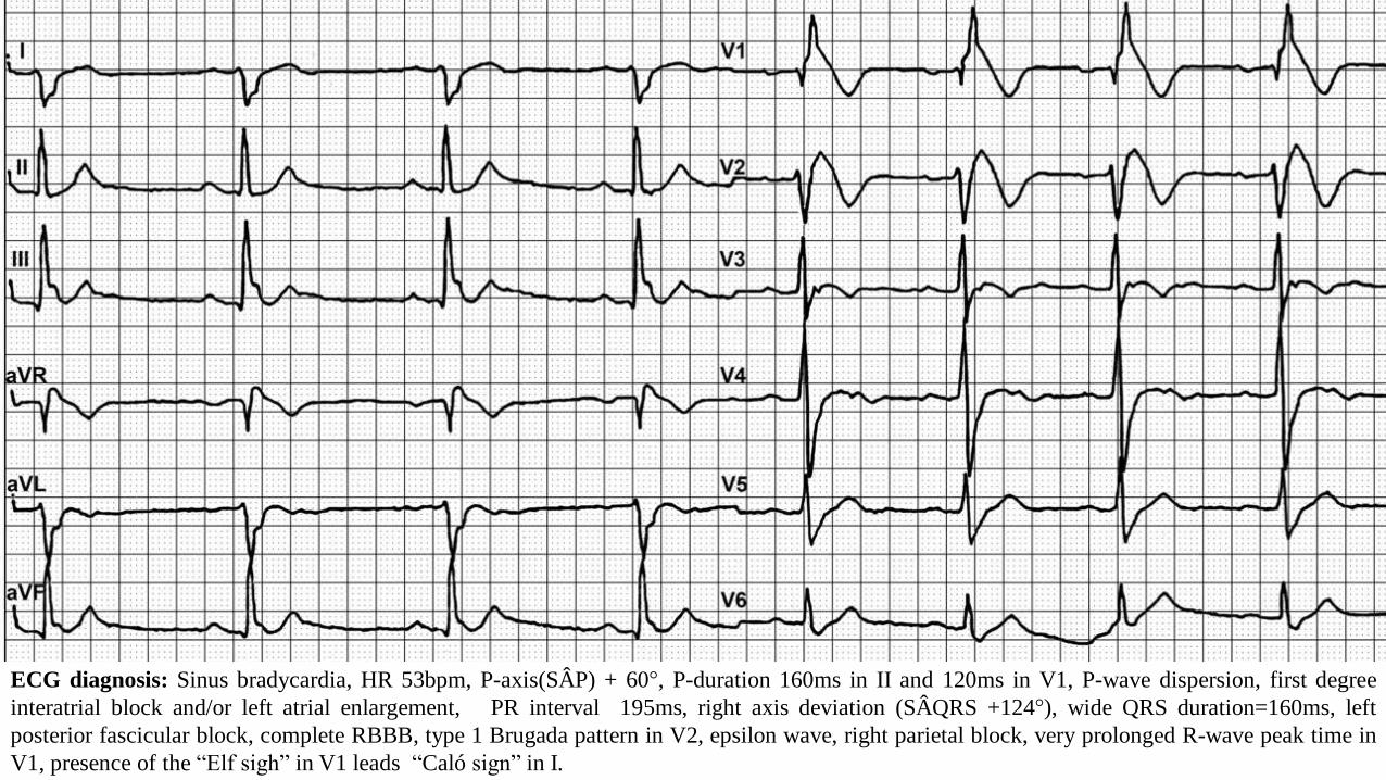
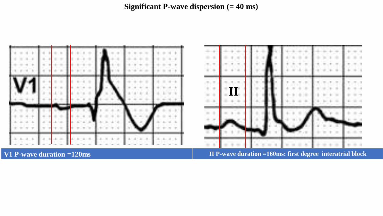
ECG diagnosis Sinus bradycardia HR 53bpm P-axis(SAcircP) + 60deg P-duration 160ms in II and 120ms in V1 P-wave dispersion first degree
interatrial block andor left atrial enlargement PR interval 195ms right axis deviation (SAcircQRS +124deg) wide QRS duration=160ms left
posterior fascicular block complete RBBB type 1 Brugada pattern in V2 epsilon wave right parietal block very prolonged R-wave peak time in
V1 presence of the ldquoElf sighrdquo in V1 leads ldquoCaloacute signrdquo in I
Main Electrocardiographic abnormalities observed in AC
bull Eventual Right atrial left atrial or biatrial enlargement and interatrial block P-wave dispersion
bull Eventual PR interval prolongation
bull Prolongation of QRS duration (QRSd) in the right precordial leads
bull QT prolongation in right precordial leads
bull Delayed S wave upstroke on right precordial leads (right parietal block)
bull Ratio of the QRSd in leads V1+V2+V3V4+V5+V6 ge 12
bull Delayed S wave upstroke(right parietal block)ge 55ms
bull Ratio of the QRSd in leads V1+V2+V3V4+V5+V6 ge 12
bull Fragmented QRS (f-QRS)
bull QRS dispersion
bull T wave inversion in the right precordial leads in patients gt 14 years of age in the absence of CRBBB It is considered a minor criteria
in the revised Task Force criteria Negative T wave in lateral leads and positive in aVR suggest LV involvement
bull Presence of epsilon waves epsilon potentials or Fontaine waves (considered a mayor criteria in the revised Task Force criteria)
bull Low-QRS voltage in the limb leads are lt5 mm because of loss of viable myocardium
bull Frequently premature ventricular contractions with LBBB pattern
bull Eventual sustained monomorphic VT with superior QRS axis (considered a mayor criteria in the revised Task Force criteria)
bull Eventual sustained monomorphic VT with inferior QRS axis (considered a minor criteria in the revised Task Force criteria)
bull Frequently supraventricular arrhythmias FA Flutter atrial
bull Incomplete RBBB or complete RBBB
Prolonged PR or PQ interval
See figure in the next slide
P-duration=160ms in II PR segment only 75
ms PR interval 195ms
P-duration=120ms in V1 PR segment only 75 ms
PR interval 195ms
PR interval 195ms Conclusion Pronged P-wave consequence of first degree interatrial block(1) andor left atrial enlargement Dynamic P-
wave duration from 160ms in II lead to 120ms I V1 In this case we observe a significant P wave dispersion (160120 ms) P-wave dispersion
(PWD) is a noninvasive ECG marker for atrial remodeling and predictor for atrial fibrillation PWD is defined as the difference between the widest
and the narrowest P-wave duration recorded from the 12 ECG leads (Peacuterez-Riera AR et al Indian Pacing Electrophysiology Journal 2016
httpswwwncbinlmnihgovpmcarticlesPMC5197451 Bayeacutes de Luna et al J Electrocardiol 2012)
Some studies report visualizing P-wave onset and offset in a minimum of 8 to 9 leads as an inclusion criterion although a minimum of three leads
has been used to determine P- dispersion (Pd) (Agarwal et al Am J Cardiol 2003) P-wave indices are manually calculated with calipers or with
digitized images Manual measurement uses paper speed at 50 mms and the voltage 1 to 2 mVcm (Aytemir et al Pacing Clin Electrophysiol
2000) with additional magnification Manual measurements have less accuracy compared with digital measurements
Interatrial blocks (IABs)
Prolonged PWD is a marker of LAE andor interatrial block both associated with myocardial fibrosis AF tendency ischemic stroke heart failure
sudden cardiac death (SCD) in the general population and all-cause death This association is independent of AF and is only partially mediated by
shared cardiovascular risk factors (Maheshwari et al Am J Cardiol 2017) IAB the Bayesacute syndrome is considered partial (P-IAB) when
the P-wave is ge120 ms and advanced (A-IAB) when the P-wave in the inferior leads II III and aVF is biphasic (+-) in addition to prolonged
PWD (Martin-Demiguel et al Am J Cardiol 2019) Biphasic (+-) plus-minus P-wave in A-IAB is due to retrograde caudo-cranial activation of
the LA induced by the presence of fibrosis in the BR in the LA ceiling This was confirmed in humans by electro-anatomic mapping (Holmqvist
et al Heart Rhythm 2008) and with advanced CMR techniques and in post-mortem specimens (Baranchuk A 2017)
IAB is a prevailing cardiac conduction abnormality that is under-recognized in clinical practice
Prolonged PWD with a cutoff of gt120 ms to gt150 ms in sinus rhythm before ablation may be associated with AF recurrence after pulmonary vein
isolation (PVI) regardless of age gender left atrial size and the presence of structural heart disease (Pranata et al Ann Noninvasive
Electrocardiol 2019) The term left atrial abnormalities was coined and widely used to encompass both atrial enlargement and IAB (Lee et al
Am J Cardiol 2007) Established ECG criteria for LAE do not reliably reflect anatomical LAE and lack sufficient predictive value to be useful
clinically P-wave abnormalities should be noted as nonspecific LA abnormalities The presence of at least one ECG criterion for LAE is sensitive
but not specific for anatomical LAE Individual criteria for LAE including P mitrale P-wave axis le30deg or NPTF- V1 gt 004 smm are highly
specific though not sensitive ECG is highly specific but insensitive for RAE Individual ECG P-wave changes do not reliably predict anatomical
atrial enlargement (Tsao et al J Cardiovasc Magn Reson 2008)
Bayes syndrome is an under-recognized arrhythmic syndrome characterized by the association of A-IAB with significant cardiogenic and
neurogenic implications such as atrial arrhythmia more specifically atrial fibrillation (Conde et al Arch Cardiol Mex 2014) vascular dementia
non-lacunar cardioembolic ischemic stroke and a possible cryptogenic stroke (as cerebral ischemia of obscure or unknown origin) (Arboix et al
World J Clin Cases 2017) Recently Bayeacutes et al classified IABs in typical and atypical (ldquoatypical A-IABrdquo) Atypical A-IAB is present when the
sinus P-wave depicts a terminal negative component in lead aVF indicating that final part of the P-waveP-loop is located in the negative
hemifield of aVF (upwards the orthogonal X leadI lead 0degplusmn180deg) and therefore a retrograde caudo-cranial activation of the LA is present
There are three morphologically atypical patterns of A-IAB Type I The terminal component of the P-wave in lead II is equiphasic Type II The
terminal component of the P-wave in lead II is biphasic ldquominus-plusrdquo (-+) and Type III P-waves in III and aVF are totally negative and biphasic
in II These cases must be differentiated from ectopic atrial rhythm where there are negative P-waves in leads III and aVF (de Luna AB et al J
Electrocardiol 2019) Table below shows the new classification of IABs (Bayes de Luna et al J Electrocardiol 2018)
Electrocardiographic classification of interatrial blocks (IABs)
1Typical IAB
1a Partial interatrial blocks (P-IABs)
a First degree PWD ge120 ms without negative terminal component in the inferior leads
b Second degree AV block when transient or intermittent (aberrancy)
1b Advanced interatrial block or third degree (A-IAB) P-wave ge 120 ms with biphasic ldquoplus-minusrdquo P-wave in the inferior leads II III and aVF
2 Atypical IAB (A-IAB)
(I) Morphological criteria
Type I P-wave ge 120ms with biphasic morphology in leads III and aVF and the final component of the P-wave in lead II is equiphasic
(isodiphasic)
Type II P-wave ge 120 ms with biphasic morphology in leads III and aVF and the final component of the P-wave in lead II is biphasic (-+)
Type III P-wave ge 120 ms with the first component of the P-wave equiphasic in leads III and aVF
(II) Duration criteria
1) P-wave lt120 ms with typical morphology (biphasic (plusmn)
2) P-wave in the inferior leads II III and aVF
Interatrial block classification and identification
Definition It is a transient pattern of partial or advanced IAB or atrial aberrancy in other words deviating from the proper or expected course of
stimulus inside the atrium Atrial aberrancy is also possible when PACs or premature parasystolic atrial beats are present(Bayeacutes de Luna A et al
J Electrocardiol 1978 Julia J Rev Esp Cardiol 1978 Breall WS et al Br Heart J 1972) Atrial aberrancy may also be present as a
transient bizarre P wave without the morphology of first or third degree IAB These changes in P wave morphology are caused by variations in the
atrial path of the sinus impulse through the atria They should be differentiated from changes induced by breathing atrial fusion beats and
artefacts including diaphragmatic contraction
Classification
1 First Degree Interatrial Block
2 Second degree or transient Interatrial Block(atrial Aberrancy) and
3 Third Degree Interatrial Block Complete or Advanced Interatrial Block
Differential diagnosis
Aberrant atrial conduction must be differentiated fromImmediatly after atrial premature contractions (60 of cases of atrial aberrancy) A
wandering atrial pacemaker aberrancy after AV junctional escape beats or AV junctional premature contraction coexisting multifocal premature
beats aberrrant atrial conduction after parasystolic beats mainly interpolated atrial parasystolic beat and several artifacts
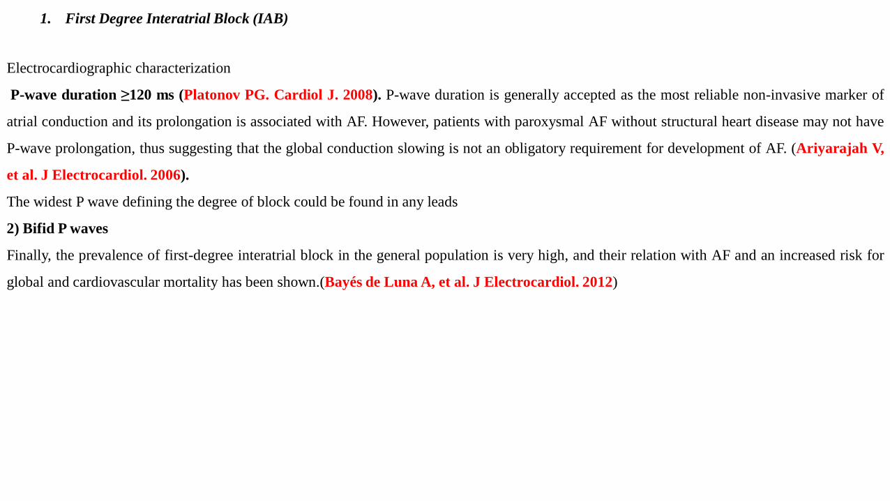
1 First Degree Interatrial Block (IAB)
Electrocardiographic characterization
P-wave duration ge120 ms (Platonov PG Cardiol J 2008) P-wave duration is generally accepted as the most reliable non-invasive marker of
atrial conduction and its prolongation is associated with AF However patients with paroxysmal AF without structural heart disease may not have
P-wave prolongation thus suggesting that the global conduction slowing is not an obligatory requirement for development of AF (Ariyarajah V
et al J Electrocardiol 2006)
The widest P wave defining the degree of block could be found in any leads
2) Bifid P waves
Finally the prevalence of first-degree interatrial block in the general population is very high and their relation with AF and an increased risk for
global and cardiovascular mortality has been shown(Bayeacutes de Luna A et al J Electrocardiol 2012)
Second-degree or transient interatrial block (atrial aberrancy)
Definition It is a transient pattern of partial or advanced IAB or atrial aberrancy in other words deviating from the proper or expected course of
stimulus inside the atrium Atrial aberrancy is also possible when premature atrial contractions or premature parasystolic atrial beats are present
(Bayes De Luna A et al Rev Esp Cardiol 1978 Julia J Rev Esp Cardiol 1978 Breall WS et al Br Heart J 1972)
Atrial aberrancy may also be present as a transient bizarre P wave without the morphology of first or third degree IAB In the figures of the next 2
slides an example of second degree IAB may be seen
These changes in P wave morphology are caused by variations in the atrial path of the sinus impulse through the atria They should be
differentiated from changes induced by breathing atrial fusion beats and artifacts including diaphragmatic contraction Second degree of IAB can
be induced by atrial or ventricular premature complexes which appear and disappear suddenly and transiently in one ECG and show a P wave that
changes morphology transiently in successive ECGs leading to misdiagnosis
Differential diagnosis
Aberrant atrial conduction must be differentiated from
1Immediatly after atrial premature contractions (60 of cases of atrial aberrancy)
2A wandering atrial pacemaker
3Aberrancy after AV junctional escape beats or AV junctional premature contraction
4Coexisting multifocal premature beats
5Aberrrant atrial conduction after parasystolic beats mainly interpolated atrial parasystolic beat
6Several artifacts
Definition In these cases the stimulus is blocked in the Bachmann region and LA is activated retrogradely with a P wave duration ge120 ms and
plus-minus(+-) plusmn P wave in inferior leads II III and aVF There is an open angle ge90deg between the vector of the first part and of the second part
of the P wave in the inferior leads Orthogonal Y lead plus-minus with a negative mode gt40 ms appear with notches and slurring in the last part of
the P loop IAB is associated in most of cases with LAE (90deg of cases) and dysfunction decreased left ventricular (LV) filling a propensity for LA
appendage thrombosis reduced atrial natriuretic peptide levels and is a predictor of paroxysmal supraventricular tachyarrhythmias such as AF
atrial flutter as well as an exacerbation of the LV failure The prevalence of first degree IAB is much higher than advanced or complete IAB
Really the ECG pattern of advanced IAB is an extremely strong marker of supraventricular tachyarrhythmia in a short period of time much more
so than the presence of first degree or partial Interatrial block Bayeacutes de Luna A et al(Bayeacutes de Luna A et al Eur Heart J 1988) studied 16
patients with ECG evidence of advanced IAB with retrograde activation of the left atrium (LA) P duration ge120 ms and plus-minus (+-) biphasic
P waves in inferior leads II III and aVF Eight patients had valvular heart disease four had dilated cardiomyopathy and four had other forms of
heart disease Patients with valvular heart disease and cardiomyopathy were compared with a control group of 22 patients with similar clinical and
echo characteristics but without IAB Patients with advanced IAB and retrograde activation of the LA had a much higher incidence of paroxysmal
AF (937) during follow-up than did the control group Eleven of 16 patients (687) with advanced IAB and retrograde activation of LA had
atrial flutter (atypical in seven cases typical in two cases and with two or more morphologies in two cases) Six patients from the control group
(277) had sustained atrial tachyarrhythmias (five AF and one typical atrial flutter) The atrial tachyarrhythmias were due more to advanced IAB
and retrograde PACs than to left atrial enlargement because the control group with a LA of the same size but without advanced IAB and
retrograde activation of LA and with less inactivation of LA and frequent PACs had a much lower incidence of paroxysmal tachycardia
Third degree complete or advanced interatrial block
Bayeacutes de Luna et al(Bayeacutes de Luna A et al Int J Cardiol 1989) demonstrated the value of preventive antiarrhythmic treatment in patients with
advanced interatrial block In this population LAE is present in 90 of cases Using drugs (amiodarone quinidine or verapamil) this percentage
was greatly lowered (25) From 81000 ECGs Bayes de Luna et al (Bayes de Luna A J Electrocardiol 1985) collected 83 cases that fulfilled
the criteria of Interatrial Conduction Disturbances with Left Atrial Retrograde Activation (IACD-LARA) (P +- in II III and VF with P width ge120
ms) The authors present the detailed study of 35 cases with surface ECG and VCG and 29 cases with orthogonal ECG leads The results are then
compared against two control groups with heart disease (30 cases) and without heart disease (25 cases) The prevalence of IACD-LARA was
nearly 1 globally and 2 among patients with valvular heart disease Arrhythmias such as AF and atrial flutter in advanced IAB is observed in
gt 90 of cases Diagnosis criteria of advanced interatrial block and retrograde activation of the LA (Bayeacutes de Luna A et al 19771988 Bayeacutes de
Luna A et al J Electrocardiol 2012) Biphasic bifid or notched ldquoplus-minusrdquo P waves in inferior leads II III and aVF of ECG and Y
orthogonal lead of VCG P duration ge120 ms angle between the first portion (RA) and end portion (LA) gt90ordm orthogonal Y lead plus-minus with
the final negative portion ge40 ms ge40 ms final portion of P loop of upstart orthogonal X and Z leads final portion of P loop delayed notches and
slurring in the last part of the P loop high Esophageal lead with positive P wave polarity and delayed low Esophageal lead with plus-minus P
wave polarity and delayed intracavitary ECG with P wave craniocaudal activation inside the RA intracavitary ECG with P wave caudal-cranial
activation inside LAThis clinical-electro-vectorcardiographic manifestation of advanced IAB should be considered a syndrome Bayeacutes syndrome
(Hernandez-Betancor I et al Curr Cardiol Rev 2017)
Third degree block complete or advanced interatrial block
LA
BB
RAA
P
M
RAJ
P loop in FP P wave in lead ldquoyrdquo
aVF
Third Degree (Advanced or complete IAB)
Electrical impulse is blockeddelayed in Bachmannrsquos muscular interatrial bundle (BB) but retrograde left atrial activation usually occurs
(Ariyarajah V et al Chest 2005) Note the existence of an open angle between the vector of the first portion of P wave (RA) and the last portion
(LA) Electrophysiological study demonstrates retrograde activation of the LA Consequently P loopwave in orthogonal lead ldquoYrdquo aVF and III is
biphasic plus-minus plusmn LA activation occurs by an alternate route rather than proceeding from right to left via the BB (Spodick DH J
Electrocardiol 2008)
Complete block in
Bachmannrsquos bundle
ge120ms
LA
RA
LA
-60deg
Y
XE
0 RALA
aVF Leads III and aVF
Plus-minus P wave
Biphasic P waves in inferior leads
Magnified P-loop (32x)
P-loop VCG in advanced IAB
E0
RA
LA
Normal P-loop wave on frontal plane
Caudo-cranial activation of
the left atrium (LA)
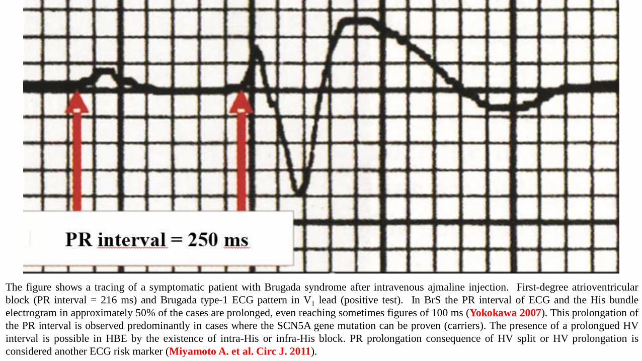
In BrS the PR interval of ECG and the His bundle electrogram in approximately 50 of the cases are prolonged even reaching sometimes figures
of 100 ms (Yokokawa et al 2007) This prolongation of the PR interval is observed predominantly in cases where the SCN5A gene mutation can
be proven (carriers) The presence of a prolonged HV interval is possible in HBE by the The presence of AF in BrS is a known risk marker
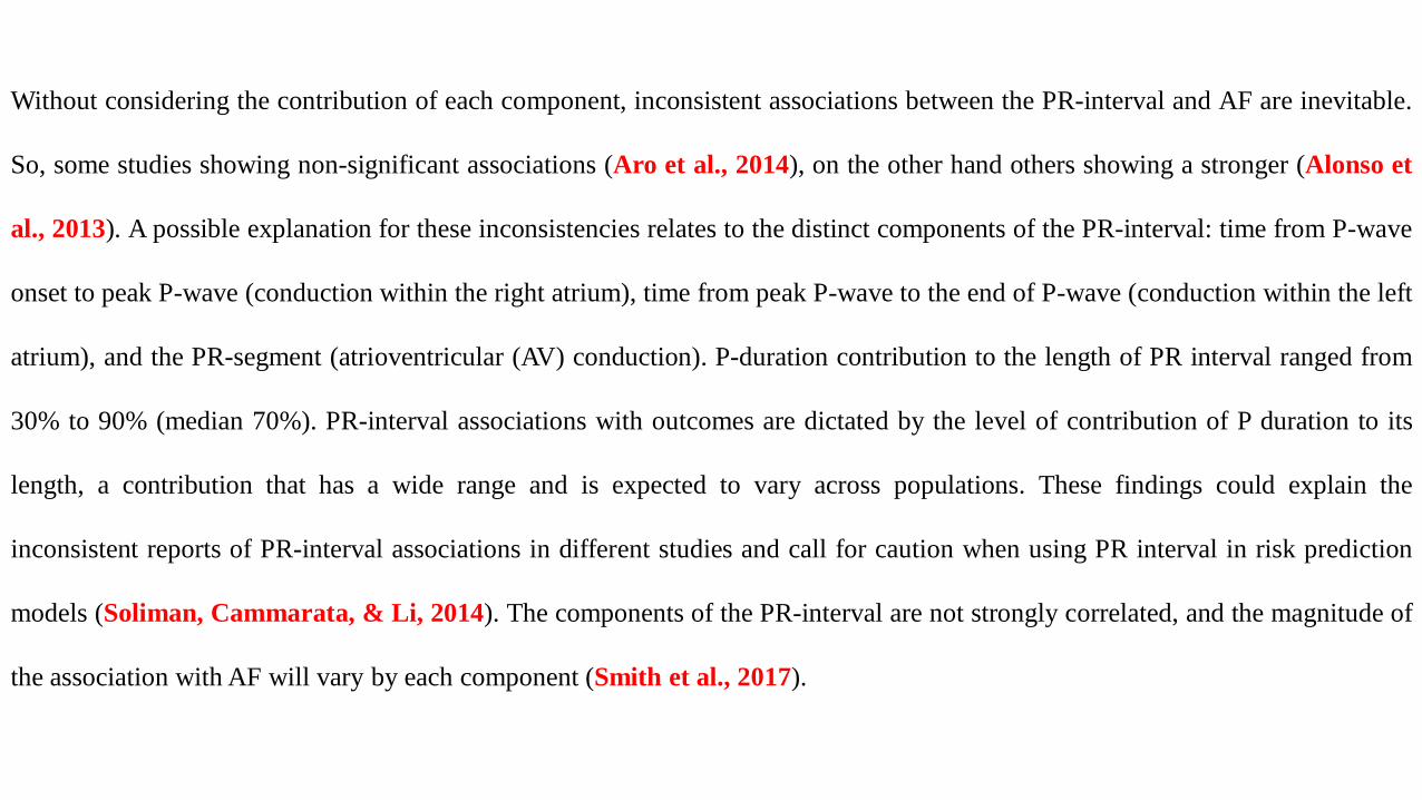
Without considering the contribution of each component inconsistent associations between the PR-interval and AF are inevitable
So some studies showing non-significant associations (Aro et al 2014) on the other hand others showing a stronger (Alonso et
al 2013) A possible explanation for these inconsistencies relates to the distinct components of the PR-interval time from P-wave
onset to peak P-wave (conduction within the right atrium) time from peak P-wave to the end of P-wave (conduction within the left
atrium) and the PR-segment (atrioventricular (AV) conduction) P-duration contribution to the length of PR interval ranged from
30 to 90 (median 70) PR-interval associations with outcomes are dictated by the level of contribution of P duration to its
length a contribution that has a wide range and is expected to vary across populations These findings could explain the
inconsistent reports of PR-interval associations in different studies and call for caution when using PR interval in risk prediction
models (Soliman Cammarata amp Li 2014) The components of the PR-interval are not strongly correlated and the magnitude of
the association with AF will vary by each component (Smith et al 2017)
IIIII
X I
aVFY
SAcircP +60deg
SAcircQRS + 124deg Right axis deviation
For the diagnosis of LPFB is necessary
1 No evidence of right ventricular
hypertrophy
2 No evidence of vertical heart in
slendersubjects This is
a cardiac electrical
position recognized in the ECG
when the QRS complex in lead aV
L resembles V1 while that in aVF
resembles V6 and
3 No evidence of a large lateral infarction
(Elizari Mval et Circulation 2007
Peacuterez-Riera AR et al Annals Non Invasive
Electrocardiol 2015 Peacuterez-Riera AR et al
Indian Pacing Electrophysiol 2018)
Caloacute sign
rS
rS
Positive Caloacute sign reflects terminal right end conduction delay (RECD) in the right ventricular outflow tract (RVOT) and subsequently more
electrical heterogeneity in the ventricular wall thickness (Calograve L et al J Am Coll Cardiol 2016)
X I
IIIIIaVF
Y
Hypothetical ECGVCG correlation on the Frontal Plane
rS pattern
in leads I
and aVL
Ventricular activation time R-wave peak time
or intrinsicoid deflection (ID) in aVFge35 ms
Notch in the descending limb of the R wave in
III middle-final notch(red arrow) This is an
hallmark of LPFB and R-IIIgtRII
SAcircQRS + 124deg Right axis deviation
The interatrial blocks by
analogy with other types of
block (sinoatrial
atrioventricular andor
bundle-branch block at the
ventricular level) may be
of first (partial) second
(transient interatrial block
is part of atrial aberrancy
second degree) or third
degree (advanced) The P
wave has a normal
electrical axis
P-wave duration =160ms
PR segment 75ms PR
interval 195ms
LPFBSAcircQRS
+124deg
SAcircP +60deg
Vectorcardiographic criteria of LPFB in the Frontal Plane
Characterization of QRS loop in the frontal plane Initial vector 20 ms vector heading above and to the left efferent limb to the left clockwise
rotation (CWR) greater area of QRS loop located in the right inferior quadrant maximal vector heading below and to the right near +110ordm (from
+80ordm to +140ordm) QRS loop of broad aspect (ldquofatrdquo looprdquo) afferent limb located in the right inferior quadrant Typical QRS loop in the frontal plane
that explains the rS pattern in I and aVL and the qR pattern in III with notch in the descending limb of the R wave and R wave in III gt R in II
Notch in the descending limb of the R wave in III (middle-final notch)
ldquofatrdquo loop
Aff
ere
nt
lim
b
1 QRS axis between +90deg-180deg in
adults
2 rS pattern in leads I and aVL
3 qR pattern in III aVF and II Q
wave is always present in III and
may be small or absent in II or aVF
4 Notch in the descending limb of the
R wave in III (middle-final notch)
5 RIII gt RII SAcircQRS closer to +120ordm
(III) than +60ordm (II) when closer to
the latter it would indicate an
incomplete form of LPFB
6 The q wave in III is always greater
than the q wave in II and aVF If
there is association with inferior
infarction the Q wave gt 40 ms
7 QRS duration less than 120 ms if
isolated (without RBBB)
8 Ventricular activation time R-wave
peak time (in aVFge35 ms
LAFB (A) causes left axis deviation in the frontal plane (usually between -45ordm and -90ordm) LPFB (B) causes right axis deviation in the frontal plane
(QRS axis asymp+120ordm between +80 to +140ordm) while left septal fascicular block (C) causes prominent anterior QRS forces in the horizontal plane
Left Posterior Fascicular Block ECGVCG correlation in the FPLeft Anterior Fascicular Block ECGVCG correlation in the FP
Left Septal Fascicular Block
ECGVCG correlation in the HP
QRSD of V1+V2+V3 V4 V5 and V6 ge 12 in approximately 65 of cases QRS prolongation located in the right precordial
leads (Nasir K Circulation 2004)
QRSD gt from V1 to V3 with 91 sensitivity 90 specificity that predicts VT in patients carriers of ARVCD (Nasir
K Pacing Clin Electrophysiol 2003)
The mechanism of the right conduction defects is not disease of the bundle branch itself but a distal block probably
situated in the RV wall This hypothesis is supported by the histological appearances of the dysplastic zones (Fontaine
G et al Arch Mal Coeur Vaiss 1984) Located prolongation has been described for QRSd interval from V1 to V3
related to V1 + V2 + V3 V4 + V5 + V6 gt12 in 97 of the cases of ARVCD and it is related with the amount of
fibrotic tissue in patients with VT that originate in the RV The sensitivity of this criterion is not known in other
entities and it speaks in favor of slow RV conduction Recent studies show that the sign is not specific since it is found
in Brugada syndrome with QT interval prolongation only from V1 to V3 (Pitzalis MV et al J Am Coll Cardiol
2003) If QT interval prolongation occurs only from V1 to V3 it is clear that this is due to depolarization time
prolongation If we admit that in Brugada syndrome there is some degree of RBBB this QT interval prolongation may
be partially due to this fact QT interval constitutes a classical measurement for ventricular repolarization however it
includes depolarization (QRS) which represents the so-called ldquoelectrical systolerdquo which includes ventricular
depolarization and repolarization In these cases of branch block and WPW it is better to measure the JT interval and
not QT (next slide)
Parietal block
QRSD of V1+V2+V3 V4 V5 and V6 ge 12 in approximately 65 of cases QRS prolongation located in the right precordial leads (Nasir K Circulation
2004)
Parietal block selective dromotropic disturbance on right precordial leads
Parietal block selective dromotropic disturbance on right precordial leads
The JT interval value and its limits
QT interval is used to measure ventricular repolarization nevertheless this parameter includes ventricular depolarization (QRS) and represents the
so-called electrical systole which is the addition of ventricular depolarization (QRS) and repolarization (STT = JT interval)
If branch block or WPW type ventricular pre-excitation occurs the QTc interval does not express ventricular repolarization correctly In these
cases JT interval measurement is more reliable (JT = QT - QRSd) than QT interval because the parameter excludes depolarization that is
prolonged as a consequence of sequential activation of the biventricular chamber (normally this activation is simultaneous)
X V6
V1
V4
V5
V2
V3
Z
3
II P-wave duration
=160ms first degre
interatrial block
P-duration=120ms in V1 PR
segment only 75 ms
PR interval 195ms
The ldquoElf signrdquo in V1 lead means
severe dromotropic disturbance across
the RVOT at the middle and the end
of the QRS complex J-wave with
Lambda like shape
P-duration=120ms in V1 PR segment only 75 ms PR interval 195ms broad QRS duration (160ms)
triphasic pattern rsRacute(CRBBB) extreme prolongation of ventricular activation time until Racute wave
apex very similar to lambda wave shape of J-wave The QRS interval is necessary measured
from QRS onset to the J point in leads II and V2 A QRS interval in lead V2 ge 120 ms was
found to be a possible predictor of a life-threatening ventricular arrhythmia andor
syncope Prolonged QRS duration as measured on a standard 12-lead ECG is associated
with ventricular arrhythmia and could serve as a simple noninvasive marker of
vulnerability to life-threatening cardiac events in patients with BrS (Ohkubo K et al Int
Heart J 2011)
A B C
HP in the present case
CRBBB+ Type 1 BrP + ldquoElf sigh in V1rdquo
probable overlapping BrS + ARC
ECG VCG correlation in the Horizontal
Plane in isolated type 1 Brugada ECG
pattern
ECGVCG in truly CRBBB
A rSRacute in V1 followed by negative T wave rSRacute in V1 followed by negative T wave rSRacute in V1 followed by negative T wave
B Unfortunately we do not have the VCG Right End Conduction Delay (RECD) in the
posterior right quadrant T-loop has finger
shape pointed to leftward and efferent limb
with slower velocity related afferent limb
RECD in the anterior right quadrant T-loop
has linear shape directed to back and
leftward and efferent limb with slower
velocity related afferent limb
C R`-wave in V1 with a notch in the ascending
ramp middle-end conduction delay The ldquoElf
sighrdquo in the right precordial leads means
severe dromotropic disturbance across the
R`-wave in V1 with oblique ascending ramp
without notching
Classical triphasic QRS pattern rSRacute with
broad final Racute wave
V1 the present case ldquoElf sign in V1rdquo
Probable overlapping BrS + AC
V1 type 1 Brugada pattern V1 complete RBBB
Ascending R` wave with notch (red arrow) followed
by negative symmetrical T-wave
Ascending R`wave without notch and
oblique ascendant followed by negative
symmetrical T-wave
With less oblique ascendant followed
by negative asymmetrical T-wave
Points 0 and J are distant (gt01 mV) indicating in
ECG J point and ST segment elevation ge02 mV
(2 mm)Efferent and afferent limbs with similar
conduction velocities repolarization mechanism
present
Points 0 and J are distant (gt01 mV) indicating in ECG J point and ST
segment elevation ge02 mV (2 mm)Efferent and afferent limbs with
similar conduction velocities repolarization mechanism present Points
J and 0 are separated ge2 mm indicating J point and ST segment
elevation When both points are distant gt1 mm it indicates ST segment
elevation which is not observed in VCG This may be indicative of
early repolarization pattern Brugada syndrome with types 1 and 2
pattern idiopathic ventricular fibrillation congenital short QT
syndrome ST-segment elevation acute coronary syndrome Prinzmetal
variant angina acute pericarditis in phase 1 left ventricular aneurysm
of anterior wall Both the afferent and efferent limbs present slow
inscription dashes very close to one another Shape elliptic or with
ldquofingerrdquo shape Direction to the left around +5deg Rotation
counterclockwise Magnitude 034 mV QRST angle 7
Points J and 0 together Shape elongated elliptic or linear
Direction to the left and front around 23deg (-14deg to +45deg)
Efferent limb of slower inscription than the afferent one
Rotation nearly always counterclockwise except for the
linear morphology Magnitude mean 034 mV (015 to 060
mV)QRST angle it could be as wide as 93deg
Trully CRBBB C
A and B
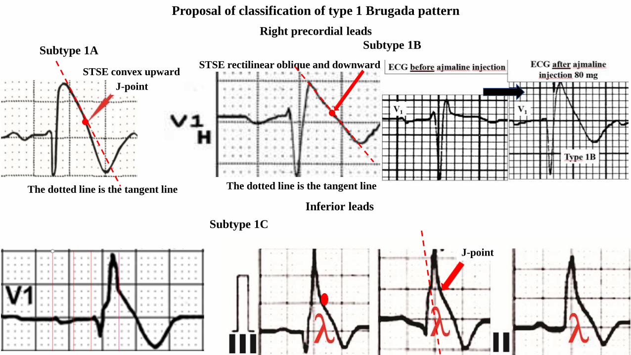
Proposal of classification of type 1 Brugada pattern
Subtype 1ASubtype 1B
STSE convex upwardSTSE rectilinear oblique and downward
Subtype 1C
Right precordial leads
Inferior leads
J-point
J-point
The dotted line is the tangent line The dotted line is the tangent line
Where is the end of the QRS complex the J point
Answer Point 2 Point 1 corresponds to high take-off and point 2 indicates the end of the QRS complex
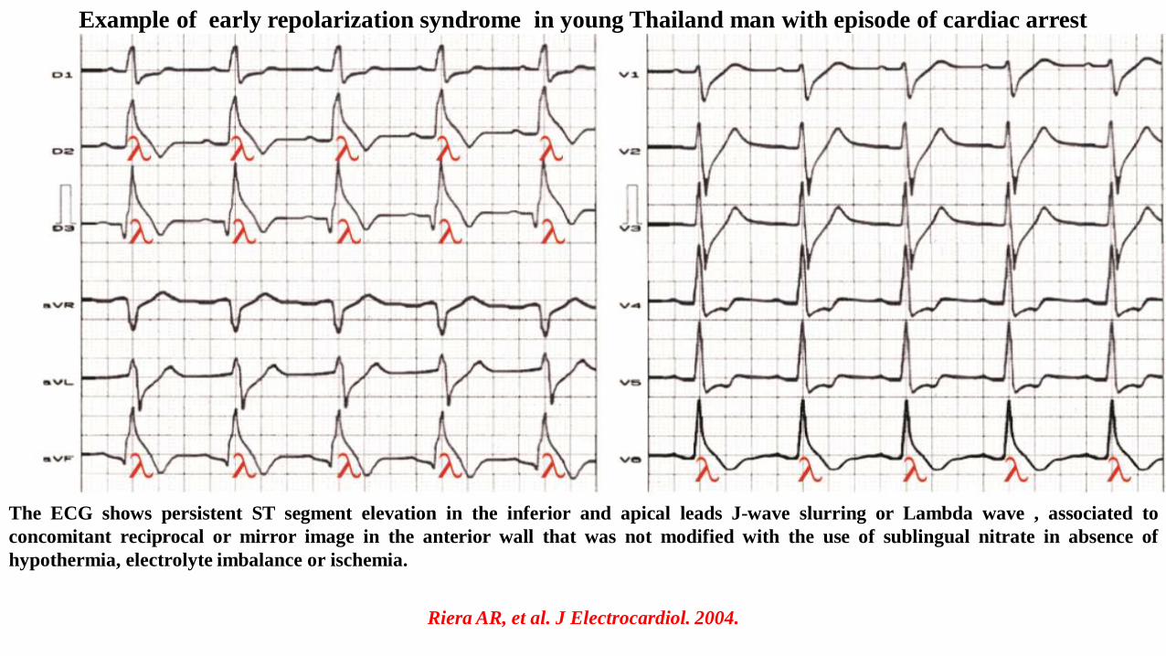
The ECG shows persistent ST segment elevation in the inferior and apical leads J-wave slurring or Lambda wave associated to
concomitant reciprocal or mirror image in the anterior wall that was not modified with the use of sublingual nitrate in absence of
hypothermia electrolyte imbalance or ischemia
Riera AR et al J Electrocardiol 2004
Example of early repolarization syndrome in young Thailand man with episode of cardiac arrest
Holter monitoring recorded the final event manifest by PVT episode with initial short-coupling ventricular premature contractions (R on T) that
ended quickly in VF and asystole Pattern 1C of repolarization has been observed in acute myocardial infarction by Kukla et al (Kukla 2007)
These authors raised the hypothesis that the ldquoLambda-like STrdquo could be a new marker of risk of acute infarction with ST segment elevation
The lambda-like J wave could be caused by ischemia although the mechanism has not been fully elucidated (Tomcsaacutenyi J et al J Electrocardiol
2019) Yagi et al reported a case of AMI that showed discrepancy between ST-T elevation with lambda-like ischemic J wave in a broad area and
coronary angiographical finding of diagonal branch occlusion (Yagi S et al J Med Invest 2019) A special form of this lambda-like ST segment
elevation accompanied by a QRS (R wave) of more than 10 mm has also been described This was termed a ldquotriangular QRS-ST-T waveformrdquo
pattern (Ciprinai A et al J Electrocardiol 2018) It is also known that this unique ECG sign does only occur in occlusive CAD (Tarantino N et
al J Electrocardiol 2018) The lambda-like ST-elevation ECG pattern is extremely rare in patients with type 2 myocardial
infarction (T2MI) triggered by variant angina or coronary spasm When this ECG pattern appears sudden cardiac death (SCD) caused by lethal
ventricular arrhythmia may occur because clinicians do not pay sufficient attention to this phenomenon The lambda-like ST-elevation pattern is
identified with other ST-elevation patterns by geometry and may be a new risk predictor for lethal ventricular arrhythmia on ECG When this
pattern is identified clinicians should adopt aggressive therapeutic strategies including ICD implantation and etiological treatment (Wang G et
al Medicine (Baltimore) 2018) Recently we described a case of ACS with transient prominent anterior QRS forces (PAF) caused by
proximal subocclusion of the LAD coronary artery before the first septal perforator branch The ECG change indicates left septal
fascicular block (LSFB) with associated slurring-type giant J-wave Currently this J-wave variant is considered as a lambda-
like wave or QRS-ST-T triangulation Its presence is indicative of poor prognosis because of the risk for cardiac arrest as a
consequence of VTVF See figure next slide
Sinus rhythm heart rate 88 bpm P-wave duration 120 ms P axis +55deg PR interval 160 ms prolonged R-wave peak time (RWPT) in V1ndashV2
QRS-axis-10deg QRS duration 120 ms No clear distinction between the end of QRS and the beginning of ST (QRSndashSTndashT ldquotriangulationrdquo)
embryonic q wave in V3ndashV4 PAF R-wave ldquoin crescendordquo from V1 to V4 and decreasing in V5ndashV6 very high J-wave of end-QRS slurring type
across all precordial leads and prolonged QTQTc interval (500588 ms) Note J-wave end-QRS slurring with lambda-like Gussak-wave
(Gussak I J Electrocardiol 2004) or triangular QRS-ST-T waveform Conclusion Left atrial enlargement LSFB (Perez-Riera de Abreu
Barbosa-Barros Nikus amp Baranchuk 2016) and giant slurring variant J-wave end-QRS
Schematic figure of J-wave variants end-QRS notching end-QRS slurring represented by the present case Early repolarization with or without
ST-segment elevation is characterized by end-QRS notching or slurring (the present case)
V1 P-wave duration =120ms II P-wave duration =160ms first degree interatrial block
II
Significant P-wave dispersion (= 40 ms)
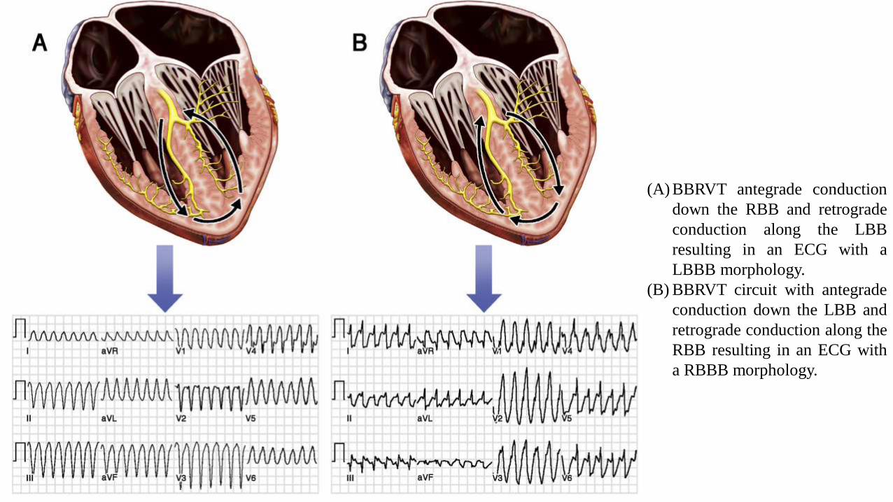
(A) BBRVT antegrade conduction
down the RBB and retrograde
conduction along the LBB
resulting in an ECG with a
LBBB morphology
(B) BBRVT circuit with antegrade
conduction down the LBB and
retrograde conduction along the
RBB resulting in an ECG with
a RBBB morphology
C Intracardiac tracing of BBRVT revealing the H-H interval preceding and predicting the subsequent ventricular-ventricular
interval BBRVT frac14 bundle branch re-entrant ventricular tachycardia H frac14 His V frac14 ventricular
1 Sinus bradycardia 2 first degree interatrial interatrial block and or Left atrial enlargement 3 P-wave dispersion 4 left posterior
fascicular block (LPFB) 5 Complete Right Bundle Branch Block 6 Parietal block 7 ldquoelf signrdquo 8 Lambda shape or J-slurring
wave 9 Epsilon wave 10 Negative T wave from V1 to V4 and 11 Induced idiopathic bundle branch re-entrant ventricular
tachycardia (BBRVT) with an underlying genetic etiology Recently Prof Melvin Scheinman group studying apparent idiopathic BBRVT
identified the first genetic culprits for this life-threatening arrhythmia providing further insight into its underlying pathophysiology and
emphasizing a potential role for genetic testing in this condition Their findings also highlight BBRVT as a novel genetic etiology of
unexplained SCD that can be definitely treated with RFCA The authors studied cases of BBRVT with normal biventricular size and function
recruited from 6 North American centers Enrollment required a clinically documented wide complex tachycardia and BBRVT proven during
EPS Study participants were screened for mutations within genes associated with cardiac conduction system disease Pathogenicity of
identified mutations was evaluated using in silico phylogenetic and physicochemical analyses and in vitro biophysical studies Among 6 cases
of idiopathic BBRVT each presented with hemodynamic compromise and 2 suffered cardiac arrests requiring resuscitation Putative culprit
mutations were identified in 3 of 6 cases including 2 in SCN5A (Ala1905Gly [novel] and c4719CgtT [splice site mutation]) and 1 in LMNA
(Leu327Val [novel]) Biophysical analysis of mutant Ala1905Gly Nav15 channels in tsA201 cells revealed significantly reduced peak current
density and positive shifts in the voltage-dependence of activation consistent with a loss-of-function The SCN5A c4719CgtT splice site
mutation has previously been reported as disease-causing in 3 cases of BrS whereas the novel LMNA Leu327Val mutation was associated
with a classic laminopathy phenotype Following catheter ablation BBRVT was noninducible in all cases and none experienced a clinical
recurrence during follow-up See figure next slide
Summary of ECG diagnosis
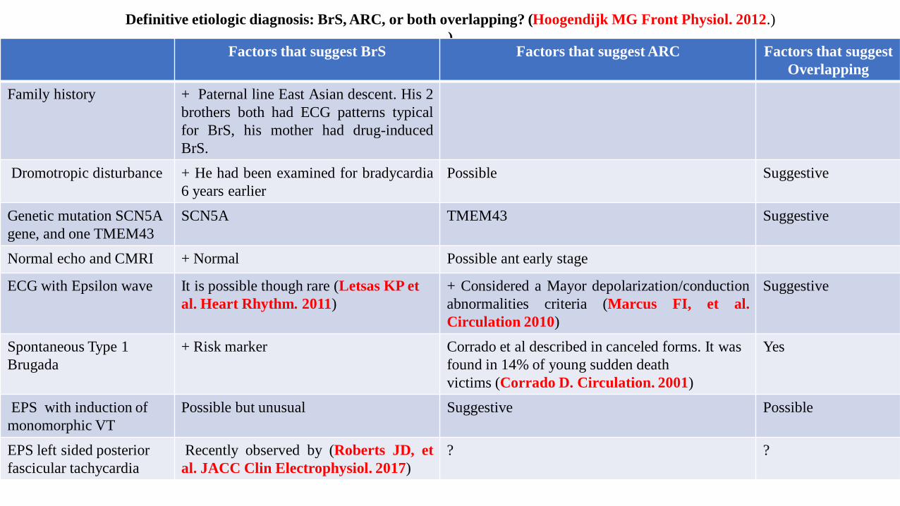
Definitive etiologic diagnosis BrS ARC or both overlapping (Hoogendijk MG Front Physiol 2012)
) Factors that suggest BrS Factors that suggest ARC Factors that suggest
Overlapping
Family history + Paternal line East Asian descent His 2
brothers both had ECG patterns typical
for BrS his mother had drug-induced
BrS
Dromotropic disturbance + He had been examined for bradycardia
6 years earlier
Possible Suggestive
Genetic mutation SCN5A
gene and one TMEM43
SCN5A TMEM43 Suggestive
Normal echo and CMRI + Normal Possible ant early stage
ECG with Epsilon wave It is possible though rare (Letsas KP et
al Heart Rhythm 2011)
+ Considered a Mayor depolarizationconduction
abnormalities criteria (Marcus FI et al
Circulation 2010)
Suggestive
Spontaneous Type 1
Brugada
+ Risk marker Corrado et al described in canceled forms It was
found in 14 of young sudden death
victims (Corrado D Circulation 2001)
Yes
EPS with induction of
monomorphic VT
Possible but unusual Suggestive Possible
EPS left sided posterior
fascicular tachycardia
Recently observed by (Roberts JD et
al JACC Clin Electrophysiol 2017)
Conclusion Phenotypic overlap between AC and BrS
The clinical features of BrS and AC are frequently in patients The first epicardial electrograms underlying the Brugada ECG pattern support the
notion that conduction disturbances in structural discontinuous myocardium underlie the BrS (Holmqvist F Heart Rhythm 2008)
(Nademanee K et al Journal of the American College of Cardiology 2015) The structural RV abnormalities in ARVC therefore most likely
predispose patients to develop the Brugada ECG pattern and the associated ventricular arrhythmias The strict exclusion of structural heart disease
in the BrS as advocated in the 2005 consensus repor appear arbitrary These conclusions plead for a broadening of the diagnostic criteria by
making a distinction in patients with the Brugada features in the presence and absence of identifiable underlying structural heart disease This
patients group is heterogeneous and incorporates various underlying cardiac conditions besides ARC the Brugada ECG pattern can also occur in
the setting of Chagasrsquo disease (Chiale PAet alAm J Cardiol 1982 Brito MR et al Europace 2010) Thus far prospective data are available
of only 17 ARVC patients with drug-induced Brugada ECG (Peters S et al Europace 2008) which is associated with a low arrhythmogenic
risk in the setting of BrS (Probst et al Only one monomorphic VT was recorded during follow-up Secondly the myocardial condition and
arrhythmogenic substrate that facilitates the Brugada features may change over time in patients with and underlying cardiomyopathy In the ARC
follow-up study by Peters the reproducibility of drug-induced Brugada ECG pattern was four out of eight Until the arrhythmogenic risk in
patients with the Brugada ECG pattern in the setting of structural heart disease has been assessed it appears reasonable to avoid or treat known
triggers arrhythmias in BrS patients such as certain pharmacological agents (Postema PG et al J Am Coll Cardiol) and fever The literature
demonstrates scientific evidence that features of both ARVC and BrS may occur in some patients
Previous clinical studies demonstrated a phenotypic overlap between the AC and BrS In 1986 Martini et al(Martini B Am Heart
J 1989) described six patients who experienced ventricular fibrillation and had clinical evidence of underlying structural abnormalities of the right
ventricle three of them exhibited a Brugada-like ECG pattern Tada et al reported right ventricular morpho-functional abnormalities andor
histologic abnormalities consistent with ARVC in 5 out of 6 Japanese men with a clinical and ECG diagnosis of BrS (Tada H et al Am J
Cardiol 1998) Corrado et al reported an Italian family with Brugada-like ST-segment elevation RV cardiomyopathic changes at
echocardiography and diagnostic morphologic features of AC at histopathologic investigation of the heart specimen of the proband with SCD
(Corrado D et al J Am Coll Cardiol 1996)
The cardiac sodium channel its mutations and their spectrum arrhythmia phenotypes
Representation of numerous phenotypes consequence of SCN5A gene mutations Early repolarization syndrome (ERS) Brugada syndrome (BrS)
Congenital long QT syndome variant 3 (LQT3) Progressive Cardiac Conduction Disease (PCCD) or Lenegravegre disease Sick Sinus Syndrome
(SSS) Sudden Unexplained Nocturnal Death Syndrome (SUNDS) Multifocal Ectopic Purkinje-related Premature Contractions (MEPPC) Sudden
Infant Death Syndrome (SIDS) Overlapping syndromes Dilated Cardiomyopathy (DCM) and Familial Atrial Fibrillation (FAF)
httpwwwrevistasuspbrjhgdarticleview122759 (Peacuterez-Riera et al J Human Growth and development 2016)
Factors modulating SCN5A gene expression H2O2 (and possibly plakophilin 2 PKP2) promotes the nuclear translocation of catenin-β1
Transcription factor 4 (TCF4) and catenin-β1 form a complex that interacts with a promoter region of SCN5A suppressing the expression of this
gene The microRNAs miR-219 and miR-200 regulate the post-transcriptional expression of SCN5A The T-box transcription factors TBX3 and
TBX5 interact with an enhancer region in SCN10A and SCN5A loci and regulate the expression of both genes The transcription factors GATA4
and GATA5 synergistically activate the expression of SCN5A through promoter binding sites ROS reactive oxygen species
BrS15ARVC9
ARVC9BrS15
Moncayo-Arlandi J Brugada R Nat Rev Cardiol 2017
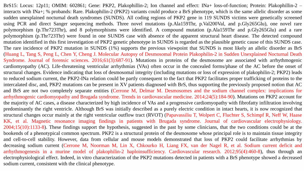
BrS15 Locus 12p11 OMIM 602861 Gene PKP2 Plakophillin-2 Ion channel and effect INa+ loss-of-function Protein Plakophillin-2 ndash
interacts with INa+ probands Rare Plakophilin-2 (PKP2) variants could produce a BrS phenotype which is the same allelic disorder as some
sudden unexplained nocturnal death syndromes (SUNDS) All coding regions of PKP2 gene in 119 SUNDS victims were genetically screened
using PCR and direct Sanger sequencing methods Three novel mutations (pAla159Thr pVal200Val and pGly265Glu) one novel rare
polymorphism (pThr723Thr) and 8 polymorphisms were identified A compound mutation (pAla159Thr and pGly265Glu) and a rare
polymorphism (pThr723Thr) were found in one SUNDS case with absence of the apparent structural heart disease The detected compound
mutation identified in this first investigation of PKP2 genetic phenotype in SUNDS is regarded as the plausible genetic cause of this SUNDS case
The rare incidence of PKP2 mutation in SUNDS (1) supports the previous viewpoint that SUNDS is most likely an allelic disorder as BrS
(Huang L Tang S Peng L Chen Y Cheng J Molecular Autopsy of Desmosomal Protein Plakophilin-2 in Sudden Unexplained Nocturnal Death
Syndrome Journal of forensic sciences 201661(3)687-91) Mutations in proteins of the desmosome are associated with arrhythmogenic
cardiomyopathy (AC) Life-threatening ventricular arrhythmias (VAs) often occur in the concealed formsphase of the AC before the onset of
structural changes Evidence indicating that loss of desmosomal integrity (including mutations or loss of expression of plakophilin-2 PKP2) leads
to reduced sodium current the PKP2-INa relation could be partly consequent to the fact that PKP2 facilitates proper trafficking of proteins to the
intercalated disc and PKP2 mutations can be present in XV patients diagnosed with BrS thus supporting the previously proposed notion that AC
and BrS are not two completely separate entities (Cerrone M Delmar M Desmosomes and the sodium channel complex implications for
arrhythmogenic cardiomyopathy and Brugada syndrome Trends in cardiovascular medicine 201424(5)184-90) Mutations on PKP2 account for
the majority of AC cases a disease characterized by high incidence of VAs and a progressive cardiomyopathy with fibrofatty infiltration involving
predominantly the right ventricle Although BrS was initially described as a purely electric condition in intact hearts it is now recognized that
structural changes occur mainly at the right ventricular outflow tract (RVOT) (Papavassiliu T Wolpert C Fluchter S Schimpf R Neff W Haase
KK et al Magnetic resonance imaging findings in patients with Brugada syndrome Journal of cardiovascular electrophysiology
200415(10)1133-8) These findings support the hypothesis suggested in the past by some clinicians that the two conditions could be at the
bookends of a phenotypical common spectrum PKP2 is a structural protein of the desmosome whose principal role is to maintain tissue integrity
and cell-to-cell stability However data from cellular and mouse models demonstrated that loss of PKP2 could facilitate arrhythmias by
decreasing sodium current (Cerrone M Noorman M Lin X Chkourko H Liang FX van der Nagel R et al Sodium current deficit and
arrhythmogenesis in a murine model of plakophilin-2 haploinsufficiency Cardiovascular research 201295(4)460-8) thus through an
electrophysiological effect Indeed in vitro characterization of the PKP2 mutations detected in patients with a BrS phenotype showed a decreased
sodium current consistent with the clinical phenotype
Super-resolution microscopy data showed that loss of PKP2 could affect proper trafficking of the sodium channel at the membrane thus
supporting the concept that proteins could have accessory roles aside from the primary one ascribed to them The role of the cardiac intercalated
disc as a functional unit with both structural and electric regulatory functions has been opening new paths of investigations on the possible
arrhythmogenic substrate in BrS (Nademanee K Raju H de Noronha SV Papadakis M Robinson L Rothery S et al Fibrosis Connexin-43 and
Conduction Abnormalities in the Brugada Syndrome Journal of the American College of Cardiology 201566(18)1976-86)
Gene Protein Frequency
in ACM
Structure Mutation type Inheritance Phenotype
ARcompou
nd
heterozygo
us
OMIM
Entry
GenotyePhe
notype
Studies
PKP2 Plakophilin-
2
20-45 Desmosome Non-missence
++(splice-site
nonsense
insdel large
del)
Missense
AD+++ AC
DCM
AD+++ AC
DCM
ARVC9 Conventional
ARVC
phenotype
(Quarta G Circulation 2011 Christensen AH J Med Genet 2010 Kapplinger JD J Am Coll Cardiol 2011 Xu T J Am Coll Cardiol 2010
Gerull B Nat Genet 2004)
ARVC9
AC BrS
Age of presentation (yrs) 15ndash30 30ndash40
Gender MgtF (31) MgtF (81)
Distribution World-wide (Italy endemic in Veneto
region)
World-wide (Southeast Asia)
Inheritance AD (AR) AD
Predominant pathogenetic genes Desmossomal genes 60 SCN5A gene 20-30
Typical symptoms Palpitations syncope cardiac arrest Syncope cardiac arrest
Imaging Structural RV (and LV) abnormalities Normal or subtle
Biopsy Fibrofatty replacement Normal minimal
ECG repolarization Right precordial TWI Right precordial high take-off ST elevation
and TWI
ECG depolarization Right precordial QRS prolongation ε
waves
RBBBLAD
AV conduction times Normal Prolonged PRHV interval
ECG changes Fixed Dynamic
Ventricular arrhythmias Monomorphic VT VF with a left bundle
branch block pattern most likely
Polymorphic VT VF
Differential diagnosis between AC and BrS
Typical mechanism
of VT
Scar- related reentry
resulting from a macro-
reentry around fibrofatty
tissue Monomorphic
Phase 2 reentry
Polymorphic very fast with short coupled interval the first VPC
Natural history Sudden death heart failure Sudden death
Event triggered Catecholamines and mostly
occur during or immediately
after exercise
Enhanced by vagotonic agents or -adrenergic blockers nocturnal
vagotony fever
Biventricular heart
failure
Possible No
Endomyocardial
biopsy
The histopathologic finding of fatty infiltration of the myocardium (non-
diagnostic for ARVC) was observed by Ohkubo et al (Ohkubo K Int
Heart J 2010) in 20 (5 of 25) and by Zumhagen et al (Zumhagen S
Circ Arrhythm Electrophysiol 2009) in 19 (4 of 21) of BrS patients
undergoing RV endomyocardial biopsy However a lower prevalence of
typical fibrofatty myocardial replacement suggestive of ARVC was reported
both in the series of Frustaci et al (Frustaci A Circulation 2005) and
Zumhagen et al (Zumhagen S Circ Arrhythm Electrophysiol 2009)
AD = autosomal dominant AR = autosomal recessive AV = atrioventricular LAD = left axis deviation LV = left ventricle RBBB = right bundle
branch block RV = right ventricle TWI = T-waves inversion VF = ventricular fibrillation VT = ventricular tachycardia
bull Age of presentation (yrs)
AC 15ndash30
BrS 30-40
bull Gender
AC Men are more frequently affected than women with an approximate ratio of 31
BrS MgtF (81)
bull Inheritance
AC Arrhythmogenic cardiomyopathy is typically inherited as an autosomal dominant(AD) pattern with variable penetrance and
incomplete expression Approximately 40 to 50 of AC patients have a mutation in genes encoding a desmosome protein The gene is
on the chromosome 14q23-q24[3]There is an autosomal recessive(AR) trait variant associated with palmoplantar keratosis and wooly
hair named Naxos disease and the Carvajal syndrome
BrS AD (asymp 25) or sporadic Cerrone et al screened by direct sequencing the PKP2 gene in a cohort of 200 patients with clinical
diagnosis of BrS and no mutations on the most prevalent genes Cerrone et al discovered five single amino acid substitutions in five
unrelated patients (Cerrone M et al Circulation 2014) This is the first systematic retrospective analysis of a patient group to define
the coexistence of sodium channelopathy and genetic PKP2 variations PKP2 mutations may be a molecular substrate leading to the
diagnosis of BrS In order to assess if this missense variant in PKP2 could affect the cardiac INa we used an HL-1 cell line stably
silenced for the endogenous PKP2 In the absence of PKP2 these cells showed a decrease in the native INa Cells transiently transfected
Differential diagnosis between AC and BrS
with each one of the PKP2 mutants associated with the BrS phenotype showed significantly decreased INa when compared with cells
transfected with wild type PKP2 Similar results were obtained when they used a line of human iPSC-derived cardiomyocytes from a patient
lacking PKP2 at the cell membrane(Kim C Nature 2013 Awad MM Human mutation 2006) In these cells INa increased upon
transfection with wild type PKP2 Transfection with one of the PKP2 mutants associated with BrS was not able to restore normal INa These
data represent the first evidence that missense mutations in PKP2 can cause a decrease in cardiac sodium channel INa and facilitate arrhythmias
even in the absence of a structural cardiomyopathy They propose that PKP2 mutations provide at least part of the molecular substrate of BrS
The inclusion of PKP2 as part of routine BrS genetic testing remains premature yet the possibility that some patients showing signs of disease
may harbor PKP2 variants should be considered when the genotype is negative for other genes associated with BrS15 Locus 12p11 OMIM
602861 Gene PKP2 Plakophillin-2 Ion channel and effect INa+ loss-of-function Protein Plakophillin-2 ndash interacts with INa+ probands
Rare Plakophilin-2 (PKP2) variants could produce a BrS phenotype which is the same allelic disorder as some sudden unexplained nocturnal
death syndromes (SUNDS) All coding regions of PKP2 gene in 119 SUNDS victims were genetically screened using PCR and direct Sanger
sequencing methods Three novel mutations (pAla159Thr pVal200Val and pGly265Glu) one novel rare polymorphism (pThr723Thr) and 8
polymorphisms were identified A compound mutation (pAla159Thr and pGly265Glu) and a rare polymorphism (pThr723Thr) were found in
one SUNDS case with absence of the apparent structural heart disease The detected compound mutation identified in this first investigation of
PKP2 genetic phenotype in SUNDS is regarded as the plausible genetic cause of this SUNDS case The rare incidence of PKP2 mutation in
SUNDS (1) supports the previous viewpoint that SUNDS is most likely an allelic disorder as BrS (Huang L 2016) Mutations in proteins of
the desmosome are associated with arrhythmogenic cardiomyopathy (AC) Life-threatening ventricular arrhythmias (VAs) often occur in the
concealed formsphase of the AC before the onset of structural changes Evidence indicating that loss of desmosomal integrity (including
mutations or loss of expression of plakophilin-2 PKP2) leads to reduced sodium current the PKP2-INa relation could be partly
consequent to the fact that PKP2 facilitates proper trafficking of proteins to the intercalated disc and PKP2 mutations can be
present in XV patients diagnosed with BrS thus supporting the previously notion that AC and BrS are not two completely
separate entities (Cerrone M Trends in cardiovascular medicine 2014) Mutations on PKP2 account for the majority of AC
cases a disease characterized by high incidence of VAs and a progressive cardiomyopathy with fibrofatty infiltration involving
predominantly the right ventricle Although BrS was initially described as a purely electric condition it is now recognized that
structural changes occur mainly at the RVOT (Papavassiliu T 2004) These findings support the hypothesis that the two
conditions could be at the bookends of a phenotypical common spectrum PKP2 is a structural protein of the desmosome whose
principal role is to maintain tissue integrity and cell-to-cell stability However data from cellular and mouse models
demonstrated that loss of PKP2 could facilitate arrhythmias by decreasing Na+ current (Cerrone M Cardiovascular research
2012) thus through an electrophysiological effect Indeed in vitro characterization of the PKP2 mutations detected in patients
with a BrS phenotype showed a decreased Na+ current consistent with the clinical phenotype Super-resolution microscopy
data showed that loss of PKP2 could affect proper trafficking of the Na+ channel at the sarcolemma thus supporting the concept
that proteins could have accessory roles aside from the primary one ascribed to them The role of the cardiac intercalated disc as
a functional unit with both structural and electric regulatory functions has been opening new paths of investigations on the
possible arrhythmogenic substrate in BrS (Nademanee K Journal of the American College of Cardiology 2015)
bull History
AC first described by the Popersquos physician Giovanni Maria Lancisi1654-1720 in his book entitled De Motu Cordis et
Aneurysmatibus (Lancisi 1736) The first comprehensive clinical description of the disease was reported by Frank Marcus et al in 1982
when he reported 24 adult cases with ventricular tachyarrhythmias with LBBB pattern (Marcus FI Circulation 1982) He described a a
family who had experienced pathologic RV heart failure and SCD in four generations Marcus et al () published a case series that for the
first time captured the clinical profile of ARVC In 24 patients they described the fibro-fatty replacement the ventricular
tachyarrhythmias and premature ventricular complexes with left bundle branch block morphology the repolarization abnormalities in the
form of inversed T-waves in the right precordial leads delayed activation on standard or signal averaged ECGs the morphological
features of increased dimensions and wall motion abnormalities of the right ventricle and the familial occurrence of this disorder The
causative mechanism was unknown and was speculated to lie in the development of the right ventricle hence it was termed
ldquoarrhythmogenic right ventricular dysplasiardquo7
The electrocardiographic features of the disease were first described including the epsilon
wave in 1984 (Fontaine G et al Arch Mal Coeur Vaiss 1984)
Guy Fontaine
BrS
bull 1953 Halfway through the 20th century (1953) Osher and Woff noticed the right bundle branch block (RBBB) electrocardiographic
pattern associated to ST segment elevation in the right precordial leads These were considered at the time as normal variants not having
been related to sudden cardiac death (SCD) (Osher HL Wolff L J Med Sci 1953)
bull 1975 Calo (Calo AA et al G Ital Cardiol 1975) reported an electrocardiographic triad that consisted of R wave ST segment
elevation and negative T wave in right precordial leads which coincide with the characteristics of the electrocardiographic pattern
currently known as Brugada-type being considered a normal variant at the time
bull 1984 A similar ECG pattern this time associated with na aborted SD and occuring in a 42-years-old male on the 2nd October 1984 was
seen in Padua Italy
bull During the 80s the Center for Disease Control in Atlanta observed an abnormally high incidence of SCD in Asian refugees who
immigrated to USA from the northeast of Thailand This form of SCD is known in this country as Lai Tai (death during sleep)
Approximately two decades later the conclusion was reached that the entity known as Sudden Unexplained Nocturnal Death Syndrome
(SUNDS) originates in an allele belonging to the same gene (SCN5A) as Brugada Disease (Vatta M et al Hum Mol Genet 2002)
bull In 1986 Prof Pedro Brugada received his first patient with typical ECG a Polish Caucasian child who suffered several episodes of
syncope The boy presented as family background his sisters SCD even though she had been treated with association of pacemaker
implantation and amiodarone In 1989 a patient with characteristic ECG was described as being a carrier of early repolarization
syndrome (Brugada P Brugada J (abstr) PACE 1991)
bull 1987 Martini B et al described six patients with apparently idiopathic VF 3 of them had upsloping WT segment elevation RBBB and
inverted T-waves They documented subtle structural abnormalities in the RV (Martini B et al Am Heart J 1987)
bull 1992 Pedro and Josep Brugada adding 2 more cases presented as an abstract in the NASPE meeting a new clinical-cardiologic
syndrome typified by the association of RBBB persistent ST segment elevation normal QT interval and SCD The Brothers described 8
cases and introduced the term Right Bundle Branch Block ST segment elevation and Sudden death syndrome The first patient in their
series with this syndrome was seen in 1986 The patient was a three years old boy from Poland With multiple episode of loss of
consciousness and the childacutes sister had died suddenly at age two after multiple episodes of aborted sudden death both had the
characteristic ECG (Brugada P et al J Am Coll Cardiol 1992)
Prof Em Dr Pedro BrugadaProf Dr Josep Brugada
bull Prevalence
AC It is estimated as 15000 (Czarnowska E et al Kardiol Pol 2003 2003) The prevalence is dependent on geographic
circumstances (Hagenah 2004) The estimated prevalence of familial AC in the general population ranges from 1 in 2000 to 1 in 5000
people Cause SCD in youth 5(US) and 25 (Italy) One of the most common cause of SD athletes AC can be sporadic or inherited
usually transmitted with an AD pattern with incomplete penetrance and variable expression or rarely as an AR pattern The AR is
frequently associated with palmoplantar keratoderma wooly hair and dystrophic nails Naxos disease and Israeli Desmoplakin-
Recesssive are observed in Arabic populations These cardio-cutaneous syndromes have been reported in the literature from Greece
Italy India Ecuador Israel and Turkey (Heidbuchel H et al Br J Sports Med 2012)
BrS worldwide pooled prevalence of BrS is 05 per 1000 The highest prevalence was reported in Southeast Asia (37 per 1000) the
prevalence of BrS in Thailand was over 146 times more common than in North America and 37 times more common than in Europe
BrS and Type-23 BrP were also more common in males than in females (Vutthikraivit W et al Acta Cardiol Sin 2018) BrS The
prevalence in Japan is 01-02 (Macfarlane PW et al J Electrocardiol 2013) From 44 unrelated index patients and family
members Schulze-Bahr et al (Schulze-Bahr E et al Hum Mutat 2003) performed a complete genetic analysis of SCN5A in BrS
The authors concluded that The sporadic cases are predominant 63 against 37 of familiar cases Disease penetrance (disease
absence in some individuals with disease gene) is complete in the SCN5A+ adult patients but incomplete in SCN5A+ children (17)
Genetic testing of SCN5A is especially useful in familial disease to identify individuals at cardiac risk In sporadic cases a genetic
basis and the value of mutation screening has to be further determined
World prevalence map of Brugada syndrome
Hawai
bull Endemic aacutereas
AC Veacuteneto area Italy Naxos Ilhas Grece
BrS World-wide (Southeast Asia) Thailand Japan Fillipines
bull Manifestations
ARC Typical Palpitations syncope cardiac arrest
BrS sincope sudden cardiac death agonal respiration
bull Typical symptoms
bull Event triggered
AC Effort (adrenergic stimulus)
BrS at rest during nocturnal sleep (vagotony) The characteristic Brugada ECG pattern is dynamic and can even disappear temporarily
(Brugada J et al J Cardiovasc Electrophysiol 1997) Prior to the initiation of VF however the Brugada ECG pattern reappears in
these patients (Kasanuki H et al Circulation 1995) The Brugada ECG pattern is most pronounced at night or at rest (Mizumaki K
et al 2004) during febrile illness (Amin AS et al Ann Intern Med 2008) and after consumption of a copious meal (Ikeda T et
al J Cardiovasc Electrophysiol 2006) which also are known triggers of VT and SCD in BrS patients Sodium channel blockers are
known to augment or provoke the Brugada ECG pattern (Miyazaki T et al J Am Coll Cardiol 1996) Other drugs can ameliorate the
Brugada ECG pattern such as isoproterenol and quinidine (Alings M et al Pacing Clin Electrophysiol 2001) and have been used to
prevent arrhythmias in observational studies and case reports (Belhassen B et al Circulation 2004 Belhassen B et al Heart
Rhythm 2017)
bull Race
AC Caucasian predominance
BrS Asian predominance in Asians was nine times more common than in Caucasians and 36 times more common than
in Hispanics
bull Physical examination
AC Eventual heart failure
BrS Normal eventual atrial fibrillation
bull Natural history
AC syncope sudden cardiac death heart failure
BrS syncope sudden cardiac death
bull Biventricular heart failure
AC LV involvement was found in 76 of hearts with AC was age dependent and was associated with clinical
arrhythmic events more severe cardiomegaly inflammatory infiltrates and heart failure ARVC can no longer be
regarded as an isolated disease of the right ventricle(Corrado D J Am Coll Cardiol 1997)
BrS No
bull Predominant pathogenic gene
AC Desmossomal 60 of cases The cardiac desmosome and proposed roles of the desmosome in (A) supporting structural stability
through cellndashcell adhesion (B) regulating transcription of genes involved in adipogenesis and apoptosis and maintaining proper
electrical conductivity through regulation of (C) gap junctions and (D) calcium homeostasis Dsc2 indicates desmocollin-2 Dsg2
desmoglein-2 Dsp desmoplakin Pkg plakoglobin Pkp2 plakophilin-2 and PM plasma membrane
BrS SC5A The first genetic association with BrS discovered was a loss-of-function mutation in the cardiac voltage-gated Na+
channel gene
SCN5A in 1998 (Chen Q et al Nature 1998) It is thought to be found in 15-30 of BrS cases There is increasing evidence that BrS is an
oligogenetic disease (Le Scouarnec S et al Hum Mol Genet 2015) with involvement of more than one genetic factor with different effect
sizes (Bezzina CR et al Nat Genet 2013) The more of these genetic factors one has the higher the likelihood of having a type 1 Brugada
pattern Currently molecular genetic testing should be limited to SCN5A SLMAP SEMA3A SCNN1A and SCN2B (Campuzano O Hum
Mutat 2019) SCN5A and in SCN5A families (presymptomatic) and counselling should include an ECG because phenotype positive
genotype negative cases have been described within these families (Probst V et al Circ Cardiovasc Genet 2009) There might be some role
for genetic testing in risk stratification (Yamagata et al Circulation 2017)
bull Chacteristic of ECG changes
bull ECG depolarization
AC Right precordial QRS prolongation (parietal block) epsilon waves This wave represent right ventricular late potentials can be
brought out by recording the ECG at double speed (50mms) double amplitude (20mmmV) and a 40 Hz filter Here epsilon waves are
evident as small notched deflections just after the QRS in lead V1 Signal averaged ECG in the absence of QRSD gt110thinspms fQRS
duration gt114thinspms orthinspDuration of terminal QRS lt40thinspμV ge38thinspms orRoot-mean-square voltage of terminal 40thinspms le20thinspμV Terminal
activation delay ge55thinspms
BrS P-wave prolongation A647D mitigates the lethal LQT3 phenotype seen with P1332L it also reduces mexilitine sensitivity and
decreases INa density These explain probands mild repolarization abnormality and prominent conduction defect in the atria and
ventricles and the expression of P1332L with A647D yields a novel disease phenotype for which mexiletine pharmacotherapy is no
longer suitable (Liu J PLoS One 2018) Augmented P-wave duration in lead II P-wave dispersion (Letsas KP et al Pacing Clin
Electrophysiol 2009) PR interval prolongation peripheric conduction delay HV prolongation split-His LAFB f-QRS end
conduction delay on RV The presence of atrial fibrillation (Kusano KF et al J Am Coll Cardiol 2008) OT territory Prolonged QRS
duration measured from lead II or lead V2 ge120 ms (Junttila 2008) Also in right precordial QRS prolongation (parietal block) QTc
interval more than 460 ms in lead V2 (Take 2011) and QT-interval prolongation in right precordial leads (Pitzalis 2003) increase in
QRS complex duration (gt110ordm) in the right precordial leads in absence of CRBBB parietal block (Pitzalis MV et al J Am Coll
Cardiol 2003) Tpeak minus Tend prolongation and Tpeak minus Tend dispersion (Castro Hevia 2006) baseline fQRS increased major arrhythmic
events up to 3-fold fQRS could be an important tool for risk assessment in patients with BrS (Rattanawong Ann Noninvasive
Electrocardiol 2018 Morita H et al Circ Arrhythm Electrophysiol 2017)
The tracing shows the P wave in a patient with BrS and positive SNC5A mutation performed before and immediately after ajmaline test (1
mgkg)P wave duration (Pd) before the injection is prolonged (Pd=135 ms) After drug administration Pd wave increases more (Pd=162 ms)
These atrial dromotropic disorders could be the substrate for reentrant atrial tachycardias such as AF
The figure shows a tracing of a symptomatic patient with Brugada syndrome after intravenous ajmaline injection First-degree atrioventricular
block (PR interval = 216 ms) and Brugada type-1 ECG pattern in V1 lead (positive test) In BrS the PR interval of ECG and the His bundle
electrogram in approximately 50 of the cases are prolonged even reaching sometimes figures of 100 ms (Yokokawa 2007) This prolongation of
the PR interval is observed predominantly in cases where the SCN5A gene mutation can be proven (carriers) The presence of a prolongued HV
interval is possible in HBE by the existence of intra-His or infra-His block PR prolongation consequence of HV split or HV prolongation is
considered another ECG risk marker (Miyamoto A et al Circ J 2011)
V1
Presence of prominent final R wave on aVR lead R wave ge 3 mm or Rq ge 075 in lead aVR (aVR sign) Slow conduction at the
RVOT may contribute to the induction of VF by PVS (Babai Bigi MA Heart Rhythm 2007)
The aVR sign Presence of prominent final R wave on aVR lead R wave ge3 mm or Rq ge075 in lead aVR (aVR sign) Slow
conduction at the RVOT may contribute to the induction of VF by PVS
AC Epsilon wave The epsilon wave of the ECG together with fragmented QRS (fQRS) the terminal conduction delay incomplete rIRBBB)
and completeadvanced RBBB (CRBBB) of peripheral origin are part of a spectrum of ventricular depolarization abnormalities AC Although
the epsilon wave is considered a major diagnostic criterion for AC since 2010 (AC Task Force Criteria) its diagnostic value is limited because
it is a sign of the later stage of the disease It would be more appropriate to say that the epsilon wave is a hallmark of AC but is of low
diagnostic sensitivity Although the epsilon wave has high specificity for AC it can be present in other pathological conditions (Peacuterez-Riera
AR et al Indian Pacing Electrophysiol J 2019) The epsilon (ε) wave can be defined as an electric signal of depolarization observed
between the end of the QRS complex and the beginning of the T wave The ε wave is found in the right precordial leads where the QRS
complex is broader than the in the left precordial leads (differencethinspge25 ms) in arrhythmogenic cardiomyopathy (AC) In patients with AC who
have left ventricular (LV) involvement the ε wave can be registered in the left andor inferior leads The ε wave represents delayed potentials
resulting from slow intraventricular conduction due to islands of surviving myocardium interspersed with fatty and fibrous tissue This
ventricular post-excitation wave consists of a slurring at the end of the QRS complex or an independent potentials after the return to the
isoelectric line The depolarization abnormality is hardly detectable by the standard 12-lead ECG (S-12-ECG) Possible causes of ε waves in
the ECG In a study with elite endurance athletes (190 senior and 157 junior athletes) an ε wave was found in 3190 senior athletes (157) and
in 1189 individuals from a sedentary control group 31ndash40 years of age CMRI showed AC findings in one of the senior athletes Pathological
ε waves in patients other than in AC CAD the ε wave has been observed in one case of acute inferior myocardial infarction (MI) associated
with RV myocardial infarction Uhls anomaly or ldquoparchment heartrdquo is an unusual myocardial abnormality first described by Henry Uhl in
1952 It is characterized by partial or complete absence of the RV myocardium with severe RV systolic and diastolic impairment Patients with
Uhls anomaly who survive to adulthood may develop right-sided heart failure or arrhythmias
The ECG shows tall and wide P waves right axis deviation frequent RBBB prominent ε waves in all QRS complexes and signs of severe
dilatation of the RV and right atrium After repair of Fallots tetralogy a case report described a patient with tetralogy of Fallot who showed
all features of the familial form of RV including an ε wave The patient had heart transplantation because of numerous episodes of ventricular
tachycardia and chronic heart failure and he had a right ventricular outflow patch aneurysm Infiltrative diseases cardiac sarcoidosis may
cause the pathological substrate required for production of ε waves Therefore differentiating AC from cardiac sarcoidosis is of clinical
importance Sickle cell anemia JW Hurst briefly mentions a probably unpublished observation of epsilon waves in a patient with sickle cell
disease with RV hypertrophy due to pulmonary arterial hypertension
Brugada syndrome (BrS) it is believed that BrS and AC are different clinical entities with respect to the clinical presentation and the genetic
predisposition The coexistence of these two relatively rare clinical entities has been reported (Hoogendijk MG Front Physiol 2012) There
may be cases where the differential diagnosis is not clear (Ozeke O et al Int J Cardiol 2018) ε waves appear to be rare in BrS and were
found in 2 of 47 patients by Letsas et al (Letsas KP et al Heart Rhythm 2011) and in 1 of a total of 12 unrelated index BrS cases included
in the study by Yu et al (Yu J et al Herz 2014)
bull ECG repolarization
AC Right precordial T-wave inversion (TWI) in right precordial leads V1 V2 and V3 in individuals over the age of 14 years And
without CRBBB is considered a major criteria (Marcus FI et al Circulation 2010)
RECD ndash Right End Conduction Delay T-loop directed to back and leftward
Fast transition from left to right
When the RV end diastolic volume is not very increased (in average 100 mlm2) The T loop presents counterclockwise rotation in the HP and axis
between +15ordm and ndash10ordm (average +5ordm)
V6
V2
HP
V3V4
V5
V1
Z
+ 90ordm+120ordm
+180ordm
SAT +15ordm
SAT -10ordm
The T loop presents counterclockwise rotation
T loop behavior in AC and its relationship with RV end diastolic volume
X
When the RV end diastolic volume is large (in average 320 mlm2) the T loop displays clockwise rotation in the HP and is located in the
right posterior quadrant which justifies the negative T wave in all precordial leads Note the presence of T loop of clockwise rotation
indicates the presence of underlying heart disease When T-wave is negative in lateral leads I aVL V5-V6 and positive in aVR it is
indictive of LV involvement
V2
HP
V3V4
V5
V1
+ 90ordm+120ordm
+180ordm
Clockwise rotation
0ordm X V6 Negative T waves from V1 to V6
T loop behavior in AC and its relationship with RV end diastolic volume
Z
T loop in 9 patients in the HP carriers of AC T loops
are arranged on the basis of progressive RVH
T loop (nordm 1) has a RV end diastolic volume of 100 mlm2
and the last loop (nordm 9) has 320 mlm2 (Nava A et al J of
Electrocardiol 1988)
Note the progressive alteration of the T loop from 1 to 9
T loop behavior in AC and its relationship with RV end diastolic volume
1 2 3
4 5 6
7 8 9
V1 V2V3
V6
Negative T waves from V1 to V6
1 The presence of horizontal (as opposed to rapidly ascending) ST segment after the J point (Takagi M et al Heart Rhythm 2013)
The presence of a J wave in multiple leads and horizontal ST-segment morphology after J wave may indicate a highly
arrhythmogenic substrate in patients with BrS
2 Augmentation of the ST segment elevation during the early recovery phase of exercise test is specific in patients with BrS and can
be a predictor of poor prognosis especially for patients with syncope alone and for asymptomatic patients (Makimoto H et al J
Am Coll Cardiol 2010)
3 Multivariate analysis revealed that a PQ interval ge170 ms and T-wave amplitude lt105 microV in lead V(1) were independent risk
stratifiers of life-threatening events Survival analysis (mean follow-up 786plusmn818 months) showed that the PQ interval and a
negative T-wave in lead V(1) were significantly associated with poor prognosis (Miyamoto A et al Circ J 2011)
4 The presence of atrial fibrillation (Kusano KF et alJ Am Coll Cardiol 2008)
5 The presence of late potentials (LPs) LPs are a noninvasive risk stratified in patients with BrS These results may support the idea
that conduction disturbance per se is arrhythmogenic (Ikeda T et alJ Am Coll Cardiol 2001)
6 A spontaneous change in ST segment is associated with the highest risk for subsequent events in subjects with a Brugada-type
ECG The presence of syncopal episodes a history of familial sudden death andor LP may increase its value (Ikeda T et al Ann
Noninvasive Electrocardiol 2005)
BrS ECG repolarization alterations
The BrS affects predominantly the right ventricle in the right ventricle outflow tract (RVOT) epicardium (Doi A et al J Cardiovasc
Electrophysiol 2010) The larger part of clinical evidence supports the presence of right end conduction delay (RECD) as part of the process of
BrS pathophysiology in the RVOT as a consequence of structural abnormalities in the heart as part of BrS (Coronel R et al Circulation 2005
Peacuterez-Riera AR Europace 2012) On the other hand in the concealed forms of AC the RECD pattern can also be observed showing type-1 ECG
pattern This pattern was shown many years ago by Guy Fontaine
Fragmented wide QRS complex in a 35-year-old Asian male patient with BrS f-QRS appears to be a marker for the substrate for spontaneous VF
in BrS and predicts patients at high risk of syncope It is a conduction abnormality within the QRS complex (Morita H et al Circulation 2008)
Fragmented QRS in Brugada Syndrome
J-point
QRSd = 120 ms
Heart rate = 68 bpm
2 Spikes
Dotted lines show onset and termination of the QRS complex
Two spikes are observed
at the upstroke of the S
wave in leads V1 and V2
High take-off
ischemic and nonischemic cardiomyopathy(Das MK et al Heart Rhythm 2010) where it represents a conduction delay of the stimulus and
is associated to an increase in mortality and arrhythmic events in these patients
Non-ischemic cardiomyopathies (Das MK Heart Rhythm 2010) In non-ischemic dilated cardiomyopathy with narrow QRS to predict
(Tigen K et al Can J Cardiol 2009)
Arrhythmogenic right ventricular cardiomyopathydysplasia (ARVCD) (Peters 2008)
Cardiac sarcoidosis (Homsi M et al Ann Noninvasive Electrocardiol 2009)
Acquired long QT syndrome The existence of fQRS plays an important role in the appearance of Torsades de Pointes (TdP) in patients with
acquired long QT interval (Moss AJ Heart Rhythm 2010 Haraoka K et al Heart Rhythm 2010)
Myocardial dysfunction pulmonary hypertension and severity in mitral stenosis(Yuce M et al Tohoku J Exp Med 2010)
Presence of a ldquonotchrdquo within a non-wide QRS complex in two adjacent leads (V1-V2) f-QRS It is a non-invasive marker of events (Das MK
Heart Rhythm 2009)
bull Atrial arrhytmias
AC late (secondary) Less is known about atrial remodeling and atrial tachyarrhythmias (ATa) in AC Wu et al performed
a cross-sectional study aimed to determine the prevalence characterization and predictors of atrial remodeling and ATa
in a large series of patients with AC From February 2004 to September 2014 294 consecutive patients who met the task
force criteria for AC were enrolled The prevalence characterization and predictors of atrial dilation and ATa were
investigated RA dilation was identified in 160 patients (544) and LA dilation in 66 patients (224) Both RA and LA
dilation were found in 44 patients (150) Twenty-five patients (85) had AF whereas 19 patients (65) had atrial
flutter (AFL) Of which 7 patients (24) had both AF and AFL Multivariate analysis showed that AFL hypertension
and RA dilation were associated with increased risk for AF AF increased the risk of AFL In conclusion atrial
remodeling and ATa were common in patients with AC (Wu et al Am J Cardiol 2016)
bull increased risk of paroxysmal atrial fibrillation Atrial fibrillation can be the first manifestation of latent BrS The ajmaline test plays an
essential role mainly in young patients with a family history of SD despite having normal finding on a base line ECG (Rodriacutegues-Mantildeero
2013) BrS is frequently associated with atrial arrhythmias AF being the most frequent Amin et al (Amin AS et al Europace 2011) verified
that patient carriers of BrS have an increased risk of developing AF because Na+
channel involvement facilitates the appearance of AF by
causing intra-atrial conduction delay and inducing structural remodeling Moreover they showed that the basal duration of the P wave is
greater in patients with BrS with the mutation compared to those without Intraatrial conduction slowing could be the substrate to maintain
AF On the contrary the presence of mutation may decrease AF by suppressing PACs PACs are more frequent in patients with BrS without
the mutation than in those with The reduction of PACs may inhibit the triggering mechanism to initiate AF Kofune et al (Kofune M et al
Int Heart J 2010) studied the atrial vulnerability in patients with BrS The authors compared 18 patients with BrS with 11 normal
individuals with similar ages without AF (control group) PES was carried out from the RA from where the following was studied The
refractory period of the RA(ERP-RA) This parameter was similar in both groups interatrial conduction time (IACT) It was significantly
higher in the group of patients with BrS Monophasic APs of the high RA (MAPs) AF inducibility with duration of gt30 seconds AF was
induced in all the patients with BrS with 1 or 2 extrastimuli and in none of the controls The maximal slope of the restitution curve
MAPD(80) (repolarization) was significantly higher in the group with BrS than in the control group VF induction occurred with PVS in all
the patients of the group of BrS carriers The authors concluded that both abnormal interatrial conduction (depolarization) and the maximal
slope of the restitution curve MAPD(80) (repolarization) may contribute to BrS arrhythmogenicity
bull Atrial arrhytmias
BrSearly (primary) Sinus rhythm is present in approximately 80 of cases but there may be a tendency for other rhythms because
electrophysiological alterations are not just confined to the ventricles Accordingly the genetic mutations could involve both the
sinoatrial node (SA node) and atrial tissue Experimental and computational analyses of the E161K mutation in the sodium channel
suggest that a loss of function in this channel is not only associated with BrS and conduction disorders but also with SA node
dysfunction (Smits JP et al J Mol Cell Cardiol 2005) Sumiyoshi et al (Sumiyoshi M et al Circ J 2005) studied 5 symptomatic
patients carriers of BrS (4 with spontaneous episodes of VF and one with syncope) of which 3 had documented sinus pauses of gt3 sec
The electrophysiological study showed a prolonged recovery time of the SA node in two patients Fazelifar et al (Fazelifar AF et al
Pacing Clin Electrophysiol 2006) presented the case of a 23-year-old man referred for ablation of atrial flutter in whom SA node
dysfunction was observed following the procedure that persisted after stopping the drugs and ruling out other causes such as organic
heart disease and electrolytic disorders The ECG showed type-1 BrS pattern with the flecainide provocative test The case clearly
shows the association of BrS with SA node disease
bull Ventricular arrhytmiasVentricular tachycardia
ARC Monomorphic VT VF with a left bundle branch block pattern most likely Catecholamines and mostly occur during or
immediately after exercise Scar- related reentry resulting from a macro-reentry around fibrofatty tissue Non-sustained or sustained VT
with LBBB pattern with superior axis is considered a Major criteria And Non-sustained or sustained VT with LBBB pattern with
inferior or unknown axis or gt500 PVCs24thinsph are considered minors criteria
SMVT with LBBB morphology and SAcircQRS with extreme deviation to the left the focus is localized in the right ventricle inflow tract the apex or
the inferior wall of the RV Heart rate 214 bpm VT with LBBB morphology and SAcircQRS to the left nearly always suggest structural heart disease
BrS Polymorphic VT with circadian pattern of presentation Enhanced by vagotonic agents or -adrenergic blockers nocturnal vagotony
fever Of all the arrhythmic events 93 occurred at night or early in the morning and 92 had pronounced ST-segment elevation These
results suggest that Bru-ECG may be associated not only with an increased risk of ventricular tachyarrhythmias but also with an increased risk
of paroxysmal AF and that the arrhythmogenesis may be related to the pronounced ST-segment elevation The PVT is characterized by a very
high rate (between 260 to 352 bpm) and very short coupling of the first ventricular premature contraction (333plusmn60 ms) that may evolve into
self-terminating or sustained ventricular fibrillation (VF)
High resolution ECG in AC
bull In AC high resolution ECG is frequently associated to late potentials (LP)
bull The wave may be observed in surface ECG however it is seen much more frequently in high resolution ECG (Gregor P Vnitr Lek 2003)
bull High resolution ECG is used to detect late potentials (LP) and waves in AC carriers
bull Patients with positive high resolution ECG (presence of LP) have statistically significant increase of S-VT andor SCD in comparison to those
with normal high resolution ECG or branch block
bull High resolution ECG with LP constitutes a marker of arrhythmic events in patients with non-ischemic dilated cardiomyopathies On the
contrary patients with dilated cardiomyopathies with normal high resolution ECG display worsening only if they develop progressive CHF
(Mancini DM et al Circulation 1993)
bull Fibro-fatty substitution of the myocardium is the substrate of slow and fragmented activation responsible for the presence of LP
bull Abnormal high resolution ECG seems to correlate with the severity of the disease
bull High resolution ECG does not seem a sensitive resource in the minor or concealed forms of the disease since in these patients there is no proper
information with this method (Oselladore L et al Am J Cardiol 1995)
bull The combination of the analysis of time domain and frequency domain of high resolution ECG may be useful for screening patients carriers of
AC This combination of both domains increases sensitivity without reducing specificity
bull Use of filters with a range between 20 and 250 Hz (substituting the classical ranges between 40 and 250 Hz) (Kinoshita O et alCirculation
1995)
bull The presence of LP in AC is found in 70 to 80 of cases These LP may identify patients with a tendency to develop VT runs in little
apparent or restricted forms and it serves to differentiate them from benign RVOT idiopathic VT with no underlying structural disease In
these cases high resolution ECG has LP in 0 to 5 of the cases as in normal patients
bull When there is structural heart disease LPs are found in 20 to 40 of cases In doubtful cases invasive studies are necessary to rule out a
limited form of cardiomyopathy (Fauchier JP et al Pacing Clin Electrophysiol 1996)
bull In absence of branch block the presence of LP in high resolution ECG is proportional to the size of the RV cavity and thus is parallel to RV
dysfunction (Mehta D et al J Am Coll Cardiol 1996)
bull In order to study the differences between benign repetitive MVT that originate in the RV and the VT from ARVCD ECG during the event and
high resolution ECG may be helpful
bull ECG during VT and high resolution ECG may be useful to differentiate both entities In the case of ARVCD VT presents QS in V1 and QRSD
related to the amount of fibrous tissue existing in the RV (Kazmierczak J et al Heart 1998)
bull There are significant differences for filtered and non-filtered QRS low duration sign and square root In absence of CLBBB these differences
become non significant for filtered or non-filtered QRS (Kazmierczak 1998)
bull There is a narrow correlation between the result from high resolution ECG and the extension of the disease with the presence of VT
bull High resolution ECG is not a valuable resource in minor forms of the disease but as this is a noninvasive method it may be useful to assess the
progression of the disease (Nava 2000)
bull In comparison to 12-lead ECG high resolution ECG detects abnormalities at higher rates in patients carriers of ARVCD (57 vs 86) High
resolution ECG is more sensitive as screening test than 12-lead ECG to detect patients carriers of ARVCD (Sekiguchi 2001)
bull The anatomopathological process of ARVCD also considers late ventricular potentials which when they are registered as LP in high resolution
ECG indicate electrical stability worsening associated to rapid progression of high resolution ECG while clinical parameters remain
unchanged This fact suggests that progression parameters in high resolution ECG are markers of electrical instability increase (Bauce 2002)
bull Sensitivity specificity predictive value and accuracy of the different criteria of high resolution ECG were estimated in comparison to SMVT
inducibility Filtered QRS duration (fQRS) in high resolution ECG is considered as predictive for the result of the electrophysiological study
and ARVCD evolution (Nasir 2003)
bull The average of presence of late potentials in AC is between 70-80 with extreme values of 47-100 The latter percentage is observed in
severe forms and with documented spontaneous VT
bull High resolution ECG is a very useful resource to follow the evolution of the disease
bull In relatives of patients high resolution ECG presents a positivity of LP between 4-16
bull Detecting posterior potentials improves by using 25 Hz filters and specificity is better observed in the orthogonal lead Z
bull High resolution ECG should be considered a standard test in the study of patients with suspicion or carriers of AC
bull Future research is necessary to confirm the value of high resolution ECG as predictor of arrhythmic risk and determining factor of progression
of the disease as well as to study the prevalence of high resolution ECG in relatives of patients thus allowing early detection
bull Multidisciplinary continuing studies on AC will help to answer some of these questions (Nasir K et al Ann Noninvasive Electrocardiol
2003)
High resolution ECG with LP
(QRSD 124 ms LAS 40 ms
RMS 1159 V)
1 Filtered QRS duration ge114 ms
2 Duration of terminal QRS 40 mV ge 38 ms
3 Root-mean-square voltage of terminal 40 ms le20 mV
High resolution criteria
Cardiac magnetic resonance imaging in a patient with AC The figure shows enlarged right ventricular volume and decreased right ventricular
ejection fraction by cardiac magnetic resonance AC arrhythmogenic cardiomyopathy RV right ventricle RVEF right ventricular ejection
fraction
bull Image
ARC As underlying heart diseases of right VTs ARC causes wall-motion abnormalities based on fibrofatty myocardial degeneration
Axial T1-weighted black blood spin-echo
images show extensive transmural fatty
replacement of the right ventricular
myocardium (RV) (arrow)
The right ventricular outflow tract (RVOT) (arrow) which is a major
criterion for the diagnosis of ARC
BrS image on cardiac magnetic resonance (CMR) feature tracking (FT) compared with ARC RVOT-VT and healthy volunteers (HV)
As underlying heart diseases of right ventricular tachyarrhythmias ARC causes wall-motion abnormalities based on fibrofatty myocardial
degeneration while RVOT-VT and BrS are thought to lack phenotypic cardiac magnetic resonance (CMR) characteristics To examine whether
CMR feature tracking (FT) in addition to ARC objectively facilitates detection of myocardial functional impairments in RVOT-VT and BrS
Cine CMR datasets of four retrospectively enrolled age-matched study groups (nthinsp=thinsp65 16 ARC 26 RVOT-VT 9 BrS 14 healthy volunteers (HV)
were independently assessed by two distinctly experienced investigators regarding myocardial function using CMR-FT Global strain () and
strain rate (s-1) in radial and longitudinal orientation were assessed at RVOT as well as for LV and RV at a basal medial and apical section with the
addition of a biventricular circumferential orientation RV longitudinal and radial basal strain () in ARC were significantly impaired compared to
RVOT-VT Synergistically RVOT endocardial radial strain () in ARC was significantly lower than in RVOT-VT For differentiation against BrS
RV basal and medial radial strain values ()) were significantly reduced when compared to HV even in case of a normal RV EF The authors
concluded that CMR-FT facilitates relevant differentiation in patients with RV tachyarrhythmias between ARC against RVOT-VT and HV as well
as between BrS with even a preserved EF against HV (Heermann P et al Clin Res Cardiol 2019)
ARC Angiogram
Conventional angiogram of the RV in a patient with ARC shows heavy trabeculation and aneurysmal bulges of the RVOT
AC Provocable coved-type ST-segment elevation in right precordial leads is an observation in approximately 16 of patients with typical
AC The value of this observation should be analyzed in a long-term follow-up of 17 patients identified by systematic ajmaline challenge
Ajmaline challenge in typical AC characterizes a subgroup of elderly predominantly female patients with the risk of developing conduction
disease Tachycardia-related events are rare The indication of ICD implantation in recurrent syncopes is critical as the rate of lead-associated
complications in a more than 3 years follow-up is high (Peters S Europace 2008) The ECG The ECG changes alone or in combination can
provide strong evidence for the diagnosis of ARVCD and helps to differentiate ARVCD from right ventricular outflow tract (RVOT)
tachycardia The typical pattern of the ECG in the Brugada syndrome is ST segment elevation in the right precordial leads This abnormality
can be dormant and elicited by administration of drugs that cause Na channel blockade such as ajmaline or type 1A or 1C antiarrhythmic
drugs Individuals who do not have the Brugada ECG findings at baseline but have this pattern induced by antiarrhythmic drugs are also at
risk for SCD Further risk stratification may be obtained in the asymptomatic patients if VF is induced at EPS (Marcus FI J Electrocardiol
2000)
BrS Spontaneously or unmasked by class IA sodium channel blockers (ie ajmaline and pilsicainide procainamide disopyramide or
propafenone have been used for the drug challenge test in BrS) Table 1 (Priori et al 2015) 12-lead surface ECG has represented the primary
source of information for diagnosis and prognosis but the specificity and accuracy (Brugada phenocopies) of the abnormal ECG pattern are
relatively low (Antzelevitch et al 2016) Day-by-day fluctuations are frequent in the ECG pattern may occur in the same patient including a
concealed Brugada ECG (normal pattern) (Richter et al 2009) Serial ECGs can assist with risk stratification based on the fraction of ECGs
Ajmaline challenge in AC versus BrS
that display a spontaneous type 1 Brugada ECG pattern (Castro Hevia J et al Int J Cardiol 2019) The table bellow shows the Sodium
channel blockers used for the drug challenge test in BrS
Drug Doses and administration
Ajmaline
Class 1a
le1mgkg IV over 5 min False-positive responses is possible with gt1mgkg (Sung AY JACC
Clin Electrophysiol 2019 )
Flecainide ClassSubclass 1c 2mgkg IV over 10min
Flecainide ClassSubclass 1c Oral at single dose of 400 mg (Dubner S et al Ann NoninvasiveElectrocardiol 2013)
Pilsicainide ClassSubclass 1c 1mgIV over 10 min
Procainamide Class Subclass 1c 10mgkg IV over 10 min
The outcome of the sodium-channel blocker challenge was significantly affected by the drug used with ajmaline more likely to provoke a type
1 Brugada electrocardiographic pattern compared with procainamide Patients undergoing the sodium-channel blocker challenge may have
contrasting results depending on the drug used with potential clinical psychosocial and socioeconomic implications(Cheung CC et al
JACC Clin Electrophysiol 2019) Additionally a positive ajmaline response was observed in a large proportion of unexplained cardiac arrest
(UCA) or sudden unexplained death (SUD) families Ajmaline has potential for confounding possibly false-positive responses in this
population particularly at high doses which could possibly lead to a misdiagnosis Clinicians should consider all alternative causes in
UCASUD and avoid ajmaline doses gt1 mgkg Ajmaline provocation testing appears to be safe and feasible in the pediatric population when
performed in an appropriate setting by an experienced team
A positive response is more common in patients with a family history of BrS in a first-degree relative and there may be an age-
related penetrance to the test (McMillan MR et al Open Heart 2014) Ajmaline challenge to rule out the presence of BrS
should be considered prior to propafenone ldquoPill-in-the-pocketrdquo (PIP) treatment with type IC drugs for cardioversion of recent-
onset AF PIP therapy in AF patients who are identified to have SCN5A R1193Q polymorphism (Li L et al Front Physiol
2019)
Examples of positive test after intravenous ajmaline injection
Before ajmaline injection After ajmaline injection
Indication Aborted SCD in patients without apparent structural heart disease Syncope of unknown origin in patients without
structural heart disease Polymorphic VT in patients without structural heart disease Family history of BrS SCD andor
recurrent syncope of unknown origin Suspicious ECG (saddle-back ST-segment elevation)
Environment Patient in fasting resting and drug-free state Presence of physician with experience in intensive-care medicine Advanced
cardiopulmonary life-support facilities available including external defibrillator intubation set and drugs (atropine
isoproterenole) Safe venous access 12 lead standard ECG Blood pressure monitoring
Performance Fractionated IV ajmaline application (10 mg every 2 min) up to target dose of 1 mgkg Continuous ECG documentation at paper
speed of 10 mms (one strip at 50 mms every 2 min) Patient and ECG supervision until normalization of ECG
Interruption
criteria
Reached target ajmaline dose Occurrence of J-point elevation or ST-segment elevation ge2 mm in at least one right precordial
lead Occurrence of frequent short-coupled premature ventricular contractions or complex ventricular arrhythmias VT sinus
dysfunction arrest or AV-block (Type II or III) (Gandjbakhch E et al Int J Cardiol 2014) QRS widening (gt130) or
interrupt the test when the QRS broadens to ge150 in patients without baseline intraventricular conduction anomalies and when
the QRS broadens to ge125 in patients with baseline intraventricular conduction prolongation (Batchvarov VN et al Heart
Rhythm 2009)
Ajmaline test mdash suggested standardized protocol (Poli S et al Europace 2018 Rolf S et al Eur Heart J 2003)
Life-threatening
ventricular
arrhythmias
I Firth approach oral quinidine or IV isoproterenol to treat electrical storms
II Treatment of Na+ channel blockers-induced cardiotoxicity56 with cardiac arrest widening of QRS complex and
hypotension refractory to intravenous fluid therapy sodium bicarbonate as an antidote the QRS duration
narrows with possible normalization of the ECG
III Peripheral extracorporeal membrane oxygenation (RCMO) (Chang et al 2016)
Indication Aborted SCD in patients without apparent structural heart disease Syncope of unknown origin in patients without
structural heart disease Polymorphic VT in patients without structural heart disease Family history of BrS SCD
andor recurrent syncope of unknown origin Suspicious ECG (saddle-back ST-segment elevation)
Environment Patient in fasting resting and drug-free state Presence of physician with experience in intensive-care medicine Advanced
cardiopulmonary life-support facilities available including external defibrillator intubation set and drugs (atropine
isoproterenol) Safe venous access 12 lead standard ECG Blood pressure monitoring
Performance Fractionated IV ajmaline application (10 mg every 2 min) up to target dose of 1 mgkg Continuous ECG documentation at
paper speed of 10 mms (one strip at 50 mms every 2 min) Patient and ECG supervision until normalization of ECG
bull Endomyocardial biopsy
ARC Epicardial fibrofatty replacement
The characteristic found is fibro-fatty replacement of right
ventricular myocardium
BrS The histopathologic finding of fatty infiltration of the myocardium (non-diagnostic for ARC) was observed by Ohkubo et al
(Ohkubo K et al Int Heart J 2010) in 20 (5 of 25) and by Zumhagen et al (Zumhagen S et al Circ Arrhythm
Electrophysiol 2009) in 19 (4 of 21) of BrS patients undergoing RV endomyocardial biopsy However a lower prevalence of typical
fibrofatty myocardial replacement suggestive of ARVC was reported both in the series of Frustaci et al (Frustaci A et
al Circulation 2005) and Zumhagen et al (Zumhagen S et al Circ Arrhythm Electrophysiol 2009) Nademanee et al verified that
BrS is associated with epicardial surface and interstitial fibrosis and reduced gap junction expression in the RVOT This collocates to
abnormal potentials and their ablation abolishes the BrS phenotype and life-threatening arrhythmias BrS is also associated with
increased collagen throughout the heart Abnormal myocardial structure and conduction are therefore responsible for BrS (Nademanee
K et al Journal of the American College of Cardiology 2015) Prolonged electrograms localized to epicardial RVOT with variable
low voltage were identified in all patients with BrS J-point and ST-segment elevation correlated with greater transmural dispersion of
late activation and is independent of total low-voltage area Despite normalization of spontaneous type 1 pattern in all patients after
ablation recurrence was still observed suggesting the ICD as the cornerstone therapy for BrS (Zhang P et al Heart Rhythm 2016)
The Revised Task Force Criteria for ARVD ARVC (Marcus FI Circulation 2010)
I Global or regional dysfunction and structural alterationslowast MajorMinor
Major By 2D echo Minor By 2D echo
Regional RV akinesia dyskinesia or aneurysm and 1 of the following
(end diastole)
PLAX RVOT ge32 mm (corrected for body size [PLAXBSA] ge19
mmm2)
PSAX RVOT ge36 mm (corrected for body size [PSAXBSA] ge21
mmm2)
or fractional area change le33
Regional RV akinesia or dyskinesia and 1 of the following (end
diastole)
PLAX RVOT ge29 to lt32 mm (corrected for body size
[PLAXBSA] ge16 to lt19 mmm2)
PSAX RVOT ge32 to lt36 mm (corrected for body size
[PSAXBSA] ge18 to lt21 mmm2)
or fractional area change gt33 to le40
By MRI By MRI
Regional RV akinesia or dyskinesia or dyssynchronous RV contraction
and 1 of the following
bull Ratio of RV end-diastolic volume to BSA ge110 mLm2 (male) or
ge100 mLm2 (female)
bull or RV ejection fraction le40
Regional RV akinesia or dyskinesia or dyssynchronous RV contraction
and 1 of the following
bull Ratio of RV end-diastolic volume to BSA ge100 to lt110 mLm2
(male) or ge90 to lt100 mLm2 (female)
bull or RV ejection fraction gt40 to le45
By RV angiography
Regional RV akinesia dyskinesia or aneurysm
II Tissue characterization of wall
Major Minor
Residual myocytes lt60 by morphometric analysis (or lt50 if
estimated) with fibrous replacement of the RV free wall myocardium in
ge1 sample with or without fatty replacement of tissue on
endomyocardial biopsy
Residual myocytes 60 to 75 by morphometric analysis (or 50 to
65 if estimated) with fibrous replacement of the RV free wall
myocardium in ge1 sample with or without fatty replacement of tissue
on endomyocardial biopsy
III Repolarization abnormalities
Major Minor
Inverted T waves in right precordial leads (V1 V2 and V3) or beyond
in individuals gt14 years of age (in the absence of complete RBB QRS
ge120 ms)
Inverted T waves in leads V1 and V2 in individuals gt14 years of age (in
the absence of complete right bundle-branch block) or in V4 V5 or V6
Inverted T waves in leads V1 V2 V3 and V4 in individuals gt14 years of
age in the presence of complete RBBB
IV Depolarizationconduction abnormalities
Major Minor
Epsilon wave (reproducible low-amplitude signals between end of QRS
complex to onset of the T wave) in the right precordial leads (V1 to V3)
bull Late potentials by SAECG in ge1 of 3 parameters in the absence of a
QRS duration of ge110 ms on the standard ECG
bull Filtered QRS duration (fQRS) ge114 ms
bull Duration of terminal QRS lt40 microV (low-amplitude signal duration)
ge38 ms
Root-mean-square voltage of terminal 40 ms le20 microV
Terminal activation duration of QRS ge55 ms measured from the nadir of
the S wave to the end of the QRS including Racute in V1 V2 or V3 in the
absence of complete right bundle-branch block
V) Ventricular Arrhythmias
Major Minor
Non-sustained or sustained ventricular tachycardia of left bundle-branch
morphology with superior axis (negative or indeterminate QRS in leads
II III and aVF and positive in lead aVL)
bull Non-SVT or S-VT of RV outflow configuration LBBB morphology
with inferior axis (positive QRS in leads II III and aVF and
negative in lead aVL) or of unknown axis
bull gt500 PVCs per 24 hours (Holter)
VI Family history
Major Minor
bull AC confirmed in a first-degree relative who meets current Task
Force criteria
bull History of AC in a first-degree relative in whom it is not possible or
practical to determine whether the family member meets current
Task Force criteria
bull AC confirmed pathologically at autopsy or surgery in a first-degree
relative
bull Premature sudden death (lt35 years of age) due to suspected AC in a
first-degree relative
bull Identification of a pathogenic mutationdagger categorized as associated or
probably associated with AC in the patient under evaluation
bull AC confirmed pathologically or by current Task Force Criteria in
second-degree relative
PLAX indicates parasternal long-axis view RVOT RV outflow tract BSA body surface area PSAX parasternal short-axis view aVF augmented
voltage unipolar left foot lead and aVL augmented voltage unipolar left arm lead
Diagnostic terminology for original criteria This diagnosis is fulfilled by the presence of 2 major or 1 major plus 2 minor criteria or 4 minor
criteria from different groups Diagnostic terminology for revised criteria definite diagnosis 2 major or 1 major and 2 minor criteria or 4 minor
from different categories borderline 1 major and 1 minor or 3 minor criteria from different categories possible 1 major or 2 minor criteria from
different categories
lowast Hypokinesis is not included in this or subsequent definitions of RV regional wall motion abnormalities for the proposed modified criteria
dagger A pathogenic mutation is a DNA alteration associated with ARVCD that alters or is expected to alter the encoded protein is unobserved or rare
in a large nonndashARVCD control population and either alters or is predicted to alter the structure or function of the protein or has demonstrated
linkage to the disease phenotype in a conclusive pedigree Eg in TMEM43 DSP PKP2 DSG2 DSC2 JUP
Source Cardiomyopathy Association httpwwwcardiomyopathyorg
Three phases have been described in ARVC disease progression (Sen-Chowdhry S et al Circulation 2007 Thiene G et al N Engl J Med
1988) In the early lsquoconcealed phasersquo individuals are often asymptomatic but are at risk of ventricular arrhythmias and sudden cardiac death1226
(Sen-Chowdhry S et al Annu Rev Med 2010) In the overt lsquoelectrical phasersquo individuals present with symptomatic arrhythmias and RV
morphological abnormalities may or may not be detectable by conventional imaging modalities (Sen-Chowdhry et al Annu Rev Med 2010)
Diffuse progressive disease may result in right left or biventricular heart failure often combined with ventricular arrhythmias
First The early concealed phase
Subtle structural changes within the RV Usually no symptoms May have VT With risk of ventricular
arrhythmias and sudden cardiac death
Second Overt electrical phase
Noticeable structuralfunctional changes within the RV Symptoms ventricular dysrhythmias
presyncope syncope palpitations Individuals present with symptomatic arrhythmias and RV
morphological abnormalities may or may not be detectable by conventional imaging modalities
Third Diffuse progressive disease
Weakening of RV
RV dilates and weakens RV failure symptoms edema of legs or ankles abdominal distension
dyspepsia anorexia
Third Phase of LV
LV dilates and weakens HF
Symptoms dyspnea on exertion orthopnea breathlessness
may result in right left or biventricular heart failure often combined with ventricular arrhythmias
Major
2D Echocardiography
Regional RV akinesia dyskinesia or aneurysm and
thinspthinspthinspthinspthinspPLAX RVOT gt32thinspmm (or PLAX RVOT gt19thinspmmm2) or
thinspthinspthinspthinspthinspPSAX RVOT gt36thinspmm (or PSAX RVOT gt21thinspmmm2) or
Fractional area change lt33
MRI
Regional RV akinesia dyskinesia or dyssynchronous RV contraction and
thinspthinspthinspRatio of RVEDV to BSA gt110thinspmlm2
(male) or gt100thinspmlm2
(female) or
Minors
2D Echocardiography
Regional RV akinesia or dyskinesia and
thinspthinspthinspthinspthinspPLAX RVOT ge29 to lt32thinspmm (or PLAX RVOT ge16 to lt19thinspmmm2) or
2D Echocardiography
bull Image
AC As underlying heart diseases of right VTs ARC causes wall-motion abnormalities based on fibrofatty myocardial degeneration
1 Agarwal YK Aronow WS Levy JA et al Association of interatrial block with development of atrial fibrillation Am J Cardiol 200391882
2 Alings M Dekker L R C Sadee A Wilde A A M (2001) Quinidine induced electrocardiographic normalization in two patients with
Brugada syndrome Pacing Clin Electrophysiol 24 1420ndash1422101046j1460-9592200101420x
3 Amin A S Meregalli P G Bardai A Wilde A A M Tan H L (2008) Fever increases the risk for cardiac arrest in the Brugada
syndrome Ann Intern Med 149 216ndash218
4 Amin AS Boink GJ Atrafi F et al Source Facilitatory and inhibitory effects of SCN5A mutations on atrial fibrillation in Brugada syndrome
Europace 2011 Jul 13968-975
5 Arboix A Marti L Dorison S et al Bayes syndrome and acute cardioembolic ischemic stroke World J Clin Cases 2017593-101doi
1012998wjccv5i393
6 Ariyarajah V Asad N Tandar A Spodick DHInteratrial block pandemic prevalence significance and diagnosisChest 2005128(2)970-5
7 Ariyarajah V Spodick DHProgression of partial to advanced interatrial blockJ Electrocardiol 2006 Apr39(2)177-9
8 Awad MM Dalal D Tichnell C James C Tucker A Abraham T Spevak PJ Calkins H Judge DP Recessive arrhythmogenic right ventricular
dysplasia due to novel cryptic splice mutation in pkp2 Human mutation 2006271157
9 Aytemir K Ozer N Atalar E et al P wave dispersion on 12-lead electrocardiography in patients with paroxysmal atrial fibrillation Pacing
Clin Electrophysiol 2000231109-12
10 Babai Bigi MA Aslani A Shahrzad SaVR sign as a risk factor for life-threatening arrhythmic events in patients with Brugada syndromeHeart
Rhythm 2007 Aug4(8)1009-12
11 Baranchuk A Interatrial block and supraventricular arrhythmias Clinical implications of Bayeacutesrsquo Syndrome Minneapolis Minnesota USA
Cardiotext Publising 2017
12 Baskin B1 Skinner JR Sanatani S Terespolsky D Krahn AD Ray PN Scherer SW Hamilton RMTMEM43 mutations associated with
arrhythmogenic right ventricular cardiomyopathy in non-Newfoundland populationsHum Genet 2013 Nov132(11)1245-52 doi
101007s00439-013-1323-2
13 Batchvarov VN Govindan M Camm AJ Behr ERSignificance of QRS prolongation during diagnostic ajmaline test in patients with
suspected Brugada syndromeHeart Rhythm 2009 May6(5)625-31 doi 101016jhrthm200901038
14 Bayeacutes de Luna A Boada FX Casellas A Crexells C Dominguez J Moll MG Juliaacute J Martret L Oter R Romaacuten M Vilaplana JConcealed
atrial electrical activity J Electrocardiol 1978 Jul11(3)301-5
References
15 Bayes De Luna A Bonnin O Ferriz J Fort De Ribot R Julia J Oter R Trilla E Roman M Vernis J Vilaplana J Ylla MDisorders of the
intra-auricular conduction with retrograde left auricular conduction Clinical and electrocardiographic study apropos of 24 casesRev Esp
Cardiol 197831(1 Pt 2)173-
16 Bayes de Luna A Fort de Ribot R Trilla E Julia J Garcia J Sadurni J Riba J Sagues FElectrocardiographic and vectorcardiographic study
of interatrial conduction disturbances with left atrial retrograde activation J Electrocardiol 1985 Jan18(1)1-13
17 Bayeacutes de Luna A Cladellas M Oter R Torner P Guindo J Martiacute V Rivera I Iturralde PInteratrial conduction block and retrograde activation
of the left atrium and paroxysmal supraventricular tachyarrhythmiaEur Heart J 1988 Oct9(10)1112-8
18 Bayeacutes de Luna A Oter MC Guindo JInteratrial conduction block with retrograde activation of the left atrium and paroxysmal
supraventricular tachyarrhythmias influence of preventive antiarrhythmic treatmentInt J Cardiol 1989 Feb22(2)147-50
19 Bayeacutes de Luna A Platonov P Cosio FG Cygankiewicz I Pastore C Baranowski R Bayeacutes-Genis A Guindo J Vintildeolas X Garcia-Niebla J
Barbosa R Stern S Spodick DInteratrial blocks A separate entity from left atrial enlargement a consensus report J Electrocardiol 2012
Sep45(5)445-51 doi 101016jjelectrocard201206029
20 Bayes de Luna A Escobar-Robledo LA Aristizabal D et al Atypical advanced interatrial blocks Definition and electrocardiographic
recognition J Electrocardiol 2018511091-3 doi 101016jjelectrocard201809004
21 Belhassen B Glick A Viskin S (2004) Efficacy of quinidine in high-risk patients with Brugada syndrome Circulation 110 1731ndash
173710116101CIR00001431593058590
22 Belhassen B1Assessing the clinical efficacy of quinidine in Brugada syndrome Mission ImpossibleHeart Rhythm 2017 Aug14(8)1155-
1156 doi 101016jhrthm201704034
23 Bezzina C R Barc J Mizusawa Y Remme C A Gourraud J B Simonet F Redon R (2013) Common variants at SCN5A-
SCN10A and HEY2 are associated with Brugada syndrome a rare disease with high risk of sudden cardiac death Nat Genet 45(9) 1044-
1049 doi 101038ng2712
24 Breall WS Chung EK Aberrant atrial conductionBr Heart J 1972 Nov34(11)1192
25 Brugada P Brugada J Right bundle branch block persistent ST segment elevation and sudden cardiac death A distinct clinical and
electrocardiographic syndrome J Am Coll Cardiol 1992201391ndash1396
26 Brugada J Brugada P (1997) Further characterization of the syndrome of right bundle branch block ST segment elevation and sudden
cardiac death J Cardiovasc Electrophysiol 8 325ndash331101111j1540-81671997tb00787
27 Brito MR Miranda CE Rabelo W Marino RLType 1 electrocardiographic Brugada pattern in a woman with Chagas disease a case
reportEuropace 2010 Sep12(9)1345-6 doi 101093europaceeuq129
28 Calo AA The triad secondary R wave RS-T segment elevation and T waves inversion in rigth precordial leads a normal electrocardiographic
variant G Ital Cardiol 19755955ndash960
29 Calograve L Giustetto C Martino A et al A New Electrocardiographic Marker of Sudden Death in Brugada Syndrome The S-Wave in Lead I J
Am Coll Cardiol 2016 Mar 2967(12)1427-1440 doi 101016jjacc201601024
30 Campuzano O Sarquella-Brugada G Fernandez-Falgueras A Cesar S Coll M Mates J Brugada R (2019) Genetic interpretation
and clinical translation of minor genes related to Brugada syndrome Hum Mutat doi 101002humu23730
31 Castro Hevia J1 Dorantes Sanchez M2 Martinez Lopez F3 Castantildeeda Chirino O4 Falcon Rodriguez R5 Puga Bravo M6 de Zayas Galguera
J7 Antzelevitch C8Multiple serial ECGs aid with the diagnosis and prognosis of Brugada syndromeInt J Cardiol 2019 Feb 15277130-135
doi 101016jijcard201808089
32 Cerrone M Noorman M Lin X Chkourko H Liang FX van der Nagel R et al Sodium current deficit and arrhythmogenesis in a murine
model of plakophilin-2 haploinsufficiency Cardiovascular research 201295(4)460-8
33 Cerrone M Delmar M Desmosomes and the sodium channel complex implications for arrhythmogenic cardiomyopathy and Brugada
syndrome Trends in cardiovascular medicine 201424(5)184-90
34 Cerrone M Lin X Zhang M Agullo-Pascual E Pfenniger A Chkourko-Gusky H Novelli V Kim C Tirasawadichai T Judge DP Rothenberg
E Chen HS Napolitano C Priori S Delmar M Missense mutations in plakophilin-2 cause sodium current deficit and associate with a brugada
syndrome phenotype Circulation 2014 Mar 11129(10)1092-103 doi 101161CIRCULATIONAHA113003077
35 Chen Q Kirsch G E Zhang D Brugada R Brugada J Brugada P Wang Q (1998) Genetic basis and molecular for idiopathic
ventricular fibrillation Nature 392(6673) 293-296 doi 10103832675
36 Cheung CC Mellor G Deyell MW Ensam B Batchvarov V Papadakis M Roberts JD Leather R Sanatani S Healey JS6 Chauhan VS
Birnie DH Champagne J9 Angaran P Klein GJ Yee R Simpson CS Talajic M Gardner M Yeung-Lai-Wah JA Chakrabarti S Laksman ZW
Sharma S Behr ER Krahn ADComparison of Ajmaline and Procainamide Provocation Tests in the Diagnosis of Brugada SyndromeJACC
Clin Electrophysiol 2019 Apr5(4)504-512 doi 101016jjacep201901026
37 Czarnowska E Włodarska EK Zaleska TArrhythmogenic right ventricular cardiomyopathy (dysplasia) etiology clinical presentation
diagnosis and treatment Kardiol Pol 2003 Jan58(1)58-63
38 Das MK Maskoun W Shen C Michael MA Suradi H Desai M Subbarao R Bhakta DFragmented QRS on twelve-lead electrocardiogram
predicts arrhythmic events in patients with ischemic and nonischemic cardiomyopathyHeart Rhythm 2010 Jan7(1)74-80 doi
101016jhrthm200909065
39 Das MK Maskoun W Shen C Michael MA Suradi H Desai M Subbarao R Bhakta DFragmented QRS on twelve-lead electrocardiogram
predicts arrhythmic events in patients with ischemic and nonischemic cardiomyopathyHeart Rhythm 2010 Jan7(1)74-80 doi
101016jhrthm200909065
40 de Luna AB Platonov PG Garcia-Niebla J et al Atypical advanced interatrial block or junctional rhythm J Electrocardiol 20195485-6
doi 101016jjelectrocard201903014
41 De Paola AAV Miyamoto MH Cirenza C et al - Teacutecnicas ablativas para o tratamento de arritmias refrataacuterias em pacientes com
cardiopatia chagaacutesica crocircnica Arq Bras Cardiol 57C3 1991
42 Dhingra RC Palileo E Strasberg B et al - Significance of HV interval in 517 patients with chronic bifascicular block Circulation
641265-1271 1981
43 Doi A Takagi M Maeda K Tatsumi H Shimeno K Yoshiyama MConduction delay in right ventricle as a marker for identifying high-risk
patients with Brugada syndromeJ Cardiovasc Electrophysiol 2010 Jun 121(6)688-96 doi 101111j1540-8167200901677x
44 Elizari MV Acunzo RS Ferreiro M Hemiblocks revisited Circulation 2007 Mar 6115(9)1154-63
45 Fauchier JP Fauchier L Babuty D Cosnay PTime-domain signal-averaged electrocardiogram in nonischemic ventricular tachycardiaPacing
Clin Electrophysiol 1996 Feb19(2)231-44
46 Fazelifar AF Haghjoo M Emkanjoo et al Brugada-type ECG association with unexpected sick sinus syndrome Pacing Clin Electrophysiol
2006 Feb 29 204-206
47 Chiale PA Przybylski J Laintildeo RA Halpern MS Saacutenchez RA Gabrieli A Elizari MV Rosenbaum MBElectrocardiographic changes evoked
by ajmaline in chronic Chagas disease with manifest myocarditisAm J Cardiol 1982 Jan49(1)14-20
48 Christensen AH Benn M Bundgaard H Tybjaerg-Hansen A Haunso S Svendsen JH Wide spectrum of desmosomal mutations in Danish
patients with arrhythmogenic right ventricular cardiomyopathy J Med Genet 201047736ndash44
49 Christensen AH Andersen CB TybjaeligrgHansen A Haunso S Svendsen JH Mutation analysis and evaluation of the cardiac localization of
TMEM43 in arrhythmogenic right ventricular cardiomyopathy Clin Genet 201180256ndash64
50 Ciprinai A DrsquoAmico G Brunello G et al The electrocardiographic bdquotriangular QRSST-T waveformrdquo pattern in patients with ST-segment
elevation myocardial infarctionIncidence pathophysiology and clinical implications J Electrocardiol 2018 518-14
51 Conde D Baranchuk A [Interatrial block as anatomical-electrical substrate for supraventricular arrhythmias Bayes syndrome] Arch Cardiol
Mex 20148432-40 doi 101016jacmx201310004]
52 Coronel R Casini S Koopmann TT Wilms-Schopman FJ Verkerk AO de Groot JR Bhuiyan Z Bezzina CR Veldkamp MW Linnenbank
AC van der Wal AC Tan HL Brugada P Wilde AA de Bakker JMRight ventricular fibrosis and conduction delay in a patient with clinical
signs of Brugada syndrome a combined electrophysiological genetic histopathologic and computational studyCirculation 2005 Nov
1112(18)2769-77
53 Corrado D Nava A Buja G Martini B Fasoli G Oselladore L Turrini P Thiene G Familial cardiomyopathy underlies syndrome of right
bundle branch block st segment elevation and sudden death J Am Coll Cardiol 199627443ndash448
54 Corrado D1 Basso C Thiene G McKenna WJ Davies MJ Fontaliran F Nava A Silvestri F Blomstrom-Lundqvist C Wlodarska EK
Fontaine G Camerini FSpectrum of clinicopathologic manifestations of arrhythmogenic right ventricular cardiomyopathydysplasia a
multicenter studyJ Am Coll Cardiol 1997 Nov 1530(6)1512-20
55 Corrado D Basso C Buja G Nava A Rossi L Thiene GRight bundle branch block right precordial st-segment elevation and sudden death in
young peopleCirculation 2001 Feb 6103(5)710-7
56 Das MK Zipes DPFragmented QRS a predictor of mortality and sudden cardiac deathHeart Rhythm 2009 Mar6(3 Suppl)S8-14 doi
101016jhrthm200810019
57 Dubner S Azocar D Gallino S Cerantonio AR Muryan S Medrano J Bruno CSingle oral flecainide dose to unmask type 1 Brugada
syndrome electrocardiographic patternAnn Noninvasive Electrocardiol 2013 May18(3)256-61 doi 101111anec12052
58 Fontaine G Frank R Guiraudon G Pavie A Tereau Y Chomette G Grosgogeat Y Significance of intraventricular conduction disorders
observed in arrhythmogenic right ventricular dysplasia Arch Mal Coeur Vaiss 1984 Aug77(8)872-9
59 Frustaci A Priori SG Pieroni M Chimenti C Napolitano C Rivolta I Sanna T Bellocci F Russo MA Cardiac histological substrate in
patients with clinical phenotype of brugada syndrome Circulation 20051123680ndash3687
60 Gerull B Heuser A Wichter T et al Mutations in the desmosomal protein plakophilin-2 are common in arrhythmogenic right ventricular
cardiomyopathy Nat Genet 2004361162ndash4
61 Gandjbakhch E Fressart V Duthoit G Marquieacute C Deharo JC Pousset F Hebert JL Simon F Himbert C Klug D Charron P Hidden-Lucet
FMalignant response to ajmaline challenge in SCN5A mutation carriers experience from a large familial studyInt J Cardiol 2014 Mar
1172(1)256-8 doi 101016jijcard201312269
62 Gandjbakhch E Redheuil A Pousset F Charron P Frank R Clinical diagnosis imaging and genetics of arrhythmogenic right ventricular
cardiomyopathydysplasia JACC state-of-the-art review J Am Coll 772 Cardiol 201872784ndash804 DOI101016jjacc201805065
63 Gregor P Electrocardiography in cardiomyopathiesVnitr Lek 2003 Sep49(9)727-9
64 Gussak I1 Bjerregaard P Kostis JElectrocardiographic lambda wave and primary idiopathic cardiac asystole a new clinical syndromeJ
Electrocardiol 2004 Apr37(2)105-7
65 Haraoka K Morita H Saito Y Toh N Miyoshi T Nishii N Nagase S Nakamura K Kohno K Kusano KF Kawaguchi K Ohe T Ito
HFragmented QRS is associated with torsades de pointes in patients with acquired long QT syndromeHeart Rhythm 2010 Dec7(12)1808-
14 doi 101016jhrthm201009008
66 Haywood AF1 Merner ND Hodgkinson KA Houston J Syrris P Booth V Connors S Pantazis A Quarta G Elliott P McKenna W Young
TLRecurrent missense mutations in TMEM43 (ARVD5) due to founder effects cause arrhythmogenic cardiomyopathies in the UK and
CanadaEur Heart J 2013 Apr34(13)1002-11 doi 101093eurheartjehs383
67 Heermann P Fritsch H Koopmann M Sporns P Paul M Heindel W Schulze-Bahr E Schuumllke CBiventricular myocardial strain analysis
using cardiac magnetic resonance feature tracking (CMR-FT) in patients with distinct types of right ventricular diseases comparing
arrhythmogenic right ventricular cardiomyopathy (ARVC) right ventricular outflow-tract tachycardia (RVOT-VT) and Brugada syndrome
(BrS)Clin Res Cardiol 2019 Mar 13 doi 101007s00392-019-01450-w
68 Heidbuchel H Prior DL Gerche AL Ventricular arrhythmias associated with long-term endurance sports what is the evidence Br J Sports
Med 201246(Suppl 1)i44ndash50
69 Hernandez-Betancor I1 Izquierdo-Gomez MM1 Garcia-Niebla J2 Laynez-Cerdena I1 Garcia-Gonzalez MJ1 Barragan-Acea A1 Irribarren-
Sarria JL3 Jimenez-Rivera JJ3 Lacalzada-Almeida J1Bayes Syndrome and Imaging TechniquesCurr Cardiol Rev 201713(4)263-273 doi
1021741573403X13666170713122600 Hodgkinson KA Connors SP Merner N et al The natural history of a genetic subtype of
arrhythmogenic right ventricular cardiomyopathy caused by a pS358L mutation in TMEM43 Clin Genet 201383321ndash31
70 Holmqvist F Husser D Tapanainen JM et al Interatrial conduction can be accurately determined using standard 12-lead electrocardiography
validation of P-wave morphology using electroanatomic mapping in man Heart Rhythm 20085413-8 doi 101016jhrthm200712017]
71 Hoogendijk MG1Diagnostic dilemmas overlapping features of brugada syndrome and arrhythmogenic right ventricular
cardiomyopathyFront Physiol 2012 May 233144 doi 103389fphys201200144
72 Homsi M Alsayed L Safadi B Mahenthiran J Das MKFragmented QRS complexes on 12-lead ECG a marker of cardiac sarcoidosis as
detected by gadolinium cardiac magnetic resonance imagingAnn Noninvasive Electrocardiol 2009 Oct14(4)319-26 doi 101111j1542-
474X200900320x
73 Huang L Tang S Peng L Chen Y Cheng J Molecular Autopsy of Desmosomal Protein Plakophilin-2 in Sudden Unexplained Nocturnal
Death Syndrome J Forensic Sci 2016 May61(3)687-91 doi 1011111556-402913027 Epub 2016 Jan 4
74 Ikeda T Sakurada H Sakabe K Sakata T Takami M Tezuka N Nakae T Noro M Enjoji Y Tejima T Sugi K Yamaguchi TAssessment of
noninvasive markers in identifying patients at risk in the Brugada syndrome insight into risk stratificationJ Am Coll Cardiol 2001
May37(6)1628-34
75 Ikeda T Takami M Sugi K Mizusawa Y Sakurada H Yoshino HNoninvasive risk stratification of subjects with a Brugada-type
electrocardiogram and no history of cardiac arrestAnn Noninvasive Electrocardiol 2005 Oct10(4)396-403) DOI 101111j1542-
474X200500055x
76 Ikeda T Abe A Yusu S Nakamura K Ishiguro H Mera H Yotsukura M Yoshino H (2006) The full stomach test as a novel diagnostic
technique for identifying patients at risk of Brugada syndrome J Cardiovasc Electrophysiol 17 602ndash607101111j1540-
8167200600424x
77 Julia J Rev Esp Cardiol 1978 31 207) Julia J Bayes De Luna A Candell J Fiol M Pons G Obrador D Oca F Trilla E Vilaplana J
Wilke MAuricular aberration apropos of 21 cases Rev Esp Cardiol 197831(2)207-14
78 Kapplinger JD Landstrom AP Salisbury BA et al Distinguishing arrhythmogenic right ventricular cardiomyopathydysplasia-associated
mutations from background genetic noise J Am Coll Cardiol 2011572317ndash27
79 Kasanuki H Ohnishi S Ohtuka M Matsuda N Nirei T Isogai R Shoda M Toyoshima Y Hosoda S (1997) Idiopathic ventricular
fibrillation induced with vagal activity in patients without obvious heart disease Circulation 95 2277ndash2285
80 Kazmierczak J De Sutter J Tavernier R Cuvelier C Dimmer C Jordaens LElectrocardiographic and morphometric features in patients
with ventricular tachycardia of right ventricular origin Heart 1998 Apr79(4)388-93
81 Kim C Wong J Wen J Wang S Wang C Spiering S Kan NG Forcales S Puri PL Leone TC Marine JE Calkins H Kelly DP Judge DP
Chen HS Studying arrhythmogenic right ventricular dysplasia with patient-specific ipscs Nature 2013494105ndash110
82 Kinoshita O Fontaine G Rosas F Elias J Iwa T Tonet J Lascault G Frank RTime- and frequency-domain analyses of the signal-averaged
ECG in patients with arrhythmogenic right ventricular dysplasiaCirculation 1995 Feb 191(3)715-21
83 Kofune M Watanabe I Ohkubo K et al Abnormal atrial repolarization and depolarization contribute to the inducibility of atrial fibrillation in
Brugada syndrome Int Heart J 2010 May 51159-165
84 Kusano KF1 Taniyama M Nakamura K Miura D Banba K Nagase S Morita H Nishii N Watanabe A Tada T Murakami M Miyaji K
Hiramatsu S Nakagawa K Tanaka M Miura A Kimura H Fuke S Sumita W Sakuragi S Urakawa S Iwasaki J Ohe TAtrial fibrillation in
patients with Brugada syndrome relationships of gene mutation electrophysiology and clinical backgroundsJ Am Coll Cardiol 2008 Mar
2551(12)1169-75 doi 101016jjacc200710060)
85 Lancisi G De Motu Cordis et Aneurysmatibus Opus Posthumum In Duas Partes Divisum Rome Giovanni Maria Salvioni 1728
86 Lee KS Appleton CP Lester SJ et al Relation of electrocardiographic criteria for left atrial enlargement to two-dimensional
87 echocardiographic left atrial volume measurements Am J Cardiol 200799113-8 doi 101016jamjcard200607073
88 Le Scouarnec S Karakachoff M Gourraud J B Lindenbaum P Bonnaud S Portero V Redon R (2015) Testing the burden of rare
variation in arrhythmia-susceptibility genes provides new insights into molecular diagnosis for Brugada syndrome Hum Mol Genet 24(10)
2757-2763 doi 101093hmgddv036
89 Letsas KP Weber R Astheimer K Mihas CC Stockinger J Blum T Kalusche D Arentz TPredictors of atrial tachyarrhythmias in subjects
with type 1 ECG pattern of Brugada syndromePacing Clin Electrophysiol 2009 Apr32(4)500-5 doi 101111j1540-8159200902311x
90 Letsas KP Efremidis M Weber R Korantzopoulos P Protonotarios N Prappa E Kounas SP Evagelidou EN Xydonas S Kalusche D Sideris
A Arentz TEpsilon-like waves and ventricular conduction abnormalities in subjects with type 1 ECG pattern of Brugada syndromeHeart
Rhythm 2011 Jun8(6)874-8 doi 101016jhrthm201101043
91 Lev M Anatomic basis for atrioventricular block Am J Med 196437742 1010160002-9343(64)90022-1
92 Li L Ruan Y1 Liu N1 Zhao Q1 Zhang M1 Li X1 Zuo S1 Le J1 Wu K1 Bai R1 Ma C1Pill-in-the-Pocket Treatment of Propafenone
Unmasks ECG Brugada Pattern in an Atrial Fibrillation Patient With a Common SCN5A R1193Q Polymorphism Front Physiol 2019 Mar
2910353 doi 103389fphys201900353
93 Liang WC Mitsuhashi H Keduka E Nonaka I Noguchi S Nishino I Hayashi YKTMEM43 mutations in Emery-Dreifuss muscular
dystrophy-related myopathyAnn Neurol 2011 Jun69(6)1005-13 doi 101002ana22338
94 Liu J Bayer JD Aschar-Sobbi R et al Complex interactions in a novel SCN5A compound mutation associated with long QT and Brugada
syndrome Implications for Na+ channel blocking pharmacotherapy for de novo conduction disease PLoS One 2018 May 2313(5)e0197273
doi 101371journalpone0197273 eCollection 2018
95 Macfarlane PW Clark EN Heng JSJ wave patterns--morphology prevalence and nomenclatureJ Electrocardiol 2013 Nov-Dec46(6)505-9
doi 101016jjelectrocard201308013 Makimoto H Nakagawa E Takaki H Yamada Y Okamura H Noda T Satomi K Suyama K Aihara
N Kurita T Kamakura S Shimizu WAugmented ST-segment elevation during recovery from exercise predicts cardiac events in patients with
Brugada syndromeJ Am Coll Cardiol 2010 Nov 256(19)1576-84 doi 101016jjacc201006033
96 McAnulty J Rahimtoola SH Murphy E et al - Natural history of high risk bundle branch block Final report of a prospective study N
Engl J Med 307137-149 1982
97 McMillan MR Day TG Bartsota M Mead-Regan S Bryant R Mangat J Abrams D Lowe M Kaski JPFeasibility and outcomes of ajmaline
provocation testing for Brugada syndrome in children in a specialist paediatric inherited cardiovascular diseases centreOpen Heart 2014 Feb
121(1)e000023 doi 101136openhrt-2013-000023
98 Mancini DM Wong KL Simson MBPrognostic value of an abnormal signal-averaged electrocardiogram in patients with nonischemic
congestive cardiomyopathyCirculation 1993 Apr87(4)1083-92
99 Marcus FI Fontaine GH Guiraudon G et al Right ventricular dysplasia a report of 24 adult cases Circulation 198265(2)384ndash98
100Marcus FIElectrocardiographic features of inherited diseases that predispose to the development of cardiac arrhythmias long QT syndrome
arrhythmogenic right ventricular cardiomyopathydysplasia and Brugada syndromeJ Electrocardiol 200033 Suppl1-10
101Marcus FI McKenna WJ Sherrill D Basso C Bauce B Bluemke DA Calkins H Corrado D Cox MG Daubert JP Fontaine G Gear K
Hauer R Maheshwari A Norby FL Soliman EZ et al Relation of Prolonged P-Wave Duration to Risk of Sudden Cardiac Death in the
General Population (from the Atherosclerosis Risk in Communities Study) Am J Cardiol 20171191302-6 [PMID 5444665 doi
101016jamjcard201701012]
102Martin-Demiguel I Nunez-Gil IJ Perez-Castellanos A et al Prevalence and Significance of Interatrial Block in Takotsubo Syndrome (from
the RETAKO Registry) Am J Cardiol 2019 doi 101016jamjcard201903028]
103Martini B Nava A Thiene G Buja GF Canciani B Scognamiglio R Daliento L Dalla Volta S Ventricular fibrillation without apparent heart
disease Description of six cases Am Heart J 19891181203ndash1209
104Merner ND Hodgkinson KA Haywood AF et al Arrhythmogenic right ventricular cardiomyopathy type 5 is a fully penetrant lethal
arrhythmic disorder caused by a missense mutation in the TMEM43 gene Am J Hum Genet 2008 82809ndash21
105Mehta D Goldman M David O Gomes JAValue of quantitative measurement of signal-averaged electrocardiographic variables in
arrhythmogenic right ventricular dysplasia correlation with echocardiographic right ventricular cavity dimensionsJ Am Coll Cardiol 1996
Sep28(3)713-9
106Miyamoto A Hayashi H Makiyama T Yoshino T Mizusawa Y Sugimoto Y Ito M Xue JQ Murakami Y Horie MRisk determinants in
individuals with a spontaneous type 1 Brugada ECG Circ J 201175(4)844-51
107Miyamoto A Hayashi H Makiyama T Yoshino T Mizusawa Y Sugimoto Y Ito M Xue JQ Murakami Y Horie MCirc J 201175(4)844-51
108Miyazaki T Mitamura H Miyoshi S Soejima K Ogawa S Aizawa Y (1996) Autonomic and antiarrhythmic drug modulation of ST
segment elevation in patients with Brugada syndrome J Am Coll Cardiol 27 1061ndash10701010160735-1097(95)00613-3
109Morita H Kusano KF Miura D Nagase S Nakamura K Morita ST Ohe T Zipes DP Wu JFragmented QRS as a marker of conduction
abnormality and a predictor of prognosis of Brugada syndromeCirculation 2008 Oct 21118(17)1697-704 doi
101161CIRCULATIONAHA108770917
110Morita H Watanabe A Morimoto Y et al Distribution and Prognostic Significance of Fragmented QRS in Patients With Brugada Syndrome
Circ Arrhythm Electrophysiol 2017 Mar10(3) pii e004765 doi 101161CIRCEP116004765
111Moss AJ Fragmented QRS the new high-risk kid on the block in acquired long QT syndromeHeart Rhythm 2010 Dec7(12)1815-6 doi
101016jhrthm201009019
112Nademanee K Raju H de Noronha SV Papadakis M Robinson L Rothery S et al Fibrosis Connexin-43 and Conduction Abnormalities in
the Brugada Syndrome Journal of the American College of Cardiology 201566(18)1976-86 doi 101016jjacc201508862
113Nasir K Rutberg J Tandri H Berger R Tomaselli G Calkins HUtility of SAECG in arrhythmogenic right ventricle dysplasiaAnn
Noninvasive Electrocardiol 2003 Apr8(2)112-20
114Nasir K Tandri H Rutberg J Tichnell C Spevak P Crossan J Baughman KL Kasper EK Tomaselli GF Berger R Calkins H Filtered QRS
duration on signal-averaged electrocardiography predicts inducibility of ventricular tachycardia in arrhythmogenic right ventricle dysplasia
Pacing Clin Electrophysiol 2003 Oct26(10)1955-60Nasir K Bomma C Tandri H Roguin A Dalal D Prakasa K Tichnell C James
C Spevak PJ Marcus F Calkins H Electrocardiographic features of arrhythmogenic right ventricular dysplasiacardiomyopathy according to
disease severity a need to broaden diagnostic criteria Circulation 2004 Sep 21110(12)1527-34
115Nava A Canciani B Buja G Martini B Daliento L Scognamiglio R Thiene GElectrovectorcardiographic study of negative T waves on
precordial leads in arrhythmogenic right ventricular dysplasia relationship with right ventricular volumesJ Electrocardiol 1988
Aug21(3)239-45
116Nava A Picard MH Protonotarios N Saffitz JE Sanborn DM Steinberg JS Tandri H Thiene G Towbin JA Tsatsopoulou A Wichter T
Zareba WDiagnosis of arrhythmogenic right ventricular cardiomyopathydysplasia proposed modification of the task force
criteriaCirculation 2010 Apr 6121(13)1533-41 doi 101161CIRCULATIONAHA108840827
117Nava A Picard MH Protonotarios N Saffitz JE Sanborn DM Steinberg JS Tandri H Thiene G Towbin JA Tsatsopoulou A Wichter T
Zareba WDiagnosis of arrhythmogenic right ventricular cardiomyopathydysplasia proposed modification of the task force
criteriaCirculation 2010 Apr 6121(13)1533-41 doi 101161CIRCULATIONAHA10884082
118Ohkubo K Watanabe I Okumura Y Takagi Y Ashino S Kofune M Sugimura H Nakai T Kasamaki Y Hirayama A Morimoto S Right
ventricular histological substrate and conduction delay in patients with brugada syndrome Int Heart J 20105117ndash23
119Oselladore L Nava A Buja G Turrini P Daliento L Livolsi B Thiene GSignal-averaged electrocardiography in familial form of
arrhythmogenic right ventricular cardiomyopathyAm J Cardiol 1995 May 1575(15)1038-41
120Osher HL Wolff L Electrocardiographic patern simulating acute myocardial injury J Med Sci 1953226541ndash545
121Ozeke O Aras D Cay S Ozcan F Acar B Topaloglu S Ajmaline-induced Epsilon wave its role is not only for diagnosis but also for risk
stratification Int J Cardiol 201826499
122Papavassiliu T Wolpert C Fluchter S Schimpf R Neff W Haase KK et al Magnetic resonance imaging findings in patients with Brugada
syndrome Journal of cardiovascular electrophysiology 200415(10)1133-8
123Peacuterez-Riera AR Baranchuk AUnusual conduction disorder left posterior fascicular block + left septal fascicular blockAnn Noninvasive
Electrocardiol 2015 Mar20(2)187-8 doi 101111anec12185 Epub 2014 Jul 7
124Peacuterez-Riera AR Daminello Raimundo R Watanabe RA Figueiredo JL de Abreu LC Cardiac sodium channel its mutations and their
spectrum of arrhythmia phenotypes J Hum Growth Dev 26(3) 281-296
125Peacuterez-Riera AR1 Barbosa-Barros R2 Daminello-Raimundo R3 de Abreu LC3 Tonussi Mendes JE3 Nikus K4 Left posterior fascicular
block state-of-the-art review A 2018 updateIndian Pacing Electrophysiol J 2018 Nov - Dec18(6)217-230 doi 101016jipej20181000
126Peacuterez-Riera AR1 Barbosa-Barros R2 Daminello-Raimundo R3 de Abreu LC3 Garciacutea-Niebla J4 de Deus Morais MJ5 Nikus K6 Marcus
FI7Epsilon wave A review of historical aspectsIndian Pacing Electrophysiol J 2019 Mar - Apr19(2)63-67 doi 101016jipej201902003
127Peters SArrhythmogenic right ventricular dysplasia-cardiomyopathy and provocable coved-type ST-segment elevation in right precordial
leads clues from long-term follow-upEuropace 2008 Jul10(7)816-20 doi 101093europaceeun03
128Pitzalis MV Anaclerio M Iacoviello M Forleo C Guida P Troccoli R Massari F Mastropasqua F Sorrentino S Manghisi A Rizzon P QT-
interval prolongation in right precordial leads an additional electrocardiographic hallmark of Brugada syndrome J Am Coll Cardiol 2003
Nov 542(9)1632-7
129Platonov PG Atrial conduction and atrial fibrillation what can we learn from surface ECG Cardiol J 200815(5)402-7
130Poli S12 Toniolo M1 Maiani M1 Zanuttini D1 Rebellato L1 Vendramin I1 Dametto E3 Bernardi G3 Bassi F4 Napolitano C5 Livi U1
Proclemer A1Management of untreatable ventricular arrhythmias during pharmacologic challenges with sodium channel blockers for
suspected Brugada syndromeEuropace 2018 Feb 120(2)234-242 doi 101093europaceeux092
131Priori SG1 Wilde AA Horie M Cho Y Behr ER Berul C Blom N Brugada J Chiang CE Huikuri H Kannankeril P Krahn A Leenhardt A
Moss A Schwartz PJ Shimizu W Tomaselli G Tracy CHRSEHRAAPHRS expert consensus statement on the diagnosis and management of
patients with inherited primary arrhythmia syndromes document endorsed by HRS EHRA and APHRS in May 2013 and by ACCF AHA
PACES and AEPC in June 2013Heart Rhythm 2013 Dec10(12)1932-63 doi 101016jhrthm201305014
132Postema P G van Dessel P F H M Kors J A Linnenbank A C van Herpen G Ritsema van Eck H J van Geloven N de Bakker J M
T Wilde A A M Tan H L (2010) Local depolarization abnormalities are the dominant pathophysiologic mechanism for type 1
electrocardiogram in Brugada syndrome a study of electrocardiograms vectorcardiograms and body surface potential maps during ajmaline
provocation J Am Coll Cardiol 55 789ndash797101016jjacc200911033
133Postema P G van Dessel P F H M Kors J A Linnenbank A C van Herpen G Ritsema van Eck H J van Geloven N de Bakker J M
T Wilde A A M Tan H L (2010) Local depolarization abnormalities are the dominant pathophysiologic mechanism for type 1
electrocardiogram in Brugada syndrome a study of electrocardiograms vectorcardiograms and body surface potential maps during ajmaline
provocation J Am Coll Cardiol 55 789ndash797101016jjacc200911033
134Probst V Wilde A A Barc J Sacher F Babuty D Mabo P Schott J J (2009) SCN5A mutations and the role of genetic
background in the pathophysiology of Brugada syndrome Circ Cardiovasc Genet 2(6) 552-557 doi 101161CIRCGENETICS1098533
135Quarta G Muir A Pantazis A et al Familial evaluation in arrhythmogenic right ventricular cardiomyopathy impact of genetics and revised
task force criteria Circulation 2011 1232701ndash9
136Quarta G Muir A Pantazis A et al Familial evaluation in arrhythmogenic right ventricular cardiomyopathy impact of genetics and revised
task force criteria Circulation 2011 1232701ndash9
137Pranata R Yonas E Vania R Prolonged P-wave duration in sinus rhythm pre-ablation is associated with atrial fibrillation recurrence after
pulmonary vein isolation-A systematic review and meta-analysis Ann Noninvasive Electrocardiol 2019e12653 doi 101111anec12653]
138Rassi S Jatene J Farina R et al - Avaliaccedilatildeo eletrofisioloacutegica do sistema His-Purkinje apoacutes sensibilizaccedilatildeo com procainamida Arq Bras
Cardiol 45106 1985
139Rattanawong P12 Riangwiwat T1 Prasitlumkum N1 Limpruttidham N1 Kanjanahattakij N3 Chongsathidkiet P4 Vutthikraivit W5 Chung
EH6Baseline fragmented QRS increases the risk of major arrhythmic events in Brugada syndrome Systematic review and meta-analysisAnn
Noninvasive Electrocardiol 2018 Mar23(2)e12507 doi 101111anec12507
140Riera AR Ferreira C Schapachnik E et al Brugada syndrome with atypical ECG downsloping ST-segment elevation in inferior leads J
Electrocardiol 2004 Apr 37 101-104
141Roberts JD Gollob MH Young C Connors SP Gray C Wilton SB Green MS Zhu DW Hodgkinson KA Poon A Li Q Orr N Tang AS
Klein GJ Wojciak J Campagna J Olgin JE Badhwar N Vedantham V Marcus GM Kwok PY Deo RC Scheinman MM Bundle Branch Re-
Entrant Ventricular Tachycardia Novel Genetic Mechanisms in a Life-Threatening ArrhythmiaJACC Clin Electrophysiol 2017 Mar3(3)276-
288 doi 101016jjacep201609019
142Rolf S1 Bruns HJ Wichter T Kirchhof P Ribbing M Wasmer K Paul M Breithardt G Haverkamp W Eckardt LThe ajmaline challenge in
Brugada syndrome diagnostic impact safety and recommended protocolEur Heart J 2003 Jun24(12)1104-12 Schulze-Bahr E1 Eckardt L
Breithardt G Seidl K Wichter T Wolpert C Borggrefe M Haverkamp WSodium channel gene (SCN5A) mutations in 44 index patients with
Brugada syndrome different incidences in familial and sporadic diseaseHum Mutat 2003 Jun21(6)651-2
143Smits JP Koopmann TT Wilders R et al A mutation in the human cardiac sodium channel (E161K) contributes to sick sinus syndrome
conduction disease and Brugada syndrome in two families J Mol Cell Cardiol 2005 Jun 38 969-981
144Spodick DH Ariyarajah VInteratrial block a prevalent widely neglected and portentous abnormalityJ Electrocardiol 2008 Jan-
Feb41(1)61-2
145Sumiyoshi M Nakazato Y Tokano T y col Sinus node dysfunction concomitant with Brugada syndrome Circ J 2005 Aug 69 946-950
146Sun AYDrug Provocation Testing in Brugada Syndrome A Test of Uncertain Significance JACC Clin Electrophysiol 2019 Apr5(4)513-515
doi 101016jjacep20190300
147Tada H Aihara N Ohe T Yutani C Hamada S Miyanuma H Takamiya M Kamakura S Arrhythmogenic right ventricular cardiomyopathy
underlies syndrome of right bundle branch block st-segment elevation and sudden death Am J Cardiol 199881519ndash522
148Takagi M Aonuma K Sekiguchi Y Yokoyama Y Aihara N Hiraoka M Japan Idiopathic Ventricular Fibrillation Study (J-IVFS)
InvestigatorsThe prognostic value of early repolarization (J wave) and ST-segment morphology after J wave in Brugada syndrome
multicenter study in JapanHeart Rhythm 2013 Apr10(4)533-9 doi 101016jhrthm201212023)
149Tarantino N Santoro F Brunetti ND Triangular bdquoshark fin-likerdquo ST modification in takotsubo syndrome Challenging the concept of ST-
elevation patterns without coronary occlusion J Electrocardiol 2018 511153-1154
150Tigen K Karaahmet T Gurel E Cevik C Nugent K Pala S Tanalp AC Mutlu B Basaran YThe utility of fragmented QRS complexes to
predict significant intraventricular dyssynchrony in nonischemic dilated cardiomyopathy patients with a narrow QRS intervalCan J Cardiol
2009 Sep25(9)517-22
151Tomcsaacutenyi J1 Boacutezsik B2Lambda-like ST-elevation with Brody EffectJ Electrocardiol 2019 Jan 24 pii S0022-0736(18)30908-7 doi
101016jjelectrocard201901087 Tsao CW Josephson ME Hauser TH et al Accuracy of electrocardiographic criteria for atrial
enlargement validation with cardiovascular magnetic resonance J Cardiovasc Magn Reson 2008107 doi 1011861532-429X-10-7
152Vatta M Dumaine R Varghese G et al Genetic and biophysical basis of sudden unexplained nocturnal death syndrome (SUNDS) a disease
allelic to Brugada syndrome Hum Mol Genet 200211337ndash345
153Vutthikraivit W1 Rattanawong P2 Putthapiban P3 Sukhumthammarat W3 Vathesatogkit P4 Ngarmukos T4 Thakkinstian A5Worldwide
Prevalence of Brugada Syndrome A Systematic Review and Meta-AnalysisActa Cardiol Sin 2018 May34(3)267-277 doi
106515ACS201805_34(3)20180302
154Wang G Zhao N Zhang C Zhong S Li XLambda-like ST-segment elevation in acute myocardial infarction triggered by coronary spasm may
be a new risk predictor for lethal ventricular arrhythmia A case report Medicine (Baltimore) 2018 Dec97(49)e13561 doi
101097MD0000000000013561
155Wu L1 Guo J1 Zheng L1 Chen G1 Ding L1 Qiao Y1 Sun W1 Yao Y2 Zhang S1Atrial Remodeling and Atrial Tachyarrhythmias in
Arrhythmogenic Right Ventricular CardiomyopathyAm J Cardiol 2016 Sep 1118(5)750-3 doi 101016jamjcard201606003
156Xu T Yang Z Vatta M et al Compound and digenic heterozygosity contributes to arrhythmogenic right ventricular cardiomyopathy J Am
Coll Cardiol 201055587ndash97
157Yagi S Ueno R Sutou K Wakatsuki T Yamaguchi K Saijo Y Hara T Ise T Kusunose K Bando M Matsuura T Tobiume T Yamada H
Fukuda D Soeki T Akaike M Sata MLambda-like J wave due to acute myocardial infarction of the diagonal branchJ Med Invest
201966(12)185-187 doi 102152jmi66185
158Yamagata K Horie M Aiba T Ogawa S Aizawa Y Ohe T Shimizu W (2017) Genotype-Phenotype Correlation of SCN5A
Mutation for the Clinical and Electrocardiographic Characteristics of Probands With Brugada Syndrome A Japanese Multicenter Registry
Circulation 135(23) 2255-2270 doi 101161CIRCULATIONAHA117027983
159Yu J Hu J Dai X SCN5A mutation in Chinese patients with arrhythmogenic right ventricular dysplasia Herz 201439271ndash275
160Yuce M Davutoglu V Ozbala B Ercan S Kizilkan N Akcay M Sari I Akkoyun C Dogan A Alici MH Yavuz FFragmented QRS is
predictive of myocardial dysfunction pulmonary hypertension and severity in mitral stenosisTohoku J Exp Med 2010 Apr220(4)279-83
161Zhang P Tung R Zhang Z Sheng X Liu Q Jiang R Sun Y Chen S Yu L Ye Y Fu G Shivkumar K Jiang CCharacterization of the
epicardial substrate for catheter ablation of Brugada syndromeHeart Rhythm 2016 Nov13(11)2151-2158 doi 101016jhrthm201607025
162Zumhagen S Spieker T Rolinck J Baba HA Breithardt G Bocker W Eckardt L Paul M Wichter T Schulze-Bahr E Absence of
pathognomonic or inflammatory patterns in cardiac biopsies from patients with brugada syndrome Circ Arrhythm Electrophysiol 2009216ndash
23

Case report
A 37-year-old man was admitted after syncope at rest with facial trauma
He had been examined for bradycardia 6 years earlier had a known complete right bundle branch block (RBBB) without apparent structural heart
disease
Familial background His 2 brothers both had ECG patterns typical for Brugada syndrome his mother had drug-induced Brugada syndrome His
father was from East Asian descent
Electrocardiography (ECG) findings (next slide)
Echo The left ventricular ejection fraction was 57 the right ventricle had normal wall thickness without dilatation
Cardiac magnetic resonance imaging (MRI) was entirely normal
EEP A monomorphic ventricular tachycardia was induced (second trace) Induction of symptomatic sustained wide complex monomorphic
tachycardia (cycle length 540ms) with one ventricular extra-stimulus on a drive train of 600ms The QRS width is 180ms with left axis and
positive complexes in V1 and V2 (ECG electrocardiography RBBB) The tachycardia was a left sided posterior fascicular tachycardia and was
easily ablated on a site with a Purkinje potential
A dual-chamber implantable cardioverter defibrillator was implanted given the bradycardia and conduction disease Genetic analysis showed 2
missense variants in the SCN5A gene and one TMEM43 variant all with unclear relation to his disease given his East Asian roots His gene
belongs to the TMEM43 family
Resting ECG Which is the diagnosis
Induction of symptomatic sustained wide complex monomorphic tachycardia (cycle length 540ms) with one ventricular extra-stimulus on a drive
train of 600ms The QRS width is 180ms with left axis and positive complexes in V1 and V2 (ECG electrocardiography RBBB right bundle
branch block)
1 How to guide us with the mutations found 2 missense variants in the SCN5A gene and one TMEM43 variant
2 Which is the clinical diagnosis
3 Which is the ECG diagnosis
4 Is this ECG compatible with Brugada syndrome
5 Are the anomalies in the right precordial leads a sign of another disease
6 Is the ventricular arrhythmia related to Brugada syndrome A sustained induced monomorphic ventricular tachycardia (second trace)
Induction of symptomatic sustained wide complex monomorphic tachycardia (cycle length 540ms) with one ventricular extra-stimulus on a
drive train of 600ms The QRS width is 180ms with left axis and positive complexes in V1 and V2 (ECG electrocardiography RBBB) The
tachycardia was a left sided posterior fascicular tachycardia and was easily ablated on a site with a Purkinje potential
Questions
Colleagues opinions
Thanks for the interesting case Baseline ECG shows Brugada pattern especially in V2in addition there are J waves in the inferior leads which point to a higher risk for arrhythmias In addition tall R in V1 and right axis deviation is noteworthy suggestive of additional fascicular disease ieleft septal block and Left posterior fascicular block The VT is certainly compatible with a left posterior fascicular focus
To try and put this all together a SCN5A mutation might explain the Brugada pattern as well as conduction delay in the fascicles leading to reentrant fascicular tachycardias We have described this in patients with bundle to bundle reentry and am aware of a case reported from Canada with SCN5A mutation and fascicular tachycardia
Professor Melvin M Sheinman
httpswwwyoutubecomwatchv=Fm3SwORdBS4
httpswwwyoutubecomwatchv=j9w86h7QvJwThe past present and future of ablation with Dr Melvin Scheinman
Dr Melvin A Scheinman is Professor of Medicine and holds the Walter HShorenstein Endowed Chair in Cardiology at the University of California SanFrancisco He has received awards including the Paul Dudley White Award forExcellence in Teaching by the American Heart Association and the DistinguishedScience Award of the American College of Cardiology He grew up in BrooklynNew York and took his undergraduate degree at Johns Hopkins Universitywhere he graduated first in his class Postgraduate medical education includedthe Albert Einstein College of Medicine residency training at the University ofNorth Carolina (Chapel Hill) and cardiology training at the University ofCalifornia San Francisco Medical Center
Dear Andreacutes Prof Melvin Scheinman brought very interesting comments
Actually his group (1) described SCN5A mutations in patients with idiopathic bundle branch reentry VT however none of the BBR-VT cases
was suggestive of the ldquoBelhassen VTrdquo type
In addition I do not find trace of the case from Canada with SCN5A mutation and fascicular VT Can you ask him to provide this reference
Thanks
Bernard Belahsen
1 Roberts JD Gollob MH Young C Connors SP Gray C Wilton SB Green MS Zhu DW Hodgkinson KA Poon A Li Q Orr N Tang
AS Klein GJ Wojciak J Campagna J Olgin JE Badhwar N Vedantham V Marcus GM Kwok PY Deo RC Scheinman
MM Bundle Branch Re-Entrant Ventricular Tachycardia Novel Genetic Mechanisms in a Life-Threatening ArrhythmiaJACC Clin
Electrophysiol 2017 Mar3(3)276-288 doi 101016jjacep201609019
Very interesting and infrequent case
With Brugada pattern in V2 early repolarization in inferior leads wide QRS complex very tall Racute wave in V1 and epsilon wave in V3In this case is important to rule out arrhythmogenic cardiomyopathy with important abnormalities in despolarization and repolarization ispossible to the MRI is currently negative because is an early stage of the myocardiopathy or it only presents with electrical abnormalities and notwith fibrofatty infiltration which is a rare presentation and it has been associated with PKP2 mutationThe genetic findings and the left ventricle VT are more associated with LV involvement of arrhythmogenic cardiomyopathy
Thanks for sharingHumberto Rodriguez -Reyes MD FACC AHA y HRS Member bull Instructor RCP Baacutesico Avanzado y Avanzado para Expertos (BLS ACLS ACLS-EP) de
la AHA bull Presidente Sociedad Cardiovascular y del Metabolismo Presidente SOMEEC 2013-2014 bull Coordinador del capiacutetulo de ReanimacioacutenCardio Pulmonar del Consejo de la Alianza contra la Muerte Suacutebita de la SIACinfocardiologicacommx
Aguas Calientes Meacutexico
Spanish Se trata de un paciente masculino joven con herencia y eacutetnica positiva para siacutendrome de Brugada que desarrolla siacutencope en reposo por
probable TV -FV Su ECG muestra ritmo sinusal bradicaacuterdico bloqueo AV de 1deg grado probable bloqueo interauricular parcial eje a la
derecha alrededor de 110 deg QRS ancho con imagen de BRD like (bifascicular BRD +BFPS) en V2 es caracteriacutestico de patroacuten de Brugada tipo
1 En V1 hay onda R alta y ondas S en precordiales izquierdas pero en las derechas en maacutes ancho El QTc es normal No veo desniveles del ST
en derivaciones inferiores ni ondas J si un ST raacutepidamente ascendente La onda R de aVR mide gt 03 mV Tampoco veo fQRS como otros
signos de peor pronoacutestico para TV-FV La TV inducida con un extra estiacutemulo es monomoacuterfica Habitualmente las TV del BrS son polimorfas
Aunque hay descriptas mono morfas (Shimada y col 1996) Me permito adjuntar un breve e improvisado diagrama sobre el ECG original
Esta descripto el gen autosoacutemico recesivo TRPM4 en el BrS ( Janin y col 2018) y el TRPM4 asociado al SCN5A en este siacutendrome (Gualandi y
col 2017) El siguiente comentario lo planteo como interrogante pensando en BRD like y habiendo hecho el estudio electrofisioloacutegico se podriacutea
haber intentado la Maniobra del Dr Pablo Chiale y asiacute desenmascarar al todo el BRD y el patroacuten de Brugada
Saludos respetuosos y muy cordiales estimado Profesor esperemos las opiniones de quienes de verdad saben mucho y desde luego su
acostumbrada resolucioacuten final
Dr Juan Carlos Manzzardo Mendoza-ArgentinaEnghish This is a male patient young with positive ethnicity and inheritance for BrS who develops syncope at rest due to probable TV-FV His
ECG shows sinus rhythm bradycardic 1st degree AV block probable partial interatrial block QRS axis around +110deg QRS width with RBBB
image like (bifascicular RBBB+ LSFB) in V2 is characteristic of Brugada type 1 pattern In V1 there is high R wave and S waves in left
precordial but in the right in more width The QTc is normal I do not see ST differences in inferior derivations or J waves if a rapidly rising ST
The R wave of aVR measuresgt 03 mV Nor do I see fQRS as other signs of worse prognosis for TV-FV TV induced with extra stimulation is
monomorphic Usually the TVs of the BrS are polymorphic Although there are described morphorphic (Shimada M1 Miyazaki T Miyoshi S
Soejima K Hori S Mitamura H Ogawa SSustained monomorphic ventricular tachycardia in a patient with Brugada syndromeJpn Circ
J 1996 Jun60(6)364-70) I allow myself to attach a brief and improvised diagram about the original ECG( see next slide) The autosomal
recessive TRPM4 gene in BrS (Janin et al 2018) and TRPM4 associated with SCN5A in this syndrome (Gualandi et al 2017) are described The
following comment I pose as a question thinking of RBBB like and having done the EPS could have tried the Chiale manouver and thus
unmask the whole RBBB and the Brugada pattern
Regards respectful and very cordial esteemed Professor lets hope the opinions of those who really know a lot and of course their usual final
resolution
Buenas noches estimado Andreacutes Good evening dear Andreacutes
Dear Juan Carlos Thank you for your lucid analysis As always denoting your intelligent mind Your analysis of the case seemed very complete to
me I will only make a few observations to see if you agree You commented that the electric axis of the QRS is around +110deg The QRS pattern in
aVR is of type QR with the Q discretely deeper than the height of the R that follows it however the latter is much wider than the Q in virtue of the
CRBBB that causes terminal conduction delay located forward and to the right just in the area of the RVOT faced by aVR therefore the area
of the final R is greater than the area of the initial Q consequently I think that the axis is minimally to the right of +120 deg Not discreetly to the left
(+ 110 deg) I observed with increase the voltage of the R of aVR and I had the impression that it does not exceed 2mm consequently I think it is
not present the aVR sign (Babai Bigi MA et al Heart Rhythm 2007) For those who do not know or do not remember these authors show the
existence of a significant correlation between a prominent R wave in aVR (aVR sign) and the risk of development of arrhythmic events in BrS In
the presence of BrS the prominent R wave in primary aVR may further reflect a delay in right ventricular conduction of the RVOT and
subsequently greater electrical heterogeneity which in turn is responsible for an increased of arrhythmiaacutes risk The aVR sign was defined as R
wave ge 03 mV or R qgt = 075 in the aVR lead In other words this sign is based on the depolarization theory of the BrS where there is a final
disturbance conduction on RVOT because of the minor expression of the connexin 43 contracted by Naademanee et al (Nademanee K et al
Journal of the American College of Cardiology 2015) You comment that the recessive TRPM4 gene in the BrS is described but you got
confused because the mutation found is TMEM43 and not TRPM4 Please look well in the description of the story
Regarding your question if it will not be interesting to perform ldquoChiale maneuverrdquo I answer that it is not necessary because in this case the
CRBBB does not eclipse the Type 1 Brugada pattern as seen in V2 The definition of the type 1 pattern currently does not require that it be present
in two or more right precordial leads as long as it has one for the diagnosis of the syndrome (Priori SG1 et al Heart Rhythm 2013)
An affectionate hug and thanks for your opinion Andreacutes
English
Querido Juan Carlos Gracias por tu lucido anaacutelisis Como siempre denotando tu mente inteligente Tu anaacutelisis del caso me parecioacute bien completo
Apenas hareacute miacutenimas observaciones para ver si concuerdas
1 Dices que el eje eleacutectrico del QRS estaacute por vuelta de los 110deg El patroacuten en aVR es de tipo QR con la Q discretamente maacutes profunda que la
altura de la R que le sigue no obstante esta uacuteltima es mucho maacutes ancha que la Q en virtud del BCRD que ocasiona retardo final de
conduccioacuten localizado adelante y a la derecha justamente en el aacuterea del RVOT enfrentada por aVR por lo tanto el aacuterea de la R final es
mayor de que el aacuterea de la Q inicial consecuentemente pienso que el eje estaacute miacutenimamente a la derecha de +120deg Y no discretamente a la
izquierda(+110deg)
2 Observeacute con aumento el voltaje de la R de aVR y tuve la impresioacuten que no pasa de 2mm consequetemte pienso que no estaacute presente o signo
de Babai Bigi MA (Babai Bigi MA et al Heart Rhythm 2007) Para los que no saben o no se recuerda estos autores muestran la existencia
de una correlacioacuten significativa entre una onda R prominente en aVR (signo aVR) y el riesgo de desarrollo de eventos arriacutetmicos en el
siacutendrome de Brugada En presencia de BrS la onda R prominente en aVR principal puede reflejar maacutes un retraso en la conduccioacuten ventricular
derecha del tracto de salida y posteriormente una mayor heterogeneidad eleacutectrica que a su vez es responsable de un mayor riesgo de arritmia
El signo aVR se definioacute como onda R ge 03 mV o R qgt = 075 en la derivacioacuten aVR En otras palabras este signo se basa en la teoriacutea de la
despolarizacioacuten del Brugada donde existe un disturbio final de conduccioacuten justamente por la menor expresioacuten de la conexiona 43 en contrada
por Naademee
3 Tu comentas que estaacute descripto el gen TRPM4 recesivo en el Brugada maacutes te confundiste porque la mutacioacuten encontrada es TMEM43 y no
TRPM4 fijate bien en la descripcioacuten de la historia
4 En relacioacuten a tu pregunta si no seraacute interesante tentar la maniobra de Chiale te respondo que no es necesario porque en este caso el BRD no
eclipsa el patroacuten Brugada como se ve en V2 La definicioacuten del patroacuten tipo 1 actualmente no exige que esteacute presente en dos o maacutes
derivaciones precordiales derechas basta que tenga una para el diagnoacutestico del siacutendrome (Priori SG1 et al Heart Rhythm 2013) Tu dices
ldquoQRS ancho con imagen de BRD like (bifascicular BRD +BFPS)rdquo Me parece que si tiene bloqueo AV de primer grado deberiacuteas decir
ldquoQRS ancho con imagen de BRD like (trifascicular BRD +BFPS)rdquo
Un abrazo afectuoso y gracias por opinar
PotroAndreacutes
Spanish
Answer to questions from Adail Paixatildeo Almeida MD
a Which is the clinical diagnosis A Unexplained syncope at rest We donrsquot know the subjacent cause Possible VT
b Which is the clinical diagnosis Syncope episode of unknow origin
c Which is the ECG diagnosis Complete right bundle branch block spontaneous Type 1 Brugada pattern with ldquoLambda shaperdquo in V1
and typical in V2 parietal block presence of epsilon wave in V3 possible long QT interval (1-3)
d EPS Monomorphic sustained ventricular tachycardia type idiopathic fascicular ventricular tachycardia
e Are the anomalies compatible with Brugada syndrome Yes see next slide
f Are the anomalies in the right precordial leads compatible with Brugada syndrome Yes
g Are the abnomalities in the right precordial leads compatible with another entity Maybe ARVCD Gene TMEM43 mutation SCN5A
BrS
h Is the arrhythmia compatible with BrS Not frequently In BrS the typical VT is polymorphic VTVF The event of SMVT is observed in
ARVC but with LBBB pattern
1 Martini B Cannas S Nava A Brugada by any other name Eur Heart J 2001 Oct22(19)1835-6
2 Peacuterez Riera AR Antzelevitch C Schapacknik E Dubner S Ferreira CIs there an overlap between Brugada syndrome and arrhythmogenic
right ventricular cardiomyopathydysplasiaJ Electrocardiol 2005 Jul38(3)260-3
3 Hoogendijk MG1Diagnostic dilemmas overlapping features of brugada syndrome and arrhythmogenic right ventricular
cardiomyopathyFront Physiol 2012 May 233144 doi 103389fphys201200144
Adail Paixatildeo Almeida
He holds a medical degree from the Federal University of Cearaacute (1974) He is currently an enrolled physician of the Regional Council of
Medicine of the Bahia State Has experience in the area of Cardiology with Specialization in Clinical Cardiology and Ergometry Titled by the
Brazilian Society of Cardiology Registration in Internal Medicine in CFM and Intensivist Lecture and regular speaker at state and regional
events of SBC Ba and AMB ABM Ba Professional Member of the American Heart Association
Adail Paixatildeo Almeida MD Vitoria da Conquista Bahia Brazil Nick name ldquoPainhordquo (Mean Caring way of Bahians and
Northeastern call their parents His electrophysiologist son calls Adail Painho
Final comment by Andreacutes Ricardo Peacuterez-Riera MD PhD
Laboratoacuterio de Delineamento de Estudos e Escrita Cientiacutefica da Faculdade de Medicina do ABC Santo Andreacute SP Brazil
Laboratory of Study Design and Scientific Writing of the Faculty of Medicine of ABC Santo Andreacute SP Brazil
ECG VCG Peacuterez-Riera | my cardiology site of scientific interests
httpsekgvcgwordpresscom
(1 Gandjbakhch E et al J Am Coll Cardiol 2018 2 Merner ND et al Am J Hum Genet 2008 3 Hodgkinson et al Clin Genet 20134 4
Christensen et al Clin Genet 20115 5 Liang WC et al Ann Neurol 2011 6 Haywood AF et al Eur Heart J 2013)
This gene belongs to the TMEM43 family Defects in this gene are the cause of familial arrhythmogenic right ventricular dysplasia type 5
(ARVD5) also known as arrhythmogenic right ventricular cardiomyopathy type 5 (ARVC5) Arrhythmogenic right ventricular dysplasia is an
inherited disorder often involving both ventricles and is characterized by MVT with LBBB patternm heart failure SCD and fibrofatty
replacement of cardiomyocytes This gene contains a response element for PPAR gamma (an adipogenic transcription factor) which may explain
the fibrofatty replacement of the myocardium a characteristic pathological finding in AC May have an important role in maintaining nuclear
envelope structure by organizing protein complexes at the inner nuclear membrane Required for retaining emerin at the inner nuclear membrane
(By similarity) Emery-Dreifuss muscular dystrophy 7 autosomal ARVC type 5
From Emery-Dreifuss muscular dystrophy 7 autosomal dominant (EDMD7) A form of Emery-Dreifuss muscular dystrophy a degenerative
myopathy characterized by weakness and atrophy of muscle without involvement of the nervous system early contractures of the elbows achilles
tendons and spine and cardiomyopathy associated with cardiac conduction defects [MIM614302] (Liang WC et al Ann Neurol 2011)
Emery-Dreifuss
muscular
dystrophy-
related
myopathy 7 (5)
TMEM43
ARVD familial 5 (ARVD5) A congenital heart disease characterized by infiltration of adipose and fibrous tissue into the RV and loss of
myocardial cells resulting in ventricular and supraventricular arrhythmias [MIM604400] Cytogenetic Location 3p251 which is the short (p)
arm of chromosome 3 at position 251
Other names for this gene
ARVC5 In 83 affected individuals with ARCD-5 (604400) from 15 unrelated Newfoundland families (Merner et al Am J Hum Genet 2008)
identified heterozygosity for a missense mutation (S358L 6120480001) in the TMEM43 gene that was not found in 47 spouses or 161 controls In
an analysis of the TMEM43 gene in 55 Danish probands who fulfilled the criteria for ARVD and 10 patients with only some features of ARVD
Christensen et al (Christensen AH et al J Med Genet 2010) identified 1 woman fulfilling the criteria who carried the S358L variant In DNA
samples from 195 unrelated individuals with suspected ARVD Baskin et al (Baskin B et alHum Genet 2013) identified 6 patients who carried
the S358L Newfoundland mutation in TMEM43 including a 43-year-old New Zealand man who was not of Newfoundland descent In addition 5
patients carried 5 different rare sequence variants in the TMEM43 (see eg 6120480004) 2 of whom also carried a variant in the PKP2 and DSP
genes respectively EDMD7 Based on the putative role for TMEM43 in the nuclear envelope Liang et al (Liang WCC et al Ann Neurol
2011) analyzed the TMEM43 gene in 41 patients with Emery-Dreifuss muscular dystrophy (EDMD) who were negative for mutations in known
EDMD-related genes and identified different heterozygous missense mutations in 2 unrelated individuals LUMA
Gene Function (Bengtsson and Otto (2008) found that TMEM43 binds A- (LMNA 150330) and B- (LMNB1 150340) type lamins and depends
on A-type lamins for its inner nuclear membrane localization The TMEM43 protein was also shown to interact with emerin (EMD 300384)
Downregulation of TMEM43 and overexpression of dominant-negative acting TMEM43 caused redistribution of emerin The findings suggested
that TMEM43 functions as a nuclear membrane organizer (Liang WCC et al Ann Neurol 2011) demonstrated that TMEM43 interacts with
SUN2 (613569) another inner nuclear membrane protein
Main Alternative Diagnosis andor Potential ACM Mimicking Diseases
Brugada syndrome
Sarcoidosis
Dilated cardiomyopathy
Athletersquos heart
Idiopathic infundibular PVCVT
Myocarditis
Uhlrsquos disease
Other causes of RV dilatation andor dysfunction
Ebsteinrsquos anomaly
Left to right shunt interatrial septal defect anomalous pulmonary venous return
Tricuspid regurgitation
Inferior infarct with RV extension
1) Arrhythmogenic right ventricular cardiomyopathy (ARVC)
2) Arrhythmogenic right ventricular dysplasiacardiomyopathy (ARVDC)
3) Arrhythmogenic cardiomyopathy (AC) Important In this presentation we will use AC nomenclature
Main nomenclatures used in the literature
Arrhythmogenic cardiomyopathy (AC) is a hereditary disease characterized by ventricular arrhythmias right ventricular andor
left ventricular dysfunction and fibrofatty replacement of cardiomyocytes Patients with AC typically present between the second
and the fourth decade of life with ventricular tachycardias However sudden cardiac death (SCD) may be the first manifestation
often at young age in the concealed stage of disease AC is diagnosed by a set of clinically applicable criteria defined by an
international
Task Force The current Task Force Criteria are the essential standard for a correct diagnosis in individuals suspected of AC The
genetic substrate for AC is predominantly identified in genes encoding desmosomal proteins
In a minority of patients a non-desmosomal mutation predisposes to the phenotype
Risk stratification in AC is imperfect at present
Genotype-phenotype correlation analysis may provide more insight into risk profiles of index patients and family members
In addition to symptomatic treatment prevention of SCD is the most important therapeutic goal in AC Therapeutic options in
symptomatic patients include antiarrhythmic drugs catheter ablation and ICD implantation Furthermore patients with AC and
also all pathogenic mutation carriers should be advised against practising competitive and endurance sports
ECG diagnosis Sinus bradycardia HR 53bpm P-axis(SAcircP) + 60deg P-duration 160ms in II and 120ms in V1 P-wave dispersion first degree
interatrial block andor left atrial enlargement PR interval 195ms right axis deviation (SAcircQRS +124deg) wide QRS duration=160ms left
posterior fascicular block complete RBBB type 1 Brugada pattern in V2 epsilon wave right parietal block very prolonged R-wave peak time in
V1 presence of the ldquoElf sighrdquo in V1 leads ldquoCaloacute signrdquo in I
Main Electrocardiographic abnormalities observed in AC
bull Eventual Right atrial left atrial or biatrial enlargement and interatrial block P-wave dispersion
bull Eventual PR interval prolongation
bull Prolongation of QRS duration (QRSd) in the right precordial leads
bull QT prolongation in right precordial leads
bull Delayed S wave upstroke on right precordial leads (right parietal block)
bull Ratio of the QRSd in leads V1+V2+V3V4+V5+V6 ge 12
bull Delayed S wave upstroke(right parietal block)ge 55ms
bull Ratio of the QRSd in leads V1+V2+V3V4+V5+V6 ge 12
bull Fragmented QRS (f-QRS)
bull QRS dispersion
bull T wave inversion in the right precordial leads in patients gt 14 years of age in the absence of CRBBB It is considered a minor criteria
in the revised Task Force criteria Negative T wave in lateral leads and positive in aVR suggest LV involvement
bull Presence of epsilon waves epsilon potentials or Fontaine waves (considered a mayor criteria in the revised Task Force criteria)
bull Low-QRS voltage in the limb leads are lt5 mm because of loss of viable myocardium
bull Frequently premature ventricular contractions with LBBB pattern
bull Eventual sustained monomorphic VT with superior QRS axis (considered a mayor criteria in the revised Task Force criteria)
bull Eventual sustained monomorphic VT with inferior QRS axis (considered a minor criteria in the revised Task Force criteria)
bull Frequently supraventricular arrhythmias FA Flutter atrial
bull Incomplete RBBB or complete RBBB
Prolonged PR or PQ interval
See figure in the next slide
P-duration=160ms in II PR segment only 75
ms PR interval 195ms
P-duration=120ms in V1 PR segment only 75 ms
PR interval 195ms
PR interval 195ms Conclusion Pronged P-wave consequence of first degree interatrial block(1) andor left atrial enlargement Dynamic P-
wave duration from 160ms in II lead to 120ms I V1 In this case we observe a significant P wave dispersion (160120 ms) P-wave dispersion
(PWD) is a noninvasive ECG marker for atrial remodeling and predictor for atrial fibrillation PWD is defined as the difference between the widest
and the narrowest P-wave duration recorded from the 12 ECG leads (Peacuterez-Riera AR et al Indian Pacing Electrophysiology Journal 2016
httpswwwncbinlmnihgovpmcarticlesPMC5197451 Bayeacutes de Luna et al J Electrocardiol 2012)
Some studies report visualizing P-wave onset and offset in a minimum of 8 to 9 leads as an inclusion criterion although a minimum of three leads
has been used to determine P- dispersion (Pd) (Agarwal et al Am J Cardiol 2003) P-wave indices are manually calculated with calipers or with
digitized images Manual measurement uses paper speed at 50 mms and the voltage 1 to 2 mVcm (Aytemir et al Pacing Clin Electrophysiol
2000) with additional magnification Manual measurements have less accuracy compared with digital measurements
Interatrial blocks (IABs)
Prolonged PWD is a marker of LAE andor interatrial block both associated with myocardial fibrosis AF tendency ischemic stroke heart failure
sudden cardiac death (SCD) in the general population and all-cause death This association is independent of AF and is only partially mediated by
shared cardiovascular risk factors (Maheshwari et al Am J Cardiol 2017) IAB the Bayesacute syndrome is considered partial (P-IAB) when
the P-wave is ge120 ms and advanced (A-IAB) when the P-wave in the inferior leads II III and aVF is biphasic (+-) in addition to prolonged
PWD (Martin-Demiguel et al Am J Cardiol 2019) Biphasic (+-) plus-minus P-wave in A-IAB is due to retrograde caudo-cranial activation of
the LA induced by the presence of fibrosis in the BR in the LA ceiling This was confirmed in humans by electro-anatomic mapping (Holmqvist
et al Heart Rhythm 2008) and with advanced CMR techniques and in post-mortem specimens (Baranchuk A 2017)
IAB is a prevailing cardiac conduction abnormality that is under-recognized in clinical practice
Prolonged PWD with a cutoff of gt120 ms to gt150 ms in sinus rhythm before ablation may be associated with AF recurrence after pulmonary vein
isolation (PVI) regardless of age gender left atrial size and the presence of structural heart disease (Pranata et al Ann Noninvasive
Electrocardiol 2019) The term left atrial abnormalities was coined and widely used to encompass both atrial enlargement and IAB (Lee et al
Am J Cardiol 2007) Established ECG criteria for LAE do not reliably reflect anatomical LAE and lack sufficient predictive value to be useful
clinically P-wave abnormalities should be noted as nonspecific LA abnormalities The presence of at least one ECG criterion for LAE is sensitive
but not specific for anatomical LAE Individual criteria for LAE including P mitrale P-wave axis le30deg or NPTF- V1 gt 004 smm are highly
specific though not sensitive ECG is highly specific but insensitive for RAE Individual ECG P-wave changes do not reliably predict anatomical
atrial enlargement (Tsao et al J Cardiovasc Magn Reson 2008)
Bayes syndrome is an under-recognized arrhythmic syndrome characterized by the association of A-IAB with significant cardiogenic and
neurogenic implications such as atrial arrhythmia more specifically atrial fibrillation (Conde et al Arch Cardiol Mex 2014) vascular dementia
non-lacunar cardioembolic ischemic stroke and a possible cryptogenic stroke (as cerebral ischemia of obscure or unknown origin) (Arboix et al
World J Clin Cases 2017) Recently Bayeacutes et al classified IABs in typical and atypical (ldquoatypical A-IABrdquo) Atypical A-IAB is present when the
sinus P-wave depicts a terminal negative component in lead aVF indicating that final part of the P-waveP-loop is located in the negative
hemifield of aVF (upwards the orthogonal X leadI lead 0degplusmn180deg) and therefore a retrograde caudo-cranial activation of the LA is present
There are three morphologically atypical patterns of A-IAB Type I The terminal component of the P-wave in lead II is equiphasic Type II The
terminal component of the P-wave in lead II is biphasic ldquominus-plusrdquo (-+) and Type III P-waves in III and aVF are totally negative and biphasic
in II These cases must be differentiated from ectopic atrial rhythm where there are negative P-waves in leads III and aVF (de Luna AB et al J
Electrocardiol 2019) Table below shows the new classification of IABs (Bayes de Luna et al J Electrocardiol 2018)
Electrocardiographic classification of interatrial blocks (IABs)
1Typical IAB
1a Partial interatrial blocks (P-IABs)
a First degree PWD ge120 ms without negative terminal component in the inferior leads
b Second degree AV block when transient or intermittent (aberrancy)
1b Advanced interatrial block or third degree (A-IAB) P-wave ge 120 ms with biphasic ldquoplus-minusrdquo P-wave in the inferior leads II III and aVF
2 Atypical IAB (A-IAB)
(I) Morphological criteria
Type I P-wave ge 120ms with biphasic morphology in leads III and aVF and the final component of the P-wave in lead II is equiphasic
(isodiphasic)
Type II P-wave ge 120 ms with biphasic morphology in leads III and aVF and the final component of the P-wave in lead II is biphasic (-+)
Type III P-wave ge 120 ms with the first component of the P-wave equiphasic in leads III and aVF
(II) Duration criteria
1) P-wave lt120 ms with typical morphology (biphasic (plusmn)
2) P-wave in the inferior leads II III and aVF
Interatrial block classification and identification
Definition It is a transient pattern of partial or advanced IAB or atrial aberrancy in other words deviating from the proper or expected course of
stimulus inside the atrium Atrial aberrancy is also possible when PACs or premature parasystolic atrial beats are present(Bayeacutes de Luna A et al
J Electrocardiol 1978 Julia J Rev Esp Cardiol 1978 Breall WS et al Br Heart J 1972) Atrial aberrancy may also be present as a
transient bizarre P wave without the morphology of first or third degree IAB These changes in P wave morphology are caused by variations in the
atrial path of the sinus impulse through the atria They should be differentiated from changes induced by breathing atrial fusion beats and
artefacts including diaphragmatic contraction
Classification
1 First Degree Interatrial Block
2 Second degree or transient Interatrial Block(atrial Aberrancy) and
3 Third Degree Interatrial Block Complete or Advanced Interatrial Block
Differential diagnosis
Aberrant atrial conduction must be differentiated fromImmediatly after atrial premature contractions (60 of cases of atrial aberrancy) A
wandering atrial pacemaker aberrancy after AV junctional escape beats or AV junctional premature contraction coexisting multifocal premature
beats aberrrant atrial conduction after parasystolic beats mainly interpolated atrial parasystolic beat and several artifacts
1 First Degree Interatrial Block (IAB)
Electrocardiographic characterization
P-wave duration ge120 ms (Platonov PG Cardiol J 2008) P-wave duration is generally accepted as the most reliable non-invasive marker of
atrial conduction and its prolongation is associated with AF However patients with paroxysmal AF without structural heart disease may not have
P-wave prolongation thus suggesting that the global conduction slowing is not an obligatory requirement for development of AF (Ariyarajah V
et al J Electrocardiol 2006)
The widest P wave defining the degree of block could be found in any leads
2) Bifid P waves
Finally the prevalence of first-degree interatrial block in the general population is very high and their relation with AF and an increased risk for
global and cardiovascular mortality has been shown(Bayeacutes de Luna A et al J Electrocardiol 2012)
Second-degree or transient interatrial block (atrial aberrancy)
Definition It is a transient pattern of partial or advanced IAB or atrial aberrancy in other words deviating from the proper or expected course of
stimulus inside the atrium Atrial aberrancy is also possible when premature atrial contractions or premature parasystolic atrial beats are present
(Bayes De Luna A et al Rev Esp Cardiol 1978 Julia J Rev Esp Cardiol 1978 Breall WS et al Br Heart J 1972)
Atrial aberrancy may also be present as a transient bizarre P wave without the morphology of first or third degree IAB In the figures of the next 2
slides an example of second degree IAB may be seen
These changes in P wave morphology are caused by variations in the atrial path of the sinus impulse through the atria They should be
differentiated from changes induced by breathing atrial fusion beats and artifacts including diaphragmatic contraction Second degree of IAB can
be induced by atrial or ventricular premature complexes which appear and disappear suddenly and transiently in one ECG and show a P wave that
changes morphology transiently in successive ECGs leading to misdiagnosis
Differential diagnosis
Aberrant atrial conduction must be differentiated from
1Immediatly after atrial premature contractions (60 of cases of atrial aberrancy)
2A wandering atrial pacemaker
3Aberrancy after AV junctional escape beats or AV junctional premature contraction
4Coexisting multifocal premature beats
5Aberrrant atrial conduction after parasystolic beats mainly interpolated atrial parasystolic beat
6Several artifacts
Definition In these cases the stimulus is blocked in the Bachmann region and LA is activated retrogradely with a P wave duration ge120 ms and
plus-minus(+-) plusmn P wave in inferior leads II III and aVF There is an open angle ge90deg between the vector of the first part and of the second part
of the P wave in the inferior leads Orthogonal Y lead plus-minus with a negative mode gt40 ms appear with notches and slurring in the last part of
the P loop IAB is associated in most of cases with LAE (90deg of cases) and dysfunction decreased left ventricular (LV) filling a propensity for LA
appendage thrombosis reduced atrial natriuretic peptide levels and is a predictor of paroxysmal supraventricular tachyarrhythmias such as AF
atrial flutter as well as an exacerbation of the LV failure The prevalence of first degree IAB is much higher than advanced or complete IAB
Really the ECG pattern of advanced IAB is an extremely strong marker of supraventricular tachyarrhythmia in a short period of time much more
so than the presence of first degree or partial Interatrial block Bayeacutes de Luna A et al(Bayeacutes de Luna A et al Eur Heart J 1988) studied 16
patients with ECG evidence of advanced IAB with retrograde activation of the left atrium (LA) P duration ge120 ms and plus-minus (+-) biphasic
P waves in inferior leads II III and aVF Eight patients had valvular heart disease four had dilated cardiomyopathy and four had other forms of
heart disease Patients with valvular heart disease and cardiomyopathy were compared with a control group of 22 patients with similar clinical and
echo characteristics but without IAB Patients with advanced IAB and retrograde activation of the LA had a much higher incidence of paroxysmal
AF (937) during follow-up than did the control group Eleven of 16 patients (687) with advanced IAB and retrograde activation of LA had
atrial flutter (atypical in seven cases typical in two cases and with two or more morphologies in two cases) Six patients from the control group
(277) had sustained atrial tachyarrhythmias (five AF and one typical atrial flutter) The atrial tachyarrhythmias were due more to advanced IAB
and retrograde PACs than to left atrial enlargement because the control group with a LA of the same size but without advanced IAB and
retrograde activation of LA and with less inactivation of LA and frequent PACs had a much lower incidence of paroxysmal tachycardia
Third degree complete or advanced interatrial block
Bayeacutes de Luna et al(Bayeacutes de Luna A et al Int J Cardiol 1989) demonstrated the value of preventive antiarrhythmic treatment in patients with
advanced interatrial block In this population LAE is present in 90 of cases Using drugs (amiodarone quinidine or verapamil) this percentage
was greatly lowered (25) From 81000 ECGs Bayes de Luna et al (Bayes de Luna A J Electrocardiol 1985) collected 83 cases that fulfilled
the criteria of Interatrial Conduction Disturbances with Left Atrial Retrograde Activation (IACD-LARA) (P +- in II III and VF with P width ge120
ms) The authors present the detailed study of 35 cases with surface ECG and VCG and 29 cases with orthogonal ECG leads The results are then
compared against two control groups with heart disease (30 cases) and without heart disease (25 cases) The prevalence of IACD-LARA was
nearly 1 globally and 2 among patients with valvular heart disease Arrhythmias such as AF and atrial flutter in advanced IAB is observed in
gt 90 of cases Diagnosis criteria of advanced interatrial block and retrograde activation of the LA (Bayeacutes de Luna A et al 19771988 Bayeacutes de
Luna A et al J Electrocardiol 2012) Biphasic bifid or notched ldquoplus-minusrdquo P waves in inferior leads II III and aVF of ECG and Y
orthogonal lead of VCG P duration ge120 ms angle between the first portion (RA) and end portion (LA) gt90ordm orthogonal Y lead plus-minus with
the final negative portion ge40 ms ge40 ms final portion of P loop of upstart orthogonal X and Z leads final portion of P loop delayed notches and
slurring in the last part of the P loop high Esophageal lead with positive P wave polarity and delayed low Esophageal lead with plus-minus P
wave polarity and delayed intracavitary ECG with P wave craniocaudal activation inside the RA intracavitary ECG with P wave caudal-cranial
activation inside LAThis clinical-electro-vectorcardiographic manifestation of advanced IAB should be considered a syndrome Bayeacutes syndrome
(Hernandez-Betancor I et al Curr Cardiol Rev 2017)
Third degree block complete or advanced interatrial block
LA
BB
RAA
P
M
RAJ
P loop in FP P wave in lead ldquoyrdquo
aVF
Third Degree (Advanced or complete IAB)
Electrical impulse is blockeddelayed in Bachmannrsquos muscular interatrial bundle (BB) but retrograde left atrial activation usually occurs
(Ariyarajah V et al Chest 2005) Note the existence of an open angle between the vector of the first portion of P wave (RA) and the last portion
(LA) Electrophysiological study demonstrates retrograde activation of the LA Consequently P loopwave in orthogonal lead ldquoYrdquo aVF and III is
biphasic plus-minus plusmn LA activation occurs by an alternate route rather than proceeding from right to left via the BB (Spodick DH J
Electrocardiol 2008)
Complete block in
Bachmannrsquos bundle
ge120ms
LA
RA
LA
-60deg
Y
XE
0 RALA
aVF Leads III and aVF
Plus-minus P wave
Biphasic P waves in inferior leads
Magnified P-loop (32x)
P-loop VCG in advanced IAB
E0
RA
LA
Normal P-loop wave on frontal plane
Caudo-cranial activation of
the left atrium (LA)
In BrS the PR interval of ECG and the His bundle electrogram in approximately 50 of the cases are prolonged even reaching sometimes figures
of 100 ms (Yokokawa et al 2007) This prolongation of the PR interval is observed predominantly in cases where the SCN5A gene mutation can
be proven (carriers) The presence of a prolonged HV interval is possible in HBE by the The presence of AF in BrS is a known risk marker
Without considering the contribution of each component inconsistent associations between the PR-interval and AF are inevitable
So some studies showing non-significant associations (Aro et al 2014) on the other hand others showing a stronger (Alonso et
al 2013) A possible explanation for these inconsistencies relates to the distinct components of the PR-interval time from P-wave
onset to peak P-wave (conduction within the right atrium) time from peak P-wave to the end of P-wave (conduction within the left
atrium) and the PR-segment (atrioventricular (AV) conduction) P-duration contribution to the length of PR interval ranged from
30 to 90 (median 70) PR-interval associations with outcomes are dictated by the level of contribution of P duration to its
length a contribution that has a wide range and is expected to vary across populations These findings could explain the
inconsistent reports of PR-interval associations in different studies and call for caution when using PR interval in risk prediction
models (Soliman Cammarata amp Li 2014) The components of the PR-interval are not strongly correlated and the magnitude of
the association with AF will vary by each component (Smith et al 2017)
IIIII
X I
aVFY
SAcircP +60deg
SAcircQRS + 124deg Right axis deviation
For the diagnosis of LPFB is necessary
1 No evidence of right ventricular
hypertrophy
2 No evidence of vertical heart in
slendersubjects This is
a cardiac electrical
position recognized in the ECG
when the QRS complex in lead aV
L resembles V1 while that in aVF
resembles V6 and
3 No evidence of a large lateral infarction
(Elizari Mval et Circulation 2007
Peacuterez-Riera AR et al Annals Non Invasive
Electrocardiol 2015 Peacuterez-Riera AR et al
Indian Pacing Electrophysiol 2018)
Caloacute sign
rS
rS
Positive Caloacute sign reflects terminal right end conduction delay (RECD) in the right ventricular outflow tract (RVOT) and subsequently more
electrical heterogeneity in the ventricular wall thickness (Calograve L et al J Am Coll Cardiol 2016)
X I
IIIIIaVF
Y
Hypothetical ECGVCG correlation on the Frontal Plane
rS pattern
in leads I
and aVL
Ventricular activation time R-wave peak time
or intrinsicoid deflection (ID) in aVFge35 ms
Notch in the descending limb of the R wave in
III middle-final notch(red arrow) This is an
hallmark of LPFB and R-IIIgtRII
SAcircQRS + 124deg Right axis deviation
The interatrial blocks by
analogy with other types of
block (sinoatrial
atrioventricular andor
bundle-branch block at the
ventricular level) may be
of first (partial) second
(transient interatrial block
is part of atrial aberrancy
second degree) or third
degree (advanced) The P
wave has a normal
electrical axis
P-wave duration =160ms
PR segment 75ms PR
interval 195ms
LPFBSAcircQRS
+124deg
SAcircP +60deg
Vectorcardiographic criteria of LPFB in the Frontal Plane
Characterization of QRS loop in the frontal plane Initial vector 20 ms vector heading above and to the left efferent limb to the left clockwise
rotation (CWR) greater area of QRS loop located in the right inferior quadrant maximal vector heading below and to the right near +110ordm (from
+80ordm to +140ordm) QRS loop of broad aspect (ldquofatrdquo looprdquo) afferent limb located in the right inferior quadrant Typical QRS loop in the frontal plane
that explains the rS pattern in I and aVL and the qR pattern in III with notch in the descending limb of the R wave and R wave in III gt R in II
Notch in the descending limb of the R wave in III (middle-final notch)
ldquofatrdquo loop
Aff
ere
nt
lim
b
1 QRS axis between +90deg-180deg in
adults
2 rS pattern in leads I and aVL
3 qR pattern in III aVF and II Q
wave is always present in III and
may be small or absent in II or aVF
4 Notch in the descending limb of the
R wave in III (middle-final notch)
5 RIII gt RII SAcircQRS closer to +120ordm
(III) than +60ordm (II) when closer to
the latter it would indicate an
incomplete form of LPFB
6 The q wave in III is always greater
than the q wave in II and aVF If
there is association with inferior
infarction the Q wave gt 40 ms
7 QRS duration less than 120 ms if
isolated (without RBBB)
8 Ventricular activation time R-wave
peak time (in aVFge35 ms
LAFB (A) causes left axis deviation in the frontal plane (usually between -45ordm and -90ordm) LPFB (B) causes right axis deviation in the frontal plane
(QRS axis asymp+120ordm between +80 to +140ordm) while left septal fascicular block (C) causes prominent anterior QRS forces in the horizontal plane
Left Posterior Fascicular Block ECGVCG correlation in the FPLeft Anterior Fascicular Block ECGVCG correlation in the FP
Left Septal Fascicular Block
ECGVCG correlation in the HP
QRSD of V1+V2+V3 V4 V5 and V6 ge 12 in approximately 65 of cases QRS prolongation located in the right precordial
leads (Nasir K Circulation 2004)
QRSD gt from V1 to V3 with 91 sensitivity 90 specificity that predicts VT in patients carriers of ARVCD (Nasir
K Pacing Clin Electrophysiol 2003)
The mechanism of the right conduction defects is not disease of the bundle branch itself but a distal block probably
situated in the RV wall This hypothesis is supported by the histological appearances of the dysplastic zones (Fontaine
G et al Arch Mal Coeur Vaiss 1984) Located prolongation has been described for QRSd interval from V1 to V3
related to V1 + V2 + V3 V4 + V5 + V6 gt12 in 97 of the cases of ARVCD and it is related with the amount of
fibrotic tissue in patients with VT that originate in the RV The sensitivity of this criterion is not known in other
entities and it speaks in favor of slow RV conduction Recent studies show that the sign is not specific since it is found
in Brugada syndrome with QT interval prolongation only from V1 to V3 (Pitzalis MV et al J Am Coll Cardiol
2003) If QT interval prolongation occurs only from V1 to V3 it is clear that this is due to depolarization time
prolongation If we admit that in Brugada syndrome there is some degree of RBBB this QT interval prolongation may
be partially due to this fact QT interval constitutes a classical measurement for ventricular repolarization however it
includes depolarization (QRS) which represents the so-called ldquoelectrical systolerdquo which includes ventricular
depolarization and repolarization In these cases of branch block and WPW it is better to measure the JT interval and
not QT (next slide)
Parietal block
QRSD of V1+V2+V3 V4 V5 and V6 ge 12 in approximately 65 of cases QRS prolongation located in the right precordial leads (Nasir K Circulation
2004)
Parietal block selective dromotropic disturbance on right precordial leads
Parietal block selective dromotropic disturbance on right precordial leads
The JT interval value and its limits
QT interval is used to measure ventricular repolarization nevertheless this parameter includes ventricular depolarization (QRS) and represents the
so-called electrical systole which is the addition of ventricular depolarization (QRS) and repolarization (STT = JT interval)
If branch block or WPW type ventricular pre-excitation occurs the QTc interval does not express ventricular repolarization correctly In these
cases JT interval measurement is more reliable (JT = QT - QRSd) than QT interval because the parameter excludes depolarization that is
prolonged as a consequence of sequential activation of the biventricular chamber (normally this activation is simultaneous)
X V6
V1
V4
V5
V2
V3
Z
3
II P-wave duration
=160ms first degre
interatrial block
P-duration=120ms in V1 PR
segment only 75 ms
PR interval 195ms
The ldquoElf signrdquo in V1 lead means
severe dromotropic disturbance across
the RVOT at the middle and the end
of the QRS complex J-wave with
Lambda like shape
P-duration=120ms in V1 PR segment only 75 ms PR interval 195ms broad QRS duration (160ms)
triphasic pattern rsRacute(CRBBB) extreme prolongation of ventricular activation time until Racute wave
apex very similar to lambda wave shape of J-wave The QRS interval is necessary measured
from QRS onset to the J point in leads II and V2 A QRS interval in lead V2 ge 120 ms was
found to be a possible predictor of a life-threatening ventricular arrhythmia andor
syncope Prolonged QRS duration as measured on a standard 12-lead ECG is associated
with ventricular arrhythmia and could serve as a simple noninvasive marker of
vulnerability to life-threatening cardiac events in patients with BrS (Ohkubo K et al Int
Heart J 2011)
A B C
HP in the present case
CRBBB+ Type 1 BrP + ldquoElf sigh in V1rdquo
probable overlapping BrS + ARC
ECG VCG correlation in the Horizontal
Plane in isolated type 1 Brugada ECG
pattern
ECGVCG in truly CRBBB
A rSRacute in V1 followed by negative T wave rSRacute in V1 followed by negative T wave rSRacute in V1 followed by negative T wave
B Unfortunately we do not have the VCG Right End Conduction Delay (RECD) in the
posterior right quadrant T-loop has finger
shape pointed to leftward and efferent limb
with slower velocity related afferent limb
RECD in the anterior right quadrant T-loop
has linear shape directed to back and
leftward and efferent limb with slower
velocity related afferent limb
C R`-wave in V1 with a notch in the ascending
ramp middle-end conduction delay The ldquoElf
sighrdquo in the right precordial leads means
severe dromotropic disturbance across the
R`-wave in V1 with oblique ascending ramp
without notching
Classical triphasic QRS pattern rSRacute with
broad final Racute wave
V1 the present case ldquoElf sign in V1rdquo
Probable overlapping BrS + AC
V1 type 1 Brugada pattern V1 complete RBBB
Ascending R` wave with notch (red arrow) followed
by negative symmetrical T-wave
Ascending R`wave without notch and
oblique ascendant followed by negative
symmetrical T-wave
With less oblique ascendant followed
by negative asymmetrical T-wave
Points 0 and J are distant (gt01 mV) indicating in
ECG J point and ST segment elevation ge02 mV
(2 mm)Efferent and afferent limbs with similar
conduction velocities repolarization mechanism
present
Points 0 and J are distant (gt01 mV) indicating in ECG J point and ST
segment elevation ge02 mV (2 mm)Efferent and afferent limbs with
similar conduction velocities repolarization mechanism present Points
J and 0 are separated ge2 mm indicating J point and ST segment
elevation When both points are distant gt1 mm it indicates ST segment
elevation which is not observed in VCG This may be indicative of
early repolarization pattern Brugada syndrome with types 1 and 2
pattern idiopathic ventricular fibrillation congenital short QT
syndrome ST-segment elevation acute coronary syndrome Prinzmetal
variant angina acute pericarditis in phase 1 left ventricular aneurysm
of anterior wall Both the afferent and efferent limbs present slow
inscription dashes very close to one another Shape elliptic or with
ldquofingerrdquo shape Direction to the left around +5deg Rotation
counterclockwise Magnitude 034 mV QRST angle 7
Points J and 0 together Shape elongated elliptic or linear
Direction to the left and front around 23deg (-14deg to +45deg)
Efferent limb of slower inscription than the afferent one
Rotation nearly always counterclockwise except for the
linear morphology Magnitude mean 034 mV (015 to 060
mV)QRST angle it could be as wide as 93deg
Trully CRBBB C
A and B
Proposal of classification of type 1 Brugada pattern
Subtype 1ASubtype 1B
STSE convex upwardSTSE rectilinear oblique and downward
Subtype 1C
Right precordial leads
Inferior leads
J-point
J-point
The dotted line is the tangent line The dotted line is the tangent line
Where is the end of the QRS complex the J point
Answer Point 2 Point 1 corresponds to high take-off and point 2 indicates the end of the QRS complex
The ECG shows persistent ST segment elevation in the inferior and apical leads J-wave slurring or Lambda wave associated to
concomitant reciprocal or mirror image in the anterior wall that was not modified with the use of sublingual nitrate in absence of
hypothermia electrolyte imbalance or ischemia
Riera AR et al J Electrocardiol 2004
Example of early repolarization syndrome in young Thailand man with episode of cardiac arrest
Holter monitoring recorded the final event manifest by PVT episode with initial short-coupling ventricular premature contractions (R on T) that
ended quickly in VF and asystole Pattern 1C of repolarization has been observed in acute myocardial infarction by Kukla et al (Kukla 2007)
These authors raised the hypothesis that the ldquoLambda-like STrdquo could be a new marker of risk of acute infarction with ST segment elevation
The lambda-like J wave could be caused by ischemia although the mechanism has not been fully elucidated (Tomcsaacutenyi J et al J Electrocardiol
2019) Yagi et al reported a case of AMI that showed discrepancy between ST-T elevation with lambda-like ischemic J wave in a broad area and
coronary angiographical finding of diagonal branch occlusion (Yagi S et al J Med Invest 2019) A special form of this lambda-like ST segment
elevation accompanied by a QRS (R wave) of more than 10 mm has also been described This was termed a ldquotriangular QRS-ST-T waveformrdquo
pattern (Ciprinai A et al J Electrocardiol 2018) It is also known that this unique ECG sign does only occur in occlusive CAD (Tarantino N et
al J Electrocardiol 2018) The lambda-like ST-elevation ECG pattern is extremely rare in patients with type 2 myocardial
infarction (T2MI) triggered by variant angina or coronary spasm When this ECG pattern appears sudden cardiac death (SCD) caused by lethal
ventricular arrhythmia may occur because clinicians do not pay sufficient attention to this phenomenon The lambda-like ST-elevation pattern is
identified with other ST-elevation patterns by geometry and may be a new risk predictor for lethal ventricular arrhythmia on ECG When this
pattern is identified clinicians should adopt aggressive therapeutic strategies including ICD implantation and etiological treatment (Wang G et
al Medicine (Baltimore) 2018) Recently we described a case of ACS with transient prominent anterior QRS forces (PAF) caused by
proximal subocclusion of the LAD coronary artery before the first septal perforator branch The ECG change indicates left septal
fascicular block (LSFB) with associated slurring-type giant J-wave Currently this J-wave variant is considered as a lambda-
like wave or QRS-ST-T triangulation Its presence is indicative of poor prognosis because of the risk for cardiac arrest as a
consequence of VTVF See figure next slide
Sinus rhythm heart rate 88 bpm P-wave duration 120 ms P axis +55deg PR interval 160 ms prolonged R-wave peak time (RWPT) in V1ndashV2
QRS-axis-10deg QRS duration 120 ms No clear distinction between the end of QRS and the beginning of ST (QRSndashSTndashT ldquotriangulationrdquo)
embryonic q wave in V3ndashV4 PAF R-wave ldquoin crescendordquo from V1 to V4 and decreasing in V5ndashV6 very high J-wave of end-QRS slurring type
across all precordial leads and prolonged QTQTc interval (500588 ms) Note J-wave end-QRS slurring with lambda-like Gussak-wave
(Gussak I J Electrocardiol 2004) or triangular QRS-ST-T waveform Conclusion Left atrial enlargement LSFB (Perez-Riera de Abreu
Barbosa-Barros Nikus amp Baranchuk 2016) and giant slurring variant J-wave end-QRS
Schematic figure of J-wave variants end-QRS notching end-QRS slurring represented by the present case Early repolarization with or without
ST-segment elevation is characterized by end-QRS notching or slurring (the present case)
V1 P-wave duration =120ms II P-wave duration =160ms first degree interatrial block
II
Significant P-wave dispersion (= 40 ms)
(A) BBRVT antegrade conduction
down the RBB and retrograde
conduction along the LBB
resulting in an ECG with a
LBBB morphology
(B) BBRVT circuit with antegrade
conduction down the LBB and
retrograde conduction along the
RBB resulting in an ECG with
a RBBB morphology
C Intracardiac tracing of BBRVT revealing the H-H interval preceding and predicting the subsequent ventricular-ventricular
interval BBRVT frac14 bundle branch re-entrant ventricular tachycardia H frac14 His V frac14 ventricular
1 Sinus bradycardia 2 first degree interatrial interatrial block and or Left atrial enlargement 3 P-wave dispersion 4 left posterior
fascicular block (LPFB) 5 Complete Right Bundle Branch Block 6 Parietal block 7 ldquoelf signrdquo 8 Lambda shape or J-slurring
wave 9 Epsilon wave 10 Negative T wave from V1 to V4 and 11 Induced idiopathic bundle branch re-entrant ventricular
tachycardia (BBRVT) with an underlying genetic etiology Recently Prof Melvin Scheinman group studying apparent idiopathic BBRVT
identified the first genetic culprits for this life-threatening arrhythmia providing further insight into its underlying pathophysiology and
emphasizing a potential role for genetic testing in this condition Their findings also highlight BBRVT as a novel genetic etiology of
unexplained SCD that can be definitely treated with RFCA The authors studied cases of BBRVT with normal biventricular size and function
recruited from 6 North American centers Enrollment required a clinically documented wide complex tachycardia and BBRVT proven during
EPS Study participants were screened for mutations within genes associated with cardiac conduction system disease Pathogenicity of
identified mutations was evaluated using in silico phylogenetic and physicochemical analyses and in vitro biophysical studies Among 6 cases
of idiopathic BBRVT each presented with hemodynamic compromise and 2 suffered cardiac arrests requiring resuscitation Putative culprit
mutations were identified in 3 of 6 cases including 2 in SCN5A (Ala1905Gly [novel] and c4719CgtT [splice site mutation]) and 1 in LMNA
(Leu327Val [novel]) Biophysical analysis of mutant Ala1905Gly Nav15 channels in tsA201 cells revealed significantly reduced peak current
density and positive shifts in the voltage-dependence of activation consistent with a loss-of-function The SCN5A c4719CgtT splice site
mutation has previously been reported as disease-causing in 3 cases of BrS whereas the novel LMNA Leu327Val mutation was associated
with a classic laminopathy phenotype Following catheter ablation BBRVT was noninducible in all cases and none experienced a clinical
recurrence during follow-up See figure next slide
Summary of ECG diagnosis
Definitive etiologic diagnosis BrS ARC or both overlapping (Hoogendijk MG Front Physiol 2012)
) Factors that suggest BrS Factors that suggest ARC Factors that suggest
Overlapping
Family history + Paternal line East Asian descent His 2
brothers both had ECG patterns typical
for BrS his mother had drug-induced
BrS
Dromotropic disturbance + He had been examined for bradycardia
6 years earlier
Possible Suggestive
Genetic mutation SCN5A
gene and one TMEM43
SCN5A TMEM43 Suggestive
Normal echo and CMRI + Normal Possible ant early stage
ECG with Epsilon wave It is possible though rare (Letsas KP et
al Heart Rhythm 2011)
+ Considered a Mayor depolarizationconduction
abnormalities criteria (Marcus FI et al
Circulation 2010)
Suggestive
Spontaneous Type 1
Brugada
+ Risk marker Corrado et al described in canceled forms It was
found in 14 of young sudden death
victims (Corrado D Circulation 2001)
Yes
EPS with induction of
monomorphic VT
Possible but unusual Suggestive Possible
EPS left sided posterior
fascicular tachycardia
Recently observed by (Roberts JD et
al JACC Clin Electrophysiol 2017)
Conclusion Phenotypic overlap between AC and BrS
The clinical features of BrS and AC are frequently in patients The first epicardial electrograms underlying the Brugada ECG pattern support the
notion that conduction disturbances in structural discontinuous myocardium underlie the BrS (Holmqvist F Heart Rhythm 2008)
(Nademanee K et al Journal of the American College of Cardiology 2015) The structural RV abnormalities in ARVC therefore most likely
predispose patients to develop the Brugada ECG pattern and the associated ventricular arrhythmias The strict exclusion of structural heart disease
in the BrS as advocated in the 2005 consensus repor appear arbitrary These conclusions plead for a broadening of the diagnostic criteria by
making a distinction in patients with the Brugada features in the presence and absence of identifiable underlying structural heart disease This
patients group is heterogeneous and incorporates various underlying cardiac conditions besides ARC the Brugada ECG pattern can also occur in
the setting of Chagasrsquo disease (Chiale PAet alAm J Cardiol 1982 Brito MR et al Europace 2010) Thus far prospective data are available
of only 17 ARVC patients with drug-induced Brugada ECG (Peters S et al Europace 2008) which is associated with a low arrhythmogenic
risk in the setting of BrS (Probst et al Only one monomorphic VT was recorded during follow-up Secondly the myocardial condition and
arrhythmogenic substrate that facilitates the Brugada features may change over time in patients with and underlying cardiomyopathy In the ARC
follow-up study by Peters the reproducibility of drug-induced Brugada ECG pattern was four out of eight Until the arrhythmogenic risk in
patients with the Brugada ECG pattern in the setting of structural heart disease has been assessed it appears reasonable to avoid or treat known
triggers arrhythmias in BrS patients such as certain pharmacological agents (Postema PG et al J Am Coll Cardiol) and fever The literature
demonstrates scientific evidence that features of both ARVC and BrS may occur in some patients
Previous clinical studies demonstrated a phenotypic overlap between the AC and BrS In 1986 Martini et al(Martini B Am Heart
J 1989) described six patients who experienced ventricular fibrillation and had clinical evidence of underlying structural abnormalities of the right
ventricle three of them exhibited a Brugada-like ECG pattern Tada et al reported right ventricular morpho-functional abnormalities andor
histologic abnormalities consistent with ARVC in 5 out of 6 Japanese men with a clinical and ECG diagnosis of BrS (Tada H et al Am J
Cardiol 1998) Corrado et al reported an Italian family with Brugada-like ST-segment elevation RV cardiomyopathic changes at
echocardiography and diagnostic morphologic features of AC at histopathologic investigation of the heart specimen of the proband with SCD
(Corrado D et al J Am Coll Cardiol 1996)
The cardiac sodium channel its mutations and their spectrum arrhythmia phenotypes
Representation of numerous phenotypes consequence of SCN5A gene mutations Early repolarization syndrome (ERS) Brugada syndrome (BrS)
Congenital long QT syndome variant 3 (LQT3) Progressive Cardiac Conduction Disease (PCCD) or Lenegravegre disease Sick Sinus Syndrome
(SSS) Sudden Unexplained Nocturnal Death Syndrome (SUNDS) Multifocal Ectopic Purkinje-related Premature Contractions (MEPPC) Sudden
Infant Death Syndrome (SIDS) Overlapping syndromes Dilated Cardiomyopathy (DCM) and Familial Atrial Fibrillation (FAF)
httpwwwrevistasuspbrjhgdarticleview122759 (Peacuterez-Riera et al J Human Growth and development 2016)
Factors modulating SCN5A gene expression H2O2 (and possibly plakophilin 2 PKP2) promotes the nuclear translocation of catenin-β1
Transcription factor 4 (TCF4) and catenin-β1 form a complex that interacts with a promoter region of SCN5A suppressing the expression of this
gene The microRNAs miR-219 and miR-200 regulate the post-transcriptional expression of SCN5A The T-box transcription factors TBX3 and
TBX5 interact with an enhancer region in SCN10A and SCN5A loci and regulate the expression of both genes The transcription factors GATA4
and GATA5 synergistically activate the expression of SCN5A through promoter binding sites ROS reactive oxygen species
BrS15ARVC9
ARVC9BrS15
Moncayo-Arlandi J Brugada R Nat Rev Cardiol 2017
BrS15 Locus 12p11 OMIM 602861 Gene PKP2 Plakophillin-2 Ion channel and effect INa+ loss-of-function Protein Plakophillin-2 ndash
interacts with INa+ probands Rare Plakophilin-2 (PKP2) variants could produce a BrS phenotype which is the same allelic disorder as some
sudden unexplained nocturnal death syndromes (SUNDS) All coding regions of PKP2 gene in 119 SUNDS victims were genetically screened
using PCR and direct Sanger sequencing methods Three novel mutations (pAla159Thr pVal200Val and pGly265Glu) one novel rare
polymorphism (pThr723Thr) and 8 polymorphisms were identified A compound mutation (pAla159Thr and pGly265Glu) and a rare
polymorphism (pThr723Thr) were found in one SUNDS case with absence of the apparent structural heart disease The detected compound
mutation identified in this first investigation of PKP2 genetic phenotype in SUNDS is regarded as the plausible genetic cause of this SUNDS case
The rare incidence of PKP2 mutation in SUNDS (1) supports the previous viewpoint that SUNDS is most likely an allelic disorder as BrS
(Huang L Tang S Peng L Chen Y Cheng J Molecular Autopsy of Desmosomal Protein Plakophilin-2 in Sudden Unexplained Nocturnal Death
Syndrome Journal of forensic sciences 201661(3)687-91) Mutations in proteins of the desmosome are associated with arrhythmogenic
cardiomyopathy (AC) Life-threatening ventricular arrhythmias (VAs) often occur in the concealed formsphase of the AC before the onset of
structural changes Evidence indicating that loss of desmosomal integrity (including mutations or loss of expression of plakophilin-2 PKP2) leads
to reduced sodium current the PKP2-INa relation could be partly consequent to the fact that PKP2 facilitates proper trafficking of proteins to the
intercalated disc and PKP2 mutations can be present in XV patients diagnosed with BrS thus supporting the previously proposed notion that AC
and BrS are not two completely separate entities (Cerrone M Delmar M Desmosomes and the sodium channel complex implications for
arrhythmogenic cardiomyopathy and Brugada syndrome Trends in cardiovascular medicine 201424(5)184-90) Mutations on PKP2 account for
the majority of AC cases a disease characterized by high incidence of VAs and a progressive cardiomyopathy with fibrofatty infiltration involving
predominantly the right ventricle Although BrS was initially described as a purely electric condition in intact hearts it is now recognized that
structural changes occur mainly at the right ventricular outflow tract (RVOT) (Papavassiliu T Wolpert C Fluchter S Schimpf R Neff W Haase
KK et al Magnetic resonance imaging findings in patients with Brugada syndrome Journal of cardiovascular electrophysiology
200415(10)1133-8) These findings support the hypothesis suggested in the past by some clinicians that the two conditions could be at the
bookends of a phenotypical common spectrum PKP2 is a structural protein of the desmosome whose principal role is to maintain tissue integrity
and cell-to-cell stability However data from cellular and mouse models demonstrated that loss of PKP2 could facilitate arrhythmias by
decreasing sodium current (Cerrone M Noorman M Lin X Chkourko H Liang FX van der Nagel R et al Sodium current deficit and
arrhythmogenesis in a murine model of plakophilin-2 haploinsufficiency Cardiovascular research 201295(4)460-8) thus through an
electrophysiological effect Indeed in vitro characterization of the PKP2 mutations detected in patients with a BrS phenotype showed a decreased
sodium current consistent with the clinical phenotype
Super-resolution microscopy data showed that loss of PKP2 could affect proper trafficking of the sodium channel at the membrane thus
supporting the concept that proteins could have accessory roles aside from the primary one ascribed to them The role of the cardiac intercalated
disc as a functional unit with both structural and electric regulatory functions has been opening new paths of investigations on the possible
arrhythmogenic substrate in BrS (Nademanee K Raju H de Noronha SV Papadakis M Robinson L Rothery S et al Fibrosis Connexin-43 and
Conduction Abnormalities in the Brugada Syndrome Journal of the American College of Cardiology 201566(18)1976-86)
Gene Protein Frequency
in ACM
Structure Mutation type Inheritance Phenotype
ARcompou
nd
heterozygo
us
OMIM
Entry
GenotyePhe
notype
Studies
PKP2 Plakophilin-
2
20-45 Desmosome Non-missence
++(splice-site
nonsense
insdel large
del)
Missense
AD+++ AC
DCM
AD+++ AC
DCM
ARVC9 Conventional
ARVC
phenotype
(Quarta G Circulation 2011 Christensen AH J Med Genet 2010 Kapplinger JD J Am Coll Cardiol 2011 Xu T J Am Coll Cardiol 2010
Gerull B Nat Genet 2004)
ARVC9
AC BrS
Age of presentation (yrs) 15ndash30 30ndash40
Gender MgtF (31) MgtF (81)
Distribution World-wide (Italy endemic in Veneto
region)
World-wide (Southeast Asia)
Inheritance AD (AR) AD
Predominant pathogenetic genes Desmossomal genes 60 SCN5A gene 20-30
Typical symptoms Palpitations syncope cardiac arrest Syncope cardiac arrest
Imaging Structural RV (and LV) abnormalities Normal or subtle
Biopsy Fibrofatty replacement Normal minimal
ECG repolarization Right precordial TWI Right precordial high take-off ST elevation
and TWI
ECG depolarization Right precordial QRS prolongation ε
waves
RBBBLAD
AV conduction times Normal Prolonged PRHV interval
ECG changes Fixed Dynamic
Ventricular arrhythmias Monomorphic VT VF with a left bundle
branch block pattern most likely
Polymorphic VT VF
Differential diagnosis between AC and BrS
Typical mechanism
of VT
Scar- related reentry
resulting from a macro-
reentry around fibrofatty
tissue Monomorphic
Phase 2 reentry
Polymorphic very fast with short coupled interval the first VPC
Natural history Sudden death heart failure Sudden death
Event triggered Catecholamines and mostly
occur during or immediately
after exercise
Enhanced by vagotonic agents or -adrenergic blockers nocturnal
vagotony fever
Biventricular heart
failure
Possible No
Endomyocardial
biopsy
The histopathologic finding of fatty infiltration of the myocardium (non-
diagnostic for ARVC) was observed by Ohkubo et al (Ohkubo K Int
Heart J 2010) in 20 (5 of 25) and by Zumhagen et al (Zumhagen S
Circ Arrhythm Electrophysiol 2009) in 19 (4 of 21) of BrS patients
undergoing RV endomyocardial biopsy However a lower prevalence of
typical fibrofatty myocardial replacement suggestive of ARVC was reported
both in the series of Frustaci et al (Frustaci A Circulation 2005) and
Zumhagen et al (Zumhagen S Circ Arrhythm Electrophysiol 2009)
AD = autosomal dominant AR = autosomal recessive AV = atrioventricular LAD = left axis deviation LV = left ventricle RBBB = right bundle
branch block RV = right ventricle TWI = T-waves inversion VF = ventricular fibrillation VT = ventricular tachycardia
bull Age of presentation (yrs)
AC 15ndash30
BrS 30-40
bull Gender
AC Men are more frequently affected than women with an approximate ratio of 31
BrS MgtF (81)
bull Inheritance
AC Arrhythmogenic cardiomyopathy is typically inherited as an autosomal dominant(AD) pattern with variable penetrance and
incomplete expression Approximately 40 to 50 of AC patients have a mutation in genes encoding a desmosome protein The gene is
on the chromosome 14q23-q24[3]There is an autosomal recessive(AR) trait variant associated with palmoplantar keratosis and wooly
hair named Naxos disease and the Carvajal syndrome
BrS AD (asymp 25) or sporadic Cerrone et al screened by direct sequencing the PKP2 gene in a cohort of 200 patients with clinical
diagnosis of BrS and no mutations on the most prevalent genes Cerrone et al discovered five single amino acid substitutions in five
unrelated patients (Cerrone M et al Circulation 2014) This is the first systematic retrospective analysis of a patient group to define
the coexistence of sodium channelopathy and genetic PKP2 variations PKP2 mutations may be a molecular substrate leading to the
diagnosis of BrS In order to assess if this missense variant in PKP2 could affect the cardiac INa we used an HL-1 cell line stably
silenced for the endogenous PKP2 In the absence of PKP2 these cells showed a decrease in the native INa Cells transiently transfected
Differential diagnosis between AC and BrS
with each one of the PKP2 mutants associated with the BrS phenotype showed significantly decreased INa when compared with cells
transfected with wild type PKP2 Similar results were obtained when they used a line of human iPSC-derived cardiomyocytes from a patient
lacking PKP2 at the cell membrane(Kim C Nature 2013 Awad MM Human mutation 2006) In these cells INa increased upon
transfection with wild type PKP2 Transfection with one of the PKP2 mutants associated with BrS was not able to restore normal INa These
data represent the first evidence that missense mutations in PKP2 can cause a decrease in cardiac sodium channel INa and facilitate arrhythmias
even in the absence of a structural cardiomyopathy They propose that PKP2 mutations provide at least part of the molecular substrate of BrS
The inclusion of PKP2 as part of routine BrS genetic testing remains premature yet the possibility that some patients showing signs of disease
may harbor PKP2 variants should be considered when the genotype is negative for other genes associated with BrS15 Locus 12p11 OMIM
602861 Gene PKP2 Plakophillin-2 Ion channel and effect INa+ loss-of-function Protein Plakophillin-2 ndash interacts with INa+ probands
Rare Plakophilin-2 (PKP2) variants could produce a BrS phenotype which is the same allelic disorder as some sudden unexplained nocturnal
death syndromes (SUNDS) All coding regions of PKP2 gene in 119 SUNDS victims were genetically screened using PCR and direct Sanger
sequencing methods Three novel mutations (pAla159Thr pVal200Val and pGly265Glu) one novel rare polymorphism (pThr723Thr) and 8
polymorphisms were identified A compound mutation (pAla159Thr and pGly265Glu) and a rare polymorphism (pThr723Thr) were found in
one SUNDS case with absence of the apparent structural heart disease The detected compound mutation identified in this first investigation of
PKP2 genetic phenotype in SUNDS is regarded as the plausible genetic cause of this SUNDS case The rare incidence of PKP2 mutation in
SUNDS (1) supports the previous viewpoint that SUNDS is most likely an allelic disorder as BrS (Huang L 2016) Mutations in proteins of
the desmosome are associated with arrhythmogenic cardiomyopathy (AC) Life-threatening ventricular arrhythmias (VAs) often occur in the
concealed formsphase of the AC before the onset of structural changes Evidence indicating that loss of desmosomal integrity (including
mutations or loss of expression of plakophilin-2 PKP2) leads to reduced sodium current the PKP2-INa relation could be partly
consequent to the fact that PKP2 facilitates proper trafficking of proteins to the intercalated disc and PKP2 mutations can be
present in XV patients diagnosed with BrS thus supporting the previously notion that AC and BrS are not two completely
separate entities (Cerrone M Trends in cardiovascular medicine 2014) Mutations on PKP2 account for the majority of AC
cases a disease characterized by high incidence of VAs and a progressive cardiomyopathy with fibrofatty infiltration involving
predominantly the right ventricle Although BrS was initially described as a purely electric condition it is now recognized that
structural changes occur mainly at the RVOT (Papavassiliu T 2004) These findings support the hypothesis that the two
conditions could be at the bookends of a phenotypical common spectrum PKP2 is a structural protein of the desmosome whose
principal role is to maintain tissue integrity and cell-to-cell stability However data from cellular and mouse models
demonstrated that loss of PKP2 could facilitate arrhythmias by decreasing Na+ current (Cerrone M Cardiovascular research
2012) thus through an electrophysiological effect Indeed in vitro characterization of the PKP2 mutations detected in patients
with a BrS phenotype showed a decreased Na+ current consistent with the clinical phenotype Super-resolution microscopy
data showed that loss of PKP2 could affect proper trafficking of the Na+ channel at the sarcolemma thus supporting the concept
that proteins could have accessory roles aside from the primary one ascribed to them The role of the cardiac intercalated disc as
a functional unit with both structural and electric regulatory functions has been opening new paths of investigations on the
possible arrhythmogenic substrate in BrS (Nademanee K Journal of the American College of Cardiology 2015)
bull History
AC first described by the Popersquos physician Giovanni Maria Lancisi1654-1720 in his book entitled De Motu Cordis et
Aneurysmatibus (Lancisi 1736) The first comprehensive clinical description of the disease was reported by Frank Marcus et al in 1982
when he reported 24 adult cases with ventricular tachyarrhythmias with LBBB pattern (Marcus FI Circulation 1982) He described a a
family who had experienced pathologic RV heart failure and SCD in four generations Marcus et al () published a case series that for the
first time captured the clinical profile of ARVC In 24 patients they described the fibro-fatty replacement the ventricular
tachyarrhythmias and premature ventricular complexes with left bundle branch block morphology the repolarization abnormalities in the
form of inversed T-waves in the right precordial leads delayed activation on standard or signal averaged ECGs the morphological
features of increased dimensions and wall motion abnormalities of the right ventricle and the familial occurrence of this disorder The
causative mechanism was unknown and was speculated to lie in the development of the right ventricle hence it was termed
ldquoarrhythmogenic right ventricular dysplasiardquo7
The electrocardiographic features of the disease were first described including the epsilon
wave in 1984 (Fontaine G et al Arch Mal Coeur Vaiss 1984)
Guy Fontaine
BrS
bull 1953 Halfway through the 20th century (1953) Osher and Woff noticed the right bundle branch block (RBBB) electrocardiographic
pattern associated to ST segment elevation in the right precordial leads These were considered at the time as normal variants not having
been related to sudden cardiac death (SCD) (Osher HL Wolff L J Med Sci 1953)
bull 1975 Calo (Calo AA et al G Ital Cardiol 1975) reported an electrocardiographic triad that consisted of R wave ST segment
elevation and negative T wave in right precordial leads which coincide with the characteristics of the electrocardiographic pattern
currently known as Brugada-type being considered a normal variant at the time
bull 1984 A similar ECG pattern this time associated with na aborted SD and occuring in a 42-years-old male on the 2nd October 1984 was
seen in Padua Italy
bull During the 80s the Center for Disease Control in Atlanta observed an abnormally high incidence of SCD in Asian refugees who
immigrated to USA from the northeast of Thailand This form of SCD is known in this country as Lai Tai (death during sleep)
Approximately two decades later the conclusion was reached that the entity known as Sudden Unexplained Nocturnal Death Syndrome
(SUNDS) originates in an allele belonging to the same gene (SCN5A) as Brugada Disease (Vatta M et al Hum Mol Genet 2002)
bull In 1986 Prof Pedro Brugada received his first patient with typical ECG a Polish Caucasian child who suffered several episodes of
syncope The boy presented as family background his sisters SCD even though she had been treated with association of pacemaker
implantation and amiodarone In 1989 a patient with characteristic ECG was described as being a carrier of early repolarization
syndrome (Brugada P Brugada J (abstr) PACE 1991)
bull 1987 Martini B et al described six patients with apparently idiopathic VF 3 of them had upsloping WT segment elevation RBBB and
inverted T-waves They documented subtle structural abnormalities in the RV (Martini B et al Am Heart J 1987)
bull 1992 Pedro and Josep Brugada adding 2 more cases presented as an abstract in the NASPE meeting a new clinical-cardiologic
syndrome typified by the association of RBBB persistent ST segment elevation normal QT interval and SCD The Brothers described 8
cases and introduced the term Right Bundle Branch Block ST segment elevation and Sudden death syndrome The first patient in their
series with this syndrome was seen in 1986 The patient was a three years old boy from Poland With multiple episode of loss of
consciousness and the childacutes sister had died suddenly at age two after multiple episodes of aborted sudden death both had the
characteristic ECG (Brugada P et al J Am Coll Cardiol 1992)
Prof Em Dr Pedro BrugadaProf Dr Josep Brugada
bull Prevalence
AC It is estimated as 15000 (Czarnowska E et al Kardiol Pol 2003 2003) The prevalence is dependent on geographic
circumstances (Hagenah 2004) The estimated prevalence of familial AC in the general population ranges from 1 in 2000 to 1 in 5000
people Cause SCD in youth 5(US) and 25 (Italy) One of the most common cause of SD athletes AC can be sporadic or inherited
usually transmitted with an AD pattern with incomplete penetrance and variable expression or rarely as an AR pattern The AR is
frequently associated with palmoplantar keratoderma wooly hair and dystrophic nails Naxos disease and Israeli Desmoplakin-
Recesssive are observed in Arabic populations These cardio-cutaneous syndromes have been reported in the literature from Greece
Italy India Ecuador Israel and Turkey (Heidbuchel H et al Br J Sports Med 2012)
BrS worldwide pooled prevalence of BrS is 05 per 1000 The highest prevalence was reported in Southeast Asia (37 per 1000) the
prevalence of BrS in Thailand was over 146 times more common than in North America and 37 times more common than in Europe
BrS and Type-23 BrP were also more common in males than in females (Vutthikraivit W et al Acta Cardiol Sin 2018) BrS The
prevalence in Japan is 01-02 (Macfarlane PW et al J Electrocardiol 2013) From 44 unrelated index patients and family
members Schulze-Bahr et al (Schulze-Bahr E et al Hum Mutat 2003) performed a complete genetic analysis of SCN5A in BrS
The authors concluded that The sporadic cases are predominant 63 against 37 of familiar cases Disease penetrance (disease
absence in some individuals with disease gene) is complete in the SCN5A+ adult patients but incomplete in SCN5A+ children (17)
Genetic testing of SCN5A is especially useful in familial disease to identify individuals at cardiac risk In sporadic cases a genetic
basis and the value of mutation screening has to be further determined
World prevalence map of Brugada syndrome
Hawai
bull Endemic aacutereas
AC Veacuteneto area Italy Naxos Ilhas Grece
BrS World-wide (Southeast Asia) Thailand Japan Fillipines
bull Manifestations
ARC Typical Palpitations syncope cardiac arrest
BrS sincope sudden cardiac death agonal respiration
bull Typical symptoms
bull Event triggered
AC Effort (adrenergic stimulus)
BrS at rest during nocturnal sleep (vagotony) The characteristic Brugada ECG pattern is dynamic and can even disappear temporarily
(Brugada J et al J Cardiovasc Electrophysiol 1997) Prior to the initiation of VF however the Brugada ECG pattern reappears in
these patients (Kasanuki H et al Circulation 1995) The Brugada ECG pattern is most pronounced at night or at rest (Mizumaki K
et al 2004) during febrile illness (Amin AS et al Ann Intern Med 2008) and after consumption of a copious meal (Ikeda T et
al J Cardiovasc Electrophysiol 2006) which also are known triggers of VT and SCD in BrS patients Sodium channel blockers are
known to augment or provoke the Brugada ECG pattern (Miyazaki T et al J Am Coll Cardiol 1996) Other drugs can ameliorate the
Brugada ECG pattern such as isoproterenol and quinidine (Alings M et al Pacing Clin Electrophysiol 2001) and have been used to
prevent arrhythmias in observational studies and case reports (Belhassen B et al Circulation 2004 Belhassen B et al Heart
Rhythm 2017)
bull Race
AC Caucasian predominance
BrS Asian predominance in Asians was nine times more common than in Caucasians and 36 times more common than
in Hispanics
bull Physical examination
AC Eventual heart failure
BrS Normal eventual atrial fibrillation
bull Natural history
AC syncope sudden cardiac death heart failure
BrS syncope sudden cardiac death
bull Biventricular heart failure
AC LV involvement was found in 76 of hearts with AC was age dependent and was associated with clinical
arrhythmic events more severe cardiomegaly inflammatory infiltrates and heart failure ARVC can no longer be
regarded as an isolated disease of the right ventricle(Corrado D J Am Coll Cardiol 1997)
BrS No
bull Predominant pathogenic gene
AC Desmossomal 60 of cases The cardiac desmosome and proposed roles of the desmosome in (A) supporting structural stability
through cellndashcell adhesion (B) regulating transcription of genes involved in adipogenesis and apoptosis and maintaining proper
electrical conductivity through regulation of (C) gap junctions and (D) calcium homeostasis Dsc2 indicates desmocollin-2 Dsg2
desmoglein-2 Dsp desmoplakin Pkg plakoglobin Pkp2 plakophilin-2 and PM plasma membrane
BrS SC5A The first genetic association with BrS discovered was a loss-of-function mutation in the cardiac voltage-gated Na+
channel gene
SCN5A in 1998 (Chen Q et al Nature 1998) It is thought to be found in 15-30 of BrS cases There is increasing evidence that BrS is an
oligogenetic disease (Le Scouarnec S et al Hum Mol Genet 2015) with involvement of more than one genetic factor with different effect
sizes (Bezzina CR et al Nat Genet 2013) The more of these genetic factors one has the higher the likelihood of having a type 1 Brugada
pattern Currently molecular genetic testing should be limited to SCN5A SLMAP SEMA3A SCNN1A and SCN2B (Campuzano O Hum
Mutat 2019) SCN5A and in SCN5A families (presymptomatic) and counselling should include an ECG because phenotype positive
genotype negative cases have been described within these families (Probst V et al Circ Cardiovasc Genet 2009) There might be some role
for genetic testing in risk stratification (Yamagata et al Circulation 2017)
bull Chacteristic of ECG changes
bull ECG depolarization
AC Right precordial QRS prolongation (parietal block) epsilon waves This wave represent right ventricular late potentials can be
brought out by recording the ECG at double speed (50mms) double amplitude (20mmmV) and a 40 Hz filter Here epsilon waves are
evident as small notched deflections just after the QRS in lead V1 Signal averaged ECG in the absence of QRSD gt110thinspms fQRS
duration gt114thinspms orthinspDuration of terminal QRS lt40thinspμV ge38thinspms orRoot-mean-square voltage of terminal 40thinspms le20thinspμV Terminal
activation delay ge55thinspms
BrS P-wave prolongation A647D mitigates the lethal LQT3 phenotype seen with P1332L it also reduces mexilitine sensitivity and
decreases INa density These explain probands mild repolarization abnormality and prominent conduction defect in the atria and
ventricles and the expression of P1332L with A647D yields a novel disease phenotype for which mexiletine pharmacotherapy is no
longer suitable (Liu J PLoS One 2018) Augmented P-wave duration in lead II P-wave dispersion (Letsas KP et al Pacing Clin
Electrophysiol 2009) PR interval prolongation peripheric conduction delay HV prolongation split-His LAFB f-QRS end
conduction delay on RV The presence of atrial fibrillation (Kusano KF et al J Am Coll Cardiol 2008) OT territory Prolonged QRS
duration measured from lead II or lead V2 ge120 ms (Junttila 2008) Also in right precordial QRS prolongation (parietal block) QTc
interval more than 460 ms in lead V2 (Take 2011) and QT-interval prolongation in right precordial leads (Pitzalis 2003) increase in
QRS complex duration (gt110ordm) in the right precordial leads in absence of CRBBB parietal block (Pitzalis MV et al J Am Coll
Cardiol 2003) Tpeak minus Tend prolongation and Tpeak minus Tend dispersion (Castro Hevia 2006) baseline fQRS increased major arrhythmic
events up to 3-fold fQRS could be an important tool for risk assessment in patients with BrS (Rattanawong Ann Noninvasive
Electrocardiol 2018 Morita H et al Circ Arrhythm Electrophysiol 2017)
The tracing shows the P wave in a patient with BrS and positive SNC5A mutation performed before and immediately after ajmaline test (1
mgkg)P wave duration (Pd) before the injection is prolonged (Pd=135 ms) After drug administration Pd wave increases more (Pd=162 ms)
These atrial dromotropic disorders could be the substrate for reentrant atrial tachycardias such as AF
The figure shows a tracing of a symptomatic patient with Brugada syndrome after intravenous ajmaline injection First-degree atrioventricular
block (PR interval = 216 ms) and Brugada type-1 ECG pattern in V1 lead (positive test) In BrS the PR interval of ECG and the His bundle
electrogram in approximately 50 of the cases are prolonged even reaching sometimes figures of 100 ms (Yokokawa 2007) This prolongation of
the PR interval is observed predominantly in cases where the SCN5A gene mutation can be proven (carriers) The presence of a prolongued HV
interval is possible in HBE by the existence of intra-His or infra-His block PR prolongation consequence of HV split or HV prolongation is
considered another ECG risk marker (Miyamoto A et al Circ J 2011)
V1
Presence of prominent final R wave on aVR lead R wave ge 3 mm or Rq ge 075 in lead aVR (aVR sign) Slow conduction at the
RVOT may contribute to the induction of VF by PVS (Babai Bigi MA Heart Rhythm 2007)
The aVR sign Presence of prominent final R wave on aVR lead R wave ge3 mm or Rq ge075 in lead aVR (aVR sign) Slow
conduction at the RVOT may contribute to the induction of VF by PVS
AC Epsilon wave The epsilon wave of the ECG together with fragmented QRS (fQRS) the terminal conduction delay incomplete rIRBBB)
and completeadvanced RBBB (CRBBB) of peripheral origin are part of a spectrum of ventricular depolarization abnormalities AC Although
the epsilon wave is considered a major diagnostic criterion for AC since 2010 (AC Task Force Criteria) its diagnostic value is limited because
it is a sign of the later stage of the disease It would be more appropriate to say that the epsilon wave is a hallmark of AC but is of low
diagnostic sensitivity Although the epsilon wave has high specificity for AC it can be present in other pathological conditions (Peacuterez-Riera
AR et al Indian Pacing Electrophysiol J 2019) The epsilon (ε) wave can be defined as an electric signal of depolarization observed
between the end of the QRS complex and the beginning of the T wave The ε wave is found in the right precordial leads where the QRS
complex is broader than the in the left precordial leads (differencethinspge25 ms) in arrhythmogenic cardiomyopathy (AC) In patients with AC who
have left ventricular (LV) involvement the ε wave can be registered in the left andor inferior leads The ε wave represents delayed potentials
resulting from slow intraventricular conduction due to islands of surviving myocardium interspersed with fatty and fibrous tissue This
ventricular post-excitation wave consists of a slurring at the end of the QRS complex or an independent potentials after the return to the
isoelectric line The depolarization abnormality is hardly detectable by the standard 12-lead ECG (S-12-ECG) Possible causes of ε waves in
the ECG In a study with elite endurance athletes (190 senior and 157 junior athletes) an ε wave was found in 3190 senior athletes (157) and
in 1189 individuals from a sedentary control group 31ndash40 years of age CMRI showed AC findings in one of the senior athletes Pathological
ε waves in patients other than in AC CAD the ε wave has been observed in one case of acute inferior myocardial infarction (MI) associated
with RV myocardial infarction Uhls anomaly or ldquoparchment heartrdquo is an unusual myocardial abnormality first described by Henry Uhl in
1952 It is characterized by partial or complete absence of the RV myocardium with severe RV systolic and diastolic impairment Patients with
Uhls anomaly who survive to adulthood may develop right-sided heart failure or arrhythmias
The ECG shows tall and wide P waves right axis deviation frequent RBBB prominent ε waves in all QRS complexes and signs of severe
dilatation of the RV and right atrium After repair of Fallots tetralogy a case report described a patient with tetralogy of Fallot who showed
all features of the familial form of RV including an ε wave The patient had heart transplantation because of numerous episodes of ventricular
tachycardia and chronic heart failure and he had a right ventricular outflow patch aneurysm Infiltrative diseases cardiac sarcoidosis may
cause the pathological substrate required for production of ε waves Therefore differentiating AC from cardiac sarcoidosis is of clinical
importance Sickle cell anemia JW Hurst briefly mentions a probably unpublished observation of epsilon waves in a patient with sickle cell
disease with RV hypertrophy due to pulmonary arterial hypertension
Brugada syndrome (BrS) it is believed that BrS and AC are different clinical entities with respect to the clinical presentation and the genetic
predisposition The coexistence of these two relatively rare clinical entities has been reported (Hoogendijk MG Front Physiol 2012) There
may be cases where the differential diagnosis is not clear (Ozeke O et al Int J Cardiol 2018) ε waves appear to be rare in BrS and were
found in 2 of 47 patients by Letsas et al (Letsas KP et al Heart Rhythm 2011) and in 1 of a total of 12 unrelated index BrS cases included
in the study by Yu et al (Yu J et al Herz 2014)
bull ECG repolarization
AC Right precordial T-wave inversion (TWI) in right precordial leads V1 V2 and V3 in individuals over the age of 14 years And
without CRBBB is considered a major criteria (Marcus FI et al Circulation 2010)
RECD ndash Right End Conduction Delay T-loop directed to back and leftward
Fast transition from left to right
When the RV end diastolic volume is not very increased (in average 100 mlm2) The T loop presents counterclockwise rotation in the HP and axis
between +15ordm and ndash10ordm (average +5ordm)
V6
V2
HP
V3V4
V5
V1
Z
+ 90ordm+120ordm
+180ordm
SAT +15ordm
SAT -10ordm
The T loop presents counterclockwise rotation
T loop behavior in AC and its relationship with RV end diastolic volume
X
When the RV end diastolic volume is large (in average 320 mlm2) the T loop displays clockwise rotation in the HP and is located in the
right posterior quadrant which justifies the negative T wave in all precordial leads Note the presence of T loop of clockwise rotation
indicates the presence of underlying heart disease When T-wave is negative in lateral leads I aVL V5-V6 and positive in aVR it is
indictive of LV involvement
V2
HP
V3V4
V5
V1
+ 90ordm+120ordm
+180ordm
Clockwise rotation
0ordm X V6 Negative T waves from V1 to V6
T loop behavior in AC and its relationship with RV end diastolic volume
Z
T loop in 9 patients in the HP carriers of AC T loops
are arranged on the basis of progressive RVH
T loop (nordm 1) has a RV end diastolic volume of 100 mlm2
and the last loop (nordm 9) has 320 mlm2 (Nava A et al J of
Electrocardiol 1988)
Note the progressive alteration of the T loop from 1 to 9
T loop behavior in AC and its relationship with RV end diastolic volume
1 2 3
4 5 6
7 8 9
V1 V2V3
V6
Negative T waves from V1 to V6
1 The presence of horizontal (as opposed to rapidly ascending) ST segment after the J point (Takagi M et al Heart Rhythm 2013)
The presence of a J wave in multiple leads and horizontal ST-segment morphology after J wave may indicate a highly
arrhythmogenic substrate in patients with BrS
2 Augmentation of the ST segment elevation during the early recovery phase of exercise test is specific in patients with BrS and can
be a predictor of poor prognosis especially for patients with syncope alone and for asymptomatic patients (Makimoto H et al J
Am Coll Cardiol 2010)
3 Multivariate analysis revealed that a PQ interval ge170 ms and T-wave amplitude lt105 microV in lead V(1) were independent risk
stratifiers of life-threatening events Survival analysis (mean follow-up 786plusmn818 months) showed that the PQ interval and a
negative T-wave in lead V(1) were significantly associated with poor prognosis (Miyamoto A et al Circ J 2011)
4 The presence of atrial fibrillation (Kusano KF et alJ Am Coll Cardiol 2008)
5 The presence of late potentials (LPs) LPs are a noninvasive risk stratified in patients with BrS These results may support the idea
that conduction disturbance per se is arrhythmogenic (Ikeda T et alJ Am Coll Cardiol 2001)
6 A spontaneous change in ST segment is associated with the highest risk for subsequent events in subjects with a Brugada-type
ECG The presence of syncopal episodes a history of familial sudden death andor LP may increase its value (Ikeda T et al Ann
Noninvasive Electrocardiol 2005)
BrS ECG repolarization alterations
The BrS affects predominantly the right ventricle in the right ventricle outflow tract (RVOT) epicardium (Doi A et al J Cardiovasc
Electrophysiol 2010) The larger part of clinical evidence supports the presence of right end conduction delay (RECD) as part of the process of
BrS pathophysiology in the RVOT as a consequence of structural abnormalities in the heart as part of BrS (Coronel R et al Circulation 2005
Peacuterez-Riera AR Europace 2012) On the other hand in the concealed forms of AC the RECD pattern can also be observed showing type-1 ECG
pattern This pattern was shown many years ago by Guy Fontaine
Fragmented wide QRS complex in a 35-year-old Asian male patient with BrS f-QRS appears to be a marker for the substrate for spontaneous VF
in BrS and predicts patients at high risk of syncope It is a conduction abnormality within the QRS complex (Morita H et al Circulation 2008)
Fragmented QRS in Brugada Syndrome
J-point
QRSd = 120 ms
Heart rate = 68 bpm
2 Spikes
Dotted lines show onset and termination of the QRS complex
Two spikes are observed
at the upstroke of the S
wave in leads V1 and V2
High take-off
ischemic and nonischemic cardiomyopathy(Das MK et al Heart Rhythm 2010) where it represents a conduction delay of the stimulus and
is associated to an increase in mortality and arrhythmic events in these patients
Non-ischemic cardiomyopathies (Das MK Heart Rhythm 2010) In non-ischemic dilated cardiomyopathy with narrow QRS to predict
(Tigen K et al Can J Cardiol 2009)
Arrhythmogenic right ventricular cardiomyopathydysplasia (ARVCD) (Peters 2008)
Cardiac sarcoidosis (Homsi M et al Ann Noninvasive Electrocardiol 2009)
Acquired long QT syndrome The existence of fQRS plays an important role in the appearance of Torsades de Pointes (TdP) in patients with
acquired long QT interval (Moss AJ Heart Rhythm 2010 Haraoka K et al Heart Rhythm 2010)
Myocardial dysfunction pulmonary hypertension and severity in mitral stenosis(Yuce M et al Tohoku J Exp Med 2010)
Presence of a ldquonotchrdquo within a non-wide QRS complex in two adjacent leads (V1-V2) f-QRS It is a non-invasive marker of events (Das MK
Heart Rhythm 2009)
bull Atrial arrhytmias
AC late (secondary) Less is known about atrial remodeling and atrial tachyarrhythmias (ATa) in AC Wu et al performed
a cross-sectional study aimed to determine the prevalence characterization and predictors of atrial remodeling and ATa
in a large series of patients with AC From February 2004 to September 2014 294 consecutive patients who met the task
force criteria for AC were enrolled The prevalence characterization and predictors of atrial dilation and ATa were
investigated RA dilation was identified in 160 patients (544) and LA dilation in 66 patients (224) Both RA and LA
dilation were found in 44 patients (150) Twenty-five patients (85) had AF whereas 19 patients (65) had atrial
flutter (AFL) Of which 7 patients (24) had both AF and AFL Multivariate analysis showed that AFL hypertension
and RA dilation were associated with increased risk for AF AF increased the risk of AFL In conclusion atrial
remodeling and ATa were common in patients with AC (Wu et al Am J Cardiol 2016)
bull increased risk of paroxysmal atrial fibrillation Atrial fibrillation can be the first manifestation of latent BrS The ajmaline test plays an
essential role mainly in young patients with a family history of SD despite having normal finding on a base line ECG (Rodriacutegues-Mantildeero
2013) BrS is frequently associated with atrial arrhythmias AF being the most frequent Amin et al (Amin AS et al Europace 2011) verified
that patient carriers of BrS have an increased risk of developing AF because Na+
channel involvement facilitates the appearance of AF by
causing intra-atrial conduction delay and inducing structural remodeling Moreover they showed that the basal duration of the P wave is
greater in patients with BrS with the mutation compared to those without Intraatrial conduction slowing could be the substrate to maintain
AF On the contrary the presence of mutation may decrease AF by suppressing PACs PACs are more frequent in patients with BrS without
the mutation than in those with The reduction of PACs may inhibit the triggering mechanism to initiate AF Kofune et al (Kofune M et al
Int Heart J 2010) studied the atrial vulnerability in patients with BrS The authors compared 18 patients with BrS with 11 normal
individuals with similar ages without AF (control group) PES was carried out from the RA from where the following was studied The
refractory period of the RA(ERP-RA) This parameter was similar in both groups interatrial conduction time (IACT) It was significantly
higher in the group of patients with BrS Monophasic APs of the high RA (MAPs) AF inducibility with duration of gt30 seconds AF was
induced in all the patients with BrS with 1 or 2 extrastimuli and in none of the controls The maximal slope of the restitution curve
MAPD(80) (repolarization) was significantly higher in the group with BrS than in the control group VF induction occurred with PVS in all
the patients of the group of BrS carriers The authors concluded that both abnormal interatrial conduction (depolarization) and the maximal
slope of the restitution curve MAPD(80) (repolarization) may contribute to BrS arrhythmogenicity
bull Atrial arrhytmias
BrSearly (primary) Sinus rhythm is present in approximately 80 of cases but there may be a tendency for other rhythms because
electrophysiological alterations are not just confined to the ventricles Accordingly the genetic mutations could involve both the
sinoatrial node (SA node) and atrial tissue Experimental and computational analyses of the E161K mutation in the sodium channel
suggest that a loss of function in this channel is not only associated with BrS and conduction disorders but also with SA node
dysfunction (Smits JP et al J Mol Cell Cardiol 2005) Sumiyoshi et al (Sumiyoshi M et al Circ J 2005) studied 5 symptomatic
patients carriers of BrS (4 with spontaneous episodes of VF and one with syncope) of which 3 had documented sinus pauses of gt3 sec
The electrophysiological study showed a prolonged recovery time of the SA node in two patients Fazelifar et al (Fazelifar AF et al
Pacing Clin Electrophysiol 2006) presented the case of a 23-year-old man referred for ablation of atrial flutter in whom SA node
dysfunction was observed following the procedure that persisted after stopping the drugs and ruling out other causes such as organic
heart disease and electrolytic disorders The ECG showed type-1 BrS pattern with the flecainide provocative test The case clearly
shows the association of BrS with SA node disease
bull Ventricular arrhytmiasVentricular tachycardia
ARC Monomorphic VT VF with a left bundle branch block pattern most likely Catecholamines and mostly occur during or
immediately after exercise Scar- related reentry resulting from a macro-reentry around fibrofatty tissue Non-sustained or sustained VT
with LBBB pattern with superior axis is considered a Major criteria And Non-sustained or sustained VT with LBBB pattern with
inferior or unknown axis or gt500 PVCs24thinsph are considered minors criteria
SMVT with LBBB morphology and SAcircQRS with extreme deviation to the left the focus is localized in the right ventricle inflow tract the apex or
the inferior wall of the RV Heart rate 214 bpm VT with LBBB morphology and SAcircQRS to the left nearly always suggest structural heart disease
BrS Polymorphic VT with circadian pattern of presentation Enhanced by vagotonic agents or -adrenergic blockers nocturnal vagotony
fever Of all the arrhythmic events 93 occurred at night or early in the morning and 92 had pronounced ST-segment elevation These
results suggest that Bru-ECG may be associated not only with an increased risk of ventricular tachyarrhythmias but also with an increased risk
of paroxysmal AF and that the arrhythmogenesis may be related to the pronounced ST-segment elevation The PVT is characterized by a very
high rate (between 260 to 352 bpm) and very short coupling of the first ventricular premature contraction (333plusmn60 ms) that may evolve into
self-terminating or sustained ventricular fibrillation (VF)
High resolution ECG in AC
bull In AC high resolution ECG is frequently associated to late potentials (LP)
bull The wave may be observed in surface ECG however it is seen much more frequently in high resolution ECG (Gregor P Vnitr Lek 2003)
bull High resolution ECG is used to detect late potentials (LP) and waves in AC carriers
bull Patients with positive high resolution ECG (presence of LP) have statistically significant increase of S-VT andor SCD in comparison to those
with normal high resolution ECG or branch block
bull High resolution ECG with LP constitutes a marker of arrhythmic events in patients with non-ischemic dilated cardiomyopathies On the
contrary patients with dilated cardiomyopathies with normal high resolution ECG display worsening only if they develop progressive CHF
(Mancini DM et al Circulation 1993)
bull Fibro-fatty substitution of the myocardium is the substrate of slow and fragmented activation responsible for the presence of LP
bull Abnormal high resolution ECG seems to correlate with the severity of the disease
bull High resolution ECG does not seem a sensitive resource in the minor or concealed forms of the disease since in these patients there is no proper
information with this method (Oselladore L et al Am J Cardiol 1995)
bull The combination of the analysis of time domain and frequency domain of high resolution ECG may be useful for screening patients carriers of
AC This combination of both domains increases sensitivity without reducing specificity
bull Use of filters with a range between 20 and 250 Hz (substituting the classical ranges between 40 and 250 Hz) (Kinoshita O et alCirculation
1995)
bull The presence of LP in AC is found in 70 to 80 of cases These LP may identify patients with a tendency to develop VT runs in little
apparent or restricted forms and it serves to differentiate them from benign RVOT idiopathic VT with no underlying structural disease In
these cases high resolution ECG has LP in 0 to 5 of the cases as in normal patients
bull When there is structural heart disease LPs are found in 20 to 40 of cases In doubtful cases invasive studies are necessary to rule out a
limited form of cardiomyopathy (Fauchier JP et al Pacing Clin Electrophysiol 1996)
bull In absence of branch block the presence of LP in high resolution ECG is proportional to the size of the RV cavity and thus is parallel to RV
dysfunction (Mehta D et al J Am Coll Cardiol 1996)
bull In order to study the differences between benign repetitive MVT that originate in the RV and the VT from ARVCD ECG during the event and
high resolution ECG may be helpful
bull ECG during VT and high resolution ECG may be useful to differentiate both entities In the case of ARVCD VT presents QS in V1 and QRSD
related to the amount of fibrous tissue existing in the RV (Kazmierczak J et al Heart 1998)
bull There are significant differences for filtered and non-filtered QRS low duration sign and square root In absence of CLBBB these differences
become non significant for filtered or non-filtered QRS (Kazmierczak 1998)
bull There is a narrow correlation between the result from high resolution ECG and the extension of the disease with the presence of VT
bull High resolution ECG is not a valuable resource in minor forms of the disease but as this is a noninvasive method it may be useful to assess the
progression of the disease (Nava 2000)
bull In comparison to 12-lead ECG high resolution ECG detects abnormalities at higher rates in patients carriers of ARVCD (57 vs 86) High
resolution ECG is more sensitive as screening test than 12-lead ECG to detect patients carriers of ARVCD (Sekiguchi 2001)
bull The anatomopathological process of ARVCD also considers late ventricular potentials which when they are registered as LP in high resolution
ECG indicate electrical stability worsening associated to rapid progression of high resolution ECG while clinical parameters remain
unchanged This fact suggests that progression parameters in high resolution ECG are markers of electrical instability increase (Bauce 2002)
bull Sensitivity specificity predictive value and accuracy of the different criteria of high resolution ECG were estimated in comparison to SMVT
inducibility Filtered QRS duration (fQRS) in high resolution ECG is considered as predictive for the result of the electrophysiological study
and ARVCD evolution (Nasir 2003)
bull The average of presence of late potentials in AC is between 70-80 with extreme values of 47-100 The latter percentage is observed in
severe forms and with documented spontaneous VT
bull High resolution ECG is a very useful resource to follow the evolution of the disease
bull In relatives of patients high resolution ECG presents a positivity of LP between 4-16
bull Detecting posterior potentials improves by using 25 Hz filters and specificity is better observed in the orthogonal lead Z
bull High resolution ECG should be considered a standard test in the study of patients with suspicion or carriers of AC
bull Future research is necessary to confirm the value of high resolution ECG as predictor of arrhythmic risk and determining factor of progression
of the disease as well as to study the prevalence of high resolution ECG in relatives of patients thus allowing early detection
bull Multidisciplinary continuing studies on AC will help to answer some of these questions (Nasir K et al Ann Noninvasive Electrocardiol
2003)
High resolution ECG with LP
(QRSD 124 ms LAS 40 ms
RMS 1159 V)
1 Filtered QRS duration ge114 ms
2 Duration of terminal QRS 40 mV ge 38 ms
3 Root-mean-square voltage of terminal 40 ms le20 mV
High resolution criteria
Cardiac magnetic resonance imaging in a patient with AC The figure shows enlarged right ventricular volume and decreased right ventricular
ejection fraction by cardiac magnetic resonance AC arrhythmogenic cardiomyopathy RV right ventricle RVEF right ventricular ejection
fraction
bull Image
ARC As underlying heart diseases of right VTs ARC causes wall-motion abnormalities based on fibrofatty myocardial degeneration
Axial T1-weighted black blood spin-echo
images show extensive transmural fatty
replacement of the right ventricular
myocardium (RV) (arrow)
The right ventricular outflow tract (RVOT) (arrow) which is a major
criterion for the diagnosis of ARC
BrS image on cardiac magnetic resonance (CMR) feature tracking (FT) compared with ARC RVOT-VT and healthy volunteers (HV)
As underlying heart diseases of right ventricular tachyarrhythmias ARC causes wall-motion abnormalities based on fibrofatty myocardial
degeneration while RVOT-VT and BrS are thought to lack phenotypic cardiac magnetic resonance (CMR) characteristics To examine whether
CMR feature tracking (FT) in addition to ARC objectively facilitates detection of myocardial functional impairments in RVOT-VT and BrS
Cine CMR datasets of four retrospectively enrolled age-matched study groups (nthinsp=thinsp65 16 ARC 26 RVOT-VT 9 BrS 14 healthy volunteers (HV)
were independently assessed by two distinctly experienced investigators regarding myocardial function using CMR-FT Global strain () and
strain rate (s-1) in radial and longitudinal orientation were assessed at RVOT as well as for LV and RV at a basal medial and apical section with the
addition of a biventricular circumferential orientation RV longitudinal and radial basal strain () in ARC were significantly impaired compared to
RVOT-VT Synergistically RVOT endocardial radial strain () in ARC was significantly lower than in RVOT-VT For differentiation against BrS
RV basal and medial radial strain values ()) were significantly reduced when compared to HV even in case of a normal RV EF The authors
concluded that CMR-FT facilitates relevant differentiation in patients with RV tachyarrhythmias between ARC against RVOT-VT and HV as well
as between BrS with even a preserved EF against HV (Heermann P et al Clin Res Cardiol 2019)
ARC Angiogram
Conventional angiogram of the RV in a patient with ARC shows heavy trabeculation and aneurysmal bulges of the RVOT
AC Provocable coved-type ST-segment elevation in right precordial leads is an observation in approximately 16 of patients with typical
AC The value of this observation should be analyzed in a long-term follow-up of 17 patients identified by systematic ajmaline challenge
Ajmaline challenge in typical AC characterizes a subgroup of elderly predominantly female patients with the risk of developing conduction
disease Tachycardia-related events are rare The indication of ICD implantation in recurrent syncopes is critical as the rate of lead-associated
complications in a more than 3 years follow-up is high (Peters S Europace 2008) The ECG The ECG changes alone or in combination can
provide strong evidence for the diagnosis of ARVCD and helps to differentiate ARVCD from right ventricular outflow tract (RVOT)
tachycardia The typical pattern of the ECG in the Brugada syndrome is ST segment elevation in the right precordial leads This abnormality
can be dormant and elicited by administration of drugs that cause Na channel blockade such as ajmaline or type 1A or 1C antiarrhythmic
drugs Individuals who do not have the Brugada ECG findings at baseline but have this pattern induced by antiarrhythmic drugs are also at
risk for SCD Further risk stratification may be obtained in the asymptomatic patients if VF is induced at EPS (Marcus FI J Electrocardiol
2000)
BrS Spontaneously or unmasked by class IA sodium channel blockers (ie ajmaline and pilsicainide procainamide disopyramide or
propafenone have been used for the drug challenge test in BrS) Table 1 (Priori et al 2015) 12-lead surface ECG has represented the primary
source of information for diagnosis and prognosis but the specificity and accuracy (Brugada phenocopies) of the abnormal ECG pattern are
relatively low (Antzelevitch et al 2016) Day-by-day fluctuations are frequent in the ECG pattern may occur in the same patient including a
concealed Brugada ECG (normal pattern) (Richter et al 2009) Serial ECGs can assist with risk stratification based on the fraction of ECGs
Ajmaline challenge in AC versus BrS
that display a spontaneous type 1 Brugada ECG pattern (Castro Hevia J et al Int J Cardiol 2019) The table bellow shows the Sodium
channel blockers used for the drug challenge test in BrS
Drug Doses and administration
Ajmaline
Class 1a
le1mgkg IV over 5 min False-positive responses is possible with gt1mgkg (Sung AY JACC
Clin Electrophysiol 2019 )
Flecainide ClassSubclass 1c 2mgkg IV over 10min
Flecainide ClassSubclass 1c Oral at single dose of 400 mg (Dubner S et al Ann NoninvasiveElectrocardiol 2013)
Pilsicainide ClassSubclass 1c 1mgIV over 10 min
Procainamide Class Subclass 1c 10mgkg IV over 10 min
The outcome of the sodium-channel blocker challenge was significantly affected by the drug used with ajmaline more likely to provoke a type
1 Brugada electrocardiographic pattern compared with procainamide Patients undergoing the sodium-channel blocker challenge may have
contrasting results depending on the drug used with potential clinical psychosocial and socioeconomic implications(Cheung CC et al
JACC Clin Electrophysiol 2019) Additionally a positive ajmaline response was observed in a large proportion of unexplained cardiac arrest
(UCA) or sudden unexplained death (SUD) families Ajmaline has potential for confounding possibly false-positive responses in this
population particularly at high doses which could possibly lead to a misdiagnosis Clinicians should consider all alternative causes in
UCASUD and avoid ajmaline doses gt1 mgkg Ajmaline provocation testing appears to be safe and feasible in the pediatric population when
performed in an appropriate setting by an experienced team
A positive response is more common in patients with a family history of BrS in a first-degree relative and there may be an age-
related penetrance to the test (McMillan MR et al Open Heart 2014) Ajmaline challenge to rule out the presence of BrS
should be considered prior to propafenone ldquoPill-in-the-pocketrdquo (PIP) treatment with type IC drugs for cardioversion of recent-
onset AF PIP therapy in AF patients who are identified to have SCN5A R1193Q polymorphism (Li L et al Front Physiol
2019)
Examples of positive test after intravenous ajmaline injection
Before ajmaline injection After ajmaline injection
Indication Aborted SCD in patients without apparent structural heart disease Syncope of unknown origin in patients without
structural heart disease Polymorphic VT in patients without structural heart disease Family history of BrS SCD andor
recurrent syncope of unknown origin Suspicious ECG (saddle-back ST-segment elevation)
Environment Patient in fasting resting and drug-free state Presence of physician with experience in intensive-care medicine Advanced
cardiopulmonary life-support facilities available including external defibrillator intubation set and drugs (atropine
isoproterenole) Safe venous access 12 lead standard ECG Blood pressure monitoring
Performance Fractionated IV ajmaline application (10 mg every 2 min) up to target dose of 1 mgkg Continuous ECG documentation at paper
speed of 10 mms (one strip at 50 mms every 2 min) Patient and ECG supervision until normalization of ECG
Interruption
criteria
Reached target ajmaline dose Occurrence of J-point elevation or ST-segment elevation ge2 mm in at least one right precordial
lead Occurrence of frequent short-coupled premature ventricular contractions or complex ventricular arrhythmias VT sinus
dysfunction arrest or AV-block (Type II or III) (Gandjbakhch E et al Int J Cardiol 2014) QRS widening (gt130) or
interrupt the test when the QRS broadens to ge150 in patients without baseline intraventricular conduction anomalies and when
the QRS broadens to ge125 in patients with baseline intraventricular conduction prolongation (Batchvarov VN et al Heart
Rhythm 2009)
Ajmaline test mdash suggested standardized protocol (Poli S et al Europace 2018 Rolf S et al Eur Heart J 2003)
Life-threatening
ventricular
arrhythmias
I Firth approach oral quinidine or IV isoproterenol to treat electrical storms
II Treatment of Na+ channel blockers-induced cardiotoxicity56 with cardiac arrest widening of QRS complex and
hypotension refractory to intravenous fluid therapy sodium bicarbonate as an antidote the QRS duration
narrows with possible normalization of the ECG
III Peripheral extracorporeal membrane oxygenation (RCMO) (Chang et al 2016)
Indication Aborted SCD in patients without apparent structural heart disease Syncope of unknown origin in patients without
structural heart disease Polymorphic VT in patients without structural heart disease Family history of BrS SCD
andor recurrent syncope of unknown origin Suspicious ECG (saddle-back ST-segment elevation)
Environment Patient in fasting resting and drug-free state Presence of physician with experience in intensive-care medicine Advanced
cardiopulmonary life-support facilities available including external defibrillator intubation set and drugs (atropine
isoproterenol) Safe venous access 12 lead standard ECG Blood pressure monitoring
Performance Fractionated IV ajmaline application (10 mg every 2 min) up to target dose of 1 mgkg Continuous ECG documentation at
paper speed of 10 mms (one strip at 50 mms every 2 min) Patient and ECG supervision until normalization of ECG
bull Endomyocardial biopsy
ARC Epicardial fibrofatty replacement
The characteristic found is fibro-fatty replacement of right
ventricular myocardium
BrS The histopathologic finding of fatty infiltration of the myocardium (non-diagnostic for ARC) was observed by Ohkubo et al
(Ohkubo K et al Int Heart J 2010) in 20 (5 of 25) and by Zumhagen et al (Zumhagen S et al Circ Arrhythm
Electrophysiol 2009) in 19 (4 of 21) of BrS patients undergoing RV endomyocardial biopsy However a lower prevalence of typical
fibrofatty myocardial replacement suggestive of ARVC was reported both in the series of Frustaci et al (Frustaci A et
al Circulation 2005) and Zumhagen et al (Zumhagen S et al Circ Arrhythm Electrophysiol 2009) Nademanee et al verified that
BrS is associated with epicardial surface and interstitial fibrosis and reduced gap junction expression in the RVOT This collocates to
abnormal potentials and their ablation abolishes the BrS phenotype and life-threatening arrhythmias BrS is also associated with
increased collagen throughout the heart Abnormal myocardial structure and conduction are therefore responsible for BrS (Nademanee
K et al Journal of the American College of Cardiology 2015) Prolonged electrograms localized to epicardial RVOT with variable
low voltage were identified in all patients with BrS J-point and ST-segment elevation correlated with greater transmural dispersion of
late activation and is independent of total low-voltage area Despite normalization of spontaneous type 1 pattern in all patients after
ablation recurrence was still observed suggesting the ICD as the cornerstone therapy for BrS (Zhang P et al Heart Rhythm 2016)
The Revised Task Force Criteria for ARVD ARVC (Marcus FI Circulation 2010)
I Global or regional dysfunction and structural alterationslowast MajorMinor
Major By 2D echo Minor By 2D echo
Regional RV akinesia dyskinesia or aneurysm and 1 of the following
(end diastole)
PLAX RVOT ge32 mm (corrected for body size [PLAXBSA] ge19
mmm2)
PSAX RVOT ge36 mm (corrected for body size [PSAXBSA] ge21
mmm2)
or fractional area change le33
Regional RV akinesia or dyskinesia and 1 of the following (end
diastole)
PLAX RVOT ge29 to lt32 mm (corrected for body size
[PLAXBSA] ge16 to lt19 mmm2)
PSAX RVOT ge32 to lt36 mm (corrected for body size
[PSAXBSA] ge18 to lt21 mmm2)
or fractional area change gt33 to le40
By MRI By MRI
Regional RV akinesia or dyskinesia or dyssynchronous RV contraction
and 1 of the following
bull Ratio of RV end-diastolic volume to BSA ge110 mLm2 (male) or
ge100 mLm2 (female)
bull or RV ejection fraction le40
Regional RV akinesia or dyskinesia or dyssynchronous RV contraction
and 1 of the following
bull Ratio of RV end-diastolic volume to BSA ge100 to lt110 mLm2
(male) or ge90 to lt100 mLm2 (female)
bull or RV ejection fraction gt40 to le45
By RV angiography
Regional RV akinesia dyskinesia or aneurysm
II Tissue characterization of wall
Major Minor
Residual myocytes lt60 by morphometric analysis (or lt50 if
estimated) with fibrous replacement of the RV free wall myocardium in
ge1 sample with or without fatty replacement of tissue on
endomyocardial biopsy
Residual myocytes 60 to 75 by morphometric analysis (or 50 to
65 if estimated) with fibrous replacement of the RV free wall
myocardium in ge1 sample with or without fatty replacement of tissue
on endomyocardial biopsy
III Repolarization abnormalities
Major Minor
Inverted T waves in right precordial leads (V1 V2 and V3) or beyond
in individuals gt14 years of age (in the absence of complete RBB QRS
ge120 ms)
Inverted T waves in leads V1 and V2 in individuals gt14 years of age (in
the absence of complete right bundle-branch block) or in V4 V5 or V6
Inverted T waves in leads V1 V2 V3 and V4 in individuals gt14 years of
age in the presence of complete RBBB
IV Depolarizationconduction abnormalities
Major Minor
Epsilon wave (reproducible low-amplitude signals between end of QRS
complex to onset of the T wave) in the right precordial leads (V1 to V3)
bull Late potentials by SAECG in ge1 of 3 parameters in the absence of a
QRS duration of ge110 ms on the standard ECG
bull Filtered QRS duration (fQRS) ge114 ms
bull Duration of terminal QRS lt40 microV (low-amplitude signal duration)
ge38 ms
Root-mean-square voltage of terminal 40 ms le20 microV
Terminal activation duration of QRS ge55 ms measured from the nadir of
the S wave to the end of the QRS including Racute in V1 V2 or V3 in the
absence of complete right bundle-branch block
V) Ventricular Arrhythmias
Major Minor
Non-sustained or sustained ventricular tachycardia of left bundle-branch
morphology with superior axis (negative or indeterminate QRS in leads
II III and aVF and positive in lead aVL)
bull Non-SVT or S-VT of RV outflow configuration LBBB morphology
with inferior axis (positive QRS in leads II III and aVF and
negative in lead aVL) or of unknown axis
bull gt500 PVCs per 24 hours (Holter)
VI Family history
Major Minor
bull AC confirmed in a first-degree relative who meets current Task
Force criteria
bull History of AC in a first-degree relative in whom it is not possible or
practical to determine whether the family member meets current
Task Force criteria
bull AC confirmed pathologically at autopsy or surgery in a first-degree
relative
bull Premature sudden death (lt35 years of age) due to suspected AC in a
first-degree relative
bull Identification of a pathogenic mutationdagger categorized as associated or
probably associated with AC in the patient under evaluation
bull AC confirmed pathologically or by current Task Force Criteria in
second-degree relative
PLAX indicates parasternal long-axis view RVOT RV outflow tract BSA body surface area PSAX parasternal short-axis view aVF augmented
voltage unipolar left foot lead and aVL augmented voltage unipolar left arm lead
Diagnostic terminology for original criteria This diagnosis is fulfilled by the presence of 2 major or 1 major plus 2 minor criteria or 4 minor
criteria from different groups Diagnostic terminology for revised criteria definite diagnosis 2 major or 1 major and 2 minor criteria or 4 minor
from different categories borderline 1 major and 1 minor or 3 minor criteria from different categories possible 1 major or 2 minor criteria from
different categories
lowast Hypokinesis is not included in this or subsequent definitions of RV regional wall motion abnormalities for the proposed modified criteria
dagger A pathogenic mutation is a DNA alteration associated with ARVCD that alters or is expected to alter the encoded protein is unobserved or rare
in a large nonndashARVCD control population and either alters or is predicted to alter the structure or function of the protein or has demonstrated
linkage to the disease phenotype in a conclusive pedigree Eg in TMEM43 DSP PKP2 DSG2 DSC2 JUP
Source Cardiomyopathy Association httpwwwcardiomyopathyorg
Three phases have been described in ARVC disease progression (Sen-Chowdhry S et al Circulation 2007 Thiene G et al N Engl J Med
1988) In the early lsquoconcealed phasersquo individuals are often asymptomatic but are at risk of ventricular arrhythmias and sudden cardiac death1226
(Sen-Chowdhry S et al Annu Rev Med 2010) In the overt lsquoelectrical phasersquo individuals present with symptomatic arrhythmias and RV
morphological abnormalities may or may not be detectable by conventional imaging modalities (Sen-Chowdhry et al Annu Rev Med 2010)
Diffuse progressive disease may result in right left or biventricular heart failure often combined with ventricular arrhythmias
First The early concealed phase
Subtle structural changes within the RV Usually no symptoms May have VT With risk of ventricular
arrhythmias and sudden cardiac death
Second Overt electrical phase
Noticeable structuralfunctional changes within the RV Symptoms ventricular dysrhythmias
presyncope syncope palpitations Individuals present with symptomatic arrhythmias and RV
morphological abnormalities may or may not be detectable by conventional imaging modalities
Third Diffuse progressive disease
Weakening of RV
RV dilates and weakens RV failure symptoms edema of legs or ankles abdominal distension
dyspepsia anorexia
Third Phase of LV
LV dilates and weakens HF
Symptoms dyspnea on exertion orthopnea breathlessness
may result in right left or biventricular heart failure often combined with ventricular arrhythmias
Major
2D Echocardiography
Regional RV akinesia dyskinesia or aneurysm and
thinspthinspthinspthinspthinspPLAX RVOT gt32thinspmm (or PLAX RVOT gt19thinspmmm2) or
thinspthinspthinspthinspthinspPSAX RVOT gt36thinspmm (or PSAX RVOT gt21thinspmmm2) or
Fractional area change lt33
MRI
Regional RV akinesia dyskinesia or dyssynchronous RV contraction and
thinspthinspthinspRatio of RVEDV to BSA gt110thinspmlm2
(male) or gt100thinspmlm2
(female) or
Minors
2D Echocardiography
Regional RV akinesia or dyskinesia and
thinspthinspthinspthinspthinspPLAX RVOT ge29 to lt32thinspmm (or PLAX RVOT ge16 to lt19thinspmmm2) or
2D Echocardiography
bull Image
AC As underlying heart diseases of right VTs ARC causes wall-motion abnormalities based on fibrofatty myocardial degeneration
1 Agarwal YK Aronow WS Levy JA et al Association of interatrial block with development of atrial fibrillation Am J Cardiol 200391882
2 Alings M Dekker L R C Sadee A Wilde A A M (2001) Quinidine induced electrocardiographic normalization in two patients with
Brugada syndrome Pacing Clin Electrophysiol 24 1420ndash1422101046j1460-9592200101420x
3 Amin A S Meregalli P G Bardai A Wilde A A M Tan H L (2008) Fever increases the risk for cardiac arrest in the Brugada
syndrome Ann Intern Med 149 216ndash218
4 Amin AS Boink GJ Atrafi F et al Source Facilitatory and inhibitory effects of SCN5A mutations on atrial fibrillation in Brugada syndrome
Europace 2011 Jul 13968-975
5 Arboix A Marti L Dorison S et al Bayes syndrome and acute cardioembolic ischemic stroke World J Clin Cases 2017593-101doi
1012998wjccv5i393
6 Ariyarajah V Asad N Tandar A Spodick DHInteratrial block pandemic prevalence significance and diagnosisChest 2005128(2)970-5
7 Ariyarajah V Spodick DHProgression of partial to advanced interatrial blockJ Electrocardiol 2006 Apr39(2)177-9
8 Awad MM Dalal D Tichnell C James C Tucker A Abraham T Spevak PJ Calkins H Judge DP Recessive arrhythmogenic right ventricular
dysplasia due to novel cryptic splice mutation in pkp2 Human mutation 2006271157
9 Aytemir K Ozer N Atalar E et al P wave dispersion on 12-lead electrocardiography in patients with paroxysmal atrial fibrillation Pacing
Clin Electrophysiol 2000231109-12
10 Babai Bigi MA Aslani A Shahrzad SaVR sign as a risk factor for life-threatening arrhythmic events in patients with Brugada syndromeHeart
Rhythm 2007 Aug4(8)1009-12
11 Baranchuk A Interatrial block and supraventricular arrhythmias Clinical implications of Bayeacutesrsquo Syndrome Minneapolis Minnesota USA
Cardiotext Publising 2017
12 Baskin B1 Skinner JR Sanatani S Terespolsky D Krahn AD Ray PN Scherer SW Hamilton RMTMEM43 mutations associated with
arrhythmogenic right ventricular cardiomyopathy in non-Newfoundland populationsHum Genet 2013 Nov132(11)1245-52 doi
101007s00439-013-1323-2
13 Batchvarov VN Govindan M Camm AJ Behr ERSignificance of QRS prolongation during diagnostic ajmaline test in patients with
suspected Brugada syndromeHeart Rhythm 2009 May6(5)625-31 doi 101016jhrthm200901038
14 Bayeacutes de Luna A Boada FX Casellas A Crexells C Dominguez J Moll MG Juliaacute J Martret L Oter R Romaacuten M Vilaplana JConcealed
atrial electrical activity J Electrocardiol 1978 Jul11(3)301-5
References
15 Bayes De Luna A Bonnin O Ferriz J Fort De Ribot R Julia J Oter R Trilla E Roman M Vernis J Vilaplana J Ylla MDisorders of the
intra-auricular conduction with retrograde left auricular conduction Clinical and electrocardiographic study apropos of 24 casesRev Esp
Cardiol 197831(1 Pt 2)173-
16 Bayes de Luna A Fort de Ribot R Trilla E Julia J Garcia J Sadurni J Riba J Sagues FElectrocardiographic and vectorcardiographic study
of interatrial conduction disturbances with left atrial retrograde activation J Electrocardiol 1985 Jan18(1)1-13
17 Bayeacutes de Luna A Cladellas M Oter R Torner P Guindo J Martiacute V Rivera I Iturralde PInteratrial conduction block and retrograde activation
of the left atrium and paroxysmal supraventricular tachyarrhythmiaEur Heart J 1988 Oct9(10)1112-8
18 Bayeacutes de Luna A Oter MC Guindo JInteratrial conduction block with retrograde activation of the left atrium and paroxysmal
supraventricular tachyarrhythmias influence of preventive antiarrhythmic treatmentInt J Cardiol 1989 Feb22(2)147-50
19 Bayeacutes de Luna A Platonov P Cosio FG Cygankiewicz I Pastore C Baranowski R Bayeacutes-Genis A Guindo J Vintildeolas X Garcia-Niebla J
Barbosa R Stern S Spodick DInteratrial blocks A separate entity from left atrial enlargement a consensus report J Electrocardiol 2012
Sep45(5)445-51 doi 101016jjelectrocard201206029
20 Bayes de Luna A Escobar-Robledo LA Aristizabal D et al Atypical advanced interatrial blocks Definition and electrocardiographic
recognition J Electrocardiol 2018511091-3 doi 101016jjelectrocard201809004
21 Belhassen B Glick A Viskin S (2004) Efficacy of quinidine in high-risk patients with Brugada syndrome Circulation 110 1731ndash
173710116101CIR00001431593058590
22 Belhassen B1Assessing the clinical efficacy of quinidine in Brugada syndrome Mission ImpossibleHeart Rhythm 2017 Aug14(8)1155-
1156 doi 101016jhrthm201704034
23 Bezzina C R Barc J Mizusawa Y Remme C A Gourraud J B Simonet F Redon R (2013) Common variants at SCN5A-
SCN10A and HEY2 are associated with Brugada syndrome a rare disease with high risk of sudden cardiac death Nat Genet 45(9) 1044-
1049 doi 101038ng2712
24 Breall WS Chung EK Aberrant atrial conductionBr Heart J 1972 Nov34(11)1192
25 Brugada P Brugada J Right bundle branch block persistent ST segment elevation and sudden cardiac death A distinct clinical and
electrocardiographic syndrome J Am Coll Cardiol 1992201391ndash1396
26 Brugada J Brugada P (1997) Further characterization of the syndrome of right bundle branch block ST segment elevation and sudden
cardiac death J Cardiovasc Electrophysiol 8 325ndash331101111j1540-81671997tb00787
27 Brito MR Miranda CE Rabelo W Marino RLType 1 electrocardiographic Brugada pattern in a woman with Chagas disease a case
reportEuropace 2010 Sep12(9)1345-6 doi 101093europaceeuq129
28 Calo AA The triad secondary R wave RS-T segment elevation and T waves inversion in rigth precordial leads a normal electrocardiographic
variant G Ital Cardiol 19755955ndash960
29 Calograve L Giustetto C Martino A et al A New Electrocardiographic Marker of Sudden Death in Brugada Syndrome The S-Wave in Lead I J
Am Coll Cardiol 2016 Mar 2967(12)1427-1440 doi 101016jjacc201601024
30 Campuzano O Sarquella-Brugada G Fernandez-Falgueras A Cesar S Coll M Mates J Brugada R (2019) Genetic interpretation
and clinical translation of minor genes related to Brugada syndrome Hum Mutat doi 101002humu23730
31 Castro Hevia J1 Dorantes Sanchez M2 Martinez Lopez F3 Castantildeeda Chirino O4 Falcon Rodriguez R5 Puga Bravo M6 de Zayas Galguera
J7 Antzelevitch C8Multiple serial ECGs aid with the diagnosis and prognosis of Brugada syndromeInt J Cardiol 2019 Feb 15277130-135
doi 101016jijcard201808089
32 Cerrone M Noorman M Lin X Chkourko H Liang FX van der Nagel R et al Sodium current deficit and arrhythmogenesis in a murine
model of plakophilin-2 haploinsufficiency Cardiovascular research 201295(4)460-8
33 Cerrone M Delmar M Desmosomes and the sodium channel complex implications for arrhythmogenic cardiomyopathy and Brugada
syndrome Trends in cardiovascular medicine 201424(5)184-90
34 Cerrone M Lin X Zhang M Agullo-Pascual E Pfenniger A Chkourko-Gusky H Novelli V Kim C Tirasawadichai T Judge DP Rothenberg
E Chen HS Napolitano C Priori S Delmar M Missense mutations in plakophilin-2 cause sodium current deficit and associate with a brugada
syndrome phenotype Circulation 2014 Mar 11129(10)1092-103 doi 101161CIRCULATIONAHA113003077
35 Chen Q Kirsch G E Zhang D Brugada R Brugada J Brugada P Wang Q (1998) Genetic basis and molecular for idiopathic
ventricular fibrillation Nature 392(6673) 293-296 doi 10103832675
36 Cheung CC Mellor G Deyell MW Ensam B Batchvarov V Papadakis M Roberts JD Leather R Sanatani S Healey JS6 Chauhan VS
Birnie DH Champagne J9 Angaran P Klein GJ Yee R Simpson CS Talajic M Gardner M Yeung-Lai-Wah JA Chakrabarti S Laksman ZW
Sharma S Behr ER Krahn ADComparison of Ajmaline and Procainamide Provocation Tests in the Diagnosis of Brugada SyndromeJACC
Clin Electrophysiol 2019 Apr5(4)504-512 doi 101016jjacep201901026
37 Czarnowska E Włodarska EK Zaleska TArrhythmogenic right ventricular cardiomyopathy (dysplasia) etiology clinical presentation
diagnosis and treatment Kardiol Pol 2003 Jan58(1)58-63
38 Das MK Maskoun W Shen C Michael MA Suradi H Desai M Subbarao R Bhakta DFragmented QRS on twelve-lead electrocardiogram
predicts arrhythmic events in patients with ischemic and nonischemic cardiomyopathyHeart Rhythm 2010 Jan7(1)74-80 doi
101016jhrthm200909065
39 Das MK Maskoun W Shen C Michael MA Suradi H Desai M Subbarao R Bhakta DFragmented QRS on twelve-lead electrocardiogram
predicts arrhythmic events in patients with ischemic and nonischemic cardiomyopathyHeart Rhythm 2010 Jan7(1)74-80 doi
101016jhrthm200909065
40 de Luna AB Platonov PG Garcia-Niebla J et al Atypical advanced interatrial block or junctional rhythm J Electrocardiol 20195485-6
doi 101016jjelectrocard201903014
41 De Paola AAV Miyamoto MH Cirenza C et al - Teacutecnicas ablativas para o tratamento de arritmias refrataacuterias em pacientes com
cardiopatia chagaacutesica crocircnica Arq Bras Cardiol 57C3 1991
42 Dhingra RC Palileo E Strasberg B et al - Significance of HV interval in 517 patients with chronic bifascicular block Circulation
641265-1271 1981
43 Doi A Takagi M Maeda K Tatsumi H Shimeno K Yoshiyama MConduction delay in right ventricle as a marker for identifying high-risk
patients with Brugada syndromeJ Cardiovasc Electrophysiol 2010 Jun 121(6)688-96 doi 101111j1540-8167200901677x
44 Elizari MV Acunzo RS Ferreiro M Hemiblocks revisited Circulation 2007 Mar 6115(9)1154-63
45 Fauchier JP Fauchier L Babuty D Cosnay PTime-domain signal-averaged electrocardiogram in nonischemic ventricular tachycardiaPacing
Clin Electrophysiol 1996 Feb19(2)231-44
46 Fazelifar AF Haghjoo M Emkanjoo et al Brugada-type ECG association with unexpected sick sinus syndrome Pacing Clin Electrophysiol
2006 Feb 29 204-206
47 Chiale PA Przybylski J Laintildeo RA Halpern MS Saacutenchez RA Gabrieli A Elizari MV Rosenbaum MBElectrocardiographic changes evoked
by ajmaline in chronic Chagas disease with manifest myocarditisAm J Cardiol 1982 Jan49(1)14-20
48 Christensen AH Benn M Bundgaard H Tybjaerg-Hansen A Haunso S Svendsen JH Wide spectrum of desmosomal mutations in Danish
patients with arrhythmogenic right ventricular cardiomyopathy J Med Genet 201047736ndash44
49 Christensen AH Andersen CB TybjaeligrgHansen A Haunso S Svendsen JH Mutation analysis and evaluation of the cardiac localization of
TMEM43 in arrhythmogenic right ventricular cardiomyopathy Clin Genet 201180256ndash64
50 Ciprinai A DrsquoAmico G Brunello G et al The electrocardiographic bdquotriangular QRSST-T waveformrdquo pattern in patients with ST-segment
elevation myocardial infarctionIncidence pathophysiology and clinical implications J Electrocardiol 2018 518-14
51 Conde D Baranchuk A [Interatrial block as anatomical-electrical substrate for supraventricular arrhythmias Bayes syndrome] Arch Cardiol
Mex 20148432-40 doi 101016jacmx201310004]
52 Coronel R Casini S Koopmann TT Wilms-Schopman FJ Verkerk AO de Groot JR Bhuiyan Z Bezzina CR Veldkamp MW Linnenbank
AC van der Wal AC Tan HL Brugada P Wilde AA de Bakker JMRight ventricular fibrosis and conduction delay in a patient with clinical
signs of Brugada syndrome a combined electrophysiological genetic histopathologic and computational studyCirculation 2005 Nov
1112(18)2769-77
53 Corrado D Nava A Buja G Martini B Fasoli G Oselladore L Turrini P Thiene G Familial cardiomyopathy underlies syndrome of right
bundle branch block st segment elevation and sudden death J Am Coll Cardiol 199627443ndash448
54 Corrado D1 Basso C Thiene G McKenna WJ Davies MJ Fontaliran F Nava A Silvestri F Blomstrom-Lundqvist C Wlodarska EK
Fontaine G Camerini FSpectrum of clinicopathologic manifestations of arrhythmogenic right ventricular cardiomyopathydysplasia a
multicenter studyJ Am Coll Cardiol 1997 Nov 1530(6)1512-20
55 Corrado D Basso C Buja G Nava A Rossi L Thiene GRight bundle branch block right precordial st-segment elevation and sudden death in
young peopleCirculation 2001 Feb 6103(5)710-7
56 Das MK Zipes DPFragmented QRS a predictor of mortality and sudden cardiac deathHeart Rhythm 2009 Mar6(3 Suppl)S8-14 doi
101016jhrthm200810019
57 Dubner S Azocar D Gallino S Cerantonio AR Muryan S Medrano J Bruno CSingle oral flecainide dose to unmask type 1 Brugada
syndrome electrocardiographic patternAnn Noninvasive Electrocardiol 2013 May18(3)256-61 doi 101111anec12052
58 Fontaine G Frank R Guiraudon G Pavie A Tereau Y Chomette G Grosgogeat Y Significance of intraventricular conduction disorders
observed in arrhythmogenic right ventricular dysplasia Arch Mal Coeur Vaiss 1984 Aug77(8)872-9
59 Frustaci A Priori SG Pieroni M Chimenti C Napolitano C Rivolta I Sanna T Bellocci F Russo MA Cardiac histological substrate in
patients with clinical phenotype of brugada syndrome Circulation 20051123680ndash3687
60 Gerull B Heuser A Wichter T et al Mutations in the desmosomal protein plakophilin-2 are common in arrhythmogenic right ventricular
cardiomyopathy Nat Genet 2004361162ndash4
61 Gandjbakhch E Fressart V Duthoit G Marquieacute C Deharo JC Pousset F Hebert JL Simon F Himbert C Klug D Charron P Hidden-Lucet
FMalignant response to ajmaline challenge in SCN5A mutation carriers experience from a large familial studyInt J Cardiol 2014 Mar
1172(1)256-8 doi 101016jijcard201312269
62 Gandjbakhch E Redheuil A Pousset F Charron P Frank R Clinical diagnosis imaging and genetics of arrhythmogenic right ventricular
cardiomyopathydysplasia JACC state-of-the-art review J Am Coll 772 Cardiol 201872784ndash804 DOI101016jjacc201805065
63 Gregor P Electrocardiography in cardiomyopathiesVnitr Lek 2003 Sep49(9)727-9
64 Gussak I1 Bjerregaard P Kostis JElectrocardiographic lambda wave and primary idiopathic cardiac asystole a new clinical syndromeJ
Electrocardiol 2004 Apr37(2)105-7
65 Haraoka K Morita H Saito Y Toh N Miyoshi T Nishii N Nagase S Nakamura K Kohno K Kusano KF Kawaguchi K Ohe T Ito
HFragmented QRS is associated with torsades de pointes in patients with acquired long QT syndromeHeart Rhythm 2010 Dec7(12)1808-
14 doi 101016jhrthm201009008
66 Haywood AF1 Merner ND Hodgkinson KA Houston J Syrris P Booth V Connors S Pantazis A Quarta G Elliott P McKenna W Young
TLRecurrent missense mutations in TMEM43 (ARVD5) due to founder effects cause arrhythmogenic cardiomyopathies in the UK and
CanadaEur Heart J 2013 Apr34(13)1002-11 doi 101093eurheartjehs383
67 Heermann P Fritsch H Koopmann M Sporns P Paul M Heindel W Schulze-Bahr E Schuumllke CBiventricular myocardial strain analysis
using cardiac magnetic resonance feature tracking (CMR-FT) in patients with distinct types of right ventricular diseases comparing
arrhythmogenic right ventricular cardiomyopathy (ARVC) right ventricular outflow-tract tachycardia (RVOT-VT) and Brugada syndrome
(BrS)Clin Res Cardiol 2019 Mar 13 doi 101007s00392-019-01450-w
68 Heidbuchel H Prior DL Gerche AL Ventricular arrhythmias associated with long-term endurance sports what is the evidence Br J Sports
Med 201246(Suppl 1)i44ndash50
69 Hernandez-Betancor I1 Izquierdo-Gomez MM1 Garcia-Niebla J2 Laynez-Cerdena I1 Garcia-Gonzalez MJ1 Barragan-Acea A1 Irribarren-
Sarria JL3 Jimenez-Rivera JJ3 Lacalzada-Almeida J1Bayes Syndrome and Imaging TechniquesCurr Cardiol Rev 201713(4)263-273 doi
1021741573403X13666170713122600 Hodgkinson KA Connors SP Merner N et al The natural history of a genetic subtype of
arrhythmogenic right ventricular cardiomyopathy caused by a pS358L mutation in TMEM43 Clin Genet 201383321ndash31
70 Holmqvist F Husser D Tapanainen JM et al Interatrial conduction can be accurately determined using standard 12-lead electrocardiography
validation of P-wave morphology using electroanatomic mapping in man Heart Rhythm 20085413-8 doi 101016jhrthm200712017]
71 Hoogendijk MG1Diagnostic dilemmas overlapping features of brugada syndrome and arrhythmogenic right ventricular
cardiomyopathyFront Physiol 2012 May 233144 doi 103389fphys201200144
72 Homsi M Alsayed L Safadi B Mahenthiran J Das MKFragmented QRS complexes on 12-lead ECG a marker of cardiac sarcoidosis as
detected by gadolinium cardiac magnetic resonance imagingAnn Noninvasive Electrocardiol 2009 Oct14(4)319-26 doi 101111j1542-
474X200900320x
73 Huang L Tang S Peng L Chen Y Cheng J Molecular Autopsy of Desmosomal Protein Plakophilin-2 in Sudden Unexplained Nocturnal
Death Syndrome J Forensic Sci 2016 May61(3)687-91 doi 1011111556-402913027 Epub 2016 Jan 4
74 Ikeda T Sakurada H Sakabe K Sakata T Takami M Tezuka N Nakae T Noro M Enjoji Y Tejima T Sugi K Yamaguchi TAssessment of
noninvasive markers in identifying patients at risk in the Brugada syndrome insight into risk stratificationJ Am Coll Cardiol 2001
May37(6)1628-34
75 Ikeda T Takami M Sugi K Mizusawa Y Sakurada H Yoshino HNoninvasive risk stratification of subjects with a Brugada-type
electrocardiogram and no history of cardiac arrestAnn Noninvasive Electrocardiol 2005 Oct10(4)396-403) DOI 101111j1542-
474X200500055x
76 Ikeda T Abe A Yusu S Nakamura K Ishiguro H Mera H Yotsukura M Yoshino H (2006) The full stomach test as a novel diagnostic
technique for identifying patients at risk of Brugada syndrome J Cardiovasc Electrophysiol 17 602ndash607101111j1540-
8167200600424x
77 Julia J Rev Esp Cardiol 1978 31 207) Julia J Bayes De Luna A Candell J Fiol M Pons G Obrador D Oca F Trilla E Vilaplana J
Wilke MAuricular aberration apropos of 21 cases Rev Esp Cardiol 197831(2)207-14
78 Kapplinger JD Landstrom AP Salisbury BA et al Distinguishing arrhythmogenic right ventricular cardiomyopathydysplasia-associated
mutations from background genetic noise J Am Coll Cardiol 2011572317ndash27
79 Kasanuki H Ohnishi S Ohtuka M Matsuda N Nirei T Isogai R Shoda M Toyoshima Y Hosoda S (1997) Idiopathic ventricular
fibrillation induced with vagal activity in patients without obvious heart disease Circulation 95 2277ndash2285
80 Kazmierczak J De Sutter J Tavernier R Cuvelier C Dimmer C Jordaens LElectrocardiographic and morphometric features in patients
with ventricular tachycardia of right ventricular origin Heart 1998 Apr79(4)388-93
81 Kim C Wong J Wen J Wang S Wang C Spiering S Kan NG Forcales S Puri PL Leone TC Marine JE Calkins H Kelly DP Judge DP
Chen HS Studying arrhythmogenic right ventricular dysplasia with patient-specific ipscs Nature 2013494105ndash110
82 Kinoshita O Fontaine G Rosas F Elias J Iwa T Tonet J Lascault G Frank RTime- and frequency-domain analyses of the signal-averaged
ECG in patients with arrhythmogenic right ventricular dysplasiaCirculation 1995 Feb 191(3)715-21
83 Kofune M Watanabe I Ohkubo K et al Abnormal atrial repolarization and depolarization contribute to the inducibility of atrial fibrillation in
Brugada syndrome Int Heart J 2010 May 51159-165
84 Kusano KF1 Taniyama M Nakamura K Miura D Banba K Nagase S Morita H Nishii N Watanabe A Tada T Murakami M Miyaji K
Hiramatsu S Nakagawa K Tanaka M Miura A Kimura H Fuke S Sumita W Sakuragi S Urakawa S Iwasaki J Ohe TAtrial fibrillation in
patients with Brugada syndrome relationships of gene mutation electrophysiology and clinical backgroundsJ Am Coll Cardiol 2008 Mar
2551(12)1169-75 doi 101016jjacc200710060)
85 Lancisi G De Motu Cordis et Aneurysmatibus Opus Posthumum In Duas Partes Divisum Rome Giovanni Maria Salvioni 1728
86 Lee KS Appleton CP Lester SJ et al Relation of electrocardiographic criteria for left atrial enlargement to two-dimensional
87 echocardiographic left atrial volume measurements Am J Cardiol 200799113-8 doi 101016jamjcard200607073
88 Le Scouarnec S Karakachoff M Gourraud J B Lindenbaum P Bonnaud S Portero V Redon R (2015) Testing the burden of rare
variation in arrhythmia-susceptibility genes provides new insights into molecular diagnosis for Brugada syndrome Hum Mol Genet 24(10)
2757-2763 doi 101093hmgddv036
89 Letsas KP Weber R Astheimer K Mihas CC Stockinger J Blum T Kalusche D Arentz TPredictors of atrial tachyarrhythmias in subjects
with type 1 ECG pattern of Brugada syndromePacing Clin Electrophysiol 2009 Apr32(4)500-5 doi 101111j1540-8159200902311x
90 Letsas KP Efremidis M Weber R Korantzopoulos P Protonotarios N Prappa E Kounas SP Evagelidou EN Xydonas S Kalusche D Sideris
A Arentz TEpsilon-like waves and ventricular conduction abnormalities in subjects with type 1 ECG pattern of Brugada syndromeHeart
Rhythm 2011 Jun8(6)874-8 doi 101016jhrthm201101043
91 Lev M Anatomic basis for atrioventricular block Am J Med 196437742 1010160002-9343(64)90022-1
92 Li L Ruan Y1 Liu N1 Zhao Q1 Zhang M1 Li X1 Zuo S1 Le J1 Wu K1 Bai R1 Ma C1Pill-in-the-Pocket Treatment of Propafenone
Unmasks ECG Brugada Pattern in an Atrial Fibrillation Patient With a Common SCN5A R1193Q Polymorphism Front Physiol 2019 Mar
2910353 doi 103389fphys201900353
93 Liang WC Mitsuhashi H Keduka E Nonaka I Noguchi S Nishino I Hayashi YKTMEM43 mutations in Emery-Dreifuss muscular
dystrophy-related myopathyAnn Neurol 2011 Jun69(6)1005-13 doi 101002ana22338
94 Liu J Bayer JD Aschar-Sobbi R et al Complex interactions in a novel SCN5A compound mutation associated with long QT and Brugada
syndrome Implications for Na+ channel blocking pharmacotherapy for de novo conduction disease PLoS One 2018 May 2313(5)e0197273
doi 101371journalpone0197273 eCollection 2018
95 Macfarlane PW Clark EN Heng JSJ wave patterns--morphology prevalence and nomenclatureJ Electrocardiol 2013 Nov-Dec46(6)505-9
doi 101016jjelectrocard201308013 Makimoto H Nakagawa E Takaki H Yamada Y Okamura H Noda T Satomi K Suyama K Aihara
N Kurita T Kamakura S Shimizu WAugmented ST-segment elevation during recovery from exercise predicts cardiac events in patients with
Brugada syndromeJ Am Coll Cardiol 2010 Nov 256(19)1576-84 doi 101016jjacc201006033
96 McAnulty J Rahimtoola SH Murphy E et al - Natural history of high risk bundle branch block Final report of a prospective study N
Engl J Med 307137-149 1982
97 McMillan MR Day TG Bartsota M Mead-Regan S Bryant R Mangat J Abrams D Lowe M Kaski JPFeasibility and outcomes of ajmaline
provocation testing for Brugada syndrome in children in a specialist paediatric inherited cardiovascular diseases centreOpen Heart 2014 Feb
121(1)e000023 doi 101136openhrt-2013-000023
98 Mancini DM Wong KL Simson MBPrognostic value of an abnormal signal-averaged electrocardiogram in patients with nonischemic
congestive cardiomyopathyCirculation 1993 Apr87(4)1083-92
99 Marcus FI Fontaine GH Guiraudon G et al Right ventricular dysplasia a report of 24 adult cases Circulation 198265(2)384ndash98
100Marcus FIElectrocardiographic features of inherited diseases that predispose to the development of cardiac arrhythmias long QT syndrome
arrhythmogenic right ventricular cardiomyopathydysplasia and Brugada syndromeJ Electrocardiol 200033 Suppl1-10
101Marcus FI McKenna WJ Sherrill D Basso C Bauce B Bluemke DA Calkins H Corrado D Cox MG Daubert JP Fontaine G Gear K
Hauer R Maheshwari A Norby FL Soliman EZ et al Relation of Prolonged P-Wave Duration to Risk of Sudden Cardiac Death in the
General Population (from the Atherosclerosis Risk in Communities Study) Am J Cardiol 20171191302-6 [PMID 5444665 doi
101016jamjcard201701012]
102Martin-Demiguel I Nunez-Gil IJ Perez-Castellanos A et al Prevalence and Significance of Interatrial Block in Takotsubo Syndrome (from
the RETAKO Registry) Am J Cardiol 2019 doi 101016jamjcard201903028]
103Martini B Nava A Thiene G Buja GF Canciani B Scognamiglio R Daliento L Dalla Volta S Ventricular fibrillation without apparent heart
disease Description of six cases Am Heart J 19891181203ndash1209
104Merner ND Hodgkinson KA Haywood AF et al Arrhythmogenic right ventricular cardiomyopathy type 5 is a fully penetrant lethal
arrhythmic disorder caused by a missense mutation in the TMEM43 gene Am J Hum Genet 2008 82809ndash21
105Mehta D Goldman M David O Gomes JAValue of quantitative measurement of signal-averaged electrocardiographic variables in
arrhythmogenic right ventricular dysplasia correlation with echocardiographic right ventricular cavity dimensionsJ Am Coll Cardiol 1996
Sep28(3)713-9
106Miyamoto A Hayashi H Makiyama T Yoshino T Mizusawa Y Sugimoto Y Ito M Xue JQ Murakami Y Horie MRisk determinants in
individuals with a spontaneous type 1 Brugada ECG Circ J 201175(4)844-51
107Miyamoto A Hayashi H Makiyama T Yoshino T Mizusawa Y Sugimoto Y Ito M Xue JQ Murakami Y Horie MCirc J 201175(4)844-51
108Miyazaki T Mitamura H Miyoshi S Soejima K Ogawa S Aizawa Y (1996) Autonomic and antiarrhythmic drug modulation of ST
segment elevation in patients with Brugada syndrome J Am Coll Cardiol 27 1061ndash10701010160735-1097(95)00613-3
109Morita H Kusano KF Miura D Nagase S Nakamura K Morita ST Ohe T Zipes DP Wu JFragmented QRS as a marker of conduction
abnormality and a predictor of prognosis of Brugada syndromeCirculation 2008 Oct 21118(17)1697-704 doi
101161CIRCULATIONAHA108770917
110Morita H Watanabe A Morimoto Y et al Distribution and Prognostic Significance of Fragmented QRS in Patients With Brugada Syndrome
Circ Arrhythm Electrophysiol 2017 Mar10(3) pii e004765 doi 101161CIRCEP116004765
111Moss AJ Fragmented QRS the new high-risk kid on the block in acquired long QT syndromeHeart Rhythm 2010 Dec7(12)1815-6 doi
101016jhrthm201009019
112Nademanee K Raju H de Noronha SV Papadakis M Robinson L Rothery S et al Fibrosis Connexin-43 and Conduction Abnormalities in
the Brugada Syndrome Journal of the American College of Cardiology 201566(18)1976-86 doi 101016jjacc201508862
113Nasir K Rutberg J Tandri H Berger R Tomaselli G Calkins HUtility of SAECG in arrhythmogenic right ventricle dysplasiaAnn
Noninvasive Electrocardiol 2003 Apr8(2)112-20
114Nasir K Tandri H Rutberg J Tichnell C Spevak P Crossan J Baughman KL Kasper EK Tomaselli GF Berger R Calkins H Filtered QRS
duration on signal-averaged electrocardiography predicts inducibility of ventricular tachycardia in arrhythmogenic right ventricle dysplasia
Pacing Clin Electrophysiol 2003 Oct26(10)1955-60Nasir K Bomma C Tandri H Roguin A Dalal D Prakasa K Tichnell C James
C Spevak PJ Marcus F Calkins H Electrocardiographic features of arrhythmogenic right ventricular dysplasiacardiomyopathy according to
disease severity a need to broaden diagnostic criteria Circulation 2004 Sep 21110(12)1527-34
115Nava A Canciani B Buja G Martini B Daliento L Scognamiglio R Thiene GElectrovectorcardiographic study of negative T waves on
precordial leads in arrhythmogenic right ventricular dysplasia relationship with right ventricular volumesJ Electrocardiol 1988
Aug21(3)239-45
116Nava A Picard MH Protonotarios N Saffitz JE Sanborn DM Steinberg JS Tandri H Thiene G Towbin JA Tsatsopoulou A Wichter T
Zareba WDiagnosis of arrhythmogenic right ventricular cardiomyopathydysplasia proposed modification of the task force
criteriaCirculation 2010 Apr 6121(13)1533-41 doi 101161CIRCULATIONAHA108840827
117Nava A Picard MH Protonotarios N Saffitz JE Sanborn DM Steinberg JS Tandri H Thiene G Towbin JA Tsatsopoulou A Wichter T
Zareba WDiagnosis of arrhythmogenic right ventricular cardiomyopathydysplasia proposed modification of the task force
criteriaCirculation 2010 Apr 6121(13)1533-41 doi 101161CIRCULATIONAHA10884082
118Ohkubo K Watanabe I Okumura Y Takagi Y Ashino S Kofune M Sugimura H Nakai T Kasamaki Y Hirayama A Morimoto S Right
ventricular histological substrate and conduction delay in patients with brugada syndrome Int Heart J 20105117ndash23
119Oselladore L Nava A Buja G Turrini P Daliento L Livolsi B Thiene GSignal-averaged electrocardiography in familial form of
arrhythmogenic right ventricular cardiomyopathyAm J Cardiol 1995 May 1575(15)1038-41
120Osher HL Wolff L Electrocardiographic patern simulating acute myocardial injury J Med Sci 1953226541ndash545
121Ozeke O Aras D Cay S Ozcan F Acar B Topaloglu S Ajmaline-induced Epsilon wave its role is not only for diagnosis but also for risk
stratification Int J Cardiol 201826499
122Papavassiliu T Wolpert C Fluchter S Schimpf R Neff W Haase KK et al Magnetic resonance imaging findings in patients with Brugada
syndrome Journal of cardiovascular electrophysiology 200415(10)1133-8
123Peacuterez-Riera AR Baranchuk AUnusual conduction disorder left posterior fascicular block + left septal fascicular blockAnn Noninvasive
Electrocardiol 2015 Mar20(2)187-8 doi 101111anec12185 Epub 2014 Jul 7
124Peacuterez-Riera AR Daminello Raimundo R Watanabe RA Figueiredo JL de Abreu LC Cardiac sodium channel its mutations and their
spectrum of arrhythmia phenotypes J Hum Growth Dev 26(3) 281-296
125Peacuterez-Riera AR1 Barbosa-Barros R2 Daminello-Raimundo R3 de Abreu LC3 Tonussi Mendes JE3 Nikus K4 Left posterior fascicular
block state-of-the-art review A 2018 updateIndian Pacing Electrophysiol J 2018 Nov - Dec18(6)217-230 doi 101016jipej20181000
126Peacuterez-Riera AR1 Barbosa-Barros R2 Daminello-Raimundo R3 de Abreu LC3 Garciacutea-Niebla J4 de Deus Morais MJ5 Nikus K6 Marcus
FI7Epsilon wave A review of historical aspectsIndian Pacing Electrophysiol J 2019 Mar - Apr19(2)63-67 doi 101016jipej201902003
127Peters SArrhythmogenic right ventricular dysplasia-cardiomyopathy and provocable coved-type ST-segment elevation in right precordial
leads clues from long-term follow-upEuropace 2008 Jul10(7)816-20 doi 101093europaceeun03
128Pitzalis MV Anaclerio M Iacoviello M Forleo C Guida P Troccoli R Massari F Mastropasqua F Sorrentino S Manghisi A Rizzon P QT-
interval prolongation in right precordial leads an additional electrocardiographic hallmark of Brugada syndrome J Am Coll Cardiol 2003
Nov 542(9)1632-7
129Platonov PG Atrial conduction and atrial fibrillation what can we learn from surface ECG Cardiol J 200815(5)402-7
130Poli S12 Toniolo M1 Maiani M1 Zanuttini D1 Rebellato L1 Vendramin I1 Dametto E3 Bernardi G3 Bassi F4 Napolitano C5 Livi U1
Proclemer A1Management of untreatable ventricular arrhythmias during pharmacologic challenges with sodium channel blockers for
suspected Brugada syndromeEuropace 2018 Feb 120(2)234-242 doi 101093europaceeux092
131Priori SG1 Wilde AA Horie M Cho Y Behr ER Berul C Blom N Brugada J Chiang CE Huikuri H Kannankeril P Krahn A Leenhardt A
Moss A Schwartz PJ Shimizu W Tomaselli G Tracy CHRSEHRAAPHRS expert consensus statement on the diagnosis and management of
patients with inherited primary arrhythmia syndromes document endorsed by HRS EHRA and APHRS in May 2013 and by ACCF AHA
PACES and AEPC in June 2013Heart Rhythm 2013 Dec10(12)1932-63 doi 101016jhrthm201305014
132Postema P G van Dessel P F H M Kors J A Linnenbank A C van Herpen G Ritsema van Eck H J van Geloven N de Bakker J M
T Wilde A A M Tan H L (2010) Local depolarization abnormalities are the dominant pathophysiologic mechanism for type 1
electrocardiogram in Brugada syndrome a study of electrocardiograms vectorcardiograms and body surface potential maps during ajmaline
provocation J Am Coll Cardiol 55 789ndash797101016jjacc200911033
133Postema P G van Dessel P F H M Kors J A Linnenbank A C van Herpen G Ritsema van Eck H J van Geloven N de Bakker J M
T Wilde A A M Tan H L (2010) Local depolarization abnormalities are the dominant pathophysiologic mechanism for type 1
electrocardiogram in Brugada syndrome a study of electrocardiograms vectorcardiograms and body surface potential maps during ajmaline
provocation J Am Coll Cardiol 55 789ndash797101016jjacc200911033
134Probst V Wilde A A Barc J Sacher F Babuty D Mabo P Schott J J (2009) SCN5A mutations and the role of genetic
background in the pathophysiology of Brugada syndrome Circ Cardiovasc Genet 2(6) 552-557 doi 101161CIRCGENETICS1098533
135Quarta G Muir A Pantazis A et al Familial evaluation in arrhythmogenic right ventricular cardiomyopathy impact of genetics and revised
task force criteria Circulation 2011 1232701ndash9
136Quarta G Muir A Pantazis A et al Familial evaluation in arrhythmogenic right ventricular cardiomyopathy impact of genetics and revised
task force criteria Circulation 2011 1232701ndash9
137Pranata R Yonas E Vania R Prolonged P-wave duration in sinus rhythm pre-ablation is associated with atrial fibrillation recurrence after
pulmonary vein isolation-A systematic review and meta-analysis Ann Noninvasive Electrocardiol 2019e12653 doi 101111anec12653]
138Rassi S Jatene J Farina R et al - Avaliaccedilatildeo eletrofisioloacutegica do sistema His-Purkinje apoacutes sensibilizaccedilatildeo com procainamida Arq Bras
Cardiol 45106 1985
139Rattanawong P12 Riangwiwat T1 Prasitlumkum N1 Limpruttidham N1 Kanjanahattakij N3 Chongsathidkiet P4 Vutthikraivit W5 Chung
EH6Baseline fragmented QRS increases the risk of major arrhythmic events in Brugada syndrome Systematic review and meta-analysisAnn
Noninvasive Electrocardiol 2018 Mar23(2)e12507 doi 101111anec12507
140Riera AR Ferreira C Schapachnik E et al Brugada syndrome with atypical ECG downsloping ST-segment elevation in inferior leads J
Electrocardiol 2004 Apr 37 101-104
141Roberts JD Gollob MH Young C Connors SP Gray C Wilton SB Green MS Zhu DW Hodgkinson KA Poon A Li Q Orr N Tang AS
Klein GJ Wojciak J Campagna J Olgin JE Badhwar N Vedantham V Marcus GM Kwok PY Deo RC Scheinman MM Bundle Branch Re-
Entrant Ventricular Tachycardia Novel Genetic Mechanisms in a Life-Threatening ArrhythmiaJACC Clin Electrophysiol 2017 Mar3(3)276-
288 doi 101016jjacep201609019
142Rolf S1 Bruns HJ Wichter T Kirchhof P Ribbing M Wasmer K Paul M Breithardt G Haverkamp W Eckardt LThe ajmaline challenge in
Brugada syndrome diagnostic impact safety and recommended protocolEur Heart J 2003 Jun24(12)1104-12 Schulze-Bahr E1 Eckardt L
Breithardt G Seidl K Wichter T Wolpert C Borggrefe M Haverkamp WSodium channel gene (SCN5A) mutations in 44 index patients with
Brugada syndrome different incidences in familial and sporadic diseaseHum Mutat 2003 Jun21(6)651-2
143Smits JP Koopmann TT Wilders R et al A mutation in the human cardiac sodium channel (E161K) contributes to sick sinus syndrome
conduction disease and Brugada syndrome in two families J Mol Cell Cardiol 2005 Jun 38 969-981
144Spodick DH Ariyarajah VInteratrial block a prevalent widely neglected and portentous abnormalityJ Electrocardiol 2008 Jan-
Feb41(1)61-2
145Sumiyoshi M Nakazato Y Tokano T y col Sinus node dysfunction concomitant with Brugada syndrome Circ J 2005 Aug 69 946-950
146Sun AYDrug Provocation Testing in Brugada Syndrome A Test of Uncertain Significance JACC Clin Electrophysiol 2019 Apr5(4)513-515
doi 101016jjacep20190300
147Tada H Aihara N Ohe T Yutani C Hamada S Miyanuma H Takamiya M Kamakura S Arrhythmogenic right ventricular cardiomyopathy
underlies syndrome of right bundle branch block st-segment elevation and sudden death Am J Cardiol 199881519ndash522
148Takagi M Aonuma K Sekiguchi Y Yokoyama Y Aihara N Hiraoka M Japan Idiopathic Ventricular Fibrillation Study (J-IVFS)
InvestigatorsThe prognostic value of early repolarization (J wave) and ST-segment morphology after J wave in Brugada syndrome
multicenter study in JapanHeart Rhythm 2013 Apr10(4)533-9 doi 101016jhrthm201212023)
149Tarantino N Santoro F Brunetti ND Triangular bdquoshark fin-likerdquo ST modification in takotsubo syndrome Challenging the concept of ST-
elevation patterns without coronary occlusion J Electrocardiol 2018 511153-1154
150Tigen K Karaahmet T Gurel E Cevik C Nugent K Pala S Tanalp AC Mutlu B Basaran YThe utility of fragmented QRS complexes to
predict significant intraventricular dyssynchrony in nonischemic dilated cardiomyopathy patients with a narrow QRS intervalCan J Cardiol
2009 Sep25(9)517-22
151Tomcsaacutenyi J1 Boacutezsik B2Lambda-like ST-elevation with Brody EffectJ Electrocardiol 2019 Jan 24 pii S0022-0736(18)30908-7 doi
101016jjelectrocard201901087 Tsao CW Josephson ME Hauser TH et al Accuracy of electrocardiographic criteria for atrial
enlargement validation with cardiovascular magnetic resonance J Cardiovasc Magn Reson 2008107 doi 1011861532-429X-10-7
152Vatta M Dumaine R Varghese G et al Genetic and biophysical basis of sudden unexplained nocturnal death syndrome (SUNDS) a disease
allelic to Brugada syndrome Hum Mol Genet 200211337ndash345
153Vutthikraivit W1 Rattanawong P2 Putthapiban P3 Sukhumthammarat W3 Vathesatogkit P4 Ngarmukos T4 Thakkinstian A5Worldwide
Prevalence of Brugada Syndrome A Systematic Review and Meta-AnalysisActa Cardiol Sin 2018 May34(3)267-277 doi
106515ACS201805_34(3)20180302
154Wang G Zhao N Zhang C Zhong S Li XLambda-like ST-segment elevation in acute myocardial infarction triggered by coronary spasm may
be a new risk predictor for lethal ventricular arrhythmia A case report Medicine (Baltimore) 2018 Dec97(49)e13561 doi
101097MD0000000000013561
155Wu L1 Guo J1 Zheng L1 Chen G1 Ding L1 Qiao Y1 Sun W1 Yao Y2 Zhang S1Atrial Remodeling and Atrial Tachyarrhythmias in
Arrhythmogenic Right Ventricular CardiomyopathyAm J Cardiol 2016 Sep 1118(5)750-3 doi 101016jamjcard201606003
156Xu T Yang Z Vatta M et al Compound and digenic heterozygosity contributes to arrhythmogenic right ventricular cardiomyopathy J Am
Coll Cardiol 201055587ndash97
157Yagi S Ueno R Sutou K Wakatsuki T Yamaguchi K Saijo Y Hara T Ise T Kusunose K Bando M Matsuura T Tobiume T Yamada H
Fukuda D Soeki T Akaike M Sata MLambda-like J wave due to acute myocardial infarction of the diagonal branchJ Med Invest
201966(12)185-187 doi 102152jmi66185
158Yamagata K Horie M Aiba T Ogawa S Aizawa Y Ohe T Shimizu W (2017) Genotype-Phenotype Correlation of SCN5A
Mutation for the Clinical and Electrocardiographic Characteristics of Probands With Brugada Syndrome A Japanese Multicenter Registry
Circulation 135(23) 2255-2270 doi 101161CIRCULATIONAHA117027983
159Yu J Hu J Dai X SCN5A mutation in Chinese patients with arrhythmogenic right ventricular dysplasia Herz 201439271ndash275
160Yuce M Davutoglu V Ozbala B Ercan S Kizilkan N Akcay M Sari I Akkoyun C Dogan A Alici MH Yavuz FFragmented QRS is
predictive of myocardial dysfunction pulmonary hypertension and severity in mitral stenosisTohoku J Exp Med 2010 Apr220(4)279-83
161Zhang P Tung R Zhang Z Sheng X Liu Q Jiang R Sun Y Chen S Yu L Ye Y Fu G Shivkumar K Jiang CCharacterization of the
epicardial substrate for catheter ablation of Brugada syndromeHeart Rhythm 2016 Nov13(11)2151-2158 doi 101016jhrthm201607025
162Zumhagen S Spieker T Rolinck J Baba HA Breithardt G Bocker W Eckardt L Paul M Wichter T Schulze-Bahr E Absence of
pathognomonic or inflammatory patterns in cardiac biopsies from patients with brugada syndrome Circ Arrhythm Electrophysiol 2009216ndash
23