Case Report Cytological Features of a Variant NUT Midline ...
6
Case Report Cytological Features of a Variant NUT Midline Carcinoma of the Lung Harboring the NSD3-NUT Fusion Gene: A Case Report and Literature Review Shiho Kuroda, 1 Shioto Suzuki, 1 Akira Kurita, 1 Mari Muraki, 1 Yoichiro Aoshima, 2 Fumihiko Tanioka, 1 and Haruhiko Sugimura 3 1 Division of Pathology, Iwata City Hospital, 512-3 Ookubo, Iwata, Shizuoka 438-8550, Japan 2 Division of Respiratory Medicine, Iwata City Hospital, 512-3 Ookubo, Iwata, Shizuoka 438-8550, Japan 3 Department of Tumor Pathology, Hamamatsu University School of Medicine, 1-20-1 Handayama, Higashi-ku, Hamamatsu, Shizuoka 431-3192, Japan Correspondence should be addressed to Shioto Suzuki; [email protected] Received 21 November 2014; Accepted 29 December 2014 Academic Editor: Imtiaz A. Chaudhry Copyright © 2015 Shiho Kuroda et al. is is an open access article distributed under the Creative Commons Attribution License, which permits unrestricted use, distribution, and reproduction in any medium, provided the original work is properly cited. Background. Nuclear protein in testis (NUT) midline carcinoma (NMC) is a very rare and aggressive malignancy. In more than two-thirds of these NMC cases, a fusion between NUT and BRD4 or BRD3 has been documented; other variants are rare. e cytology of NMC itself has been sparsely documented and that of variant NMC has never been reported. Case Presentation.A 36-year-old woman was admitted because of a rapidly progressing lung tumor with metastases to the breast and bone. We recently reported this patient as the first case of a variant NMC of the lung harboring an NSD3-NUT fusion, based on immunohistochemical and genetic analyses. Cytological material was available for the present review. A highly cellular smear contained a predominantly noncohesive pattern of monomorphic cells with diameters 2–2.5 times greater than those of small lymphocytes, with a round- to-oval nucleus, slightly irregular nuclear contours, variably prominent nucleoli, scant cytoplasm, and identifiable mitotic figures. Foci of stratification and overt pearl formation, including a dyskeratocyte, were occasionally observed. e necrotic background contained naked nuclei, karyorrhectic debris, apoptotic cells, and macrophages phagocytizing karyorrhectic debris; nuclear crushing was noted. Conclusion. e cytological features of a variant NMC of the lung are described for the first time. 1. Introduction Nuclear protein in testis (NUT) midline carcinoma (NMC) is a recently recognized entity that is characterized by undif- ferentiated morphological features and immunoreactivity to NUT [1]. is disease is a very rare [2–4] and aggressive [3, 4] malignancy that most oſten occurs in the midline of the body, including the head and neck and the mediastinum [2, 4]. Currently, the diagnosis of NMC depends on the identification of a rearrangement involving the NUT locus at 15q14 that generates a specific fusion transcript with a mem- ber of the bromodomain-containing protein (BRD) family, such as BRD4 located on chromosome 19p13.1. In more than two-thirds of NMC cases, a gene fusion between NUT and BRD4 or BRD3 has been documented [2, 5–7]; other variant fusions are rare [6]. Recently, we described a variant NMC in which an NSD3- (nuclear receptor binding SET domain 3- ) NUT rearrangement was identified in the primary tissue using 5 -rapid amplification of the cDNA end (RACE); the fusion was validated using fluorescence in situ hybridization (FISH) [8]. Little information is available on the cytological features of NMC. ree reports have described the cytological features of 4 common NMC cases [9, 10] and another NMC case in which the gene rearrangement was not analyzed [11], and no information on the cytology of variant NMC is presently Hindawi Publishing Corporation Case Reports in Pathology Volume 2015, Article ID 572951, 5 pages http://dx.doi.org/10.1155/2015/572951
Transcript of Case Report Cytological Features of a Variant NUT Midline ...

Case ReportCytological Features of a Variant NUT Midline Carcinoma ofthe Lung Harboring the NSD3-NUT Fusion Gene A Case Reportand Literature Review
Shiho Kuroda1 Shioto Suzuki1 Akira Kurita1 Mari Muraki1 Yoichiro Aoshima2
Fumihiko Tanioka1 and Haruhiko Sugimura3
1Division of Pathology Iwata City Hospital 512-3 Ookubo Iwata Shizuoka 438-8550 Japan2Division of Respiratory Medicine Iwata City Hospital 512-3 Ookubo Iwata Shizuoka 438-8550 Japan3Department of Tumor Pathology Hamamatsu University School of Medicine 1-20-1 Handayama Higashi-kuHamamatsu Shizuoka 431-3192 Japan
Correspondence should be addressed to Shioto Suzuki shiosuzuki-pathuminnet
Received 21 November 2014 Accepted 29 December 2014
Academic Editor Imtiaz A Chaudhry
Copyright copy 2015 Shiho Kuroda et al This is an open access article distributed under the Creative Commons Attribution Licensewhich permits unrestricted use distribution and reproduction in any medium provided the original work is properly cited
Background Nuclear protein in testis (NUT) midline carcinoma (NMC) is a very rare and aggressive malignancy In more thantwo-thirds of these NMC cases a fusion between NUT and BRD4 or BRD3 has been documented other variants are rare Thecytology of NMC itself has been sparsely documented and that of variant NMC has never been reported Case Presentation A36-year-old woman was admitted because of a rapidly progressing lung tumor with metastases to the breast and bone We recentlyreported this patient as the first case of a variant NMCof the lung harboring anNSD3-NUT fusion based on immunohistochemicaland genetic analyses Cytological material was available for the present review A highly cellular smear contained a predominantlynoncohesive pattern of monomorphic cells with diameters 2ndash25 times greater than those of small lymphocytes with a round-to-oval nucleus slightly irregular nuclear contours variably prominent nucleoli scant cytoplasm and identifiable mitotic figuresFoci of stratification and overt pearl formation including a dyskeratocyte were occasionally observed The necrotic backgroundcontained naked nuclei karyorrhectic debris apoptotic cells and macrophages phagocytizing karyorrhectic debris nuclearcrushing was noted Conclusion The cytological features of a variant NMC of the lung are described for the first time
1 Introduction
Nuclear protein in testis (NUT) midline carcinoma (NMC)is a recently recognized entity that is characterized by undif-ferentiated morphological features and immunoreactivity toNUT [1] This disease is a very rare [2ndash4] and aggressive[3 4] malignancy that most often occurs in the midline ofthe body including the head and neck and the mediastinum[2 4] Currently the diagnosis of NMC depends on theidentification of a rearrangement involving the NUT locus at15q14 that generates a specific fusion transcript with a mem-ber of the bromodomain-containing protein (BRD) familysuch as BRD4 located on chromosome 19p131 In more than
two-thirds of NMC cases a gene fusion between NUT andBRD4 or BRD3 has been documented [2 5ndash7] other variantfusions are rare [6] Recently we described a variant NMCin which an NSD3- (nuclear receptor binding SET domain 3-) NUT rearrangement was identified in the primary tissueusing 51015840-rapid amplification of the cDNA end (RACE) thefusion was validated using fluorescence in situ hybridization(FISH) [8]
Little information is available on the cytological featuresofNMCThree reports have described the cytological featuresof 4 common NMC cases [9 10] and another NMC case inwhich the gene rearrangement was not analyzed [11] andno information on the cytology of variant NMC is presently
Hindawi Publishing CorporationCase Reports in PathologyVolume 2015 Article ID 572951 5 pageshttpdxdoiorg1011552015572951
2 Case Reports in Pathology
availableWeherein describe for the first time the cytologicalfeatures of a variant NMC of the lung harboring an NSD3-NUT fusion gene
2 Clinical Summary
A 36-year-old woman sought medical advice because of acough accompanied by wheezing with a 2-month durationAn enhanced computed tomography scan performed at thetime of hospitalization revealed a mass (75 times 38 times 35mmin size) in the left lung that extended to the middle medi-astinum metastatic lesions in the liver breast bones andlymph nodes were also detected [8] A transbronchial biopsy(TBB) of the lung tumor and an endobronchial ultrasound-guided transbronchial needle aspiration (EBUS-TBNA) of alower mediastinal lymph node with lung tumor involvementwere performed
3 Materials and Methods
The aspiration material obtained from the lymph node wasseparated into two parts its small portion was availablefor cytological investigation The specimen was air-driedand Hemacolor-stained (MERCK Darmstadt Germany) atbedside according to the manufacturerrsquos instructions anda portion was fixed in 95 ethanol and stained withPapanicolaou as ordinary methods The larger part of theaspiration material obtained from the lymph node and thebiopsy material taken from the lung tumor were imme-diately immersed in 20 buffered neutral formalin fixedovernight and embedded in paraffin These specimens werethen sectioned and used for hematoxylin-eosin stainingimmunohistochemistry with antibodies for EMA (DAKOGlostrup Denmark) p63 (Santa Cruz Biotechnology Dal-las TX USA) cytokeratin AE1AE3 (DAKO) cytokeratinCAM 52 (Becton Dickinson and Company CA USA)CD138 (DAKO) vimentin (DAKO) and others and FISHas reported previously [8]
31 Cytological Findings An overview of the Papanicolaou-stained smear showed the specimen to be highly cellular withloosely cohesive cells andor isolated cells (Figure 1)The cellswere 2ndash25 times greater in diameter than that of a smalllymphocyte The nuclei were round to oval in shape andhad slightly irregular contours with one or more prominentnucleoli (Figure 1) The chromatin was hyperchromatic andfinely granular in most of the cells or vesicular in occasionalcells The main cells which had scant cytoplasm and anindistinct cell border often formed loosely cohesive clusters(Figure 2) These clusters were in contact with foci of strat-ification (Figure 2) which consisted of occasional cells witha clear cell border and moderately delicate cytoplasm oftenwith cytoplasmic coarse vacuoles (Figure 3 arrowhead) thatwere negative for epithelial mucin In addition a few cellsshowed an overt pearl formation including a dyskeratocyte(Figure 3 arrow) whereas occasional small apoptotic cellswith orange G-colored cytoplasm were scattered throughoutthe specimen A glandular structure was not observed
Figure 1 Papanicolaou-stained smear showing a high cellularitywith loosely cohesive cells or isolated cells The cells were 2ndash25times greater in diameter than that of a small lymphocyte Thenuclei were round to oval in shape with slightly irregular contoursand contained one or more prominent nucleoli The chromatin washyperchromatic and finely granular
Figure 2 Papanicolaou-stained smear showing loosely cohesiveclusters and stratification The main cells exhibited scant cytoplasmand an indistinct cell border forming loosely cohesive clusters (rightside) that were in contact with foci of stratification (center-left side)which consisted of occasional cells with a clear cell border andmoderately delicate cytoplasm
Figure 3 Papanicolaou-stained smear showing pearl formation Afew cells showed overt pearl formation including a dyskeratocyte(arrow) implying keratinization Occasional cells with moderatelydelicate cytoplasmand cytoplasmic coarse vacuoles (arrowhead) arevisible
Case Reports in Pathology 3
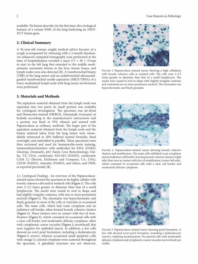
Figure 4 Papanicolaou-stained smear showing a necrotic back-ground containing naked nuclei karyorrhectic debris apoptoticcells and macrophages phagocytizing karyorrhectic debris
Figure 5 Hemacolor-stained smear showing cells bearingbasophilic cytoplasm focally with several cytoplasmic fine vacuoles
Pleomorphism of the cells was not prominentMitotic figureswere often identified The necrotic background containednaked nuclei karyorrhectic debris apoptotic cells andmacrophages phagocytizing karyorrhectic debris (Figure 4)nuclear crushing was noted
On the other hand the Hemacolor-stained smear showedoccasional cells bearing delicate basophilic cytoplasm focallywith several cytoplasmic fine vacuoles (Figure 5) whereasthese cytoplasmic fine vacuoles were not identified in thePapanicolaou-stained smear material
32 Histological Immunohistological and FISH FindingsDetails of the histological and molecular features of thiscase have been previously reported [8] Briefly only a smallamount of biopsy was available for histological investigationand it revealed an undifferentiated neoplasm with necrosis(Figure 6) In this material squamous differentiation whichis a possible characteristic of variant NMC was not apparent
Immunohistochemical staining demonstrated focal posi-tivity for EMA p63 cytokeratin AE1AE3 cytokeratin CAM52 CD138 and vimentin In addition a nuclear stainingpattern for NUT was evident (Figure 7) Furthermore FISHanalyses revealed an NSD3-NUT rearrangement (Figure 8)whereas BRD34-NUT fusion genes were not identified
Figure 6 Representative histological image showing sheets ofundifferentiated malignant cells with focal necrosis
Figure 7 Immunohistochemistry showing a nuclear staining pat-tern for NUT
33 Treatment and Follow-Up Thepatient received chemora-diation therapy for 10 months after her diagnosis Howeverthe patient died with disease progression at 10 months afterthe diagnosis Autopsy was not permitted
4 Discussion
The histological features of NMCs range from entirelyundifferentiated carcinomas to carcinomas with prominentsquamous differentiation [2 7 12ndash15] Thus a diagnosis ofNMC based solely on morphology can be difficult Previousstudies have described the cytological features of 4 commonNMCs harboring a BRD34-NUT fusion gene [9 10] andanother NMC case in which the gene rearrangement was notanalyzed [11] the cytological characteristics of these NMCsshowed a highly cellular predominantly noncohesive patternof relatively small cells with a round nucleus scant cytoplasmirregular nuclear contours variably prominent nucleoli andidentifiablemitotic figuresThese findings were also observedin the present variant NMC case Thus the previouslyreported findings imply that the cytological characteristics ofNMC are nonspecific and similar to those of undifferentiatedcarcinoma
Keratinization has not been identified in previous studiesexamining the cytology of NMCs [9ndash11] In theory overt ker-atinization is very important and sometimes pathognomonicfor a scrutinized diagnosis of NMC that is it is assumed
4 Case Reports in Pathology
Figure 8 A dual-color FISH analysis showing the fusion gene asa single yellow (overlapping) signal (arrows) including a green(NSD3) and an orange (NUT) signal
to be a relatively characteristic finding of NMC harboringa NUT gene rearrangement involving a gene other thanBRD4 [16] Keratinization in surgical material however isnotorious for evading observation because its distributionis often very focal [6 16] and sampling biases often occurThe cytology of the present NMC variant showed overt pearlformation including a dyskeratocyte and the stratificationin contact with loosely cohesive clusters we believe that thisfinding corresponds to abrupt keratinization which has beenreported as a peculiar characteristic of variant NMC [16] butwas not recognized in our corresponding histology specimen[8] Thus the combination of cytological and histologicalfindings may help to reveal keratinization concurrent withstratification in undifferentiated carcinoma which is one cluefor a diagnosis of NMC especially variant NMC rather thanother tumors
The Hemacolor-stained smear showed occasional cellspossessing delicate basophilic cytoplasm with several finevacuoles which were not observed in our Papanicolaou-stained smear In a previous NMC case harboring a BRD3-NUT fusion gene the cells were described as possessingdelicate to finely vacuolated cytoplasm although we couldnot observe the cytoplasm in detail because of the lowmagnification of the published figures [10] On the otherhand these cytoplasmic fine vacuoles were not observed inNMC cases harboring a BRD4-NUT fusion gene [9] Furtherstudies are needed to clarify whether these cytoplasmic finevacuoles are specific for NMC harboring a fusion geneinvolving NUT and a gene other than BRD4
In conclusion although the distinction of NMC fromother poorly differentiated carcinomas based solely onmorphology is difficult cytological investigation is help-ful especially for identifying abrupt keratinization whichhistopathological investigations canmiss because of samplingbiases Our experience has shown that the identificationof the following clues may suggest a diagnosis of NMCovert pearl formation including a dyskeratocyte stratifi-cation and cytoplasmic fine vacuoles especially in caseswhere the initial suspected diagnosis was ldquoundifferentiatedor poorly differentiated carcinomawith little pleomorphismrdquoFurthermore the identification of this entity is critical
and immunohistochemistry or FISH studies should be con-sidered for the identification of NUT gene rearrangements
Conflict of Interests
The authors have no conflicts of interest to declare
Acknowledgments
The authors appreciate the technical assistance of all theclinical and technical staffmembers of the PathologyDivisionof Iwata City Hospital This contribution was supported inpart by Grants-in-Aid for the US-Japan CooperativeMedicalScience Program the National Cancer Center Research andDevelopment Fund a grant for priority areas from theJapanese Ministry of Education Culture Sports Scienceand Technology (221S0001) and Grants-in-Aid for CancerResearch from the Japanese Ministry of Health Labourand Welfare (23120201 and 10103838) the Smoking ResearchFoundation and the Princess Takamatsu Cancer ResearchFund
References
[1] H Haack L A Johnson C J Fry et al ldquoDiagnosis of NUTmidline carcinomausing aNUT-specificmonoclonal antibodyrdquoThe American Journal of Surgical Pathology vol 33 no 7 pp984ndash991 2009
[2] C A French ldquoNUT midline carcinomardquo Cancer Genetics andCytogenetics vol 203 no 1 pp 16ndash20 2010
[3] D E Bauer CMMitchell KM Strait et al ldquoClinicopathologicfeatures and long-term outcomes of NUT midline carcinomardquoClinical Cancer Research vol 18 no 20 pp 5773ndash5779 2012
[4] E B Stelow ldquoA review of NUT midline carcinomardquo Head andNeck Pathology vol 5 no 1 pp 31ndash35 2011
[5] C A French I Miyoshi I Kubonishi H E Grier A R Perez-Atayde and J A Fletcher ldquoBRD4-NUT fusion oncogene anovel mechanism in aggressive carcinomardquo Cancer Researchvol 63 no 2 pp 304ndash307 2003
[6] C A French ldquoPathogenesis of NUT midline carcinomardquoAnnual Review of Pathology Mechanisms of Disease vol 7 pp247ndash265 2012
[7] S Suzuki N Kurabe H Minato et al ldquoA rare Japanese casewith a NUTmidline carcinoma in the nasal cavity a case reportwith immunohistochemical and genetic analysesrdquo PathologyResearch and Practice vol 210 no 6 pp 383ndash388 2014
[8] S Suzuki N Kurabe I Ohnishi et al ldquoNSD3-NUT-expressingmidline carcinoma of the lung first characterization of primarycancer tissuerdquo PathologymdashResearch and Practice In press
[9] A M Bellizzi C Bruzzi C A French and E B StelowldquoThe cytologic features of NUT midline carcinomardquo CancerCytopathology vol 117 no 6 pp 508ndash515 2009
[10] B Zhu W Laskin Y Chen et al ldquoNUT midline carcinoma aneoplasm with diagnostic challenges in cytologyrdquo Cytopathol-ogy vol 22 no 6 pp 414ndash417 2011
[11] H S Park Y S Bae S O Yoon et al ldquoUsefulness of nuclearprotein in testis (NUT) immunohistochemistry in the cytodi-agnosis of NUT midline carcinoma a brief case reportrdquo TheKorean Journal of Pathology vol 48 no 4 pp 335ndash338 2014
Case Reports in Pathology 5
[12] A G Evans C A French M J Cameron et al ldquoPathologiccharacteristics of NUT midline carcinoma arising in the medi-astinumrdquo American Journal of Surgical Pathology vol 36 no 8pp 1222ndash1227 2012
[13] J A Bishop andWHWestra ldquoNUTmidline carcinomas of thesinonasal tractrdquo American Journal of Surgical Pathology vol 36no 8 pp 1216ndash1221 2012
[14] B N Davis R G Karabakhtsian A L Pettigrew S M ArnoldC A French and Y M Brill ldquoNuclear protein in testis midlinecarcinomas a lethal and underrecognized entityrdquo Archives ofPathology and Laboratory Medicine vol 135 no 11 pp 1494ndash1498 2011
[15] E B StelowAM Bellizzi K Taneja et al ldquoNUT rearrangementin undifferentiated carcinomas of the upper aerodigestive tractrdquoThe American Journal of Surgical Pathology vol 32 no 6 pp828ndash834 2008
[16] CA French J L KutokWC Faquin et al ldquoMidline carcinomaof children and young adults withNUT rearrangementrdquo Journalof Clinical Oncology vol 22 no 20 pp 4135ndash4139 2004
Submit your manuscripts athttpwwwhindawicom
Stem CellsInternational
Hindawi Publishing Corporationhttpwwwhindawicom Volume 2014
Hindawi Publishing Corporationhttpwwwhindawicom Volume 2014
MEDIATORSINFLAMMATION
of
Hindawi Publishing Corporationhttpwwwhindawicom Volume 2014
Behavioural Neurology
EndocrinologyInternational Journal of
Hindawi Publishing Corporationhttpwwwhindawicom Volume 2014
Hindawi Publishing Corporationhttpwwwhindawicom Volume 2014
Disease Markers
Hindawi Publishing Corporationhttpwwwhindawicom Volume 2014
BioMed Research International
OncologyJournal of
Hindawi Publishing Corporationhttpwwwhindawicom Volume 2014
Hindawi Publishing Corporationhttpwwwhindawicom Volume 2014
Oxidative Medicine and Cellular Longevity
Hindawi Publishing Corporationhttpwwwhindawicom Volume 2014
PPAR Research
The Scientific World JournalHindawi Publishing Corporation httpwwwhindawicom Volume 2014
Immunology ResearchHindawi Publishing Corporationhttpwwwhindawicom Volume 2014
Journal of
ObesityJournal of
Hindawi Publishing Corporationhttpwwwhindawicom Volume 2014
Hindawi Publishing Corporationhttpwwwhindawicom Volume 2014
Computational and Mathematical Methods in Medicine
OphthalmologyJournal of
Hindawi Publishing Corporationhttpwwwhindawicom Volume 2014
Diabetes ResearchJournal of
Hindawi Publishing Corporationhttpwwwhindawicom Volume 2014
Hindawi Publishing Corporationhttpwwwhindawicom Volume 2014
Research and TreatmentAIDS
Hindawi Publishing Corporationhttpwwwhindawicom Volume 2014
Gastroenterology Research and Practice
Hindawi Publishing Corporationhttpwwwhindawicom Volume 2014
Parkinsonrsquos Disease
Evidence-Based Complementary and Alternative Medicine
Volume 2014Hindawi Publishing Corporationhttpwwwhindawicom
2 Case Reports in Pathology
availableWeherein describe for the first time the cytologicalfeatures of a variant NMC of the lung harboring an NSD3-NUT fusion gene
2 Clinical Summary
A 36-year-old woman sought medical advice because of acough accompanied by wheezing with a 2-month durationAn enhanced computed tomography scan performed at thetime of hospitalization revealed a mass (75 times 38 times 35mmin size) in the left lung that extended to the middle medi-astinum metastatic lesions in the liver breast bones andlymph nodes were also detected [8] A transbronchial biopsy(TBB) of the lung tumor and an endobronchial ultrasound-guided transbronchial needle aspiration (EBUS-TBNA) of alower mediastinal lymph node with lung tumor involvementwere performed
3 Materials and Methods
The aspiration material obtained from the lymph node wasseparated into two parts its small portion was availablefor cytological investigation The specimen was air-driedand Hemacolor-stained (MERCK Darmstadt Germany) atbedside according to the manufacturerrsquos instructions anda portion was fixed in 95 ethanol and stained withPapanicolaou as ordinary methods The larger part of theaspiration material obtained from the lymph node and thebiopsy material taken from the lung tumor were imme-diately immersed in 20 buffered neutral formalin fixedovernight and embedded in paraffin These specimens werethen sectioned and used for hematoxylin-eosin stainingimmunohistochemistry with antibodies for EMA (DAKOGlostrup Denmark) p63 (Santa Cruz Biotechnology Dal-las TX USA) cytokeratin AE1AE3 (DAKO) cytokeratinCAM 52 (Becton Dickinson and Company CA USA)CD138 (DAKO) vimentin (DAKO) and others and FISHas reported previously [8]
31 Cytological Findings An overview of the Papanicolaou-stained smear showed the specimen to be highly cellular withloosely cohesive cells andor isolated cells (Figure 1)The cellswere 2ndash25 times greater in diameter than that of a smalllymphocyte The nuclei were round to oval in shape andhad slightly irregular contours with one or more prominentnucleoli (Figure 1) The chromatin was hyperchromatic andfinely granular in most of the cells or vesicular in occasionalcells The main cells which had scant cytoplasm and anindistinct cell border often formed loosely cohesive clusters(Figure 2) These clusters were in contact with foci of strat-ification (Figure 2) which consisted of occasional cells witha clear cell border and moderately delicate cytoplasm oftenwith cytoplasmic coarse vacuoles (Figure 3 arrowhead) thatwere negative for epithelial mucin In addition a few cellsshowed an overt pearl formation including a dyskeratocyte(Figure 3 arrow) whereas occasional small apoptotic cellswith orange G-colored cytoplasm were scattered throughoutthe specimen A glandular structure was not observed
Figure 1 Papanicolaou-stained smear showing a high cellularitywith loosely cohesive cells or isolated cells The cells were 2ndash25times greater in diameter than that of a small lymphocyte Thenuclei were round to oval in shape with slightly irregular contoursand contained one or more prominent nucleoli The chromatin washyperchromatic and finely granular
Figure 2 Papanicolaou-stained smear showing loosely cohesiveclusters and stratification The main cells exhibited scant cytoplasmand an indistinct cell border forming loosely cohesive clusters (rightside) that were in contact with foci of stratification (center-left side)which consisted of occasional cells with a clear cell border andmoderately delicate cytoplasm
Figure 3 Papanicolaou-stained smear showing pearl formation Afew cells showed overt pearl formation including a dyskeratocyte(arrow) implying keratinization Occasional cells with moderatelydelicate cytoplasmand cytoplasmic coarse vacuoles (arrowhead) arevisible
Case Reports in Pathology 3
Figure 4 Papanicolaou-stained smear showing a necrotic back-ground containing naked nuclei karyorrhectic debris apoptoticcells and macrophages phagocytizing karyorrhectic debris
Figure 5 Hemacolor-stained smear showing cells bearingbasophilic cytoplasm focally with several cytoplasmic fine vacuoles
Pleomorphism of the cells was not prominentMitotic figureswere often identified The necrotic background containednaked nuclei karyorrhectic debris apoptotic cells andmacrophages phagocytizing karyorrhectic debris (Figure 4)nuclear crushing was noted
On the other hand the Hemacolor-stained smear showedoccasional cells bearing delicate basophilic cytoplasm focallywith several cytoplasmic fine vacuoles (Figure 5) whereasthese cytoplasmic fine vacuoles were not identified in thePapanicolaou-stained smear material
32 Histological Immunohistological and FISH FindingsDetails of the histological and molecular features of thiscase have been previously reported [8] Briefly only a smallamount of biopsy was available for histological investigationand it revealed an undifferentiated neoplasm with necrosis(Figure 6) In this material squamous differentiation whichis a possible characteristic of variant NMC was not apparent
Immunohistochemical staining demonstrated focal posi-tivity for EMA p63 cytokeratin AE1AE3 cytokeratin CAM52 CD138 and vimentin In addition a nuclear stainingpattern for NUT was evident (Figure 7) Furthermore FISHanalyses revealed an NSD3-NUT rearrangement (Figure 8)whereas BRD34-NUT fusion genes were not identified
Figure 6 Representative histological image showing sheets ofundifferentiated malignant cells with focal necrosis
Figure 7 Immunohistochemistry showing a nuclear staining pat-tern for NUT
33 Treatment and Follow-Up Thepatient received chemora-diation therapy for 10 months after her diagnosis Howeverthe patient died with disease progression at 10 months afterthe diagnosis Autopsy was not permitted
4 Discussion
The histological features of NMCs range from entirelyundifferentiated carcinomas to carcinomas with prominentsquamous differentiation [2 7 12ndash15] Thus a diagnosis ofNMC based solely on morphology can be difficult Previousstudies have described the cytological features of 4 commonNMCs harboring a BRD34-NUT fusion gene [9 10] andanother NMC case in which the gene rearrangement was notanalyzed [11] the cytological characteristics of these NMCsshowed a highly cellular predominantly noncohesive patternof relatively small cells with a round nucleus scant cytoplasmirregular nuclear contours variably prominent nucleoli andidentifiablemitotic figuresThese findings were also observedin the present variant NMC case Thus the previouslyreported findings imply that the cytological characteristics ofNMC are nonspecific and similar to those of undifferentiatedcarcinoma
Keratinization has not been identified in previous studiesexamining the cytology of NMCs [9ndash11] In theory overt ker-atinization is very important and sometimes pathognomonicfor a scrutinized diagnosis of NMC that is it is assumed
4 Case Reports in Pathology
Figure 8 A dual-color FISH analysis showing the fusion gene asa single yellow (overlapping) signal (arrows) including a green(NSD3) and an orange (NUT) signal
to be a relatively characteristic finding of NMC harboringa NUT gene rearrangement involving a gene other thanBRD4 [16] Keratinization in surgical material however isnotorious for evading observation because its distributionis often very focal [6 16] and sampling biases often occurThe cytology of the present NMC variant showed overt pearlformation including a dyskeratocyte and the stratificationin contact with loosely cohesive clusters we believe that thisfinding corresponds to abrupt keratinization which has beenreported as a peculiar characteristic of variant NMC [16] butwas not recognized in our corresponding histology specimen[8] Thus the combination of cytological and histologicalfindings may help to reveal keratinization concurrent withstratification in undifferentiated carcinoma which is one cluefor a diagnosis of NMC especially variant NMC rather thanother tumors
The Hemacolor-stained smear showed occasional cellspossessing delicate basophilic cytoplasm with several finevacuoles which were not observed in our Papanicolaou-stained smear In a previous NMC case harboring a BRD3-NUT fusion gene the cells were described as possessingdelicate to finely vacuolated cytoplasm although we couldnot observe the cytoplasm in detail because of the lowmagnification of the published figures [10] On the otherhand these cytoplasmic fine vacuoles were not observed inNMC cases harboring a BRD4-NUT fusion gene [9] Furtherstudies are needed to clarify whether these cytoplasmic finevacuoles are specific for NMC harboring a fusion geneinvolving NUT and a gene other than BRD4
In conclusion although the distinction of NMC fromother poorly differentiated carcinomas based solely onmorphology is difficult cytological investigation is help-ful especially for identifying abrupt keratinization whichhistopathological investigations canmiss because of samplingbiases Our experience has shown that the identificationof the following clues may suggest a diagnosis of NMCovert pearl formation including a dyskeratocyte stratifi-cation and cytoplasmic fine vacuoles especially in caseswhere the initial suspected diagnosis was ldquoundifferentiatedor poorly differentiated carcinomawith little pleomorphismrdquoFurthermore the identification of this entity is critical
and immunohistochemistry or FISH studies should be con-sidered for the identification of NUT gene rearrangements
Conflict of Interests
The authors have no conflicts of interest to declare
Acknowledgments
The authors appreciate the technical assistance of all theclinical and technical staffmembers of the PathologyDivisionof Iwata City Hospital This contribution was supported inpart by Grants-in-Aid for the US-Japan CooperativeMedicalScience Program the National Cancer Center Research andDevelopment Fund a grant for priority areas from theJapanese Ministry of Education Culture Sports Scienceand Technology (221S0001) and Grants-in-Aid for CancerResearch from the Japanese Ministry of Health Labourand Welfare (23120201 and 10103838) the Smoking ResearchFoundation and the Princess Takamatsu Cancer ResearchFund
References
[1] H Haack L A Johnson C J Fry et al ldquoDiagnosis of NUTmidline carcinomausing aNUT-specificmonoclonal antibodyrdquoThe American Journal of Surgical Pathology vol 33 no 7 pp984ndash991 2009
[2] C A French ldquoNUT midline carcinomardquo Cancer Genetics andCytogenetics vol 203 no 1 pp 16ndash20 2010
[3] D E Bauer CMMitchell KM Strait et al ldquoClinicopathologicfeatures and long-term outcomes of NUT midline carcinomardquoClinical Cancer Research vol 18 no 20 pp 5773ndash5779 2012
[4] E B Stelow ldquoA review of NUT midline carcinomardquo Head andNeck Pathology vol 5 no 1 pp 31ndash35 2011
[5] C A French I Miyoshi I Kubonishi H E Grier A R Perez-Atayde and J A Fletcher ldquoBRD4-NUT fusion oncogene anovel mechanism in aggressive carcinomardquo Cancer Researchvol 63 no 2 pp 304ndash307 2003
[6] C A French ldquoPathogenesis of NUT midline carcinomardquoAnnual Review of Pathology Mechanisms of Disease vol 7 pp247ndash265 2012
[7] S Suzuki N Kurabe H Minato et al ldquoA rare Japanese casewith a NUTmidline carcinoma in the nasal cavity a case reportwith immunohistochemical and genetic analysesrdquo PathologyResearch and Practice vol 210 no 6 pp 383ndash388 2014
[8] S Suzuki N Kurabe I Ohnishi et al ldquoNSD3-NUT-expressingmidline carcinoma of the lung first characterization of primarycancer tissuerdquo PathologymdashResearch and Practice In press
[9] A M Bellizzi C Bruzzi C A French and E B StelowldquoThe cytologic features of NUT midline carcinomardquo CancerCytopathology vol 117 no 6 pp 508ndash515 2009
[10] B Zhu W Laskin Y Chen et al ldquoNUT midline carcinoma aneoplasm with diagnostic challenges in cytologyrdquo Cytopathol-ogy vol 22 no 6 pp 414ndash417 2011
[11] H S Park Y S Bae S O Yoon et al ldquoUsefulness of nuclearprotein in testis (NUT) immunohistochemistry in the cytodi-agnosis of NUT midline carcinoma a brief case reportrdquo TheKorean Journal of Pathology vol 48 no 4 pp 335ndash338 2014
Case Reports in Pathology 5
[12] A G Evans C A French M J Cameron et al ldquoPathologiccharacteristics of NUT midline carcinoma arising in the medi-astinumrdquo American Journal of Surgical Pathology vol 36 no 8pp 1222ndash1227 2012
[13] J A Bishop andWHWestra ldquoNUTmidline carcinomas of thesinonasal tractrdquo American Journal of Surgical Pathology vol 36no 8 pp 1216ndash1221 2012
[14] B N Davis R G Karabakhtsian A L Pettigrew S M ArnoldC A French and Y M Brill ldquoNuclear protein in testis midlinecarcinomas a lethal and underrecognized entityrdquo Archives ofPathology and Laboratory Medicine vol 135 no 11 pp 1494ndash1498 2011
[15] E B StelowAM Bellizzi K Taneja et al ldquoNUT rearrangementin undifferentiated carcinomas of the upper aerodigestive tractrdquoThe American Journal of Surgical Pathology vol 32 no 6 pp828ndash834 2008
[16] CA French J L KutokWC Faquin et al ldquoMidline carcinomaof children and young adults withNUT rearrangementrdquo Journalof Clinical Oncology vol 22 no 20 pp 4135ndash4139 2004
Submit your manuscripts athttpwwwhindawicom
Stem CellsInternational
Hindawi Publishing Corporationhttpwwwhindawicom Volume 2014
Hindawi Publishing Corporationhttpwwwhindawicom Volume 2014
MEDIATORSINFLAMMATION
of
Hindawi Publishing Corporationhttpwwwhindawicom Volume 2014
Behavioural Neurology
EndocrinologyInternational Journal of
Hindawi Publishing Corporationhttpwwwhindawicom Volume 2014
Hindawi Publishing Corporationhttpwwwhindawicom Volume 2014
Disease Markers
Hindawi Publishing Corporationhttpwwwhindawicom Volume 2014
BioMed Research International
OncologyJournal of
Hindawi Publishing Corporationhttpwwwhindawicom Volume 2014
Hindawi Publishing Corporationhttpwwwhindawicom Volume 2014
Oxidative Medicine and Cellular Longevity
Hindawi Publishing Corporationhttpwwwhindawicom Volume 2014
PPAR Research
The Scientific World JournalHindawi Publishing Corporation httpwwwhindawicom Volume 2014
Immunology ResearchHindawi Publishing Corporationhttpwwwhindawicom Volume 2014
Journal of
ObesityJournal of
Hindawi Publishing Corporationhttpwwwhindawicom Volume 2014
Hindawi Publishing Corporationhttpwwwhindawicom Volume 2014
Computational and Mathematical Methods in Medicine
OphthalmologyJournal of
Hindawi Publishing Corporationhttpwwwhindawicom Volume 2014
Diabetes ResearchJournal of
Hindawi Publishing Corporationhttpwwwhindawicom Volume 2014
Hindawi Publishing Corporationhttpwwwhindawicom Volume 2014
Research and TreatmentAIDS
Hindawi Publishing Corporationhttpwwwhindawicom Volume 2014
Gastroenterology Research and Practice
Hindawi Publishing Corporationhttpwwwhindawicom Volume 2014
Parkinsonrsquos Disease
Evidence-Based Complementary and Alternative Medicine
Volume 2014Hindawi Publishing Corporationhttpwwwhindawicom