Cholangiocarcinoma: Introduction - Johns Hopkins Medicine, based
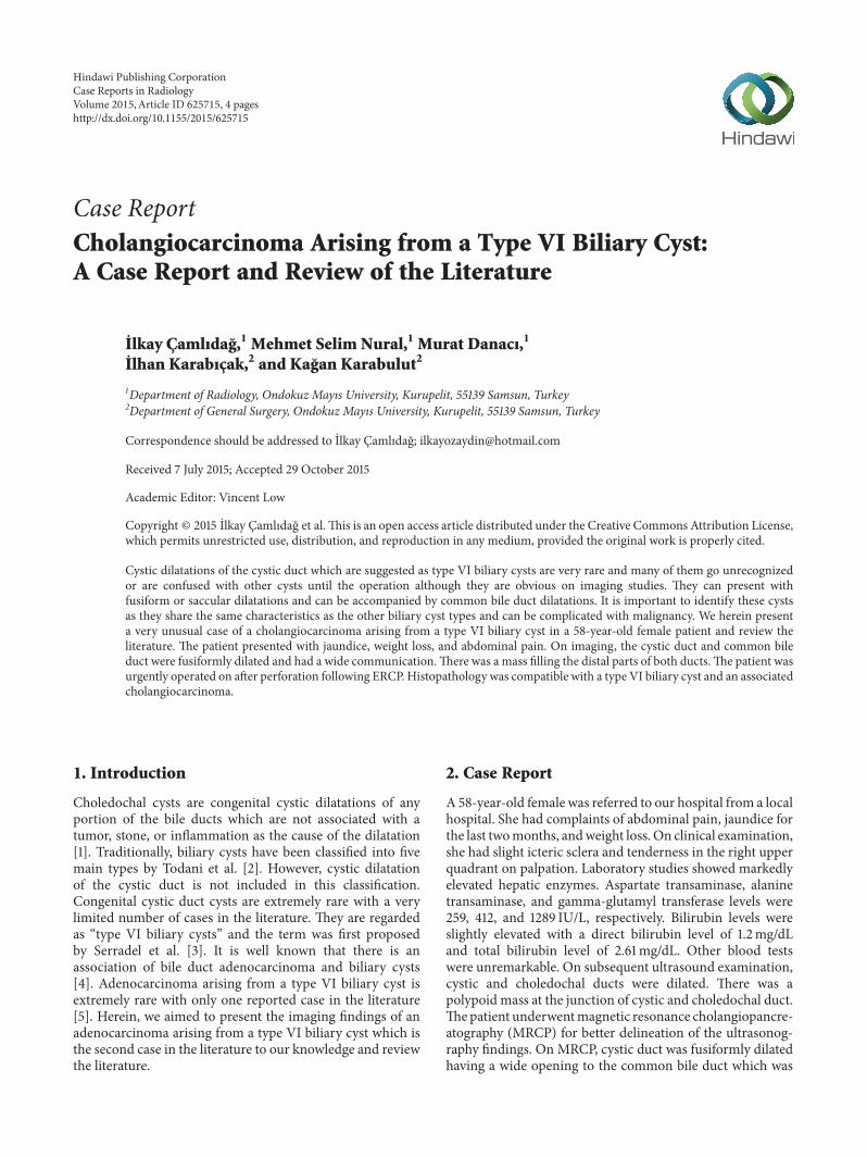
Case ReportCholangiocarcinoma Arising from a Type VI Biliary Cyst:A Case Report and Review of the Literature
Elkay ÇamlJdaL,1 Mehmet Selim Nural,1 Murat DanacJ,1
Elhan KarabJçak,2 and KaLan Karabulut2
1Department of Radiology, Ondokuz Mayıs University, Kurupelit, 55139 Samsun, Turkey2Department of General Surgery, Ondokuz Mayıs University, Kurupelit, 55139 Samsun, Turkey
Correspondence should be addressed to Ilkay Camlıdag; [email protected]
Received 7 July 2015; Accepted 29 October 2015
Academic Editor: Vincent Low
Copyright © 2015 Ilkay Camlıdag et al.This is an open access article distributed under the Creative Commons Attribution License,which permits unrestricted use, distribution, and reproduction in any medium, provided the original work is properly cited.
Cystic dilatations of the cystic duct which are suggested as type VI biliary cysts are very rare and many of them go unrecognizedor are confused with other cysts until the operation although they are obvious on imaging studies. They can present withfusiform or saccular dilatations and can be accompanied by common bile duct dilatations. It is important to identify these cystsas they share the same characteristics as the other biliary cyst types and can be complicated with malignancy. We herein presenta very unusual case of a cholangiocarcinoma arising from a type VI biliary cyst in a 58-year-old female patient and review theliterature. The patient presented with jaundice, weight loss, and abdominal pain. On imaging, the cystic duct and common bileduct were fusiformly dilated and had a wide communication.There was a mass filling the distal parts of both ducts.The patient wasurgently operated on after perforation following ERCP. Histopathology was compatible with a type VI biliary cyst and an associatedcholangiocarcinoma.
1. Introduction
Choledochal cysts are congenital cystic dilatations of anyportion of the bile ducts which are not associated with atumor, stone, or inflammation as the cause of the dilatation[1]. Traditionally, biliary cysts have been classified into fivemain types by Todani et al. [2]. However, cystic dilatationof the cystic duct is not included in this classification.Congenital cystic duct cysts are extremely rare with a verylimited number of cases in the literature. They are regardedas “type VI biliary cysts” and the term was first proposedby Serradel et al. [3]. It is well known that there is anassociation of bile duct adenocarcinoma and biliary cysts[4]. Adenocarcinoma arising from a type VI biliary cyst isextremely rare with only one reported case in the literature[5]. Herein, we aimed to present the imaging findings of anadenocarcinoma arising from a type VI biliary cyst which isthe second case in the literature to our knowledge and reviewthe literature.
2. Case Report
A 58-year-old female was referred to our hospital from a localhospital. She had complaints of abdominal pain, jaundice forthe last twomonths, andweight loss. On clinical examination,she had slight icteric sclera and tenderness in the right upperquadrant on palpation. Laboratory studies showed markedlyelevated hepatic enzymes. Aspartate transaminase, alaninetransaminase, and gamma-glutamyl transferase levels were259, 412, and 1289 IU/L, respectively. Bilirubin levels wereslightly elevated with a direct bilirubin level of 1.2mg/dLand total bilirubin level of 2.61mg/dL. Other blood testswere unremarkable. On subsequent ultrasound examination,cystic and choledochal ducts were dilated. There was apolypoid mass at the junction of cystic and choledochal duct.Thepatient underwentmagnetic resonance cholangiopancre-atography (MRCP) for better delineation of the ultrasonog-raphy findings. On MRCP, cystic duct was fusiformly dilatedhaving a wide opening to the common bile duct which was
Hindawi Publishing CorporationCase Reports in RadiologyVolume 2015, Article ID 625715, 4 pageshttp://dx.doi.org/10.1155/2015/625715

2 Case Reports in Radiology
GB
CBD
CD
(a)
GB CBDCD
(b)
Figure 1: A 58-year-old female with type VI biliary cyst and associated cholangiocarcinoma. (a, b) Axial and coronal MRCP images showfusiformly dilated cystic duct (CD) with a wide opening to the common bile duct (CBD) which is also fusiformly dilated. Intrahepatic bileducts are slightly dilated in the centre.There is a hypointense, heterogeneous mass filling distal parts of both the cystic and choledochal ducts(GB: gallbladder).
(a)
CBD
CD
(b)
Figure 2: CT examination performed with the suspicion of perforation following ERCP. (a) Axial contrast enhanced CT image showsintraperitoneal free air (short arrow) and periduodenal contrast extravasation (long arrow) suggesting perforation. (b) Multiplanarreconstructed CT image shows fusiform cystic dilatation of the cystic duct having a wide opening to the extrahepatic bile duct which isalso fusiformly dilated and the enhancing mass.
also fusiformly dilated. There was a hypointense, heteroge-neous mass filling the distal parts of both ducts. Intrahepaticbile ducts were minimally dilated in the central part but werein normal caliber in the remainder (Figure 1). An endoscopicretrograde cholangiopancreatography (ERCP) procedurewasperformed for mass sampling. ERCP confirmed the findingsof MRCP. After the procedure, the patient complained ofsevere abdominal pain and underwent computed tomog-raphy (CT) which showed free intra-abdominal air andperiduodenal contrast leakage (Figure 2(a)) consistent withperforation along with the other findings (Figure 2(b)).Therewas no sign of lymphadenopathy or distant metastasis on CT.The patient urgently underwent Whipple procedure. It wasnot until then that all the findings of fusiform dilatation ofthe cystic and choledochal ducts were interpreted as a cysticduct cyst which was proposed as a type VI biliary cyst. Onhistopathological examination, there was a polypoid masswhich was characterized by atypical epithelial cells lining afibrovascular core extending from the cyst wall that was linedby columnar epithelium (Figure 3). The histopathological
Figure 3: Atypical epithelial cells lining a fibrovascular core extend-ing from the cyst wall that is lined by columnar epithelium are seenon the histopathological specimen.
diagnosis of the polypoid mass was consistent with cholan-giocarcinoma. To our knowledge, this is the second case ofcholangiocarcinoma arising from a type VI biliary cyst.

Case Reports in Radiology 3
Figure 4: Schematic illustration of the type VI biliary cysts reported in the literature to date.
3. Discussion
Biliary cysts are cystic dilatations of the bile ducts and arerarely seen with an incidence of 1 : 100000 to 150000 in west-ern countries and an increased incidence of 1 : 1000 in Asianpopulation. It comprises 1% of all benign biliary diseases [4].Traditionally, biliary cysts are classified into five categoriesas described by Todani et al. Type I is cyst of the CBD; typeII is a cystic diverticulum of the extrahepatic bile duct; typeIII is a cyst in the intraduodenal portion of the commonbile duct; type IV refers to multiple cysts in the intra- andextrahepatic biliary tract; and type V is single or multiplecysts in the intrahepatic ducts alone which is known as Carolidisease [2]. Although not included in this classification ofbiliary cysts, cystic dilatation of the cystic duct can also beencountered; however, it is extremely rare with very limitednumber of reports in the literaturemainly consisting of singlecase reports and only one review to our knowledge [5].Serradel et al. were the first to propose calling these cysts astype VI biliary cysts [3]. Due to very little acquaintance withthe condition, diagnosis is challenging with most of the casesbeing undiagnosed as our case ormisdiagnosed as other types[3, 6, 7]. The correct diagnosis was made intraoperatively inmost of the cases in the literature [8].
Type VI biliary cyst can be encountered in differentforms. The cystic dilatation can be limited to the cysticduct either fusiform or saccular without any associatedfindings in the other bile ducts [3, 5, 6, 9, 10] or can beaccompanied by fusiform common bile duct dilatation withvarying size of communication between the cyst and dilatedCBD (see [5, 10, 11], Figure 4).
The most common theory for the development of biliarycysts is the anomalous pancreaticobiliary junction (APBJ).In the APBJ the biliary and pancreatic duct join proximal to
the sphincter of Oddi forming a long common channel thatcauses the pancreatic enzymes to flow upstream into the bileduct resulting in the weakening of the CBD wall and leadsto the formation of a cyst. However, not all biliary cysts areaccompanied by APBJ and APBJ is also encountered in theabsence of biliary cysts. So, more than one mechanism mustbe responsible for the occurrence of biliary cysts [1].
Presenting features and complications of cysts of thecystic duct are similar to other types of biliary cysts.They might be asymptomatic and incidentally detected orpresent with varying degrees of abdominal pain, jaundice,and complications such as cholangitis, calculus disease, andmalignancy with the last one being the most serious andfeared one [1, 4]. Most of the reported cases of biliary cystassociated cancers are cholangiocarcinomas arising from thecyst itself. However, they also can arise from the gallbladderor anywhere else in the biliary ducts especially in the presenceof APBJ [1, 12, 13]. To our knowledge, there is only onereported case of cholangiocarcinoma in the literature arisingfrom a type VI biliary cyst reported by Maheshwari et al.[5]. In their case, cholangiocarcinoma arose from a sacculartype cystic duct cyst. The information of the presence ofAPBJ was not given. In our case, the mass originated fromthe fusiformly dilated cystic and common bile ducts andthere was no evidence of APBJ. Two more cases of typeVI biliary cyst related cholangiocarcinoma arising from thegallbladder without any APBJ [10] and left hepatic bileduct in the presence of APBJ have also been reported[12].
Radiological imaging is very useful in the diagnosis ofbiliary cysts. Ultrasonography (USG) is the modality ofchoice for initial evaluation and frequently provides enoughinformation to make the diagnosis of a biliary cyst. In case ofa dilated nonvascular cystic structure near the porta hepatis,

4 Case Reports in Radiology
attempt should be made to delineate its connection and rela-tionship with the biliary tract and the gallbladder. However,USG may fail to depict these relationships as it is operator-dependent and sensitivity is decreased in the presence ofoverlying bowel gas and inflammatory conditions. ERCP isthe gold standard in the diagnosis of biliary cysts but it isinvasive, always requires sedation, and comes with inherentrisks such as pancreatitis, perforation, hemorrhage, contrastallergy, cholangitis, and biliary sepsis. Given its relativelymoderate risk profile and lower cost, MRCP should bethe diagnostic test of choice when preoperatively evaluat-ing biliary cysts and their associated anomalies. MRCP isequivalent to ERCP in determining biliary cyst type and ishelpful in diagnosing related pancreaticobiliary anomalies,cholangiocarcinoma, and choledocholithiasis. ERCP shouldbe used when MRCP inadequately visualizes the terminalcommon bile duct or the pancreaticobiliary junction or whena therapeutic procedure is needed. Amultidetector computedtomography (MDCT) with reformatted imaging is anotherimportant technique which has the ability to demonstratethe anatomic details of the biliary tree and the ABPJ. It canshow the presence of an associated cancer and is useful forstaging. The disadvantages are high-dose radiation exposureand contrast agent administration especially in children [1, 4,5, 9, 13].
Preferred treatment for typeVI biliary cysts is the surgicalexcision as for the other types. Early and proper diagnosisis important to avoid complications and choose the appro-priate surgical method. Cholecystectomy and transection ofthe cystic duct are sufficient in the presence of a narrowcommon duct junction. However, in the presence of a wideopening to the common bile duct, hepaticoduodenostomy orhepaticojejunostomy should be performed [7, 11].
In conclusion, we had a very typical but unrecognizedcase of type VI biliary cyst with obvious findings on allimaging modalities. Because of the unfamiliarity with thecondition, the cysts were interpreted as dilatations secondaryto mass obstruction. However, disproportionate and onlymild dilatation of the central intrahepatic bile ducts shouldhave warned us that it was not result of obstruction asthey would be much more dilated given the degree of theextrahepatic bile duct dilatation. We think that increasedfamiliarity with cystic duct cysts is crucial as early treat-ment can prevent complications like inflammation and moreimportantly malignancy. We think that cystic duct cystsshould officially be included in the Todani et al. classificationas type VI biliary cyst with all its subtypes because it has allthe characteristics of the other biliary cysts in terms of clinicalfindings and complications.
Conflict of Interests
The authors declare that there is no conflict of interestsregarding the publication of this paper.
References
[1] K. L. Hae, J. P. Seong, H. Y. Bum, A. L. Lee, H. M. Jong, and W.C. Yun, “Imaging features of adult choledochal cysts: a pictorial
review,” Korean Journal of Radiology, vol. 10, no. 1, pp. 71–80,2009.
[2] T. Todani, Y. Watanabe, M. Narusue, K. Tabuchi, and K.Okajima, “Congenital bile duct cysts. Classification, operativeprocedures, and review of thirty-seven cases including cancerarising from choledochal cyst,”TheAmerican Journal of Surgery,vol. 134, no. 2, pp. 263–269, 1977.
[3] A. F. S. Serradel, E. S. Linares, and R. H. Goepfert, “Cysticdilatation of the cystic duct: a new type of biliary cyst,” Surgery,vol. 109, no. 3, pp. 320–322, 1991.
[4] B. Jabłonska, “Biliary cysts: etiology, diagnosis and manage-ment,” World Journal of Gastroenterology, vol. 18, no. 35, pp.4801–4810, 2012.
[5] P.Maheshwari, “Cysticmalformation of cystic duct: 10 cases andreview of literature,”World Journal of Radiology, vol. 4, no. 9, pp.413–417, 2012.
[6] W. E. Bode and J. B. Aust, “Isolated cystic dilatation of the cysticduct,”The American Journal of Surgery, vol. 145, no. 6, pp. 828–829, 1983.
[7] E. S. Chan, E. D. Auyang, and E. S. Hungness, “Laparoscopicmanagement of a cystic duct cyst,” Journal of the Society ofLaparoendoscopic Surgeons, vol. 13, no. 3, pp. 436–440, 2009.
[8] U. De, S. Das, and S. Sarkar, “Type VI choledochal cystrevisited,” Singapore Medical Journal, vol. 52, no. 5, pp. e91–e93,2011.
[9] C. Goya, M. S. Arslan, A. Yavuz et al., “A rare anomaly of biliarysystem: MRCP evidence of a cystic duct cyst,” Case Reports inRadiology, vol. 2014, Article ID 291071, 4 pages, 2014.
[10] J.-H. Yoon, “Magnetic resonance cholangiopancreatographydiagnosis of choledochal cyst involving the cystic duct: reportof three cases,” British Journal of Radiology, vol. 84, no. 997, pp.e18–e22, 2011.
[11] T. K. Loke, S. H. Lam, and C. S. Chan, “Choledochal cyst: anunusual type of cystic dilatation of the cystic duct,” AmericanJournal of Roentgenology, vol. 173, pp. 619–620, 1999.
[12] R. Noun, R. Sayegh, C. Tohme-Noun, K. Honein, T. Smayra,and N. Aoun, “Extracystic biliary carcinoma associated withanomalous pancreaticobiliary junction and cysts,” Journal ofHepato-Biliary-Pancreatic Surgery, vol. 13, no. 6, pp. 577–579,2006.
[13] R. Sugita, “Magnetic resonance evaluations of biliary malig-nancy and condition at high-risk for biliary malignancy: cur-rent status,”World Journal of Hepatology, vol. 5, no. 12, pp. 654–665, 2013.

Submit your manuscripts athttp://www.hindawi.com
Stem CellsInternational
Hindawi Publishing Corporationhttp://www.hindawi.com Volume 2014
Hindawi Publishing Corporationhttp://www.hindawi.com Volume 2014
MEDIATORSINFLAMMATION
of
Hindawi Publishing Corporationhttp://www.hindawi.com Volume 2014
Behavioural Neurology
EndocrinologyInternational Journal of
Hindawi Publishing Corporationhttp://www.hindawi.com Volume 2014
Hindawi Publishing Corporationhttp://www.hindawi.com Volume 2014
Disease Markers
Hindawi Publishing Corporationhttp://www.hindawi.com Volume 2014
BioMed Research International
OncologyJournal of
Hindawi Publishing Corporationhttp://www.hindawi.com Volume 2014
Hindawi Publishing Corporationhttp://www.hindawi.com Volume 2014
Oxidative Medicine and Cellular Longevity
Hindawi Publishing Corporationhttp://www.hindawi.com Volume 2014
PPAR Research
The Scientific World JournalHindawi Publishing Corporation http://www.hindawi.com Volume 2014
Immunology ResearchHindawi Publishing Corporationhttp://www.hindawi.com Volume 2014
Journal of
ObesityJournal of
Hindawi Publishing Corporationhttp://www.hindawi.com Volume 2014
Hindawi Publishing Corporationhttp://www.hindawi.com Volume 2014
Computational and Mathematical Methods in Medicine
OphthalmologyJournal of
Hindawi Publishing Corporationhttp://www.hindawi.com Volume 2014
Diabetes ResearchJournal of
Hindawi Publishing Corporationhttp://www.hindawi.com Volume 2014
Hindawi Publishing Corporationhttp://www.hindawi.com Volume 2014
Research and TreatmentAIDS
Hindawi Publishing Corporationhttp://www.hindawi.com Volume 2014
Gastroenterology Research and Practice
Hindawi Publishing Corporationhttp://www.hindawi.com Volume 2014
Parkinson’s Disease
Evidence-Based Complementary and Alternative Medicine
Volume 2014Hindawi Publishing Corporationhttp://www.hindawi.com