Case Report Appendix and Uterus Metastasis of Squamous...
4
Hindawi Publishing Corporation Case Reports in Obstetrics and Gynecology Volume 2013, Article ID 474891, 3 pages http://dx.doi.org/10.1155/2013/474891 Case Report Appendix and Uterus Metastasis of Squamous Cell Carcinoma Arising from Mature Cystic Teratoma of the Ovary GülGah BalJk, 1 IGJk Üstüner, 1 Recep Bedir, 2 Ülkü Mete Ural, 1 Mehmet KaLJtçJ, 1 and Emine Seda GüvendaL Güven 1 1 Department of Obstetrics and Gynecology, Recep Tayyip Erdo˘ gan University School of Medicine, ˙ Islampas ¸a Mah. Tıp Fak¨ ultesi Dekanlı, Merkez, 53100 Rize, Turkey 2 Department of Pathology, Recep Tayyip Erdo˘ gan University School of Medicine, 53100 Rize, Turkey Correspondence should be addressed to G¨ uls ¸ah Balık; gulsahbalı[email protected] Received 10 April 2013; Accepted 2 June 2013 Academic Editors: S. Z. A. Badawy, B. Piura, and S. Rasmussen Copyright © 2013 G¨ uls ¸ah Balık et al. is is an open access article distributed under the Creative Commons Attribution License, which permits unrestricted use, distribution, and reproduction in any medium, provided the original work is properly cited. Mature cystic teratoma of the ovary rarely undergoes malignant transformation. ere is no consensus for a treatment modality because of the rarity of the disease. Herein we present a case of squamous cell carcinoma (SCC) arising in a mature cystic teratoma (MCT) in a 66-year-old patient. e patient underwent total hysterectomy and bilateral salpingo-oophorectomy, omentectomy, appendectomy, and bilateral pelvic + paraaortic lymph node dissection. e histopathological examination revealed malignant invasion of the appendix and uterus. e patient, who refused the continuation of treatment initiated with the administration of a single dose of cisplatin, died 5 months later because of the disease. It is imperative that gynecologists consider appendectomy in SCC arising from MCT cases. 1. Introduction Mature cystic teratomas (MCTs) of the ovary, which are a type of germ cell tumors, account for 20%–25% of all ovarian neoplasms. ey are the most common benign germ cell tumors of the ovary in younger women (<45 years). ey may be composed of mature or immature tissues deriving from the 3 germ cell layers [1]. Most patients with MCTs are asymptomatic but mass compression effect can lead to pain and abdominal disten- sion. e complications of MCT are rupture, torsion, and malignant transformation [1]. Malignant transformation is very rare in MCT’s compo- nents (2%). Squamous cell carcinoma (SCC) arising from the ectoderm is the most common type (approximately 80%), which is followed by adenocarcinoma and carcinoids [2, 3]. Herein we present a case of SCC arising in an MCT with appendix metastasis and uterus infiltration in a 66-year- old woman and review the published literature to determine possible risk/prognostic factors and treatments. 2. Case Presentation A 66-year-old multipara patient was referred to our clinic with abdominopelvic mass and abdominal pain. Pelvic exam- ination revealed a large immobile pelvic mass with regular contours extending up to the umbilicus. Ultrasound and computed tomography imaging showed a 20 × 18 cm cystic mass with solid components and calcifications in the leſt adnexal region extending up to umbilical area with severe ascites. e uterus and right ovarium were normal. e patient’s tumor marker profile was as follows: CA-125: 33.1 U/mL (normal <35 U/mL), CA 19-9: 10.54 U/mL (normal <37 U/mL), and -HcG was negative. e Pap smear being negative for malignancy, and endometrial biopsy revealed atrophy. e patient underwent exploratory laparotomy for ovarian malignancy. 1500 mL serous ascites was noted and used for cytology. e pelvic mass at this time was identified as arising from the leſt ovary. e posterior surface densely adhered to the pelvic sidewall. e tumor also had massive
Transcript of Case Report Appendix and Uterus Metastasis of Squamous...

Hindawi Publishing CorporationCase Reports in Obstetrics and GynecologyVolume 2013, Article ID 474891, 3 pageshttp://dx.doi.org/10.1155/2013/474891
Case ReportAppendix and Uterus Metastasis of Squamous Cell CarcinomaArising from Mature Cystic Teratoma of the Ovary
GülGah BalJk,1 IGJk Üstüner,1 Recep Bedir,2 Ülkü Mete Ural,1
Mehmet KaLJtçJ,1 and Emine Seda GüvendaL Güven1
1 Department of Obstetrics and Gynecology, Recep Tayyip Erdogan University School of Medicine,Islampasa Mah. Tıp Fakultesi Dekanlı, Merkez, 53100 Rize, Turkey
2Department of Pathology, Recep Tayyip Erdogan University School of Medicine, 53100 Rize, Turkey
Correspondence should be addressed to Gulsah Balık; gulsahbalı[email protected]
Received 10 April 2013; Accepted 2 June 2013
Academic Editors: S. Z. A. Badawy, B. Piura, and S. Rasmussen
Copyright © 2013 Gulsah Balık et al. This is an open access article distributed under the Creative Commons Attribution License,which permits unrestricted use, distribution, and reproduction in any medium, provided the original work is properly cited.
Mature cystic teratoma of the ovary rarely undergoes malignant transformation. There is no consensus for a treatment modalitybecause of the rarity of the disease. Herein we present a case of squamous cell carcinoma (SCC) arising in a mature cystic teratoma(MCT) in a 66-year-old patient. The patient underwent total hysterectomy and bilateral salpingo-oophorectomy, omentectomy,appendectomy, and bilateral pelvic + paraaortic lymph node dissection. The histopathological examination revealed malignantinvasion of the appendix and uterus. The patient, who refused the continuation of treatment initiated with the administration ofa single dose of cisplatin, died 5 months later because of the disease. It is imperative that gynecologists consider appendectomy inSCC arising fromMCT cases.
1. Introduction
Mature cystic teratomas (MCTs) of the ovary, which are atype of germ cell tumors, account for 20%–25% of all ovarianneoplasms. They are the most common benign germ celltumors of the ovary in younger women (<45 years).Theymaybe composed ofmature or immature tissues deriving from the3 germ cell layers [1].
Most patients with MCTs are asymptomatic but masscompression effect can lead to pain and abdominal disten-sion. The complications of MCT are rupture, torsion, andmalignant transformation [1].
Malignant transformation is very rare in MCT’s compo-nents (2%). Squamous cell carcinoma (SCC) arising from theectoderm is the most common type (approximately 80%),which is followed by adenocarcinoma and carcinoids [2, 3].
Herein we present a case of SCC arising in an MCTwith appendix metastasis and uterus infiltration in a 66-year-old woman and review the published literature to determinepossible risk/prognostic factors and treatments.
2. Case Presentation
A 66-year-old multipara patient was referred to our clinicwith abdominopelvicmass and abdominal pain. Pelvic exam-ination revealed a large immobile pelvic mass with regularcontours extending up to the umbilicus. Ultrasound andcomputed tomography imaging showed a 20 × 18 cm cysticmass with solid components and calcifications in the leftadnexal region extending up to umbilical area with severeascites. The uterus and right ovarium were normal. Thepatient’s tumor marker profile was as follows: CA-125:33.1 U/mL (normal<35U/mL), CA 19-9: 10.54U/mL (normal<37U/mL), and 𝛽-HcG was negative. The Pap smear beingnegative for malignancy, and endometrial biopsy revealedatrophy. The patient underwent exploratory laparotomy forovarian malignancy. 1500mL serous ascites was noted andused for cytology. The pelvic mass at this time was identifiedas arising from the left ovary. The posterior surface denselyadhered to the pelvic sidewall. The tumor also had massive
2 Case Reports in Obstetrics and Gynecology
Figure 1: The cyst wall was thickened with a firm mass.
Figure 2: Squamous cell carcinoma, moderately differentiated(H&E ×200).
adhesions to the bladder from its anterior. Total abdomi-nal hysterectomy and bilateral salpingo-oophorectomy wereperformed. Analysis of a frozen section revealed SCC aris-ing within an MCT. Based on this finding, omentectomy,appendectomy, and bilateral pelvic + paraaortic lymph nodedissection were performed. There was no evidence of grossresidual tumor.
Gross examination of the resulting specimen revealed asolid-cystic mass measured 20 × 15 × 4 cm with a nonintactcapsule (Figure 1). The cyst was filled with amorphous debrisand the sectioned surface of the cystic mass contained hair,sebaceous material, and focal calcification. The thickness ofthe cystic wall ranged from 1.5 to 2.4 cm. The tumor invadedthe uterine corpus.
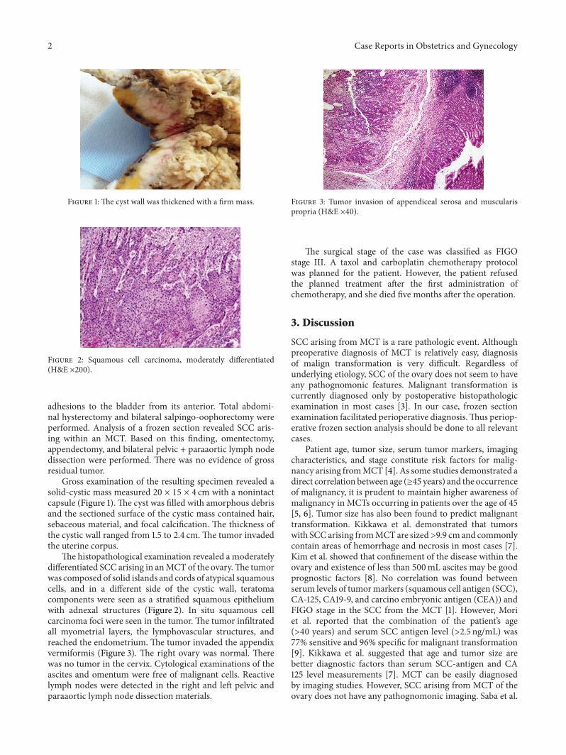
The histopathological examination revealed a moderatelydifferentiated SCC arising in anMCT of the ovary.The tumorwas composed of solid islands and cords of atypical squamouscells, and in a different side of the cystic wall, teratomacomponents were seen as a stratified squamous epitheliumwith adnexal structures (Figure 2). In situ squamous cellcarcinoma foci were seen in the tumor. The tumor infiltratedall myometrial layers, the lymphovascular structures, andreached the endometrium. The tumor invaded the appendixvermiformis (Figure 3). The right ovary was normal. Therewas no tumor in the cervix. Cytological examinations of theascites and omentum were free of malignant cells. Reactivelymph nodes were detected in the right and left pelvic andparaaortic lymph node dissection materials.
Figure 3: Tumor invasion of appendiceal serosa and muscularispropria (H&E ×40).
The surgical stage of the case was classified as FIGOstage III. A taxol and carboplatin chemotherapy protocolwas planned for the patient. However, the patient refusedthe planned treatment after the first administration ofchemotherapy, and she died five months after the operation.
3. Discussion
SCC arising from MCT is a rare pathologic event. Althoughpreoperative diagnosis of MCT is relatively easy, diagnosisof malign transformation is very difficult. Regardless ofunderlying etiology, SCC of the ovary does not seem to haveany pathognomonic features. Malignant transformation iscurrently diagnosed only by postoperative histopathologicexamination in most cases [3]. In our case, frozen sectionexamination facilitated perioperative diagnosis.Thus periop-erative frozen section analysis should be done to all relevantcases.
Patient age, tumor size, serum tumor markers, imagingcharacteristics, and stage constitute risk factors for malig-nancy arising fromMCT [4]. As some studies demonstrated adirect correlation between age (≥45 years) and the occurrenceof malignancy, it is prudent to maintain higher awareness ofmalignancy in MCTs occurring in patients over the age of 45[5, 6]. Tumor size has also been found to predict malignanttransformation. Kikkawa et al. demonstrated that tumorswith SCC arising fromMCTare sized>9.9 cm and commonlycontain areas of hemorrhage and necrosis in most cases [7].Kim et al. showed that confinement of the disease within theovary and existence of less than 500mL ascites may be goodprognostic factors [8]. No correlation was found betweenserum levels of tumormarkers (squamous cell antigen (SCC),CA-125, CA19-9, and carcino embryonic antigen (CEA)) andFIGO stage in the SCC from the MCT [1]. However, Moriet al. reported that the combination of the patient’s age(>40 years) and serum SCC antigen level (>2.5 ng/mL) was77% sensitive and 96% specific for malignant transformation[9]. Kikkawa et al. suggested that age and tumor size arebetter diagnostic factors than serum SCC-antigen and CA125 level measurements [7]. MCT can be easily diagnosedby imaging studies. However, SCC arising from MCT of theovary does not have any pathognomonic imaging. Saba et al.

Case Reports in Obstetrics and Gynecology 3
showed that the gross appearance of MCTs with malignanttransformation was similar to benign MCTs but with a moresolid component [10]. Usually this solid component tendsto extend transmurally with direct invasion of the pouch ofDouglas, uterus, and vagina. Thus, advanced age (66 years),tumor size (20 × 15 × 4 cm), 1500mL of ascites, and solidcomponent of the cyst were suggestive of malignancy in ourcase.
Prognostic factors for SCC include capsular invasion/rupture, tumor dissemination, ascites, abdominalpelvic ad-hesions and tumor types [11]. The gross appearance ofnodular, papillary, or “cauliflower-like” growths invading intothe cyst cavities, nodules, or plaques within the cyst wallsmayalso be malignancy indicators [12]. Invasion of the uterus,metastasis into the appendix, ascites, adhesions to bladder,and posterior pelvic sidewall were the bad prognostic factorsin our case.
Surgical cytoreduction with proper staging, adjuvanttherapy with platinum-based or paclitaxel-based chemother-apy, and concurrent whole pelvic radiation have been rec-ommended as methods of treatment in advanced stage andrecurrent disease of SCC arising from MCT [13]. Fertilitysparing surgery may be preferred in young patients desiringto retain fertility [13, 14].
If squamous cell lesions of the ovary are detected in apatient, the differential diagnosis should include SCC arisingfrom MCT and metastatic carcinomas. The primary focus ofmetastasis of squamous cell carcinoma to the ovary is oftenfrom uterus and vagina.
Metastasis of the appendix accounts for 4%-5% in earlystage cases of epithelial ovarian tumors. Ayhan et al. reportedappendix metastasis in 100 of 285 patients with ovariancancers, and they recommended appendectomy in all ovariancarcinomas for best cytoreduction and surgical staging [14].Appendectomy is recommended in SCC arising from MCTas the other epithelial ovarian tumors.
The prognosis is poor in SCC arising from MCT. How-ever, early stage of the disease and performing an optimalcytoreductive surgery are good prognostic factors [15].
In conclusion, although malign transformation arisingfrom MCT is extremely rare, clinicians must be awareof this risk before surgery. Because surgical procedure ishighly different between benign teratomas and malign trans-formation arising from MCTs, physicians should considerappendectomy for SCC arising fromMCT cases.
Conflict of Interests
No conflict of interests was declared by the authors.
References
[1] A. Hackethal, D. Brueggmann, M. K. Bohlmann, F. E. Franke,H.-R. Tinneberg, and K. Munstedt, “Squamous-cell carcinomain mature cystic teratoma of the ovary: systematic review andanalysis of published data,” The Lancet Oncology, vol. 9, no. 12,pp. 1173–1180, 2008.
[2] A. Iwasa, Y. Oda, E. Kaneki et al., “Squamous cell carcinomaarising in mature cystic teratoma of the ovary: an immunohis-tochemical analysis of its tumorigenesis,”Histopathology, vol. 51,no. 1, pp. 98–104, 2007.
[3] V. Ulker, C. Numanoglu, O. Akbayir et al., “Malignant trans-formation arising from mature cystic teratoma of the ovary:a report of six cases,” Journal of Obstetrics and GynaecologyResearch, vol. 38, no. 5, pp. 849–853, 2012.
[4] L. Dos Santos, E. Mok, A. Iasonos et al., “Squamous cellcarcinoma arising in mature cystic teratoma of the ovary: a caseseries and review of the literature,” Gynecologic Oncology, vol.105, no. 2, pp. 321–324, 2007.
[5] F. Kikkawa, H. Ishikawa, K. Tamakoshi, A. Nawa, N. Suganuma,and Y. Tomoda, “Squamous cell carcinoma arising frommaturecystic teratoma of the ovary: a clinicopathologic analysis,”Obstetrics and Gynecology, vol. 89, no. 6, pp. 1017–1022, 1997.
[6] M. Kashimura, M. Shinohara, T. Hirakawa, T. Kamura, andK. Matsukuma, “Clinicopathologic study of squamous cellcarcinoma of the ovary,”Gynecologic Oncology, vol. 34, no. 1, pp.75–79, 1989.
[7] F. Kikkawa, A. Nawa, K. Tamakoshi et al., “Diagnosis ofsquamous cell carcinoma arising from mature cystic teratomaof the ovary,” Cancer, vol. 82, no. 11, pp. 2249–2255, 1998.
[8] H. S. Kim, J. W. Kim, H. H. Chung, N.-H. Park, Y.-S. Song,and S.-B. Kang, “Disease confined within the ovary and smalleramount of ascites are good prognostic factors for survival ofpatients with squamous cell carcinoma arising from maturecystic teratoma of the ovary: a case series in Korea and reviewof the published reports,” Journal of Obstetrics and GynaecologyResearch, vol. 35, no. 1, pp. 99–105, 2009.
[9] Y. Mori, H. Nishii, K. Takabe et al., “Preoperative diagnosis ofmalignant transformation arising from mature cystic teratomaof the ovary,” Gynecologic Oncology, vol. 90, no. 2, pp. 338–341,2003.
[10] L. Saba, S. Guerriero, R. Sulcis, B. Virgilio, G. Melis, and G.Mallarini, “Mature and immature ovarian teratomas: CT, USand MR imaging characteristics,” European Journal of Radiol-ogy, vol. 72, no. 3, pp. 454–463, 2009.
[11] G. W. H. Stamp and E. M. McConnell, “Malignancy arising incystic ovarian teratomas. A report of 24 cases,” British Journal ofObstetrics and Gynaecology, vol. 90, no. 7, pp. 671–675, 1983.
[12] H. M. Brammer III, J. L. Buck, W. S. Hayes, S. Sheth, and F.A. Tavassoli, “From the archives of the AFIP. Malignant germcell tumors of the ovary: radiologic-pathologic correlation,”Radiographics, vol. 10, no. 4, pp. 715–724, 1990.
[13] A.-J. Chiang, V. La, J. Peng, K.-J. Yu, and N. N. H. Teng,“Squamous cell carcinoma arising from mature cystic teratomaof the ovary,” International Journal of Gynecological Cancer, vol.21, no. 3, pp. 466–474, 2011.
[14] A. Ayhan, M. Gultekin, C. Taskiran et al., “Routine appendec-tomy in epithelial ovarian carcinoma: is it necessary?”Obstetricsand Gynecology, vol. 105, no. 4, pp. 719–724, 2005.
[15] F. A. Zakkouri, S. Ouaouch, S. Boutayeb et al., “Squamous cellcarcinoma in situ arising inmature cystic teratoma of the ovary:a case report,” Journal of Ovarian Research, vol. 4, no. 1, article5, 2011.

Submit your manuscripts athttp://www.hindawi.com
Stem CellsInternational
Hindawi Publishing Corporationhttp://www.hindawi.com Volume 2014
Hindawi Publishing Corporationhttp://www.hindawi.com Volume 2014
MEDIATORSINFLAMMATION
of
Hindawi Publishing Corporationhttp://www.hindawi.com Volume 2014
Behavioural Neurology
EndocrinologyInternational Journal of
Hindawi Publishing Corporationhttp://www.hindawi.com Volume 2014
Hindawi Publishing Corporationhttp://www.hindawi.com Volume 2014
Disease Markers
Hindawi Publishing Corporationhttp://www.hindawi.com Volume 2014
BioMed Research International
OncologyJournal of
Hindawi Publishing Corporationhttp://www.hindawi.com Volume 2014
Hindawi Publishing Corporationhttp://www.hindawi.com Volume 2014
Oxidative Medicine and Cellular Longevity
Hindawi Publishing Corporationhttp://www.hindawi.com Volume 2014
PPAR Research
The Scientific World JournalHindawi Publishing Corporation http://www.hindawi.com Volume 2014
Immunology ResearchHindawi Publishing Corporationhttp://www.hindawi.com Volume 2014
Journal of
ObesityJournal of
Hindawi Publishing Corporationhttp://www.hindawi.com Volume 2014
Hindawi Publishing Corporationhttp://www.hindawi.com Volume 2014
Computational and Mathematical Methods in Medicine
OphthalmologyJournal of
Hindawi Publishing Corporationhttp://www.hindawi.com Volume 2014
Diabetes ResearchJournal of
Hindawi Publishing Corporationhttp://www.hindawi.com Volume 2014
Hindawi Publishing Corporationhttp://www.hindawi.com Volume 2014
Research and TreatmentAIDS
Hindawi Publishing Corporationhttp://www.hindawi.com Volume 2014
Gastroenterology Research and Practice
Hindawi Publishing Corporationhttp://www.hindawi.com Volume 2014
Parkinson’s Disease
Evidence-Based Complementary and Alternative Medicine
Volume 2014Hindawi Publishing Corporationhttp://www.hindawi.com