
Cardiovascular_Anatomy___Physiology.pdf
22
Presented by: RN.com 12400 High Bluff Drive San Diego, CA 92130 This course has been approved for two (2.0) contact hours. This course expires on September 24, 2006. Copyright © 2004 by RN.com. All Rights Reserved. Reproduction and distribution of these materials are prohibited without the express written authorization of RN.com. First Published: September 24, 2004 RN.com’s Assessment Series: Cardiovascular Anatomy & Physiology
-
Upload
mahesh-chendake -
Category
Documents
-
view
3 -
download
0
Transcript of Cardiovascular_Anatomy___Physiology.pdf
Presented by:
RN.com
12400 High Bluff Drive San Diego, CA 92130
This course has been approved for two (2.0) contact hours.
This course expires on September 24, 2006.
Copyright © 2004 by RN.com.
All Rights Reserved. Reproduction and distribution of these materials are prohibited without the
express written authorization of RN.com.
First Published: September 24, 2004
RN.com’s Assessment Series: Cardiovascular Anatomy &
Physiology

1
Acknowledgements________________________________________________________________________ 2 Purpose & Objectives _____________________________________________________________________ 3 Introduction _____________________________________________________________________________ 4 Cardiac Structures________________________________________________________________________ 5
Layers of the Heart _____________________________________________________________________ 5 Cardiac Chambers & Blood flow through the Heart __________________________________________ 5
The Conduction System____________________________________________________________________ 9 Depolarization and Repolarization_________________________________________________________ 9 Sinoatrial (SA) Node ___________________________________________________________________ 10 Sinoatrial (SA) Node ___________________________________________________________________ 11 Atrioventricular (AV) Node and AV Junction ______________________________________________ 11 Bundle of His _________________________________________________________________________ 11 Purkinje Fibers________________________________________________________________________ 12 Summary of Pacemaker Functions________________________________________________________ 12
Coronary Circulation_____________________________________________________________________ 13 Coronary Arteries _____________________________________________________________________ 13 Coronary Blood Flow___________________________________________________________________ 14
Systemic Circulation _____________________________________________________________________ 15 Arteries ______________________________________________________________________________ 15 Capillaries ____________________________________________________________________________ 15 Venous System ________________________________________________________________________ 15
Neurohormonal Control of the Heart and Blood Vessels________________________________________ 16 Sympathetic Nervous System of the Heart__________________________________________________ 16 Parasympathetic Nervous System of the Heart______________________________________________ 16 Receptor Control ______________________________________________________________________ 16 Vasomotor Center _____________________________________________________________________ 17 Hormonal Influences on the Heart and Blood Vessels ________________________________________ 17
Conclusion______________________________________________________________________________ 19 References ______________________________________________________________________________ 20 Post Test Viewing Instructions _____________________________________________________________ 21

2
ACKNOWLEDGEMENTS
RN.com acknowledges the valuable contributions of… … Lori Constantine MSN, RN, C-FNP, author of RN.com’s Assessment Series: Cardiovascular Anatomy and Physiology. Lori is a nurse of nine years with a broad range of clinical experience. She has worked as a staff nurse, charge nurse and nurse preceptor on many different medical surgical units including vascular, neurology, neurosurgery, urology, gynecology, ENT, general medicine, geriatrics, oncology and blood and marrow transplantation. She received her Bachelors in Nursing in 1994 and a Masters in Nursing in 1998, both from West Virginia University. Additionally, in 1998, she was certified as a Family Nurse Practitioner. She has worked in staff development as a Nurse Clinician and Education Specialist since 1999 at West Virginia University Hospitals, Morgantown, WV.

3
PURPOSE & OBJECTIVES
The focus of this cardiovascular anatomy and physiology course is to teach nurses about the structures and functions of the cardiovascular system. The anatomical structures of the cardiovascular system work together to achieve two major goals: the transport of oxygenated blood and nutrients to the cells of the body, and transport of carbon dioxide and wastes from the cells to organs that will eliminate the waste. Understanding the fundamental structures and functions of the cardiovascular system will allow you to provide care for all patients you encounter and intervene effectively for those with alterations in cardiovascular status. After successful completion of this course, you will be able to:
1. Identify the functions of various anatomical structures within the cardiovascular system.
2. Discuss the functions of the cardiovascular system.
3. Discuss the physiology of how the cardiovascular system works.

4
INTRODUCTION
Cardiovascular anatomy and physiology is an example of both a mechanical and an electrical organ system. Although the heart is essentially a “pump”, the complex anatomy and physiology that make it able to successfully keep a person alive is truly amazing. Add to that the hormonal influences on the cardiovascular system and you have a truly complicated system of structures and events that need to operate correctly and efficiently to maintain homeostasis.

5
CARDIAC STRUCTURES
Layers of the Heart The human heart is protected by two layers that envelope it. The outer layer is called the pericardium. It covers the heart. It folds in on itself at the aorta forming the epicardium of the heart. Between these layers is a small amount of fluid (10-50 mL) that provides the layers with a non-stick surface (American Association of Critical Care Nurses, 1998). While the epicardium forms the outer layer of the heart, the myocardium forms the middle layer and the endocardium the innermost layer. The coronary arteries travel across the epicardium. The muscular myocardium is the thickest layer and the workhorse of the heart.
The endocardium has a smooth inner surface to allow blood to flow easily through the heart’s chambers. Within the endocardium of the atrium are pockets known as trabiculae. These pockets are sometimes the sites of pathologic clot formation known as mural (wall) thrombi.
The heart’s valves are covered by the endocardium. The endocardium also has an endocrine function, releasing hormones such as endocardin - a substance that prolongs myocardial contraction.
Cardiac Chambers & Blood flow through the Heart The human heart is a four-chambered pump made up of two receiving chambers called atria and two pumping chambers called ventricles.
Right Atrium (RA) The Right Atrium receives oxygen-depleted blood returning from the body through the superior and inferior vena cava. It is a thin walled, low-pressure system. Normal pressures in the Right Atrium are typically 0–8 mmHg. It is home to the Sinoatrial, or SA Node, the pacemaker of the heart (American Association of Critical Care Nurses, 1998).
Aorta
R Atrium
Pulmonary Arteries
L Atrium
L Ventricle
R Ventricle
Practice Pearl When infection is within the pericardial sac the inner surfaces of these layers begin to stick. This is known as pericarditis. This
causes friction and pain. A friction rub may result.
An accumulation of relatively small
amounts of fluid in this pericardial sac is known as a pericardial effusion. If the fluid
accumulates enough to affect the heart’s ability to contract, it is known as cardiac
tamponade. Practice Pearl
Mural thrombi typically form when
blood is allowed to pool in these pockets. This usually happens due to an inability of the heart to effectively pump blood from the atria, such as in
atrial fibrillation.

6
Right Ventricle (RV) The Right Ventricle is also a thin walled, low-pressure chamber. It receives blood from the Right Atrium when the atrioventricular valve dividing the Right Atrium and ventricle (the Tricuspid Valve) is open. When this valve is open, and the chamber is resting (filling with blood [diastole]) typical right ventricular pressures are equal to that in the Right Atrium, 0-8 mmHg. However, when the valve closes and contraction (systole) begins, pressures are 15-25 mmHg, enough to pump blood forward to the lungs via the right and left pulmonary arteries. The blood is then oxygenated in the lungs (American Association of Critical Care Nurses, 1998).
Left Atrium (LA) Another thin walled, low-pressure chamber is the Left atrium. It receives oxygen-rich blood from the pulmonary circuit, via the right and left pulmonary veins. Normal resting pressures (diastolic pressures) in the Left atrium are 4-12 mmHg, less than that of the lungs. Because pressure is less in this chamber during diastole, blood is more easily returned from the higher-pressure pulmonary circuit (American Association of Critical Care Nurses, 1998).
Left Ventricle (LV) The Left Ventricle is a thick walled chamber that receives blood from the Left atrium, and is approximately three times thicker than the Right Ventricle. When the atrioventricular valve dividing the Left atrium and ventricle (the Mitral Valve) is open and the chamber is resting, or filling with blood (diastole) typical left ventricular pressures are equal to that in the Left atrium, 4-12 mmHg. However, when the valve closes and contraction (systole) begins, pressures must be generated to overcome the body’s systemic vascular resistance (SVR). These pressures are typically 110-130 mmHg (American Association of Critical Care Nurses, 1998). When the ventricle generates enough pressure to overcome the SVR, blood moves out the semilunar valve known as the Aortic Valve into the aorta. There it is transported throughout the body via a network of arteries, capillaries, and veins. Eventually the blood will return to the Right Atrium where the oxygenation process starts all over again.
Cardiac Output (CO) About two-thirds of the atrial blood flows passively from the atria into the ventricles. When atrial contraction occurs, the atrial blood is pushed down into the ventricles. This atrial contribution is called atrial kick and accounts for approximately thirty percent of the cardiac output (American Association of Critical Care Nurses, 1998). Cardiac output is the amount of blood ejected by the Left Ventricle every minute. Cardiac output equals the stroke volume times the heart rate. The heart rate is the number of times that the heart beats per minute. Heart rate increases or decreases based upon the metabolic and oxygen demands of the body. The stroke volume is the amount of blood pumped by the heart per cardiac cycle. It is measured in ml/beat. A decreased stroke volume may indicate impaired cardiac contractility or valve dysfunction and may result in heart failure. It may also indicate decreased circulating volume. Increased stroke volume may be caused by an increase in circulating volume or an increase in inotropy, the contractile force of the ventricle.
Practice Pearl
The right and left atria and ventricular chambers are separated by a septal wall
or septum

7
When the heart rate or the stroke volume (amount of blood ejected with each contraction) increases, cardiac output increases. When the heart rate or the stroke volume decreases, cardiac output decreases. Cardiac output varies according to body mass, but is typically between 4-8 liters per minute.
Cardiac index is cardiac output normalized for body surface area. There are several methods for measuring cardiac output. Typical cardiac indices are between 2.5-4.0 liters of blood per minute per meter2 (American Association of Critical Care Nurses, 1998).
Cardiac Valves When blood flows through the heart, it follows a unidirectional pattern. There are four different valves within the myocardium and their functions are to assure blood flows from the right to left side of the heart and always in a “forward” direction. The two valves found between the atria and ventricles are appropriately called atrioventricular (A-V) valves. The Tricuspid Valve separates the Right Atrium from the Right Ventricle. The Tricuspid Valve is named so because of its three (tri) leaflets (cusps). Similarly, the Mitral Valve separates the Left atrium from the Left Ventricle. The Mitral Valve is a two-leaflet valve, named after a bishop’s miter. The two remaining valves are called semilunar valves (because they look like half moons). The valve located where the pulmonary artery meets the Right Ventricle is called the Pulmonic Valve. The Aortic Valve is located at the juncture of the Left Ventricle and aorta. Both semilunar valves prevent backflow of blood into the ventricles.
Valve Type Valve Name Location Atrioventricular (AV) Tricuspid
Mitral
Separates Right Atrium and Right Ventricle Separates Left atrium and Left Ventricle
Semilunar Pulmonic
Aortic
Between Right Ventricle and pulmonary artery Between Left Ventricle and aorta
(Sherwood, 1997).
Tricuspid Valve
Pulmonic Valve
Mitral Valve
Aortic Valve
8
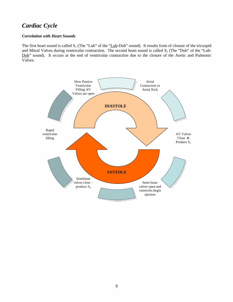
Cardiac Cycle Correlation with Heart Sounds The first heart sound is called S1 (The “Lub” of the “Lub-Dub” sound). It results from of closure of the tricuspid and Mitral Valves during ventricular contraction. The second heart sound is called S2 (The “Dub” of the “Lub-Dub” sound). It occurs at the end of ventricular contraction due to the closure of the Aortic and Pulmonic Valves.
Atrial Contraction or
Atrial Kick
AV Valves Close &
Produce S1
Slow Passive Ventricular Filling AV
Valves are open
Rapid ventricular
filling
Semilunar valves close –
produce S2
Semi-lunar
valves open and ventricles begin
ejection
DIASTOLE
SYSTOLE

9
THE CONDUCTION SYSTEM
Depolarization and Repolarization In a cardiac cell, two primary chemicals provide the electrical charges: sodium (Na+) and potassium (K+). In the resting cell, the potassium is mostly on the inside, while the sodium is mostly on the outside. This results in a negatively charged cell at rest (the interior of the cardiac cell is mostly negative or polarized at rest). When depolarized, the interior cell becomes positively charged and the cardiac cell will contract. In summary, the polarized or resting cell will carry a negative charge on the inside. When depolarized, the opposite will occur. This is due to the movement of sodium and potassium across the cell membrane. Depolarization moves an electrical wave through the myocardium. As the wave of depolarization stimulates the heart’s cells, they become positive and begin to contract. This cell-to-cell conduction of depolarization through the myocardium is carried by the fast moving sodium ions. Repolarization is the return of electrical charges to their original state. This process must happen before the cells can be ready conduct again. Note the depolarization and repolarization phases as they are represented on the ECG.
K+
Na+
++
+
++
+
K
Na
K
Na
Polarization(cell at rest)
Depolarization(cell will contract)
Repolarization(return to baseline)
V e n t r i c u l a rd e p o l a r i z a t i o n
V e n t r i c u l a rr e p o l a r i z a t i o nA t r i a l
d e p o l a r i z a t i o n

10
These electrical cells in the heart are arranged in a system of pathways called the conduction system. These specialized electrical cells and structures guide the wave of myocardial depolarization. Two distinct components must occur for the heart to be able to contract and pump blood. These components are an electrical impulse and a mechanical response to the impulse The heart also has two distinct types of cells. There are electrical (conductive) cells, which initiate electrical activity and conduct it through the heart. There are also mechanical (contracting) cells, which respond to the electrical stimulus and contract to pump blood.
The physical layout of the conduction system is shown in the picture below. It is important that you understand the sequence of events within this conduction system. Knowledge of the layout helps you to understand normal and abnormal rhythms. The conduction system consists of the Sinoatrial Node (SA Node), Atrioventricular Node (AV Node), Bundle of His (also called the AV Junction), Right and Left Bundle Branches, and Purkinje Fibers.
The electrical cells are responsible forconducting impulses through themyocardial tissue and electrical pathwaysof the heart. They are responsible for theheart rate and rhythm. An ECG tracing isdesigned to give a graphic display of theelectrical activity in the heart.
The contracting or myocardial “workingcells” contain contractile filaments. Whenthese cells are electrically stimulated,these filaments slide together and themyocardial cell contracts and the atria orventricular chambers contract. This ishow we get our pulse and blood pressure.
The electrical impulse tells the heart tobeat. This property is calledautomaticity. Automaticity means thatthese specialized cells within the heart candischarge an electrical current without anexternal pacemaker, or stimulus from thebrain via the spinal cord.
The mechanical beating or contractionof the heart occurs after the electricalstimulation. When the mechanicalcontraction occurs, the person will haveboth a heart rate and a blood pressure.
Electrical Impulse Mechanical Response

11
Sinoatrial (SA) Node The Sinoatrial Node (also called the SA Node or Sinus Node) is a group of specialized cells located in the posterior wall of the Right Atrium. The SA Node normally depolarizes or paces more rapidly than any other part of the conduction system. It sets off impulses that trigger atrial depolarization and contraction. Because the SA Node discharges impulses quicker than any other part of the heart, it is commonly known as the natural pacemaker of the heart. The SA Node normally fires at a rate of 60-100 beats per minute. After the SA Node fires, a wave of cardiac cells begin to depolarize. Depolarization occurs throughout both the right and left atria (similar to the ripple effect when a rock is thrown into a pond). This impulse travels through the atria by way of inter-nodal pathways down to the next structure, which is called the AV Node. Do you remember the term mentioned before called “atrial kick”? Atrial kick occurs when the atria contract and dump their blood into the ventricles. This atrial contraction contributes up to 30% of the cardiac output, which is obviously an important element toward maintaining our blood pressure. So remember... the SA Node is not only the primary pacemaker of the heart but also triggers atrial depolarization and the contribution of the atrial kick. The heart is truly an amazing organ. Not only does it have one dominant pacemaker (the SA Node) it also has two back-up pacemakers. A back-up pacer is located in the area near the Bundle of His. The final back-up pacer is located in the ventricles along the Purkinje fibers. More interesting information on this later.
Atrioventricular (AV) Node and AV Junction The next area of conductive tissue along the conduction pathway is at the site of the atrioventricular (AV) Node. This node is a cluster of specialized cells located in the lower portion of the Right Atrium, above the base of the Tricuspid Valve. The AV Node itself possesses no pacemaker cells. The AV Node has two functions. The first function is to DELAY the electrical impulse in order to allow the atria time to contract and complete filling of the ventricles. The second function is to receive an electrical impulse and conduct it down to the ventricles via the AV junction and Bundle of His.
Bundle of His After passing through the AV Node, the electrical impulse enters the Bundle of His (also referred to as the common bundle). The Bundle of His is located in the upper portion of the interventricular septum and connects the AV Node with the two bundle branches. If the SA Node should become diseased or fail to function properly, the Bundle of His has pacemaker cells, which are capable of discharging at an intrinsic rate of 40-60 beats per minute. This back-up pacemaker function can really come in handy! The AV Node and the bundle of His are referred to collectively as the AV junction. The Bundle of His conducts the electrical impulse down to the right and left bundle branches. The right bundle branch spreads the wave of depolarization to the Right Ventricle. Likewise, the left bundle branch spreads the wave of depolarization to both the interventricular septum and the Left Ventricle. The left bundle further divides into 3 branches or fascicles. The bundle branches further divide into Purkinje fibers.

12
Purkinje Fibers We are now coming to the end of this amazing cardiac conduction system. At the terminal ends of the bundle branches, smaller fibers distribute the electrical impulses to the muscle cells, which stimulate contraction. This web of fibers is called the Purkinje fibers. The Purkinje fibers penetrate about 1/4 to 1/3 of the way into the ventricular muscle mass and then become continuous with the cardiac muscle fibers. The electrical impulse spreads rapidly through the right and left bundle branches and Purkinje fibers to reach the ventricular muscle, causing ventricular contraction, or systole. These Purkinje fibers within the ventricles also have intrinsic pacemaker ability. This third and final pacemaker site of the myocardium can only pace at a rate of 20-40 beats per minute. You have probably noticed that the further you travel away from the SA Node, the slower the backup pacemakers become. As common sense tells you, if you only have a heart rate of 30 (from the ventricular back-up pacemaker), your blood pressure is likely to be low and you might be quite symptomatic.
Summary of Pacemaker Functions The heart is designed with a system of one dominant and two back-up pacing systems.
Summary of Pacemaker Function Pacemaker Hierarchy Location Pacing Rate
Level 1 (normal) SA Node 60-100 beats/minute
Level II (back-up system) Bundle of His/ AV Node/ Junction 40-60 beats/minute
Level III (lowest back-up system)
Purkinje Fibers within Ventricles (typically called the Ventricular Pacemaker)
20-40 beats/minute

13
CORONARY CIRCULATION
Coronary Arteries The coronary arteries receive their name for the crown they form over the heart. Two main arteries arise off the aorta at the Sinus of Valsava. These arteries are named the Right Coronary Artery (RCA) and Left Coronary Artery (LCA) When the Aortic Valve closes at the beginning of diastole, the Sinus of Valsalva distends and blood flows into the RCA and LCA.
RCA (Right Coronary Artery) The RCA arises from right side of aorta and follows a groove between Right Ventricle and Left Ventricle. It extends to back of heart, forming the posterior descending artery. The RCA is the main blood supply to Right Atrium and Right Ventricle, much of conduction system and the inferior and posterior Left Ventricle. The RCA supplies blood to the SA Node (in fifty-five percent of the population) and the AV Node (in ninety percent of the population). Occlusion of the Right Coronary Artery leads to inferior and posterior myocardial infarctions (MI). Bradycardia and arrhythmias are commonly seen in these MIs.
Occlusions of RCA Inferior or Posterior MI

14
LCA (Left Coronary Artery) The LCA arises off of the left side of the aorta. It quickly forms the left main that divides into the left anterior descending (LAD) artery and the circumflex artery.
LCA
Left Main
LAD Circumflex
LAD (Left Anterior Descending) Artery The LAD travels down the heart between the Right Ventricle and Left Ventricle to the apex and turns back up the heart. It is the main blood supply to Left Ventricle, septum, and anterior wall. Diagonal arteries arise off of the LAD.
Occlusions of LAD anterior MIs
Circumflex (CFX) Artery The circumflex artery curves around the left side of the heart between the Left Atrium and the Left Ventricle. It supplies blood to the posterior surface of the heart. It supplies blood to the SA Node (in forty-five percent of the population) and the AV Node (in ten percent of the population). Its marginal branches provides the left lateral ventricle and the posterior Left Ventricle with its blood supply.
Occlusions of Circumflex Lateral or Posterior MI
Coronary Blood Flow Two-thirds of coronary blood flow occurs during diastole, or when the heart is at rest. Five percent of cardiac output goes to the coronary arteries. Seventy percent of oxygen is extracted by the myocardial tissues of the heart, in comparison to the rest of the body at twenty-five percent. During times of extreme demand, the coronary arteries can dilate up to four times greater than normal to increase supply of oxygen to the myocardial tissues.
The de-oxygenated blood from the myocardium is collected in the coronary sinus. This large vein then returns the de-oxygenated blood to the Right Atrium.
Practice Pearl
Bradycardias increase diastolic filling time. This is why cardiac
patients can tolerate bradycardias better than
tachycardias.
Practice Pearl
Patients with Coronary Artery Disease have fixedlesions that cannot dilate to meet increased demand. This leads to angina and coronary dysfunction, which may eventually lead to
myocardial infarction.

15
Practice Pearl
In shock, pre-capillary sphincters dilate and post-
capillary sphincters contract in an attempt to supply cells with more needed nutrients due to
decreased blood supply.
Practice Pearl
Drugs that increase venous capacity (diuretics, morphine, and
nitroglycerin) will decrease preload, thus decreasing the amount of blood returning to the right side
of the heart. High fowlers also increases venous capacity &
decreases preload. The supine position decreases venous capacity
and increases preload, or the amount of blood returning to the
right side of the heart.
SYSTEMIC CIRCULATION
Arteries The arterial system carries about thirteen percent of the body’s blood volume at any given time. The heart pumps blood out through one major artery – the aorta. The aorta branches and these branches further divide into smaller arteries known as arterioles. Arterioles contain smooth muscle. They are innervated by the autonomic nervous system and can constrict and dilate to regulate blood supply to tissues. Arterioles are largely responsible for our systemic vascular resistance (SVR). Eventually, the arterioles divide enough that they become capillaries – where the exchange of oxygen, carbon dioxide, and nutrients occurs. Arteries are composed of three layers: the intima (the inner layer of epithelial cells), the media (the muscular middle layer), and the adventitia (the tough outer layer). The media layer helps the heart pump the blood. When the heart beats, the artery expands as it fills with blood. When the heart relaxes, the artery contracts exerting a force that it strong enough to push the blood forward. This rhythm between the heart and the artery results in successful circulation of the blood to the body.
Capillaries Blood spends only 0.5 seconds in capillaries. Our capillaries are only one cell thick. The exchange of oxygen, carbon dioxide, and nutrients takes place through this very thin wall. At this cellular level, the red blood cells inside the capillaries free their oxygen. This oxygen then passes through the wall and into the surrounding tissue. Simultaneously, the tissues free their waste products, like carbon dioxide. These wastes pass through the wall and into the red blood cells, where the red blood cells transport the wastes to the lungs, liver and other “cleansing” organs for their excretion. Additionally, capillaries have both pre and post capillary sphincters that have a high degree of intrinsic tone and are independent of neurohormonal controls. Capillaries auto-regulate to meet metabolic needs of surrounding tissues.
Venous System Blood leaving the capillaries returns to the heart through the venous system. The path begins with the venules and progresses to larger and larger veins which lead to the superior and inferior vena cavae which then enter the Right Atrium. Veins are highly distensible, thin walled vessels. They act as a volume reservoir for circulatory systems. At any given time, the veins carry about fifty percent of the blood volume of the body. Veins are very much like arteries, however they transport blood at a lower pressure than arteries. The veins transport blood back to the lungs and heart. Veins have valves that are located inside the veins that keep blood moving back to the heart. The vein valves also provide footholds for the blood as it travels against gravity towards the heart. For example, blood returning to the heart from the foot has to travel against gravity. The venous valves and muscle contractions of the leg prevents backflow of blood (American Association of Critical Care Nurses, 1998).
Practice Pearl
Coronary Artery Disease (CAD) is characterized by damage to the intima, or internal layer of arteries.

16
NEUROHORMONAL CONTROL OF THE HEART AND BLOOD VESSELS
The brain and central nervous system control the body through two pathways - the somatic & autonomic nervous systems. The somatic nervous system is typically under voluntary control. In contrast, the autonomic nervous system is not voluntary. Due to this involuntary system, we don’t have to think about every heart beat, the amount of blood delivered to specific tissues, the dilation of our pupils, and how much digestive motility our GI tract needs. In other words, the autonomic nervous system regulates the activities of the internal organs. The autonomic nervous system has two main parts, the sympathetic and the parasympathetic systems. These two “opposite” systems often operate in opposition to each other. Many internal organs are stimulated by both systems. When one stimulates an organ, the other tends to depress the organ. The sympathetic nervous system is responsible for the “fight-or-flight" response. This response prepares us for emergency situations. The parasympathetic nervous system, oppositely, tends to inhibit these reactions. The response of our body depends on the proportionate strength of stimulation supplied by each system at any given instance.
Sympathetic Nervous System of the Heart Activation of the sympathetic nervous system increases heart rate (positive chronotropy), increases contractility (positive inotropy), and increases conduction velocity (positive dromotropy). Additionally, in blood vessels, sympathetic activation constricts arteries and arterioles. This increases systemic vascular resistance (SVR, increases central blood flow and decreases distal blood flow. In other words blood is shunted away from the periphery to the heart, brain and skeletal muscles. Sympathetic stimulation also produces an effect on the body’s venous system. It decreases venous blood volume, and increases venous pressure (Tortora, 1987). The overall effect of sympathetic activation is to increase cardiac output, systemic vascular resistance (both arteries and veins), and arterial blood pressure. Enhanced sympathetic activity is particularly important during exercise, emotional stress, and during hemorrhagic shock.
Parasympathetic Nervous System of the Heart When the parasympathetic system is activated it works to decrease heart rate (negative chronotropy), decrease contractility (negative inotropy), and decrease conduction velocity (negative dromotropy) via the Vagus Nerve. Most blood vessels in the body do not have parasympathetic innervation. However, parasympathetic nerves do innervate salivary glands, gastrointestinal glands, and genital erectile tissue where they cause vasodilation (Tortora, 1987).
Receptor Control
Baroreceptors Baroreceptors are located in the aortic arch, the carotid bodies of the external carotid arteries, the pulmonary artery, and the atria. They respond to changes in blood pressure. The baroreceptors of the aortic arch have a high threshold pressure and are less sensitive than the carotid sinus receptors.

17
The baroreceptors of the carotid sinus typically respond to pressures ranging from 60-180 mmHg. These receptors are the dominant receptors. These receptors work by sensing the “mean” arterial blood pressure. This "set point" changes during hypertension, heart failure, and other chronic disease states. However, when there is an acute increase or decrease in mean arterial pressure, the baroreceptors alter their firing rate. Under normal physiological conditions, decreased pressure leads to decreased baroreceptor firing. This also inhibits sympathetic stimulation from the brain (medulla) (American Association of Critical Care Nurses, 1998). For example, hypotension decreases the firing rate of the carotid baroreceptors. The brain’s normal inhibition of sympathetic response decreases, thereby increasing sympathetic activity which leads to increased blood pressure by increasing vasoconstriction (increased SVR), increasing heart rate, and increasing the force of contraction of the heart. These changes result in the net effect of increased arterial pressure. Alternatively, an acute increase in arterial pressure increases the firing rate of the baroreceptors. This increases the inhibition of sympathetic activity in the brain (medulla). When sympathetic stimulation is inhibited, bradycardia, decreased conductivity, and decreased contractility of the myocardium result.
Chemoreceptors
Chemoreceptors are located both peripherally and centrally. Their role in the body is to detect abnormal changes in oxygen, carbon dioxide, and hydrogen ion concentration in the blood stream.
The body’s major chemoreceptors are located in the carotid bodies of the external carotid arteries near the bifurcation of the internal carotid arteries. These carotid bodies continually sense oxygen, carbon dioxide and hydrogen ion concentration in the blood. When these receptors are stimulated, respiratory activity is incited to change to correct the “sensed” disturbance.
Respiratory arrest and circulatory shock dramatically increase chemoreceptor activity leading to increased sympathetic stimulation to the heart and vasculature via activation of the vasomotor center.
Vasomotor Center The vasomotor center in the medulla of the brain is responsible for the overall control of blood distribution and pressure throughout the body. Impulses from the vasomotor center typically cause vasoconstriction everywhere except for the coronary and skeletal arteries, where they cause vasodilation.
Hormonal Influences on the Heart and Blood Vessels Certain hormones and substances help the body auto-regulate its blood pressure. Vasopressin or Anti Diuretic Hormone (ADH) is released from the pituitary gland in the brain when the baroreceptors sense a fall in blood pressure. Its effect on vessels is to cause vasoconstriction – which usually results in an increased blood pressure. Endothelin is released from the endothelial cells of the vasculature after vascular damage. It produces vasoconstriction of the underlying vascular smooth muscle and prevents blood loss

18
Atrial natriuretic peptide (ANP) is released from the atria of the heart and endothelium due to an increase in pressure or venous return to the atria and excess stretching of the vessels. Its net effect is to relax vascular smooth muscle and decrease blood pressure. Nitric oxide is also released from the endothelium of the vasculature when stretching increases, producing a net effect of vasodilation Renin Angiotension System When the kidneys sense a decrease on blood pressure the renin-angiotension system is activated. This system has the net effect of increasing organ perfusion and arterial blood pressure. This system is very effective when there is blood loss. However, when the system is activated due to a pathologic condition such as heart failure, the system itself is pathologic to the body, resulting in increased blood pressure that may be detrimental. The mechanism of the system is summarized below.
Practice Pearl
Nitric Oxide is the basis of the therapeutic action of nitroglycerine. It
vasodilates coronary arteries.
It also acts as the basis of action for the drug Viagra. It blocks the destruction
of the chemical messenger that is produced when nitric oxide dilates the vascular smooth muscles of the penis –
net effect vasoconstriction of the vessels of the penis and subsequently
an erection.
Decreased renal blood flow
Renin Release
Angiotensin
Aldosterone Release Vasoconstriction
Angiotensin I
Angiotensin II
Sodium & water retention
Increased Blood Pressure
Increased Organ Perfusion
Practice Pearl The mechanisms of actions
of many drugs we use to control hypertension work by interfering with specific
pathways within this system, such as ACE Inhibitors and Beta
Blockers.

19
CONCLUSION
Knowledge of the anatomy and physiology of the complex structures and mechanisms of the cardiovascular system involves not only the physical structures but also the electrical and hormonal influences that make them work. This knowledge will help you assess and care for all your patients. Please Read: This publication is intended solely for the use of healthcare professionals taking this course, for credit, from RN.com It is designed to assist healthcare professionals, including nurses, in addressing many issues associated with healthcare. The guidance provided in this publication is general in nature, and is not designed to address any specific situation. This publication in no way absolves facilities of their responsibility for the appropriate orientation of healthcare professionals. Hospitals or other organizations using this publication as a part of their own orientation processes should review the contents of this publication to ensure accuracy and compliance before using this publication. Hospitals and facilities that use this publication agree to defend and indemnify, and shall hold RN.com, including its parent(s), subsidiaries, affiliates, officers/directors, and employees from liability resulting from the use of this publication. The contents of this publication may not be reproduced without written permission from RN.com.

20
REFERENCES
American Association of Critical Care Nurses (1998). The Cardiovascular System. In J. Alspach (Ed.), Core curriculum for critical care nursing (5th ed., Rev., pp. 137-338). Philadelphia: Saunders. Sherwood, L. (Ed.). (1997). Human physiology: From cells to systems (3rd ed.). Belmont, California: Wadsworth. Tortora, G. (1989). The autonomic nervous system. In E. Dollinger (Ed.), Principals of human anatomy (5th ed., pp. 533-547). New York: Harper & Row. © Copyright 2004, AMN Healthcare, Inc.

21
POST TEST VIEWING INSTRUCTIONS In order to view the post test you may need to minimize this window and click “TAKE TEST”. You can then restore the window in order to review the course material if needed.


