Cardiovascular System Personal history : Special habits:- Special habits:- Smoking Cor pulmonale...
51
Cardiovascular System Cardiovascular System Personal history : Personal history : Special habits:- Special habits:- Smoking Cor Smoking Cor pulmonale pulmonale Coronary H. D. Coronary H. D. Athrosclerosis Athrosclerosis Arrhythmias Arrhythmias
-
Upload
gloria-adams -
Category
Documents
-
view
219 -
download
1
Transcript of Cardiovascular System Personal history : Special habits:- Special habits:- Smoking Cor pulmonale...

Cardiovascular SystemCardiovascular System
Personal history :Personal history :
Special habits:-Special habits:-
Smoking Cor pulmonaleSmoking Cor pulmonale
Coronary H. D.Coronary H. D.
Athrosclerosis Athrosclerosis
ArrhythmiasArrhythmias
I. V. Addiction Endocarditis (fungal,Staph) I. V. Addiction Endocarditis (fungal,Staph)

History of present illnessHistory of present illness
A- Analysis of complaintA- Analysis of complaint
B- Leading questions (Cardiovascular symptomsB- Leading questions (Cardiovascular symptoms ): ):
1-Symptoms of pulmonary venous 1-Symptoms of pulmonary venous CCongestion.ongestion.
2-Symptoms of systemic venous 2-Symptoms of systemic venous CCongestion.ongestion.
3- Symptoms of low 3- Symptoms of low CCardiac out put.ardiac out put.
4-4-CChest pain.hest pain.
5-5-CCyanosis & jaundice .yanosis & jaundice .
6-6-PPalpitation .alpitation .
7-Symptoms of 7-Symptoms of PPeripheral vascular disease .eripheral vascular disease .
8-Toxic symptoms8-Toxic symptoms

Pulmonary Venous CongestionPulmonary Venous Congestion Causes :- L V F.Causes :- L V F. -M. S.-M. S.
Manifestations : - DyspnoeaManifestations : - Dyspnoea -Orthopnoea-Orthopnoea - P.N. Dyspnoea- P.N. Dyspnoea -Cardiac asthma -Cardiac asthma -Cough-Cough -Haemoptysis -Haemoptysis

Cardiac DyspnoeaCardiac Dyspnoea Dyspnoea = Breathlessness =uncomfortable awareness of Dyspnoea = Breathlessness =uncomfortable awareness of
breathing.breathing. It is mainly due t reduced elastic properties of the lung It is mainly due t reduced elastic properties of the lung
(reduced pulmonary compliance).(reduced pulmonary compliance). Dyspnea on effort is usually the 1Dyspnea on effort is usually the 1stst symptom of pulmonary symptom of pulmonary
congestion.congestion. Grades of dyspnoea:Grades of dyspnoea:Grade 1 : no breathlessnessGrade 1 : no breathlessnessGrade 2 : Breathlessness on severe exertion.Grade 2 : Breathlessness on severe exertion.Grade 3 : Breathlessness on mild exertion.Grade 3 : Breathlessness on mild exertion.Grade 4 : Breathlessness at rest .Grade 4 : Breathlessness at rest .(The New York Heart Association )(The New York Heart Association )

OrthopnoeaOrthopnoea Breathlessness that occurs when the patient lies flat.Breathlessness that occurs when the patient lies flat.
Pathogenesis :Pathogenesis :
1- 1- Venous return,which in case of LVF, increase Venous return,which in case of LVF, increase pulmonary congestion.pulmonary congestion.
2-Recumbence cause abdominal contents to press up 2-Recumbence cause abdominal contents to press up against diaphragm. against diaphragm.

Paroxysmal nocturnal dyspnoeaParoxysmal nocturnal dyspnoea
The patient wake up from sleep with severe The patient wake up from sleep with severe breathlessness and cough with expectoration,the breathlessness and cough with expectoration,the attack usually lasts for 5 to 10 minutes by setting or attack usually lasts for 5 to 10 minutes by setting or getting up and inhalation of fresh air.getting up and inhalation of fresh air.
Wheezing, due to bronchial endothelial edema is Wheezing, due to bronchial endothelial edema is common (common (Cardiac asthmaCardiac asthma ) )
Pulmonary oedema:Pulmonary oedema:
Dyspnoea become very marked,with prominent Dyspnoea become very marked,with prominent central cyanosis ,cough and expectoration of big central cyanosis ,cough and expectoration of big amount of frothy sputum tinged with blood.amount of frothy sputum tinged with blood.

CoughCough
Dry ,or productive of frothy sputum ,occurs on Dry ,or productive of frothy sputum ,occurs on exertion or lying flat , accompanied or preceded by exertion or lying flat , accompanied or preceded by dyspnoea.dyspnoea.
Haemptysis :Haemptysis :-Blood tinged sputum : Pulmonary oedma.-Blood tinged sputum : Pulmonary oedma.
-Frank haemoptysis : M. S. -Frank haemoptysis : M. S.
Pulmonary embolism andPulmonary embolism and
infarction infarction

Systemic Venous CongestionSystemic Venous CongestionCausesCauses : R V F. : R V F. Pericardial diseases.Pericardial diseases.Manifestations :Manifestations :
--Oedema of L.LOedema of L.L (bilateral pitting ,start in dependent (bilateral pitting ,start in dependent
parts,always precedes ascites except in T.V or parts,always precedes ascites except in T.V or pericardial diseases(Ascites precox ) pericardial diseases(Ascites precox )
--Pain in Rt. Hypochondrium & epigastriumPain in Rt. Hypochondrium & epigastrium ( hepatic congestion.)( hepatic congestion.) --Anorexia & nauseaAnorexia & nausea ( G.I. T congestion. ) ( G.I. T congestion. )
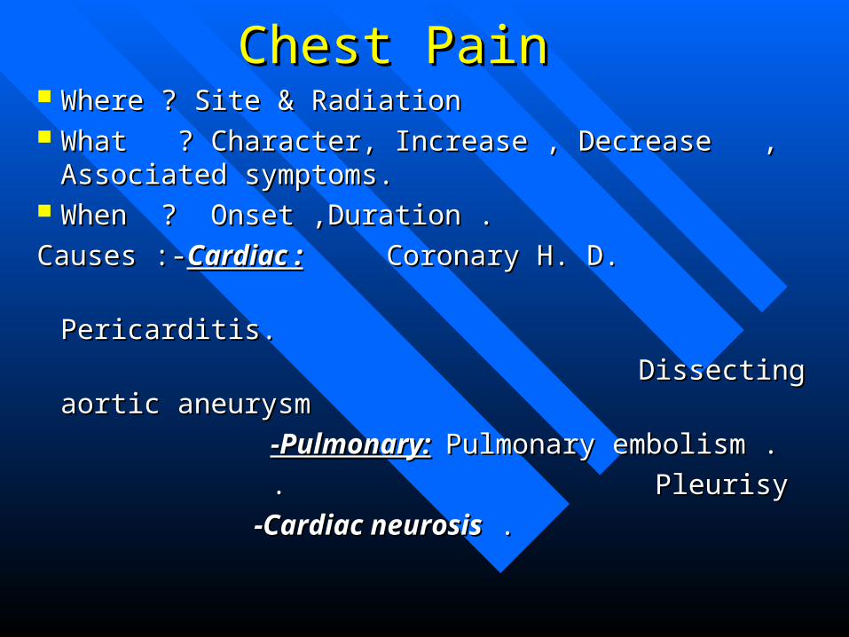
Chest PainChest Pain Where ? Site & RadiationWhere ? Site & Radiation What ? Character, Increase , Decrease , What ? Character, Increase , Decrease ,
Associated symptoms.Associated symptoms. When ? Onset ,Duration .When ? Onset ,Duration .
Causes :-Causes :-Cardiac :Cardiac : Coronary H. D. Coronary H. D.
Pericarditis. Pericarditis.
Dissecting aortic aneurysm Dissecting aortic aneurysm
-Pulmonary:-Pulmonary: Pulmonary embolism .Pulmonary embolism .
. . PleurisyPleurisy
-Cardiac neurosis-Cardiac neurosis . .

Palpitation Palpitation ( awareness of heart beats)( awareness of heart beats) Causes : -DysrrhythmiasCauses : -Dysrrhythmias - Hyperdynamic circulation.- Hyperdynamic circulation. -Volume overload (AR, MR,VSD )-Volume overload (AR, MR,VSD ) -Anxiety .-Anxiety . Ask about :Ask about : -Is it regular or irregular ?-Is it regular or irregular ? -Is it spontaneous.?-Is it spontaneous.? -Onset , Offset , and duration.-Onset , Offset , and duration. -Associated symptoms . -Associated symptoms . Example :Spontaneous regular palpitation with Example :Spontaneous regular palpitation with
sudden onset and offset sudden onset and offset PSVT PSVT

Symptoms suggestive of Peripheral Vascular diseaseSymptoms suggestive of Peripheral Vascular disease
-Coldness of extremities-Coldness of extremities-Claudication.-Claudication.-Ulcers of the leg .-Ulcers of the leg .
Toxic symptoms :Toxic symptoms : Fever,sweating, loss of weight ,loss of appetite.Fever,sweating, loss of weight ,loss of appetite. Fever in cardiac patients :Fever in cardiac patients :
- - R. F. ,infective endocarditis R. F. ,infective endocarditis - M.I.,pulm. embolism- M.I.,pulm. embolism - pericarditis ,collagen diseases - pericarditis ,collagen diseases

Low Cardiac out putLow Cardiac out put Causes: -Stenotic valve lesions :M.S.,A.S.Causes: -Stenotic valve lesions :M.S.,A.S.
-Pulmonary hypertension.-Pulmonary hypertension.
-Heart failure.-Heart failure.
Manifestations :Manifestations :
-Easy fatigue,blurring of vision,-Easy fatigue,blurring of vision,
- syncopal attacks.- syncopal attacks.
-Coldness of extremities .-Coldness of extremities .

CyanosisCyanosis Bluish ting of skin and m.m. ,due to presence of Bluish ting of skin and m.m. ,due to presence of
excessive amount of reduced Hb(excessive amount of reduced Hb(5g/dl ) in the 5g/dl ) in the underlying blood vessel.underlying blood vessel.
Cardiac causesCardiac causes Central - Congenital cyanotic H.D.Central - Congenital cyanotic H.D.
-C H F.-C H F. Peripheral - Low COP.Peripheral - Low COP. -Peripheral vascular D.-Peripheral vascular D. -C H F.-C H F. Ask about: -Onset, is it since birth ?Ask about: -Onset, is it since birth ? -Is it permanent .?-Is it permanent .? -Site -Associated symptoms.-Site -Associated symptoms.

Local Cardiac ExaminationLocal Cardiac Examination
Inspection and Palpation.Inspection and Palpation.
Percussion .Percussion .
Auscultation . Auscultation .

Inspection and PalpationInspection and Palpation A- A- Shape of the precordium:Shape of the precordium:
--Precordial bulgePrecordial bulgedenotes cardiac denotes cardiac enlargement since childhood.enlargement since childhood.
--Skeletal deformitiesSkeletal deformities : : As kyphosis ,scoliosis or pectus excavatum.As kyphosis ,scoliosis or pectus excavatum.
These may cause alteration of the position of the These may cause alteration of the position of the heart and great vessel which may predispose to heart and great vessel which may predispose to
heart failureheart failure..

B-Apex beatB-Apex beat Examine for :Examine for :
1-Visible or not. (causes of invisible apex ? )1-Visible or not. (causes of invisible apex ? )
2-2-Site:Site:(Apex beat= the outermost,lowermost palpable (Apex beat= the outermost,lowermost palpable impulse on the chest wall. )impulse on the chest wall. )
* Normally in the Lt.5* Normally in the Lt.5thth intercostal space ,just inside intercostal space ,just inside the MCL (9 cm from mid line ).the MCL (9 cm from mid line ).
*Abnormalities in site :*Abnormalities in site :
-Outward displacement-----Outward displacement----RV enlargement ,chest disease.RV enlargement ,chest disease.
-Outward and downward -Outward and downward LV enlargement,vent. AneurysmLV enlargement,vent. Aneurysm
-Displacement to Rt.--------Displacement to Rt.-------cong. Dextrocardia,chest diseasecong. Dextrocardia,chest disease

Apex beat (cont.)Apex beat (cont.)
3-Extent3-Extent : :
Localized apexLocalized apex :Normal apical impulse do not exceed :Normal apical impulse do not exceed an an inch in diameter ( Lt. V. apex )an an inch in diameter ( Lt. V. apex )
Diffuse apexDiffuse apex :apex occupying more than one :apex occupying more than one intercostal space ,extending to parasternal area intercostal space ,extending to parasternal area
Rt.V. enlargement .Rt.V. enlargement . 4- Character:4- Character:
Heaving sustained : Heaving sustained : Pressure overload Pressure overload AS,HTN.AS,HTN.
Hyperdynamic apexHyperdynamic apexVolume overload Volume overload AR,MR,TRAR,MR,TR
Slapping apex Slapping apex Palpable S1 Palpable S1 M.S.M.S.

Apex beat (cont. )Apex beat (cont. )
5-Thrill5-Thrill
-Murmurs may be so loud as to be palpable as thrill.-Murmurs may be so loud as to be palpable as thrill.
It is a palpable vibration of he chest wall similar to It is a palpable vibration of he chest wall similar to feeling back of a purring cat.feeling back of a purring cat.
- It should be timed with apex beat, either systolic or It should be timed with apex beat, either systolic or diastolic.diastolic.
- Diastolic thrill Diastolic thrill M.S.M.S.- Systolic thrill Systolic thrill M. R. M. R.- It is most easily felt when the patient turns on to the It is most easily felt when the patient turns on to the
left side.left side.


Inspection &Palpation (cont.)Inspection &Palpation (cont.)C-Other cardiac areas :C-Other cardiac areas :
Left arsenalLeft arsenal: -Pulsation LV Hypertrophy: -Pulsation LV Hypertrophy
huge Lt. huge Lt. Atrium .Atrium .
-Thrill VSD -Thrill VSD
Pulmonary Pulmonary -Pulsation Dilated pul. Art.-Pulsation Dilated pul. Art.
-Palpable P-Palpable P2 2 Pulm. Hyperten.Pulm. Hyperten.
((Diastolic shock)Diastolic shock)
Aortic Aortic Pulsation Pulsation Aneurysm of asc. Aorta. Aneurysm of asc. Aorta.
-Thrill-Thrill A.S. A.S.

Inspection &palpation (cont. )Inspection &palpation (cont. ) Epigastric PulsationEpigastric Pulsation : May be due to: : May be due to:
-Aortic pulsation: located in midline (normal in thin -Aortic pulsation: located in midline (normal in thin individual). individual).
-Hepatic pulsation: located to the Rt. Better felt by -Hepatic pulsation: located to the Rt. Better felt by bimanual palpation of liver,found in T. R.bimanual palpation of liver,found in T. R.
-Rt. Ventricular enlargement :better felt by tips of -Rt. Ventricular enlargement :better felt by tips of finger below left costal margin.finger below left costal margin.

Percussion Surface anatomy of heartSurface anatomy of heartAA = 1.5 inch from midline on the = 1.5 inch from midline on the
lower border of 2lower border of 2ndnd left costal cartilage. left costal cartilage.
BB =1 inch from midline on the upper=1 inch from midline on the upper
border of the 3border of the 3rdrd Rt. Costal cartilage. Rt. Costal cartilage.
CC =3.5 inch from midline in the =3.5 inch from midline in the
left 5left 5thth intercostal space. intercostal space.
DD =0.5 inch from midline on the =0.5 inch from midline on the
Rt. 6Rt. 6thth costal cartilage . costal cartilage .

Percussion ( cont. )Percussion ( cont. )
How to percuss the heart ?How to percuss the heart ?
-Percussion of the right border of the heart .-Percussion of the right border of the heart .
-Percussion of the base of the heart .-Percussion of the base of the heart .
-Percussion for dullness outside the apex .-Percussion for dullness outside the apex .
-Percussion of the bare area of the heart .-Percussion of the bare area of the heart .

Percussion of the heartPercussion of the heart 1-1-Rt. Border:Rt. Border:
First per cuss the upper border of the liver in Rt. MCLFirst per cuss the upper border of the liver in Rt. MCL
,then one space above the upper border of the liver, ,then one space above the upper border of the liver, from Rt. to Lt.,parallel to sternum.from Rt. to Lt.,parallel to sternum.
- Normally no dullness to the Rt. of the sternum.Normally no dullness to the Rt. of the sternum.- Causes of dullness to the Rt. Of the sternum :Causes of dullness to the Rt. Of the sternum :
* Rt. Atrial enlargement.* Rt. Atrial enlargement.
* Pericardial effusion.* Pericardial effusion.
*Aneurysm of the ascending aorta . *Aneurysm of the ascending aorta .

2- Base of the heart (upper border )2- Base of the heart (upper border ) Percuss the 2Percuss the 2ndnd Rt. And Lt. Intercostal spaces from Rt. And Lt. Intercostal spaces from
MCL to the sternum .MCL to the sternum . Normally both are resonant .Normally both are resonant . Causes of dullness in the Lt. 2Causes of dullness in the Lt. 2ndnd space space : : -Dilated pulmonary artery as in, pulm. HTN ,VSD -Dilated pulmonary artery as in, pulm. HTN ,VSD
ASD ,PDA.,pulm. Aneurysm.ASD ,PDA.,pulm. Aneurysm. -Pericardial effusion.-Pericardial effusion. -space occupying lesions in superior mediastinum.-space occupying lesions in superior mediastinum. Causes of dullness in the 2Causes of dullness in the 2ndnd Rt. Space: Rt. Space: --Dilatation of ascending aorta ,huge aneurysm in aortic arch.Dilatation of ascending aorta ,huge aneurysm in aortic arch.
-pericardial. Effusion and space occupying lesion in superior -pericardial. Effusion and space occupying lesion in superior mediation. mediation.

3-Left border3-Left border:: Normally there is no dullness outside the apex .Normally there is no dullness outside the apex . Dullness outside apex = pericardial effusion .Dullness outside apex = pericardial effusion .
4- Bare area :4- Bare area :
By light percussion in the4th &5By light percussion in the4th &5 thth intercostal intercostal spaces ,between the parasternal and midlines.spaces ,between the parasternal and midlines.
Causes of widening of bare area :Causes of widening of bare area :
RV enlargement ,pericardial effusion ,lung collaseRV enlargement ,pericardial effusion ,lung collase
Cause of resonance in bare areaCause of resonance in bare area: Emphysema.: Emphysema.

AuscultationAuscultation Ascultatory Areas :Ascultatory Areas :
Mitral areaMitral area
Pulmonary area.Pulmonary area.
First Aortic area .First Aortic area .
Second Aortic area. Second Aortic area.
Tricuspid area . Tricuspid area .


Auscultation (cont. )Auscultation (cont. ) Comment on :Comment on :
a- Heart sounds.a- Heart sounds.
b-Additional sounds .b-Additional sounds .
c- Murmurs .c- Murmurs .
d- Pericardial rub .d- Pericardial rub .

A-Heart sounds :A-Heart sounds : First H. sound ( S1 )= closure of A- V valve .First H. sound ( S1 )= closure of A- V valve .
-Best heard at Mitral area,at the beginning of syst-Best heard at Mitral area,at the beginning of syst
-Accentuated S1 M. S., T.S.-Accentuated S1 M. S., T.S.
hyperdynamic circ. States .hyperdynamic circ. States .
-Weak S1 M. R. , T. R.-Weak S1 M. R. , T. R.
calcified mitral valve.calcified mitral valve.
Severe heart failureSevere heart failure
-Variable S1 A. F.-Variable S1 A. F.
A-V dissociation . A-V dissociation .

Second heart sound (S2 )Second heart sound (S2 ) = Closure of aortic &pulm. valves = Closure of aortic &pulm. valves Best heard at base of the heart, at beginning of diastole .Best heard at base of the heart, at beginning of diastole .
Normally splitted on P area,splitting increase with inspirationNormally splitted on P area,splitting increase with inspiration . . Abnormalities :Abnormalities :On pulmonary areaOn pulmonary area : : -Accentuated pulmonary hypertension-Accentuated pulmonary hypertension accentuated A2accentuated A2 -weak P2 P. S.-weak P2 P. S. -Wide splitting P. S. ,RBBB .-Wide splitting P. S. ,RBBB . -Reversed split LBBB ,tight A.S.,HOCM-Reversed split LBBB ,tight A.S.,HOCM On Aortic area :On Aortic area : -Accentuated A2 HTN ,Hyperdynamic states-Accentuated A2 HTN ,Hyperdynamic states -Weak A2 A. S. -Weak A2 A. S.

SS3 3
*Produced by sudden distension of ventricles due to rapid *Produced by sudden distension of ventricles due to rapid filling with blood .filling with blood .
Low pitched sound ,best heard at apex with lightly Low pitched sound ,best heard at apex with lightly applied stethoscope bell.applied stethoscope bell.
Normal finding in young adult .Normal finding in young adult .
Abnormal LV S3 Abnormal LV S3 --MR,VSD ,PDA MR,VSD ,PDA
--LVF.LVF.
RVS3 RVS3 --TR TR
(on T area)(on T area) --RVF RVF

SS44 It is due to atrial contraction .It is due to atrial contraction .LV S4 LV S4 (best heard inside the apex ) (best heard inside the apex ) Causes : - HTN.Causes : - HTN. -IHD .-IHD . -A.S.-A.S.RV S4 RV S4 (best hearted at left sternal edge )(best hearted at left sternal edge ) Causes : -Pulmonary hypertension.Causes : -Pulmonary hypertension. -pulmonary embolism .-pulmonary embolism . -P. S. -P. S.

B-Additional heart soundsB-Additional heart sounds1-Gallop rhythm1-Gallop rhythm
It is hearing of 3 sounds (1It is hearing of 3 sounds (1stst , 2 , 2ndnd ,and extra sound ) ,and extra sound ) in the presence of tachycardia(like galloping in the presence of tachycardia(like galloping horse)horse)
Types :Types :
-Diastolic gallop (S1,S2,S3 ) -Diastolic gallop (S1,S2,S3 )
-Presystolic gallop (S1,S2, S$ )-Presystolic gallop (S1,S2, S$ )
-Summation gallop (S1,S2,S3,S4 )-Summation gallop (S1,S2,S3,S4 )

2-Opening snap2-Opening snap
A high-pitched sound occurs with M. S.when the A high-pitched sound occurs with M. S.when the stenosed valve moves downward towards L. V. at stenosed valve moves downward towards L. V. at the beginning of diastole.the beginning of diastole.
Best heard between the apex and sternum.Best heard between the apex and sternum. It occurs after S2 by o.o7-o.1 seconds.It occurs after S2 by o.o7-o.1 seconds. It disappear when the cusps become calcified .It disappear when the cusps become calcified . The earlier it occur ,the higher the pressure in the The earlier it occur ,the higher the pressure in the
atrium,(i.e more severe M. S.)atrium,(i.e more severe M. S.)

3-Ejection clicks3-Ejection clicks Sharp high pitched sounds closely follow the S1.Sharp high pitched sounds closely follow the S1. Occur with opening of the semilunar valve.Occur with opening of the semilunar valve. Best heard at the base of the heart.Best heard at the base of the heart. Causes :Causes :
A.S.,P. S.A.S.,P. S.
HTN ,Pulm. Hypertension. HTN ,Pulm. Hypertension.
Mid-systolic click : Mid-systolic click : occur in MVP . occur in MVP .

Prosthetic valve soundsProsthetic valve sounds Artificial mechanical valve produce 2 sounds for Artificial mechanical valve produce 2 sounds for
each cardiac cycle: a quiet opening click and each cardiac cycle: a quiet opening click and louder closing sound.louder closing sound.
Mitral prosthesis closing sound accompany and Mitral prosthesis closing sound accompany and largely constitutes) S1and opening click follow largely constitutes) S1and opening click follow S2 in similar position to opening snap .S2 in similar position to opening snap .
Conversely aortic prosthesis opening sound Conversely aortic prosthesis opening sound follow S1 in similar position to ejection follow S1 in similar position to ejection click ,while the closing sound accompanies S2click ,while the closing sound accompanies S2
Disappearance or muffling of opening sound is an Disappearance or muffling of opening sound is an early indication of thrombosis of the valve .early indication of thrombosis of the valve .

C-Murmurs :C-Murmurs : Murmurs are produced by excessive turbulence of Murmurs are produced by excessive turbulence of
blood flow within the circulation.blood flow within the circulation. Description of murmursDescription of murmurs 1-Timing (systolic ,diastolic ,or continuous).1-Timing (systolic ,diastolic ,or continuous).2-Site of maximum intensity.2-Site of maximum intensity.3-Propagation (in the direction of blood flow )3-Propagation (in the direction of blood flow )4-Quality (character:soft ,harsh ,or rumbling ) .4-Quality (character:soft ,harsh ,or rumbling ) .5-Intensity (Grades form I-VI ).5-Intensity (Grades form I-VI ).6-Relation to respiration (insp .6-Relation to respiration (insp .Rt. sided murmurs )Rt. sided murmurs )7- Position of the patient in which murmur is best 7- Position of the patient in which murmur is best
heard (Lt.lateral position heard (Lt.lateral position murmurs of mitral v.) murmurs of mitral v.)

Grades of murmurs :Grades of murmurs : Grade I :just audible in quiet room and patient.Grade I :just audible in quiet room and patient.
Grade II :Quiet or soft .Grade II :Quiet or soft .
Grade III :Moderately loud .Grade III :Moderately loud .
Grade IV :loud and accompanied by thrill.Grade IV :loud and accompanied by thrill.
Grade V :Very loud Grade V :Very loud
Grade VI : So loud ,audible with stethoscope lifted from Grade VI : So loud ,audible with stethoscope lifted from the chest wall . the chest wall .

MurmursMurmurs A- Systolic murmursA- Systolic murmurs1-1-Ejection systolic murmurEjection systolic murmur
-It begins shortly after S1and ends-It begins shortly after S1and ends
before S2 .before S2 .
-It-Itto a crescendo about the middleto a crescendo about the middle
of systole, then diminish of systole, then diminish
-Result from turbulent blood flow through sensed semilunar -Result from turbulent blood flow through sensed semilunar valves or valves or blood flow through normal v.blood flow through normal v.
-Examples : A. S. ( Aortic area )-Examples : A. S. ( Aortic area )
P. S. ,ASD .(pulmonary area ).P. S. ,ASD .(pulmonary area ).
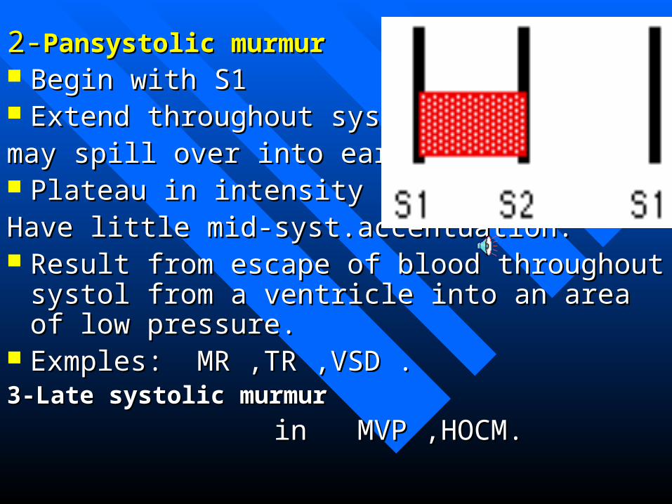
2-2-Pansystolic murmurPansystolic murmur Begin with S1Begin with S1 Extend throughout systol &Extend throughout systol &may spill over into early diast.may spill over into early diast. Plateau in intensity but may Plateau in intensity but may Have little mid-syst.accentuation.Have little mid-syst.accentuation. Result from escape of blood throughout systol from Result from escape of blood throughout systol from
a ventricle into an area of low pressure.a ventricle into an area of low pressure. Exmples: MR ,TR ,VSD .Exmples: MR ,TR ,VSD .3-Late systolic murmur3-Late systolic murmur
in MVP ,HOCM.in MVP ,HOCM.

B- Diastolic murmursB- Diastolic murmurs 1-Early diastolic murmur1-Early diastolic murmur-It starts immediately after S2-It starts immediately after S2-Soft blowing .-Soft blowing .-loudest at its onset and dies away -loudest at its onset and dies away Before end of diastole(Decrescendo)Before end of diastole(Decrescendo)-Best heard when patient lean forward and breath in -Best heard when patient lean forward and breath in
expiration.expiration.-Result from leaking semilunar valves.-Result from leaking semilunar valves.Causes : AR on Second artic area.Causes : AR on Second artic area. PR on P.(functional in severe pulm.hypertension = PR on P.(functional in severe pulm.hypertension =
Graham Steell m.) Graham Steell m.)

2-Mid-diastolic murmur2-Mid-diastolic murmur
It begins some times after S2It begins some times after S2 May be short or may extendMay be short or may extend
Long in diastoleLong in diastole Low pitched and rumbling inLow pitched and rumbling in
Character Character
Causes :Causes :
-MS best heard at apex,localized .-MS best heard at apex,localized .
-T S Max. at the T area &-T S Max. at the T area & with inspiration with inspiration
3-Presystolic murmur3-Presystolic murmur
Begin in late diastole.Begin in late diastole.
Rises to crescendo just before S1.Rises to crescendo just before S1.
The murmur is due to atrial contraction which The murmur is due to atrial contraction which cause a sharp rise in pressure gradient that increase cause a sharp rise in pressure gradient that increase blood flow through narrowed A-V valve blood flow through narrowed A-V valve (M.S.,T.S.).(M.S.,T.S.).
Murmur disappear in A.F. Murmur disappear in A.F.

C- Continous murmurC- Continous murmur
– These are continuous throughout systole and These are continuous throughout systole and diastolediastole
– Heard over the base of the heart.Heard over the base of the heart.
– Causes :Causes :
-PDA-PDA
Arteri-venous aneurysm of the lung .Arteri-venous aneurysm of the lung .
-D.D : Venous hum. -D.D : Venous hum.

Functional murmursFunctional murmurs These are murmurs which arise in absence of organic These are murmurs which arise in absence of organic
heart disease at their site of origin.heart disease at their site of origin. They are mostly systolic ,soft,of low intensity,not They are mostly systolic ,soft,of low intensity,not
accompanied with thrill and change with change of accompanied with thrill and change with change of posture of patient.posture of patient.
Examples :Examples :
-Pulmonary and aortic ejection systolic m. ,in -Pulmonary and aortic ejection systolic m. ,in hyperdynamic circulatory states.hyperdynamic circulatory states.
-Ejection systolic m. at pulmonary area in ASD -Ejection systolic m. at pulmonary area in ASD
Pericardial rubPericardial rub
Scratching sound like friction between rough Scratching sound like friction between rough surfaces and has a superficial to and fro quality.surfaces and has a superficial to and fro quality.
Best heard to the left of the lower sternumBest heard to the left of the lower sternum
It is accentuated when patient leans forward and It is accentuated when patient leans forward and by pressure with stethoscope.by pressure with stethoscope.
Cause : Acute retardatesCause : Acute retardates

Exocrdiac “Noises “Exocrdiac “Noises “
These are sounds recurring with each heartbeat but These are sounds recurring with each heartbeat but originate outside the heart.originate outside the heart.
Examples :Pericardial rubsExamples :Pericardial rubs
Clicking sound of small pneumothorax Clicking sound of small pneumothorax . . Mediastinal Crunch in pneumomediastinal: Mediastinal Crunch in pneumomediastinal: sounds like a sounds like a
loud systolic murmurloud systolic murmur

Diagnosis of cardiac caseDiagnosis of cardiac case
Aetiological diagnosis.Aetiological diagnosis. Pathological diagnosis(Anatomical ).Pathological diagnosis(Anatomical ). Functional diagnosis.Functional diagnosis. complications.complications.