Cardiovascular Pathophysiology: Right to Left · PDF fileCardiovascular Pathophysiology: Right...
21
12/9/2009 1 Cardiovascular Pathophysiology: Right to Left Shunts aka Cyanotic Lesions Ismee A Williams MD MS Ismee A. Williams, MD, MS [email protected] Pediatric Cardiology Learning Objectives • To discuss the hemodynamic significance f i htt l ft h t of right to left shunts • To describe the common cyanotic cardiac lesions in the newborn • To understand the different causes of cyanosis: obstruction to pulmonary blood cyanosis: obstruction to pulmonary blood flow vs mixing
-
Upload
phunghuong -
Category
Documents
-
view
223 -
download
3
Transcript of Cardiovascular Pathophysiology: Right to Left · PDF fileCardiovascular Pathophysiology: Right...

12/9/2009
1
Cardiovascular Pathophysiology:
Right to Left Shuntsaka Cyanotic Lesions
Ismee A Williams MD MSIsmee A. Williams, MD, [email protected] Cardiology
Learning Objectives
• To discuss the hemodynamic significance f i ht t l ft h tof right to left shunts
• To describe the common cyanotic cardiac lesions in the newborn
• To understand the different causes of cyanosis: obstruction to pulmonary bloodcyanosis: obstruction to pulmonary blood flow vs mixing

12/9/2009
2
Importance of Congenital Heart Disease
• Incidence 6 to 8 per 1000 births
• 15% are life threatening
• 25% are discharged without diagnosis
• 1/3 have cyanosis
What is Cyanosis?• Bluish discoloration of skin that occurs
when the amount of deoxygenated h l bi 5 /dL i ill ihemoglobin ≥ 5 g/dL in capillaries
• Central Cyanosis: decreased systemic oxygen delivery
• Peripheral Cyanosis: increased oxygen extraction by tissue

12/9/2009
3
Factors affecting detection of Cyanosis• Total hemoglobin concentration affects the level of O2
saturation at which cyanosis is observed– Hgb conc = 9 g/dL, need an O2 Sat of 67% to have 3-5 g/dL of
reduced hemoglobin and see cyanosis– Hgb conc = 20 g/dL, see cyanosis at O2 Sat of 85%– Decreased O2 sat may not be recognized in the setting of
anemia
• Skin pigmentation
• Factors that shift the oxygen dissociation curve to the left result in oxygen binding more tightly to Hgb and decreased release to the tissue at a given O2 tension (PO2)– Therefore, will be harder to see cyanosis (get 5 g/dL of
deoxygenated Hgb) at any given PO2
Won’t see cyanosis if anemic
12/9/2009
4
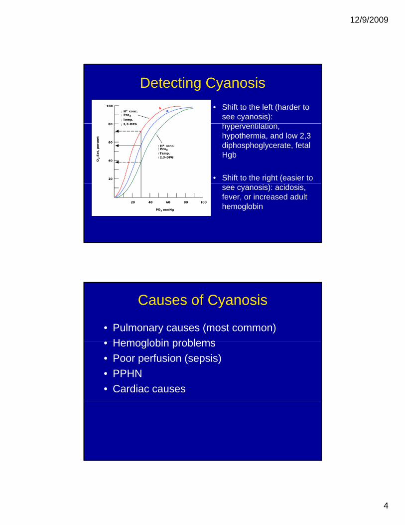
Detecting Cyanosis• Shift to the left (harder to
see cyanosis): h til tihyperventilation, hypothermia, and low 2,3 diphosphoglycerate, fetal Hgb
• Shift to the right (easier to see cyanosis): acidosis, fever, or increased adult hemoglobin
Causes of Cyanosis
• Pulmonary causes (most common)• Hemoglobin problems• Hemoglobin problems• Poor perfusion (sepsis)• PPHN• Cardiac causes
12/9/2009
5
Persistent Pulmonary Hypertension of the Newborn
• Used to be called Persistent Fetal Circulation• Abnormal pulmonary vasoconstriction or failure• Abnormal pulmonary vasoconstriction or failure
to “relax” leads to right to left shunting at the foramen ovale and the ductus arteriosus
• Profound cyanosis• Associated with neonatal asphyxia, maternal
infection• Apgar scores are low• Usually self-limited with NO and ECMO
treatment
Cardiac Causes of Cyanosis
• Decreased/obstructed pulmonary blood flow
• Systemic and Pulmonary venous Mixing g

12/9/2009
6
Decreased Pulmonary Blood Flow
• Obligatory intracardiac right to left h tishunting
• Pulmonary blood flow is provided by an alternative path – usually the ductus arteriosus
• Very cyanotic• Very cyanotic
Cardiac Lesions causing cyanosis due to decreased pulmonary blood flow
P l i• Pulmonary stenosis• Pulmonary atresia• Tricuspid atresia• Tetralogy of Fallot

12/9/2009
7
Pulmonary Stenosis
• Location of obstruction varies:obstruction varies:
• RV outflow• Pulmonary Valve • Main Pulmonary
Most common
Artery
Pulmonary Stenosis• 25-30% of CHD
– Isolated PS in 8-10% of CHD• Hemodynamic consequence: pressure
overload and hypertrophy of the RV• PE: cyanosis, systolic ejection murmur at
LUSB• Tx: Balloon vs surgery

12/9/2009
8
Pulmonary Atresia
• Obligate right to left flow across theflow across the foramen ovale
• Pulmonary blood flow supplied by the ductus arteriosusductus arteriosus “ductal dependent”
Pulmonary atresia • 3% of CHD (0.041 per 1000 live births)• Size of the RV varies• PE: cyanosis no systolic ejection murmur (noPE: cyanosis, no systolic ejection murmur (no
flow)– may have holosystolic murmur at LLSB associated
with tricuspid regurgitation• CXR: black lungs• Treatment depends on “flavor” of PA/IVS
– balloon of pulmonary valve if RV size adequate– aortico-pulmonary shunt to increase pulmonary blood
flow– staged surgery to a Fontan if RV too small– Heart transplant if RV dependent coronary sinusoids

12/9/2009
9
Tricuspid Atresia
• Obligatory right to left shunt at the PFOshunt at the PFO
• Typically have a VSD that allows blood into the RV and out the PA– Obstruction to
pulmonary flow relatedpulmonary flow related to size of VSD
• Hypoplastic right ventricle
Tricuspid Atresia
• 3% of CHD (0.056 per 1000 live births)
• 25% have transposed great vessels and problems with aortic/systemic blood flow
• PE: systolic murmur, cyanosisPE: systolic murmur, cyanosis
• Tx: staged surgery to a Fontan

12/9/2009
10
Tetralogy of Fallot
Single defect: anteriorl li t f thmalalignment of the
interventricular septum
• VSD • Aortic override • Pulmonary Stenosis • RVH
Tetralogy of Fallot
• 3.5-9% of CHD (0.26-0.8 per 1000 live births)
• Commonly associated with other defects– DiGeorge Syndrome in 25%
• Degree of pulmonary obstruction varies• Symptoms depend on amount of
obstruction to pulmonary blood flowobstruction to pulmonary blood flow– cyanosis, tet spells
• PE: systolic ejection murmur at LUSB • Tx: Surgical repair of VSD and PS

12/9/2009
11
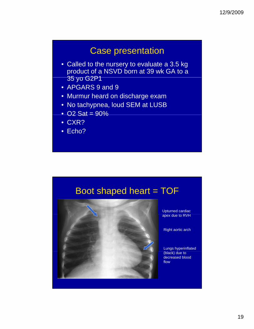
Boot shaped heart = TOF
Upturned cardiac d RVHapex due to RVH
Right aortic arch
Lungs hyperinflated (bl k) d(black) due to decreased blood flow
Mixing of Systemic and Pulmonary Venous Return
• No obstruction to pulmonary blood flow– Pulmonary flow may be greater than normal
• See both right to left AND left to right intracardiac shunting
• Associated with pulmonary HTN and ventricular failureventricular failure
• Cyanosis typically less intense than with pulmonary obstruction
12/9/2009
12
Cyanosis due to Mixing
• Truncus arteriosus• Total anomalous pulmonary venous return
(TAPVR) • Transposition of the Great Arteries (TGA)
• Mixing with Heart Failure– HLHS, Aortic stenosis, coarctation
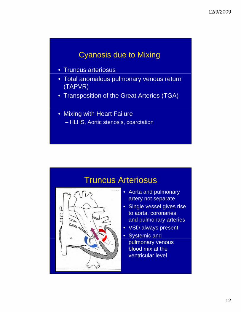
Truncus Arteriosus• Aorta and pulmonary
artery not separate• Single vessel gives rise
to aorta, coronaries, and pulmonary arteries
• VSD always present• Systemic and y
pulmonary venous blood mix at the ventricular level

12/9/2009
13
Truncus arteriosus• 1-2.5% of CHD (0.08 per 1000 live births) • Truncal valve usually very dysplasticTruncal valve usually very dysplastic • Commonly associated lesions
– Coronary anomalies, interrupted aortic arch– 25% DiGeorge
• PE: cyanosis and murmur of regurgitationy g g• High risk to develop pulm HTN over time• Tx: surgical repair in infancy
TAPVR• Pulmonary veins return
to the right heart– Via supracardiac,
intracardiac, or infradiaphragmatic path
• Pulmonary venous blood mixes with systemic venous blood at the atrial level
• Obligatory right to left shunt at atrial level to support systemic flow

12/9/2009
14
TAPVR• 2-3% of CHD (0.058 per 1000 live births)• Failure of the left atrium to incorporate the
pulmonary veins during developmentpulmonary veins during development• Obstruction to pulmonary venous flow is
common– Can occur at different levels– Most common in infradiaphragmatic TAPVR
Leads to pulmonary congestion and death– Leads to pulmonary congestion and death• PE: cyanosis, respiratory distress, CXR
white out with small heart• Tx: no PGE, surgical repair
Transposition of the Great Arteries• Great arteries are
“switched”• Systemic venous return
goes back to the body• Pulmonary venous
return goes back to the lungs
• Survival dependent on mixing between the two parallel circulations

12/9/2009
15
Transposition of the Great Arteries• Most common cyanotic CHD (0.22 per
1000 live births) • Fetal circulation allows mixing • Problems after birth• Mixing via PFO/ASD, VSD (1/3), or PDA• PE: severe cyanosis, no murmur• Tx: balloon atrial septostomy to maximize• Tx: balloon atrial septostomy to maximize
mixing at the atrial level – surgical arterial switch
Cyanosis due Mixing with Heart Failure
• Obstruction to systemic outflow, mixing, cyanosis poor perfusioncyanosis, poor perfusion
• Depend on PDA to supply systemic blood flow• As PDA closes, see poor perfusion, acidosis,
death
• Hypoplastic left heart syndrome (HLHS)• Critical valvar Aortic Stenosis• Interrupted aortic arch/Coarctation of the Aorta

12/9/2009
16
HLHS• Left side of the heart
too small/absent• Classic form is mitralClassic form is mitral
and aortic atresia• Pulmonary venous
blood shunts left to right at PFO and mixes with systemic venous returnreturn
• Blood going out the RV into the PA passes through the PDA to feed the body
HLHS• 0.16-0.27 per 1000 live births
• Severe form of single ventricle
• PE: no murmur, cyanosis, poor pulses
• Tx: PGE, Surgery: Norwood, Glenn, Fontan

12/9/2009
17
Evaluation of the cyanotic newborn• History: family hx, prenatal testing,
peripartum information• Vital Signs: HR, RR, O2 sat, 4 ext BP• Physical exam: observation of skin,
movement, respirations, palpation and ausculation of chest, palpation of femoral pulses capillary perfusionpulses, capillary perfusion
• Laboratory testing: ABG, CBC, BLCx, CXR, EKG, Echo
Hyperoxia Test: Heart vs Lungs?• Cardiac lesions typically have fully saturated
pulmonary venous bloodHigh FiO2 has little effect on PO2 and O2 Sat– High FiO2 has little effect on PO2 and O2 Sat
• Pulmonary lesions typically have pulmonary venous desaturation– Higher FiO2 increases pulmonary venous oxygen
levels and PO2 and O2 Sat• Administer 100% FiO2 for 10 minutes and• Administer 100% FiO2 for 10 minutes and
compare the PO2 at baseline and after oxygen– PO2 > 150 mm Hg = pulmonary cause– PO2 < 150 mm Hg = cardiac cause

12/9/2009
18
Case presentation• Called to the nursery to evaluate a 3.5 kg
product of a NSVD born at 39 wk GA to a 35 yo G2P1G2P1
• APGARS 9 and 9• At four hours of life RN noted the infant
appeared “dusky”• Central cyanosis, no tachypnea, no murmur• O2 Sat = 70%, PO2 = 40 mm Hg on RA, and O2
Sat = 82%, PO2 = 50 mm Hg on 100% FiO2• CXR NL
Transposition of the Great Arteries
• Prostaglandin E1
• Emergent balloon atrial septostomy
• O2 sat increases to 85%85%
• Arterial switch operation next day
12/9/2009
19
Case presentation• Called to the nursery to evaluate a 3.5 kg
product of a NSVD born at 39 wk GA to a 35 yo G2P135 yo G2P1
• APGARS 9 and 9• Murmur heard on discharge exam • No tachypnea, loud SEM at LUSB• O2 Sat = 90%O2 Sat 90%• CXR?• Echo?
Boot shaped heart = TOF
Upturned cardiac d RVHapex due to RVH
Right aortic arch
Lungs hyperinflated (bl k) d(black) due to decreased blood flow

12/9/2009
20
Tetralogy of FallotEducate parents about
tet spells
Genetic testing for DiGeorge
Frequent follow up to check O2 satcheck O2 sat
Plan elective surgical repair at 4 - 6 months
Case presentation• Get a call from an outside pediatrician • 10 day old infant with grunting and poor y g g p
perfusion – presumed sepsis• APGARS 9 and 9, no prenatal US• In ER: Grey infant, O2 Sat = 90%, no
femoral pulses, no murmur• Echo?

12/9/2009
21
HLHS• Prostaglandin E1
• Pressors• Pressors
• Intubate FiO2 21%
• Sedate and hypoventilatehypoventilate
• Norwood Stage I when stable
Summary• Cyanosis when 3-5 gm/dl of desaturated
Hgb – hard to see if anemicHgb hard to see if anemic• Mutliple causes• Cardiac causes are EMERGENCIES• Decreased pulmonary blood flow vs
Mixingg• Prostoglandin E2 to keep ductus
arteriosus OPEN