Cardiovascular System Two divisions Cardiovascular System Two divisions: pulmonary.

Cardiovascular Biology of the Incretin System
John R. Ussher and Daniel J. Drucker
Department of Medicine, Samuel Lunenfeld Research Institute, Mt. Sinai Hospital, University of Toronto, Toronto,Ontario M5G 1X5, Canada
Glucagon-like peptide-1 (GLP-1) is an incretin hormone that enhances glucose-stimulated insulin secretionand exerts direct and indirect actions on the cardiovascular system. GLP-1 and its related incretin hormone,glucose-dependent insulinotropic polypeptide, are rapidly inactivated by the enzyme dipeptidyl peptidase4 (DPP-4), a key determinant of incretin bioactivity. Two classes of medications that enhance incretin action,GLP-1 receptor (GLP-1R) agonists and DPP-4 inhibitors, are used for the treatment of type 2 diabetesmellitus. We review herein the cardiovascular biology of GLP-1R agonists and DPP-4 inhibitors, includingdirect and indirect effects on cardiomyocytes, blood vessels, adipocytes, the control of blood pressure, andpostprandial lipoprotein secretion. Both GLP-1R activation and DPP-4 inhibition exert multiple cardiopro-tective actions in preclinical models of cardiovascular dysfunction, and short-term studies in human subjectsappear to demonstrate modest yet beneficial actions on cardiac function in subjects with ischemic heartdisease. Incretin-based agents control body weight, improve glycemic control with a low risk of hypogly-cemia, decrease blood pressure, inhibit the secretion of intestinal chylomicrons, and reduce inflammationin preclinical studies. Nevertheless, there is limited information on the cardiovascular actions of theseagents in patients with diabetes and established cardiovascular disease. Hence, a more complete under-standing of the cardiovascular risk to benefit ratio of incretin-based therapies will require completion oflong-term cardiovascular outcome studies currently underway in patients with type 2 diabetes mellitus.(Endocrine Reviews 33: 0000 – 0000, 2012)
I. IntroductionII. The Incretin Axis
A. Glucagon-like peptide-1B. The GLP-1 receptorC. GLP-1-mediated regulation of insulin and gluca-
gon secretionD. Glucose-dependent insulinotropic polypeptideE. The GIP receptorF. Dipeptidyl peptidase-4 and incretin degradation
III. Role of the GLP-1R in the Cardiovascular SystemA. GLP-1R expression and signal transduction in the
myocardiumB. GLP-1R in the vasculatureC. GLP-1 action and dyslipidemiaD. Direct vs. indirect effects of GLP-1R agonism on
cardiac functionE. Cardiovascular biology of GLP-1 (9–36)
IV. GLP-1 and Cardiovascular DiseaseA. GLP-1 and hypertensionB. GLP-1 action in experimental models of
atherosclerosisC. GLP-1R activation in ischemic heart diseaseD. GLP-1R agonists in heart failure
V. DPP-4 Inhibition and Cardiovascular FunctionA. DPP-4 expression in the cardiovascular systemB. Potential cardioactive DPP-4 substratesC. DPP-4 in hypertensionD. DPP-4 in atherosclerosisE. DPP-4 in ischemic heart diseaseD. DPP-4 in heart failure and cardiomyopathy
VI. Clinical TrialsA. GLP-1R agonists and cardiovascular outcomesB. DPP-4 inhibitors and cardiovascular outcomes
VII. Future DirectionsA. Potential pitfalls of incretin-based therapyB. Novel avenues of researchC. Summary
ISSN Print 0021-972X ISSN Online 1945-7197Printed in U.S.A.Copyright © 2012 by The Endocrine Societydoi: 10.1210/er.2011-1052 Received November 28, 2011. Accepted January 18, 2012.
Abbreviations: AMI, Acute myocardial infarction; AMPK, 5�-AMP-activated protein kinase;ApoB-48, apolipoprotein B-48; BNP, B-type (brain) natriuretic peptide; CD26, cluster ofdifferentiation 26; CNS, central nervous system; CoA, coenzyme A; DBP, diastolic bloodpressure; DPP-4, dipeptidyl peptidase-4; eNOS, endothelial nitric oxide synthase; FFA, freefatty acid; G-CSF, granulocyte colony-stimulating factor; GIP, glucose-dependent insuli-notropic polypeptide; GIPR, GIP receptor; GLP-1, glucagon-like peptide-1; GLP-1R, GLP-1receptor; GLUT4, glucose transporter 4; GPCR, G protein-coupled receptor; HCAEC, hu-man coronary artery endothelial cells; HR, heart rate; HUVEC, human umbilical vein en-dothelial cells; icv, intracerebroventricular; LAD, left anterior descending; LDL, low-densitylipoprotein; LV, left ventricular; LVDP, LV developed pressure; LVEF, LV ejection fraction;NPY, neuropeptide Y; PI3K, phosphatidylinositol-3 kinase; PKA, protein kinase A; PPAR�,peroxisome proliferator-activated receptor-�; PYY, peptide YY; SBP, systolic blood pres-sure; SDF-1�, stromal cell-derived factor-1�; TAG, triacylglycerol; T2DM, type 2 diabetesmellitus; VLDL, very-low-density lipoprotein.
R E V I E W
Endocrine Reviews, April 2012, 33(2):0000–0000 edrv.endojournals.org 1
Endocrine Reviews. First published ahead of print February 8, 2012 as doi:10.1210/er.2011-1052
Copyright (C) 2012 by The Endocrine Society
I. Introduction
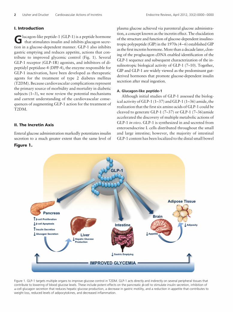
Glucagon-like peptide-1 (GLP-1) is a peptide hormonethat stimulates insulin and inhibits glucagon secre-
tion in a glucose-dependent manner. GLP-1 also inhibitsgastric emptying and reduces appetite, actions that con-tribute to improved glycemic control (Fig. 1). SeveralGLP-1 receptor (GLP-1R) agonists, and inhibitors of di-peptidyl peptidase-4 (DPP-4), the enzyme responsible forGLP-1 inactivation, have been developed as therapeuticagents for the treatment of type 2 diabetes mellitus(T2DM). Because cardiovascular complications representthe primary source of morbidity and mortality in diabeticsubjects (1–3), we now review the potential mechanismsand current understanding of the cardiovascular conse-quences of augmenting GLP-1 action for the treatment ofT2DM.
II. The Incretin Axis
Enteral glucose administration markedly potentiates insulinsecretion to a much greater extent than the same level of
plasma glucose achieved via parenteral glucose administra-tion, a concept known as the incretin effect. The elucidationof the structure and function of glucose-dependent insulino-tropic polypeptide (GIP) in the 1970s (4–6) established GIPas the first incretin hormone. More than a decade later, clon-ing of the proglucagon cDNA enabled identification of theGLP-1 sequence and subsequent characterization of the in-sulinotropic biological activity of GLP-1 (7–10). Together,GIP and GLP-1 are widely viewed as the predominant gut-derived hormones that promote glucose-dependent insulinsecretion after meal ingestion.
A. Glucagon-like peptide-1Although initial studies of GLP-1 assessed the biolog-
ical activity of GLP-1 (1–37) and GLP-1 (1–36) amide, therealization that the first six amino acids of GLP-1 could becleaved to generate GLP-1 (7–37) or GLP-1 (7–36)amideaccelerated the discovery of multiple metabolic actions ofGLP-1 in vivo. GLP-1 is synthesized in and secreted fromenteroendocrine L cells distributed throughout the smalland large intestine; however, the majority of intestinalGLP-1 content has been localized to the distal small bowel
Figure 1.
Figure 1. GLP-1 targets multiple organs to improve glucose control in T2DM. GLP-1 acts directly and indirectly on several peripheral tissues thatcontribute to lowering of blood glucose levels. These include potent effects on the pancreatic �-cell to stimulate insulin secretion, inhibition of�-cell glucagon secretion that reduces hepatic glucose production, a decrease in gastric motility, and a reduction in appetite that contributes toweight loss, reduced levels of adipocytokines, and decreased inflammation.
2 Ussher and Drucker Cardiovascular Actions of Incretins Endocrine Reviews, April 2012, 33(2):0000–0000
and colon. GLP-1 is also produced in the central nervoussystem (CNS), predominantly in the brainstem, and sub-sequently transported to a large number of regions withinthe CNS. GLP-1 is secreted from the gut at low basal levelseven in the fasted state, and plasma levels of GLP-1 in-crease 2- to 3-fold after nutrient ingestion. The biology ofGLP-1 synthesis and secretion and the multiple metabolicand extrapancreatic actions of GLP-1 and GIP have beenextensively reviewed (11–15).
B. The GLP-1 receptor
A single G protein-coupled receptor (GPCR), with con-siderable amino acid homology to related class B familyGPCR (16), transduces the majority of GLP-1 actions invivo and represents the only specific high-affinity GLP-1Ridentified to date (17). The GLP-1R was originally iden-tified in islet �-cells but is widely expressed in extrapancre-atic tissues, including the lung, kidney, CNS, enteric andperipheral nervous system, lymphocytes, blood vessels,and heart (17, 18). Multiple actions of GLP-1 and struc-turally related GLP-1R agonists have been reported in cellsand tissues that do not express the classical GLP-1R, hencethere continues to be analysis of mechanisms and path-ways capable of transducing actions of GLP-1 indepen-dent of the known GLP-1R (19).
C. GLP-1-mediated regulation of insulin andglucagon secretion
GLP-1 directly potentiates insulin secretion in a glucose-dependent manner, minimizing the risk of hypoglycemia indiabetic subjects treated with GLP-1R agonists. GLP-1 alsoinduces glucose competence in previously unresponsive�-cells (20) and rapidly improves �-cell glucose sensitivity,thereby restoring insulin secretion toward normal levels inhuman patients with T2DM (21). GLP-1 also inhibits glu-cagonsecretioninaglucose-dependentmanner(22).Becausethe majority of �-cells do not express the GLP-1R, andGLP-1 inhibits glucagon secretion even in C-peptide-nega-tive subjects with type 1 diabetes, the glucagonostatic effectsofGLP-1are likely indirect,mediated throughsomatostatin-dependent mechanisms (23).
D. Glucose-dependent insulinotropic polypeptide
GIP is a 42-amino-acid peptide synthesized in and se-creted from enteroendocrine K cells localized to the prox-imal small bowel. Like GLP-1, GIP is secreted at low basallevels in the fasted state, and plasma levels of GIP increasewithin minutes of nutrient ingestion. Although the majoraction of GIP is the glucose-dependent stimulation of in-sulin secretion, GIP also promotes lipid uptake and ex-pansion of adipocyte mass and exerts a number of extra-
pancreatic actions in the brain, bone, and adrenal gland,delineated predominantly in preclinical studies (24).
E. The GIP receptorThe GIP receptor (GIPR) is a member of the class B
family of GPCR, and GIPR activation leads to cAMP gen-eration and insulin secretion from islet �-cells. The GIPRis widely expressed in extrapancreatic tissues, includingthe gastrointestinal tract, adipose tissue, heart, pituitary,adrenal cortex, and multiple regions of the CNS (25). Dis-ruption or attenuation of GIP action is associated withdiminished weight gain, resistance to diet-induced obesity,and improved insulin sensitivity in preclinical studies (24,26), whereas genetic variation within the human Giprgene is linked to control of postprandial glucose and bodyweight (27, 28).
F. Dipeptidyl peptidase-4 and incretin degradationDPP-4, originally described as a lymphocyte cell sur-
face protein, cluster of differentiation 26 (CD26), is amembrane-spanning exopeptidase that cleaves dipep-tides from the N terminus of proteins or peptides, im-mediately after a position 2 proline or alanine, althoughDPP-4 can also cleave peptides containing other posi-tion 2 residues (29, 30). DPP-4 exists in two molecularforms that both exhibit proteolytic activity: a mem-brane-spanning protein with a short intracellular tailand a circulating protein devoid of the membrane-span-ning and intracellular regions. The biology of DPP-4 ishighly complex, because both the membrane-spanningprotein and the soluble circulating form exert multiplebiological activities independent of the catalytic activity ofthe enzyme (31). The observation that DPP-4 cleaves bothGLP-1 and GIP at the N terminus (32–34), followed bydemonstrations that chemical inhibition or genetic inac-tivation of DPP-4 increases the circulating levels of intactGLP-1 and GIP (35, 36), firmly established DPP-4 as amajor regulator of incretin degradation.
III. Role of the GLP-1R in theCardiovascular System
Elucidation of GLP-1 actions in the cardiovascular systemhas important implications for the treatment of subjectswith T2DM. Studies in animals and humans have ex-plored the biological actions of GLP-1 on the myocardiumand the vasculature, as well as on cardiovascular risk fac-tors that will be discussed below in Section III.
A. GLP-1R expression and signal transduction inthe myocardium
Glp1r mRNA transcripts have been detected by RT-PCR in the rat and mouse heart (18, 37) and in the human
Endocrine Reviews, April 2012, 33(2):0000–0000 edrv.endojournals.org 3

heart using ribonuclease protection assays (38). Immuno-histochemistry detected GLP-1R protein in mouse cardi-omyocytes and endocardium, whereas Western blottingrevealed GLP-1R protein expression in all chambers of themouse heart (37). Furthermore, immunoreactive GLP-1Rprotein has been detected in sarcolemmal membranesfrom canine myocardium by Western blotting (39).
1. GLP-1R signaling in primary cultured cardiomyocytes andcardiomyocyte cell lines
Studies in cardiomyocytes have delineated signalingpathways transduced downstream of the cardiac GLP-1R(Fig. 2). Direct treatment of adult rat cardiomyocytes withnative GLP-1 (10 nM) for 20 min increased cAMP levels,consistent with actions of GLP-1 in pancreatic �-cells (40).Surprisingly, the GLP-1-induced increase in intracellularcAMP was not coupled to an increase in intracellular Ca2�
and subsequent cardiomyocyte contractility as would beexpected for a cAMP-generating agent in the heart (40).Whole-cell patch clamping of adult canine ventricularmyocytes demonstrated a protein kinase A (PKA)-depen-
dent increase in L-type Ca2� channel current and actionpotential duration in response to 5 nM GLP-1 that peaked15 min after treatment (41). In cultured neonatal mousecardiomyocytes, treatment with exendin-4 (3 nM) for 20min increased Akt and ERK phosphorylation (42), bothwell-characterized regulators of cardiomyocyte growthand glucose metabolism (43, 44).
GLP-1R activation in primary cultures of neonatalmouse cardiomyocytes is antiapoptotic, because pretreat-ment of cells with the GLP-1R agonist liraglutide for 1 h(10–1000 nM) reduced caspase-3 cleavage induced byTNF� (100 ng/ml for 24 h) (45). Furthermore, nativeGLP-1 (10 nM) reduced apoptosis in neonatal rat cardio-myocytes after 16 h hypoxia (92% N2/5% CO2/3% O2)/4h reoxygenation (95% CO2/5% O2) (46). These effectswere prevented by coincubation with the phosphatidyl-inositol-3 kinase (PI3K) inhibitor LY294002 or the ERKinhibitor UO126.
GLP-1 (200 nM) also directly activated PI3K/Akt andERK in HL-1 cardiomyocytes, reducing the extent of apo-
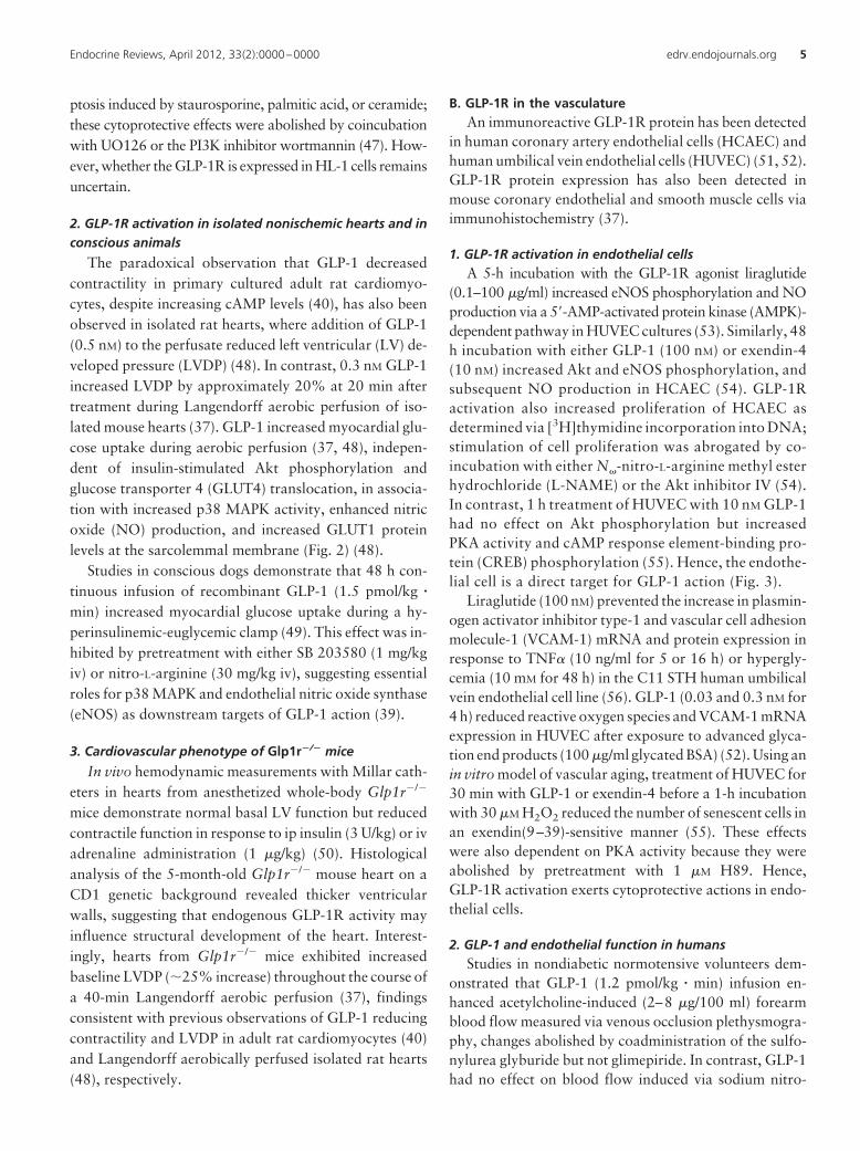
Figure 2.
Figure 2. GLP-1R-dependent intracellular signal transduction pathways in the cardiomyocyte. The signaling pathways engaged downstream of thecardiomyocyte GLP-1R lead to a reduction in apoptosis and increase in glucose uptake independent of the classical insulin-dependent pathway.ROS, Reactive oxygen species. AC, Adenylate cyclase.
4 Ussher and Drucker Cardiovascular Actions of Incretins Endocrine Reviews, April 2012, 33(2):0000–0000
ptosis induced by staurosporine, palmitic acid, or ceramide;these cytoprotective effects were abolished by coincubationwith UO126 or the PI3K inhibitor wortmannin (47). How-ever, whether the GLP-1R is expressed in HL-1 cells remainsuncertain.
2. GLP-1R activation in isolated nonischemic hearts and inconscious animals
The paradoxical observation that GLP-1 decreasedcontractility in primary cultured adult rat cardiomyo-cytes, despite increasing cAMP levels (40), has also beenobserved in isolated rat hearts, where addition of GLP-1(0.5 nM) to the perfusate reduced left ventricular (LV) de-veloped pressure (LVDP) (48). In contrast, 0.3 nM GLP-1increased LVDP by approximately 20% at 20 min aftertreatment during Langendorff aerobic perfusion of iso-lated mouse hearts (37). GLP-1 increased myocardial glu-cose uptake during aerobic perfusion (37, 48), indepen-dent of insulin-stimulated Akt phosphorylation andglucose transporter 4 (GLUT4) translocation, in associa-tion with increased p38 MAPK activity, enhanced nitricoxide (NO) production, and increased GLUT1 proteinlevels at the sarcolemmal membrane (Fig. 2) (48).
Studies in conscious dogs demonstrate that 48 h con-tinuous infusion of recombinant GLP-1 (1.5 pmol/kg �
min) increased myocardial glucose uptake during a hy-perinsulinemic-euglycemic clamp (49). This effect was in-hibited by pretreatment with either SB 203580 (1 mg/kgiv) or nitro-L-arginine (30 mg/kg iv), suggesting essentialroles for p38 MAPK and endothelial nitric oxide synthase(eNOS) as downstream targets of GLP-1 action (39).
3. Cardiovascular phenotype of Glp1r�/� mice
In vivo hemodynamic measurements with Millar cath-eters in hearts from anesthetized whole-body Glp1r�/�
mice demonstrate normal basal LV function but reducedcontractile function in response to ip insulin (3 U/kg) or ivadrenaline administration (1 �g/kg) (50). Histologicalanalysis of the 5-month-old Glp1r�/� mouse heart on aCD1 genetic background revealed thicker ventricularwalls, suggesting that endogenous GLP-1R activity mayinfluence structural development of the heart. Interest-ingly, hearts from Glp1r�/� mice exhibited increasedbaseline LVDP (�25% increase) throughout the course ofa 40-min Langendorff aerobic perfusion (37), findingsconsistent with previous observations of GLP-1 reducingcontractility and LVDP in adult rat cardiomyocytes (40)and Langendorff aerobically perfused isolated rat hearts(48), respectively.
B. GLP-1R in the vasculatureAn immunoreactive GLP-1R protein has been detected
in human coronary artery endothelial cells (HCAEC) andhuman umbilical vein endothelial cells (HUVEC) (51, 52).GLP-1R protein expression has also been detected inmouse coronary endothelial and smooth muscle cells viaimmunohistochemistry (37).
1. GLP-1R activation in endothelial cellsA 5-h incubation with the GLP-1R agonist liraglutide
(0.1–100 �g/ml) increased eNOS phosphorylation and NOproduction via a 5�-AMP-activated protein kinase (AMPK)-dependent pathway in HUVEC cultures (53). Similarly, 48h incubation with either GLP-1 (100 nM) or exendin-4(10 nM) increased Akt and eNOS phosphorylation, andsubsequent NO production in HCAEC (54). GLP-1Ractivation also increased proliferation of HCAEC asdetermined via [3H]thymidine incorporation into DNA;stimulation of cell proliferation was abrogated by co-incubation with either N�-nitro-L-arginine methyl esterhydrochloride (L-NAME) or the Akt inhibitor IV (54).In contrast, 1 h treatment of HUVEC with 10 nM GLP-1had no effect on Akt phosphorylation but increasedPKA activity and cAMP response element-binding pro-tein (CREB) phosphorylation (55). Hence, the endothe-lial cell is a direct target for GLP-1 action (Fig. 3).
Liraglutide (100 nM) prevented the increase in plasmin-ogen activator inhibitor type-1 and vascular cell adhesionmolecule-1 (VCAM-1) mRNA and protein expression inresponse to TNF� (10 ng/ml for 5 or 16 h) or hypergly-cemia (10 mM for 48 h) in the C11 STH human umbilicalvein endothelial cell line (56). GLP-1 (0.03 and 0.3 nM for4 h) reduced reactive oxygen species and VCAM-1 mRNAexpression in HUVEC after exposure to advanced glyca-tion end products (100 �g/ml glycated BSA) (52). Using anin vitro model of vascular aging, treatment of HUVEC for30 min with GLP-1 or exendin-4 before a 1-h incubationwith 30 �M H2O2 reduced the number of senescent cells inan exendin(9–39)-sensitive manner (55). These effectswere also dependent on PKA activity because they wereabolished by pretreatment with 1 �M H89. Hence,GLP-1R activation exerts cytoprotective actions in endo-thelial cells.
2. GLP-1 and endothelial function in humansStudies in nondiabetic normotensive volunteers dem-
onstrated that GLP-1 (1.2 pmol/kg � min) infusion en-hanced acetylcholine-induced (2–8 �g/100 ml) forearmblood flow measured via venous occlusion plethysmogra-phy, changes abolished by coadministration of the sulfo-nylurea glyburide but not glimepiride. In contrast, GLP-1had no effect on blood flow induced via sodium nitro-
Endocrine Reviews, April 2012, 33(2):0000–0000 edrv.endojournals.org 5
prusside (0.5- 2 �g/100 ml) (57). Similarly, fasted subjectswith T2DM and stable coronary artery disease (n � 12)exhibited improved endothelial function in response toGLP-1 infusion (2 pmol/kg � min), as indicated by an in-crease in flow-mediated vasodilation of the brachial ar-tery, independent of changes in systolic blood pressure(SBP) and diastolic blood pressure (DBP) during a hyper-insulinemic clamp (51). A study of 16 subjects with T2DMand 12 nondiabetic controls demonstrated that infusion of0.4 pmol/kg � min GLP-1 (plasma concentration of �180pg/ml) during a 2-h hyperglycemic clamp improved flow-mediated vasodilation in both groups. However, the GLP-1-mediated improvements in blood flow were consider-ablyattenuatedaftera2-monthperiodof improvedglycemiccontrol (58). Whether the effects of native GLP-1 on bloodflow are mimicked by degradation-resistant GLP-1R ago-nists remains unclear; GLP-1 but not exenatide producedvasodilation and increased cGMP release from isolated pre-constricted blood vessels ex vivo (37), and exenatide had noeffect on endothelial function in rat conduit arteries ex vivoafter infusion with intralipid in vivo, whereas native GLP-1
and GLP-1 (9–36) both exerted vasodilatory actions in con-trol experiments (59).
3. GLP-1-mediated control of heart rate (HR) andBP in animals
GLP-1R-dependent control of HR and BP is complexand species specific. Synthetic human GLP-1 (0.1–1000ng) administered into the jugular vein of male rats acutelyincreased SBP and DBP and HR; BP and HR returned tobasal levels by 25 min after GLP-1 administration (60).These effects were independent of catecholamines and ad-renergic receptors, because pretreatment with either pro-pranolol (1 mg/kg iv) or phentolamine (0.1 mg/kg iv) didnot prevent the increases in HR and BP (60). GLP-1 alsoincreased HR and BP in streptozotocin-induced diabeticrats, although plasma insulin levels were not measured(60). The increased BP and HR observed after adminis-tration of GLP-1 or exendin-4 in rats was dependent onGLP-1R signaling and abolished by iv infusion of exen-din(9–39) (61). The GLP-1-stimulated increase in HR andBP in rodents involves dual pathways originating from boththe CNS and periphery, with the neural pathway requiring
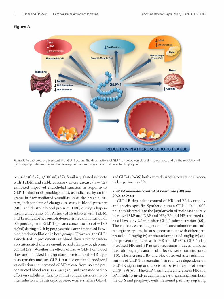
Figure 3.
Figure 3. Antiatherosclerotic potential of GLP-1 action. The direct actions of GLP-1 on blood vessels and macrophages and on the regulation ofplasma lipid profiles may impact the development and/or progression of atherosclerotic plaques.
6 Ussher and Drucker Cardiovascular Actions of Incretins Endocrine Reviews, April 2012, 33(2):0000–0000

intact vagus nerve transmission, because intracerebroven-tricular (icv) GLP-1 (100 ng) failed to increase HR and BP invagotomized rats (62). Support for a role of the CNS in theGLP-1R-dependent control of HR and BP derives from te-lemetry studies of freely moving rats, whereby GLP-1Ragonist-mediated increases in HR and BP were coupled toactivation of autonomic control centers in the CNS (63).Administration of icv exendin-4 induced Fos-like immu-noreactivity in neurons innervating sympathetic pregan-glionic neurons in the paraventricular hypothalamus, thearcuate nucleus, and the lateral hypothalamic area in therat brain (63). Furthermore, double-labeling immunohis-tochemistry after icv exendin-4 detected induction of Fos-like and tyrosine hydroxylase immunoreactivity in cat-echolaminergic neurons in the nucleus of the solitary tractand locus coeruleus (63); icv exendin-4 also activated ty-rosine hydroxylase transcription in adrenal medullary cat-echolamine neurons (63). Continuous infusion of the se-lective �2-adrenoceptor antagonist ICI 118551 or thenonselective �-adrenoceptor antagonist propranololabolished the effects of exendin-4 (250 ng/kg iv) on HR inrats (64). Furthermore, exendin-4 (250 ng/kg iv) was un-able to increase HR in adrenalectomized rats (25). Takentogether, considerable data suggest that GLP-1 engagesthe rodent sympathetic nervous system to modify HR andBP (63–65). Nevertheless, other investigators have re-ported that the vasoconstrictor properties of exendin-4were independent of the autonomic nervous system, be-cause the increase in mean arterial pressure after exen-din-4 administration persisted despite continuous infu-sion with propranolol or the �-adrenoceptor antagonistphentolamine (24). Furthermore, iv GLP-1 (20 nmol)acutely increased HR over a 1-h period in adult male ratsthat had previously undergone either adrenalectomy orvagotomy (66).
GLP-1R activation in neurons that innervate cardiacvagal neurons in the nucleus ambiguous, resulted in di-minished HR variability, and reduced parasympatheticmodulation of the heart (67). GLP-1 may also increase BPin rodents through the vasopressin system, because anintraarterially injected vasopressin receptor antagonist, B-mercapto (10 �g/kg), abolished the rise in BP after 100 ngicv GLP-1 (68). In contrast to findings of increased BPin mice and rats after acute GLP-1R activation, GLP-1(30–60 �mol/kg body weight � min) transiently increasedHR yet had no effect on aortic BP in conscious calves (69).Hence, the mechanisms underlying the acute effects ofGLP-1R agonists to increase HR and BP are complex, mayinvolve both the sympathetic and parasympathetic ner-vous system, and appear species specific.
4. GLP-1-mediated control of HR and BP in humansStudies of GLP-1 effects on BP in humans have yielded
results quite different from those obtained acutely in ro-dents. Infusion of GLP-1 (2.4 pmol/kg � min for 10 minfollowed by 1.2 pmol/kg � min) for 65 min in 55 healthyhuman subjects (13 men and 42 women, mean age 31 yr)had no effect on SBP or DBP, plasma norepinephrine lev-els, or HR variability calculated at both low and highfrequency, indices of cardiac sympathetic and parasym-pathetic activity, respectively (70). In contrast, acute scinjection of GLP-1 (80 nmol/ml) into the anterior abdom-inal wall transiently increased HR (64 � 2 vs. 54 � 2beats/min at 40 min after injection) and BP (83 � 5 vs.77 � 4 mm Hg at 20 min after injection) in 10 healthyhuman subjects; however, values returned to near baselineby approximately 50–60 min after injection (71). Theeffects of GLP-1R activation in patients with elevated BPalso contrasts from those reported in animals, which willbe described in detail in Section IV.A.2.
C. GLP-1 action and dyslipidemia
1. Studies of GLP-1 action on lipoprotein synthesisand secretion
Rats infusedwithGLP-1(20pmol/kg �min)viathe jugularvein for 6 h exhibited a reduction in triacylglycerol (TAG)absorption, decreased intestinal lymph flow, and reducedintestinal apolipoprotein B-48 (ApoB-48) production (72).Furthermore, in studies of fructose-fed hamsters, the DPP-4inhibitor sitagliptin (5 mg/kg via oral gavage for 2 or 3 wk)decreased fasting plasma TAG, predominantly in the very-low-density lipoprotein (VLDL) fraction, whereas both ex-endin-4 (24 nmol/kg ip) and sitagliptin (10 mg/kg via oralgavage) acutely decreased postprandial TAG and ApoB-48levels in male C57BL/6J mice administered olive oil and Tri-ton WR1339 after a 5 h fast (73). Exendin-4 also reducedTAGandApoB-48secretionwhenadministered1hafter theoral fat load, suggesting the effect on postprandial lipidmetabolism was not related to delayed gastric emptying(73). Moreover, exendin-4 (0.1 nM) directly decreasedApoB-48 secretion in primary cultured hamster entero-cytes, as assessed by reduced levels of 35S-labeledApoB-48 in the media over a 90-min time course,whereas Glp1r�/� mice exhibited enhanced appearanceof plasma TAG after olive oil administration. Thesefindings support a direct essential role for GLP-1 in thecontrol of chylomicron secretion (Fig. 3) independent ofchanges in gastric emptying (73).
2. GLP-1 action, on the liver and dyslipidemiaChronic administration of the DPP-4 inhibitor vilda-
gliptin (1 mM in drinking water) for 8 wk reduced fastingplasma TAG and cholesterol levels in wild-type mice fed a
Endocrine Reviews, April 2012, 33(2):0000–0000 edrv.endojournals.org 7

high-fat diet for 14 wk (45% kcal from lard) but failed tolower plasma lipid levels in mice lacking both the Glp1rand Gipr (74). Vildagliptin also reduced hepatic expres-sion of long-chain acyl coenzyme A (CoA) synthetasemRNA in wild-type but not in Glp1r�/�:Gipr�/� mice,suggesting that DPP-4 inhibition may reduce plasma TAGlevels via prevention of VLDL assembly (74). Ob/ob micetreated for 60 d with exendin-4 (10 �g/kg every 24 h forthe first 14 d, followed by either 10 or 20 �g/kg every 12 hfor the remaining 46 d) exhibited a marked reduction inhepatic lipid content as assessed by oil red O staining,associated with reduced steroyl CoA desaturase and sterolresponse element-binding protein-1c mRNA expressionand increased peroxisome proliferator-activated recep-tor-� (PPAR�) mRNA expression (75). Furthermore,daily liraglutide (200 �g/kg ip) treatment for 4 wk in micefed a high-fat and high-fructose corn syrup diet for 8 wkreversed hepatic steatosis assessed by oil red O staining(76). This effect was associated with increased mRNA ex-pression of acyl CoA oxidase and increased protein ex-pression of long-chain acyl CoA dehydrogenase, suggest-ing an elevation in peroxisomal and mitochondrial fattyacid �-oxidation, respectively (76). In primary culturedhuman hepatocytes, 24 h treatment with 10 nM exendin-4in the presence of 0.8 mM oleate, elaidate, or palmitatereduced lipid stores assessed by oil red O staining as wellas caspase-3 cleavage and DNA condensation, resultingin improved cell survival (77). Evidence for reducedendoplasmic reticulum stress and increased macroau-tophagy was also observed, because exendin-4 in-creased protein expression of glucose-regulated protein78, reduced expression of CCAAT-enhancer-bindingprotein homologous protein, and increased autophagicvacuole formation.
Daily treatment of high-fat diet-fed mice (6 wk, 44% kcalfrom fat) for 4 wk with the GLP-1R agonist CNT0736 (0.1or 1 mg/kg ip) reduced fasting VLDL production in a bodyweight-independent manner; however, exendin-4 adminis-tration (7.1 �g/kg ip) did not affect these parameters (78).Nevertheless, whether GLP-1 acts directly on the liver tocontrol lipoprotein synthesis and secretion remains unclearbecause treatment of primary murine hepatocytes for 16 hwith 40 nM exendin-4 had no effect on TAG or cholesterolsynthesis/secretion, and Glp1r mRNA transcripts corre-sponding to the entire open reading frame of the GLP-1Rwere not detected in isolated hepatocytes (74). In contrast,a 142-bp Glp1r mRNA transcript and immunoreactiveGLP-1R protein were detected in RNA from human liverbiopsies and human hepatoma HepG2 cells via RT-PCRand Western blotting, respectively (79). ImmunoreactiveGLP-1R protein detected by Western blotting was alsoidentified in both isolated human and rat hepatocytes,
and exenatide directly increased mRNA expression ofPPAR� and PPAR� in human hepatocytes (75, 79, 80).Because data from multiple groups has yielded contrast-ing results on the presence of hepatic GLP-1R expres-sion in rodent or human hepatocytes, the mechanismthrough which GLP-1 acts on the liver requires furtherelucidation (18, 74, 81, 82).
3. GLP-1 action in adipose tissueWhether the classical GLP-1R is expressed in the adi-
pocyte is uncertain. GLP-1 binding sites were detectedin solubilized membranes from both human and rat ad-ipose tissue using radioligand binding assays (83, 84),although others have been unable to detect Glp1rmRNA expression in adipose tissue from humans andrats (18, 38). Nevertheless, GLP-1 treatment of isolatedrat adipocytes increased lipolysis (85) and exogenousGLP-1 exerted both lipolytic and lipogenic effects in hu-man adipocytes (86). On the other hand, in situ microdi-alysis in nine healthy volunteers failed to demonstrate alipolytic action of GLP-1 (87). Hence, whether GLP-1 ex-erts direct actions in adipose tissue depots through theGLP-1R remains unclear.
4. GLP-1 and dyslipidemia in human subjectsGLP-1 infusion (1.2 pmol/kg � min) for 6.5 h in 14
healthy volunteers inhibited the postprandial increasein plasma TAG and free fatty acid (FFA) levels after a250-kcal solid test meal (88). Short-term studies in 12subjects with T2DM demonstrated that addition ofGLP-1 (sc injection of 25 nmol directly before meals) toinsulin therapy for 5 d, followed by administration ofGLP-1 alone for the last 2 d, decreased plasma concen-trations of VLDL-TAG (1.30 � 0.36 vs. 2.08 � 1.11mM) while increasing the size of low-density lipoprotein(LDL) cholesterol particles (mean diameter of 22.9 vs.22.3 nm) (89). A study in 50 subjects with T2DM in-fused for 4 h with GLP-1 (1.2 pmol/kg � min) after anovernight 10-h fast revealed a reduction in plasma FFAlevels (�0.25 mM decrease throughout the first 2 h)(90). A hyperglycemic clamp followed by a hyperinsu-linemic-euglycemic clamp in 16 elderly patients withT2DM treated for 3 months with a continuous sc GLP-1infusion (100 pmol/kg � h) also demonstrated decreasedplasma FFA levels during the hyperinsulinemic portionof the clamp, which was associated with a significantincrease in plasma insulin levels (91). A double-blindcrossover study in 35 patients with recent-onsetT2DM demonstrated that a single sc injection of ex-enatide (10 �g) markedly reduced postprandial levels ofTAG and ApoB-48, as well as plasma remnant lipopro-tein cholesterol, for up to 8 h in subjects fed a high-
8 Ussher and Drucker Cardiovascular Actions of Incretins Endocrine Reviews, April 2012, 33(2):0000–0000
calorie (600 kcal/m2 body surface area), fat-enriched(45%) breakfast meal after an overnight fast (92). Theseeffects were already observed during the first 4 h afterthe breakfast meal, when no changes in plasma insulinwere observed. Interpretation of studies demonstratingthat acute GLP-1R activation lowers postprandial li-pemia requires consideration of whether the findingsare due in part to the acute effect of GLP-1R agonists toinhibit gastric emptying and gut motility, actions thatare diminished with more sustained GLP-1R activation(93, 94).
DPP-4 inhibitors have also demonstrated favorable ef-fects on postprandial dyslipidemia in T2DM. A 4-wktreatment with vildagliptin (50 mg twice daily) reducedpostprandial elevations of plasma TAG, chylomicron-TAG, chylomicron-ApoB-48, and chylomicron-choles-terol for up to 8 h in response to a fat-rich test meal (1000kcal, 72 g fat) after an overnight fast (95). Similarly, adouble-blind crossover study in 36 subjects with T2DMtreated with sitagliptin (100 mg/d taken during the morn-ing meal) for 6 wk also demonstrated a reduction in post-prandial elevations of plasma TAG, ApoB-48, and FFA
levels for up to 8 h in response to a test meal (1003 kcal,68.6 g fat) after a 12-h overnight fast (96). Because DPP-4inhibitors reduce postprandial lipemia without inhibitionof gastric emptying or weight loss, these findings are con-sistent with a role for local GLP-1 in the control of intes-tinal chylomicron secretion (73).
D. Direct vs. indirect effects of GLP-1R agonism oncardiac function
One of the challenges in understanding the effects ofGLP-1 on the heart involves elucidation of the direct vs.indirect pleiotropic metabolic properties of GLP-1 (Figs.1, 2, and 4). Section III.D.1 will briefly highlight some ofthe key indirect actions/effectors of GLP-1 and their po-tential impact on cardiac function.
1. GLP-1 and insulinActivation of the �-cell GLP-1R during hyperglycemia
usually increases plasma insulin levels, leading to in-creased myocardial glucose uptake and glycogen synthesisand decreased fatty acid oxidation (Fig. 4) (97). Further-more, increased insulin receptor signaling in the heart
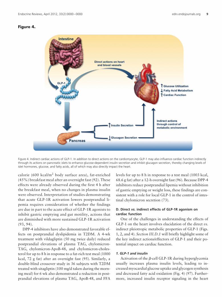
Figure 4.
Figure 4. Indirect cardiac actions of GLP-1. In addition to direct actions on the cardiomyocyte, GLP-1 may also influence cardiac function indirectlythrough its actions on pancreatic islets to enhance glucose-dependent insulin secretion and inhibit glucagon secretion, thereby changing levels ofislet hormones, glucose, and fatty acids, all of which may also directly impact the heart.
Endocrine Reviews, April 2012, 33(2):0000–0000 edrv.endojournals.org 9
leads to activation of the PI3K/Akt pathway, increasedeNOS activity, and inhibition of AMPK (98, 99). Insulinalso inhibits lipolysis and reduces plasma FFA levels (100,101), and this effect, coupled with the improvement inmyocardial glucose metabolism, supports the rationale forevaluating glucose-insulin-potassium infusions in subjectswith acute myocardial infarction (AMI) (102). Hence,whether the metabolic actions of GLP-1 on the heart invivo reflect direct and/or indirect mechanisms requirescareful consideration.
2. GLP-1, obesity, and adiponectinGLP-1R activation in the hypothalamus reduces appe-
tite and leads to weight loss (103–105). Obesity is a sig-nificant risk factor for the development of cardiovasculardiseases, with every 1 kg/m2 increment in body mass indexresulting in a 5 and 7% increase in the risk of heart failurein men and women, respectively (106). Thus, weight lossassociated with GLP-1R agonists may contribute to po-tential cardioprotective effects. Furthermore, weight lossis associatedwith increasedplasmaadiponectin levels, andadiponectin protects against both AMI and cardiac hy-pertrophy (107, 108). Indeed, the reduction in body massin 9-month-old spontaneously hypertensive, and heartfailure-prone rats infused ip with 1.5 pmol/kg � min GLP-1for 3 months is associated with improved LV function,increased survival, and an approximately 50% increase incirculating adiponectin levels (109). Consistent with thesefindings, exendin-4 (2.5–5 nM) directly increased adi-ponectin mRNA expression in 3T3-L1 adipocytes, an ef-fect blocked by the GLP-1R antagonist exendin (9–39) orthe PKA inhibitor H89 (110). Weight loss arising fromtherapy with GLP-1R agonists is also associated with re-ductions in dyslipidemia (111). Hence, attribution of di-rect vs. indirect effects to the cardiovascular actions ofGLP-1 may be difficult and requires careful analyses.
E. Cardiovascular biology of GLP-1 (9–36)GLP-1 (9–36) is the primary GLP-1 metabolite in vivo,
and circulating levels of GLP-1 (9–36) are greater than thoseof intact bioactive GLP-1 (34, 112). GLP-1 (9–36) binds tothe classical GLP-1R with low affinity and at pharmacolog-ical doses acts as a weak competitive antagonist of the �-celland gastrointestinal tract GLP-1R in vivo. However, iv ad-ministration of GLP-1 (9–36) in combination with glucosehas no effect on insulin secretion, glucose elimination, orinsulin-independent glucose disposal in either wild-type orGlp1r�/� mice (113). In contrast, GLP-1 (9–36) enhancedinsulin-independent glucose clearance in anesthetized pigs(114). Similarly, infusion of GLP-1 (9–36) into healthyfasted humans in conjunction with a test meal significantlyreduced postprandial glycemia, independently of changes
in insulin or glucagon secretion or gastric emptying (115).The magnitude of these effects was minor in comparisonwith those of native GLP-1. Moreover, a separate study inhealthy human subjects found that GLP-1 (9–36) infusionhad no direct effect on glucose tolerance, insulin secretionor sensitivity, or GLP-1 action (116). Furthermore, simul-taneous infusion of GLP-1 (9–36) with GLP-1 (7–36) didnot alter the magnitude of GLP-1 (7–36)-stimulated insu-lin secretion (116). Intriguingly, infusion of GLP-1 (9–36)had no effect on insulin sensitivity in lean subjects butsuppressed hepatic glucose production, in associationwith increased plasma insulin levels, in obese subjects(117). The complexity of GLP-1 (9–36) action is under-scored by studies demonstrating that a secondary cleavageproduct derived from GLP-1 (9–36), GLP-1 (28–36), lo-calizes to mitochondria and also reduces hepatic glucoseproduction in hepatocytes (118). It is important to notethat the actions of both GLP-1 (9–36) and GLP-1 (28–36)appear at pharmacological concentrations and are mostprominent in stressed metabolic states such as insulin re-sistance (118, 119). Hence, further research is required toascertain the biological relevance of GLP-1 (9–36) andGLP-1 (28–36) in the human endocrine and cardiovascu-lar system.
1. GLP-1 (9–36) action in cardiomyocytesGLP-1 (9–36) improved the viability of both Glp1r�/�
and Glp1r�/� mouse neonatal cardiomyocytes during re-oxygenation after 48 h hypoxia; preincubation with 0.3nM GLP-1 (9–36) for 20 min before exposure to hydrogenperoxide for 7 h also improved cell viability (42). Unex-pectedly, the actions of GLP-1 (9–36) were blocked bypretreatment with the classical GLP-1R antagonist exen-din (9–39). In contrast, exendin-4 (3 nM) had no effect oncell viability in hypoxic Glp1r�/� neonatal myocytes sub-jected to reoxygenation or in cardiomyocytes treated withhydrogen peroxide (42).
2. GLP-1 (9–36) activity in the isolated heart and inconscious animals
A 48-h continuous infusion of GLP-1 (9–36) (1.5 pmol/kg � min) in conscious dogs with pacing-induced dilated car-diomyopathy significantly reduced LV end diastolic pres-sure, and increased LV contractility and myocardial glucoseuptake during a hyperinsulinemic-euglycemic clamp (120).Consistent with the hypothesis that GLP-1 (9–36) exerts itsactions through a separate receptor, pretreatment with na-tive GLP-1 (0.3 nM) enhanced the recovery of LVDP duringreperfusion after 30 min global ischemia in hearts fromGlp1r�/� mice (37).Notably,preventionof the formationofGLP-1 (9–36) with the DPP-4 inhibitor sitagliptin preventedthe recovery of LVDP during reperfusion in hearts from
10 Ussher and Drucker Cardiovascular Actions of Incretins Endocrine Reviews, April 2012, 33(2):0000–0000

Glp1r�/� mice. Interestingly, exendin-4 (5 nM) also mod-estly improved the recovery of LVDP during reperfusionafter 30 min global ischemia in Glp1r�/� mice (37), add-ing further support for a second receptor capable of rec-ognizing GLP-1R agonists in the heart. Related studiesdemonstrated that 0.03–3 nM GLP-1 (9–36) added to theperfusate for the first 15 min of a 120-min reperfusionperiod after a 45-min ischemic insult in isolated rat heartsresulted in a significant improvement in LVDP, althoughit did not reduce infarct size (121). However, follow-upstudies from this same group could not reproduce thesefindings, demonstrating that native GLP-1, but not GLP-1(9–36), was cardioprotective in isolated ischemic rathearts ex vivo (122). Although pharmacological levels ofGLP-1 (9–36) are cardioprotective, results using DPP-4inhibitors, which markedly lower levels of GLP-1 (9–36),demonstrate significant cardioprotection after DPP-4 in-hibition in both normal and diabetic rodent models and inshort-term human studies (123–125).
IV. GLP-1 and Cardiovascular Disease
A. GLP-1 and hypertension
1. Animal studiesAlthough acute GLP-1R activation increases HR and
BP in rodents, GLP-1 actions on endothelial cells, such asincreased NO production (Fig. 3), would be predicted tobe antihypertensive (54). Indeed, db/db mice chronicallytreated with exendin-4 (ip 20 nmol/kg twice daily) for 12wk displayed a marked reduction in SBP; exendin-4 alsoattenuated the increase in SBP (�10 vs. �25 mm Hg in-crease) in db/db mice provided with 2% salt in their drink-ing water for 2 wk (126). In male C57BL/6J mice infusedwith angiotensin II (1 �g/kg � min for 2 wk), twice-dailyexendin-4 (20 nmol/kg ip) also reduced SBP (126). A 4-htreatment with exendin-4 (10 nM) prevented acute angio-tensin II-induced ERK phosphorylation in kidney proxi-mal tubular cells, raising the possibility that GLP-1R signal-ing directly modifies the actions of exogenous angiotensin II(126).A7-d sc infusionof exendin-4 (1 �g/kg �d)viaosmoticpumps also reversed corticosterone-induced increases in SBPand DBP in rats independent of changes in body weight andcaloric intake (127). Hence, although acute GLP-1R activa-tion transiently increases BP in preclinical studies, sustainedGLP-1R activation reduces BP in different animal models.
2. Studies in subjects with diabetes and/or heart diseaseInfusion of GLP-1 (0.7 pmol/kg � min) for 48 h in 15
nondiabetic human subjects with heart failure (New YorkHeart Association class II/III) resulted in small increases inHR (67 � 2 vs. 65 � 2 beats/min) and DBP (71 � 2 vs. 68 �
2 mm Hg) (128). Exenatide administered twice daily todiabetic subjects for 12 wk produced small (�2 beats/min)but nonsignificant changes in HR, and a trend toward areduction in SBP, associated with a modest weight loss of1.8 kg (129). In a 26-wk head-to-head study of liraglutidevs. sitagliptin in subjects with T2DM, HR increased withliraglutide (3.94 beats/min) but not with sitagliptin, andreductions in SBP and DBP were actually greater with sita-gliptin compared with liraglutide (130). A greater reduc-tion in DBP with sitagliptin was also observed after 1 yr oftreatment in comparison with liraglutide (131), and BPreductions were also modestly greater with sitagliptincompared with exenatide once weekly in the Results fromthe Diabetes Therapy Utilization: Researching Changes inA1C, Weight and Other Factors Through Intervention withExenatide Once Weekly (DURATION-4) study (132). Nev-ertheless, acute administration of the high-molecular-weightexendin-transferrin fusion protein in subjects with T2DMproduced significant increases in HR and DBP (mean in-crease of 10 mm Hg) (133). Hence, further analysis of theeffects of structurally distinct GLP-1R agonists on HR andBP in diabetic subjects is warranted.
Nevertheless, the majority of clinical trials investigat-ing the antidiabetic actions of GLP-1R agonists have re-ported reductions in BP. Results from the DURATION-1trial demonstrated that patients treated with exenatidecontinuously for 52 wk had a significant reduction in SBP(134). Approximately 50% of patients with a SBP of atleast 130 mm Hg at baseline achieved a normal SBP by wk52. Consistent with these findings, 314 overweight pa-tients receiving exenatide 10 �g twice daily for 82 wk alsoexperienced improvement in both SBP and DBP (135), anda retrospective analysis of 6280 patients reported a sig-nificant reduction in both SBP and DBP with exenatidetherapy (111). Similarly, combination therapy with dailyliraglutide (0.6, 1.2, or 1.8 mg)/metformin (2000 mg) for16 wk in 928 Asian subjects with T2DM reduced SBP (�3mm Hg decrease) in comparison with glimepiride (4 mg)/metformin (2000 mg) administration (136). A retrospec-tive analysis of 110 patients with T2DM treated with li-raglutide (0.6, 1.2, or 1.8 mg daily) for a mean duration of7.5 months (range, 6 months to 1.1 yr) demonstrated areduction in SBP (5 � 2 mm Hg) in the first 3 months oftreatment (137). In an analysis encompassing three ran-domized phase 3 trials totaling 2665 patients, 26 wk treat-ment with once-daily liraglutide (1.2 or 1.8 mg) in com-bination with metformin, glimepiride, or metformin androsiglitazone reduced SBP by 2.29–6.71 mm Hg (138).Furthermore, a study of 268 obese nondiabetic patientswho completed a 20-wk treatment with once-daily sc li-raglutide (1.2, 1.8, 2.4, or 3.0 mg) followed by a non-blinded 2-yr extension (final dose of 3.0 mg) demonstrated
Endocrine Reviews, April 2012, 33(2):0000–0000 edrv.endojournals.org 11
a mean 4.6 mm Hg decrease in SBP (139). The improve-ment in BP in the majority of these studies was associatedwith reductions in body weight. Nevertheless, the reduc-tions in SBP with liraglutide appear rapidly, often beforesignificant weight loss is observed (136, 138).
B. GLP-1 action in experimental modelsof atherosclerosis
Both direct and indirect actions of GLP-1 may con-tribute to the potential reduction of atherogenesis (Fig.3). GLP-1R has been localized to mouse aortic smoothmuscle and endothelial cells, as well as monocytes andmacrophages, using immunocytochemistry and West-ern blotting (140). Continuous infusion of exendin-4(300 pmol/kg � d or 24 nmol/kg � d) in nondiabeticC57BL/6 and ApoE�/� mice reduced monocyte adhesionto aortic endothelial cells at 24 d, associated with a re-duction in atherosclerotic lesion size after 28 d treatment.Furthermore, treatment for 1 h with exendin-4 (0.03–3nM) reduced levels of mRNA transcripts for the inflam-matory markers monocyte chemoattractant protein-1 andTNF� in response to 1 h lipopolysaccharide (1 �g/kg) incultured peritoneal macrophages harvested from mice 3 dafter injection of 3% thioglycolate (140). Continuous in-fusion of exendin-4 (24 nmol/kg � d) for 4 wk in C57BL/6mice also reduced neointimal formation in response toendothelial denudation of the femoral artery (141). In-triguingly, 12 h pretreatment with 10 nM exendin-4 re-duced platelet-derived growth factor-induced (25 ng/ml)bromodeoxyuridine incorporation into DNA of primarycultured mouse aortic smooth muscle cells (141). Na-gashima et al. (142) reported that continuous GLP-1 orGIP administration (4 wk osmotic mini-pump infusion of2.2 nmol/kg � d) prevented atherosclerotic lesion develop-ment in ApoE�/� mice, which may involve reduced foamcell formation in macrophages, because peritoneal mac-rophages harvested from exendin-4-treated mice exhib-ited reduced CD36 protein expression and decreased cho-lesterol ester accumulationafter18hexposure to10 �g/mloxidized LDL. Despite these intriguing findings in ani-mals, data on the long-term effects of incretin-based ther-apy on atherosclerosis-associated outcomes in diabetichumans is not yet available.
C. GLP-1R activation in ischemic heart disease
1. Animal studiesMultiple preclinical studies have demonstrated cardio-
protective effects of native GLP-1 and GLP-1R agonists inexperimental models of ischemic heart disease (37, 45, 48,143, 144). Both GLP-1 and exendin-4 improved recoveryof LVDP in isolated perfused rat and mouse hearts duringreperfusion after ischemia (37, 48, 121). Similarly, iv in-
fusion of 4.8 pmol/kg � min GLP-1 decreased infarct sizeafter 30 min ischemia induced by temporary occlusion ofthe left anterior descending (LAD) coronary artery in rats(143), and exendin-4 (10 �g iv and sc 5 min before theonset of reperfusion) decreased infarct size and improvedLV systolic function 72 h following 75 min LAD coronaryartery occlusion in pigs (145). In contrast, recombinantGLP-1 administered via the jugular vein at 3 pmol/kg � min15 min before the onset of ischemia failed to reduce infarctsize in a pig model of ischemia secondary to 60 min leftcircumflex coronary artery occlusion (146). Furthermore,liraglutide (10 �g/kg) administered to pigs for 3 d beforeLAD coronary artery occlusion did not reduce infarct sizeor improve LV function (147). The GLP-1R agonist albi-glutide, injected sc for 3 d at 3 or 10 mg/kg � d, reducedinfarct size assessed 24 h after 30 min temporary LADcoronary artery occlusion in normoglycemic rats (148).The benefit of albiglutide was attributed to improved car-diac energetics, because in vivo 13C nuclear magnetic res-onance studies demonstrated decreased fatty acid oxida-tion and increased glucose oxidation rates.
A GLP-1-transferrin protein also limited infarct size after30 min LAD coronary artery occlusion in rabbits, whethergiven sc at a dose of 10 mg/kg 12 h before the onset of isch-emia or iv at the onset of ischemia (149). Furthermore, lira-glutide administration (75 �g/kg ip twice daily) for 1 wkbefore LAD occlusion improved survival and cardiac outputassessed 4 wk after permanent occlusion of the LAD coro-nary artery in both nondiabetic and diabetic male mice(45). Interestingly, 30 nM liraglutide administered incoronary arteries at the onset of reperfusion did notprotect against reperfusion injury in isolated perfusedmouseheartsafter30minofglobal ischemia,but liraglutidedidimprove recovery of LVDP during reperfusion if injected intothe mouse in vivo before ischemia/reperfusion ex vivo. Thisobservation suggests that liraglutide may achieve cardio-protection in part through mechanisms requiring the heartto receive its normal neural, humoral, and vascular input.Furthermore, the observations that GLP-1R activationdoes not universally produce cardioprotection in preclin-ical studies highlights the importance of future studies de-signed to understand the precise biological mechanismsand cellular sites of action for different GLP-1R agonistsin the cardiovascular system.
2. Studies in subjects with ischemic heart diseaseThe observation that a 72-h infusion of GLP-1 (1.5
pmol/kg � min) initiated approximately 3.5 h after angio-plasty within approximately 6.5 h from symptom onset inpatients with AMI enhanced LV ejection fraction (LVEF)(29 � 2 vs. 39 � 2%) and infarct zone-related regionalwall motion engendered considerable interest in the car-
12 Ussher and Drucker Cardiovascular Actions of Incretins Endocrine Reviews, April 2012, 33(2):0000–0000
dioprotective actions of GLP-1 in humans (150). How-ever, this report was a single-center nonrandomized pilotstudy in a small number of patients (n � 10). Nevertheless,subsequent studies have confirmed that GLP-1 may becardioprotective. An iv infusion of GLP-1 (1.2 pmol/kg �
min) 30 min before dobutamine stress echocardiographyand continuing for 30 min into recovery in 14 patients(four with T2DM) with known coronary artery disease,protected against LV dysfunction, and mitigated postisch-emic myocardial stunning (151). These same investigatorsdemonstrated a reduction in LV dysfunction and myocar-dial stunning during dual inflation coronary balloon oc-clusion in 20 nondiabetic patients with single-vessel dis-ease in the LAD coronary artery, in whom GLP-1 wasinfused at 1.2 pmol/kg � min after completion of the firstballoon occlusion (152). A larger randomized, double-blind, placebo-controlled trial investigating the effects ofa 6-h exenatide infusion initiated 15 min before onset ofreperfusion in 172 patients undergoing primary angio-plasty to treat ST-segment elevation MI also demonstrateda reduction in the ischemic area at risk (153). Exenatidewas infused to achieve a target plasma concentration from0.03–0.3 nM (mean concentration 0.177 � 0.069 nM) andreduced reperfusion injury in these patients as determinedby an increase in myocardial salvage index and decrease ininfarct size relative to the ischemic area at risk assessed bycardiac magnetic resonance at approximately 90 d afterinfusion. However, mortality and LV contractility werenot different in patients receiving exenatide (153). Al-though accumulating data on GLP-1 action and cardio-vascular function in humans with ischemic heart diseaseappears promising with respect to safety and potentialbenefit, much larger double-blinded randomized trials arenecessary to determine whether GLP-1R agonists are car-dioprotective in a wide range of subjects with T2DM.
D. GLP-1R agonists in heart failure
1. Preclinical models of heart failureStudies in animals illustrate that GLP-1R activation
may produce beneficial effects on the failing heart (49,154). In a dog model of 28-d rapid pacing-induced heartfailure, a 48-h infusion of GLP-1 (1.5 pmol/kg � min) ex-erted insulinomimetic properties on the heart, increasingglucose uptake during a hyperinsulinemic-euglycemicclamp (49). Furthermore, GLP-1 decreased HR and in-creased LV systolic function, and decreased plasma levelsof norepinephrine and glucagon. In the spontaneously hy-pertensive and heart failure-prone rat, a 3-month ip infu-sion of 1.5 pmol/kg � min GLP-1 improved survival andpreserved LV contractile function, an effect associatedwith reduced cardiomyocyte apoptosis (109). Similarly,
liraglutide administered for 1 wk (75 �g/kg ip twice daily)to mice before permanent occlusion of the LAD coronaryartery reduced cardiac hypertrophy, decreased LV struc-tural remodeling, and improved cardiac output 4 wk afterinduction of ischemia and LV dysfunction (45). In a ratmodel of chronic heart failure, an 11-wk sc infusion ofGLP-1 (2.5 or 25 pmol/kg � min) or the exenatide analogAC3174 (1.7 or 5 pmol/kg � min) 2 wk after permanentLAD coronary artery occlusion significantly enhanced LVfunction (increased LVEF and fractional shortening),while also reducing adverse LV remodeling (decreased LVend systolic and diastolic dimensions) and improving sur-vival (155). Taken together, the available data support abeneficial role for both native GLP-1 and GLP-1R agonistsin preclinical models of heart failure.
2. Studies in human subjects with heart failureInitial trials in humans demonstrated salutary effects of
GLP-1 in subjects with heart failure; a 5-wk infusion ofGLP-1 (2.5 pmol/kg � min) in 12 patients with New YorkHeart Association class III/IV heart failure improvedLVEF, oxygen consumption, and 6-min walk test scores(154). However, this was a single-center nonrandomizedtrial,whoseresultsrequirereplication.A48-hinfusionofGLP-1(0.7 pmol/kg � min) in 15 humans with congestive heart failurebutwithoutdiabetesproducednobeneficialeffectsonLVfunc-tionandactuallyresultedinminorincreasesinHR(2beats/min)and DBP (3 mm Hg) (128). Although this particular study wasrandomized and double-blinded, a brief duration of GLP-1 in-fusion may be insufficient to increase function in a decompen-sated failing heart.
V. DPP-4 Inhibition and Cardiovascular Function
This section will discuss the cardiovascular biology of DPP-4and actions of DPP-4 inhibitors in the regulation of BP, thedevelopment of atherosclerosis, the setting of AMI, and thefailing heart, illustrating differences between DPP-4 inhibi-tors and GLP-1R agonists where appropriate.
A. DPP-4 expression in the cardiovascular systemDPP-4 is a widely expressed enzyme and has been lo-
calized to smooth muscle and endothelial cells in differentspecies (156, 157). Short-term exposure to high glucoseinduces DPP-4 activity in microvascular endothelial cells(158). Although the precise biological role of DPP-4 in thecardiomyocyte, endothelial, or coronary smooth musclecell requires further study, DPP-4 is also a circulating pro-tein, and thus DPP-4 activity in the systemic and coronarycirculation may influence intact levels of GLP-1 and othervasoactive DPP-4 substrates reaching the myocardium
Endocrine Reviews, April 2012, 33(2):0000–0000 edrv.endojournals.org 13

and vasculature (Fig. 5). The soluble form of DPP-4 mayalso interact with the mannose 6-phosphate receptor onhuman endothelial cells, promoting endothelial cell trans-migration of T cells (159).
B. Potential cardioactive DPP-4 substratesAlthough GLP-1 is classically viewed as the primary
DPP-4 substrate capable of influencing cardiovascularfunction, DPP-4 cleaves multiple peptides, many of whichalso have direct actions on the heart and blood vessels(Table 1 and Fig. 5), as discussed below in Section V.B.1.
1. Glucose-dependent insulinotropic polypeptideThe GIPR has been detected by immunohistochemistry
in both rat atrial and ventricular tissue (25). Although thebiological function, if any, of GIP in the heart is unknown,studies in rodents have linked GIPR activation to increasedadipogenesis, enhanced adipokine expression, and obesity(160–163). Hence, DPP-4-inhibition and the subsequent in-crease in plasma levels of intact bioactive GIP may have po-tential direct or indirect effects on the heart. The GIPR is alsoexpressed in endothelial cells (164), and GIPR activation
promotes endothelial cell proliferation in an endothelin-1-dependent manner in HUVEC cultures (165). The observa-tion that GIP activates different signal transduction path-ways in endothelial cells isolated from the hepatic arteryvs. the portal vein further highlights the need for moreinvestigation of the actions of GIP in different vascularbeds (166). GIPR protein has also been detected in mouseaortic smooth muscle cells and peritoneal macrophagesextracted from ApoE�/� mice (142). Moreover, adminis-tration of native GIP (25 nmol/kg � d) via continuous in-fusion with osmotic mini-pumps prevented atheroscle-rotic lesion development and macrophage infiltration inthe aortic wall of ApoE�/� mice (142). Because modula-tion of GIPR activity is being pursued for the treatment ofdiabetes and perhaps obesity (24), and DPP-4 inhibitorsprevent the degradation of biologically active levels of in-tact GIP, a greater understanding of GIP action in thecardiovascular system seems prudent.
2. Stromal cell-derived factor-1� (SDF-1�)SDF-1� is a 7.97-kDa chemokine secreted from multi-
ple cell types that promotes homing of endothelial pro-
Figure 5.
Figure 5. DPP-4 substrates that directly or indirectly regulate cardiovascular function. Multiple DPP-4 substrates have been identified that act onmultiple peripheral tissues that influence the cardiovascular system. For a summary of these direct effects on target tissues, refer to Table 1. SP,Substance P.
14 Ussher and Drucker Cardiovascular Actions of Incretins Endocrine Reviews, April 2012, 33(2):0000–0000

genitor cells to sites of cellular injury. It plays an importantrole in myelopoiesis and development of the embryonicheart, as well as in the homing of hematopoietic stem cellsand neural progenitors during embryonic development(167, 168). SDF-1 is essential for the migration of endog-enous and transplanted stem cells in rodents and humansand promotes healing of injured blood vessels and myo-cardium (169–171). For example, SDF-1 is secreted fromendothelial cells in ischemic tissue in response to activa-tion of hypoxia-inducible factor-1, promoting migrationand homing of C-X-C chemokine receptor type 4-positiveprogenitor cells to ischemic tissue (172). Because SDF-1 issubject to inactivation via either DPP-4- or matrix metal-loproteinase-mediated cleavage (169, 173), DPP-4 inhib-itors have been used to enhance SDF-1 activity and in-crease stem cell number in both preclinical and clinicalstudies of cardiovascular injury (169, 174, 175).
3. Neuropeptide Y (NPY)NPY is a 36-amino-acid neuropeptide with potent
orexigenic properties, increasing appetite and food intake.Cleavage of NPY (1–36) to NPY (3–36) by DPP-4 changesthe receptor preference and biological activity of the NPYsystem (176). NPY receptors have been detected in cardi-omyocytes and blood vessels (177, 178). Hence, inhibitionof DPP-4-mediated NPY cleavage may have a number ofeffects on the myocardium, including modulation of ioncurrents (179, 180) and induction of local coronary arteryvasoconstriction (181). Y also exerts potent angiogenicactions mediated by both Y1 and Y2 receptors, and gen-
eration of NPY (3–36) by DPP-4 enhances the angiogenicactivity of NPY via the Y2 receptor (182). Furthermoreactivation of the Y2 receptor stimulates fat angiogenesis,macrophage infiltration, and the proliferation and differ-entiation of adipocytes, promoting abdominal obesity anda metabolic syndrome-like condition in preclinical studies(183). Because both NPY and NPY (3–36) appear to in-fluence the cardiovascular system via different NPY re-ceptor subtypes, DPP-4-mediated control of the ratio ofNPY (1–36) to (3–36) may have potential implications forregulation of blood flow, BP, cardiomyocyte signal trans-duction, adiposity, and inflammation, (184).
4. Peptide YY (PYY)PYY is a 36-amino-acid peptide released from L cells
located predominantly in the ileum and colon. PYY and itsDPP-4-mediated cleavage product PYY (3–36) are bothagonists for NPY receptors. Preventing DPP-4-mediatedPYY cleavage has been shown to enhance angiotensin II-induced vasoconstriction of isolated perfused kidneysfrom spontaneously hypertensive rats (185), hence the po-tential effects of PYY on cardiac function require furtherstudy.
5. B-type (brain) natriuretic peptide (BNP)BNP is a 32-amino-acid peptide secreted from the ven-
tricle in response to increased myocyte stretch and/or vol-ume overload (186, 187) and is used as a diagnosticmarker for acute heart failure. BNP is cleaved by bothpurified human DPP-4 and endogenous DPP-4 present in
TABLE 1. DPP-4 cardioactive substrates and their effects on peripheral tissues that influence thecardiovascular system
Peptide Target tissue Effect Ref.
GLP-1 (7-36) Heart Increased cardiac function, glucose uptake, decreased contractility, apoptosis 42, 48, 49, 120Blood vessel Increased NO production, decreased inflammation 37, 46, 47, 52–54, 56Brain Decreased appetite 103–105
GLP-1 (9-36) Heart Increased cardiac function, glucose uptake, decreased apoptosis 37, 42, 120Blood vessel Increased vasodilation 37
GIP (1-42) Adipose tissue Increased lipogenesis and adipogenesis 160–163GLP-2 (1-33) Blood vessel Increased blood flow, HR, and BP 192SDF-1 (1-68) Progenitor cell Increased homing of progenitor cells to ischemic myocardium, increased angiogenesis 167–171BNP (1-32) Heart Decreased LV remodeling 189
Blood vessel Increased vasodilation 224Kidney Increased natriuresis 224
BNP (3-32) Kidney Increased natriuresis 224SP (1-11) Heart Decreased chronotropy and inotropy 196
Brain Altered cardiac adrenergic tone 225NPY (1-36) Heart Increased �Ca2 � i current 179, 180
Brain Increased appetite 226Adipose tissue Increased adipocyte differentiation, decreased lipolysis 227, 228
NPY (3-36) Adipose tissue Increased lipogenesis 183Blood vessel Increased angiogenesis 182
PYY (1-36) Blood vessel Increased collateral blood flow 229, 230Adipose tissue Decreased lipolysis
*SP, Substance P.
Endocrine Reviews, April 2012, 33(2):0000–0000 edrv.endojournals.org 15

human plasma (188). BNP binds to natriuretic peptidereceptors that are linked to activation of guanylyl cyclaseand subsequent cGMP production, inducing arterial andvenous vasodilation. Furthermore, direct intramyocardialinjection of a human BNP-expressing adenovirus beforepermanent occlusion of the LAD coronary artery or afterinfusion of angiotensin II improves LV function and re-duces adverse LV remodeling in rats (189). Recombinanthuman BNP, nesiritide, has been investigated for the man-agement of acute decompensated heart failure; however,the Acute Study of Clinical Effectiveness of Nesiritide inDecompensated Heart Failure Trial suggests that nesirit-ide does not improve clinical outcomes (190). Both BNP(1–32) and BNP (3–32) produce natriuretic and vasodi-latory actions, hence the extent to which reduction ofDPP-4 activity influences BP and LV function throughmodulation of BNP in diabetic subjects requires furtherstudy.
6. Glucagon-like peptide-2
GLP-2 is a 33-amino-acid DPP-4-sensitive peptide(191) coencoded together with glucagon and GLP-1 in theproglucagon gene. GLP-2 administration in humans in-creases mesenteric artery blood flow, which may result ina compensatory increase in HR and cardiac output (192).An immunoreactiveGLP-2 receptorproteinhasbeen iden-tified in rat heart ventricles via Western blotting, andGLP-2 treatment of Langendorff aerobic-perfused rathearts increased contractility at 10�12
M concentrationsbut decreased contractility at escalating concentrations(10�10 to 10�7
M) (193). In contrast, Northern blot andRT-PCR analyses failed to detect the GLP-2R in mouseand rat heart, respectively (194), hence the mechanismsthrough which GLP-2 regulates cardiovascular functionrequire additional scrutiny.
7. Other cardioactive DPP-4 substrates
DPP-4 cleaves substance P (195), and exogenous sub-stance P administration exerts negative chronotropic andinotropic effects in Langendorff perfused guinea pig hearts(196). Substance P may also modulate adrenergic activity inthe heart. Bradykinin is cleaved in part by DPP-4 and pre-dominantlybyaminopeptidaseP.WhetherDPP-4 inhibitionsignificantly modulates bradykinin activity, thereby poten-tially contributing to the pathophysiology of angiotensin-converting enzyme inhibitor-associated angioedema, re-quires further study (197). Because plasma levels of variousDPP-4 substrates are often low and difficult to quantify, de-lineating the contributions of different substrates to changesobserved in cardiovascular biology pursuant to DPP-4 inhi-bition remains challenging.
C. DPP-4 in hypertension
1. DPP-4 inhibition and BP control in animalsAnalysis of 20-wk-old Zucker diabetic fatty rats orally
gavaged with sitagliptin (10 mg/kg � d) for 6 wk revealedsignificant reductions in SBP (�25 mm Hg decrease) andDBP (�18 mm Hg decrease) (198). Furthermore, 5-wk-old male spontaneously hypertensive rats treated withsitagliptin (40 mg/kg by gavage) for 8 d demonstratedsignificant reductions in both SBP (�26 mm Hg decrease)and DBP (�16 mm Hg decrease), although no effect on BPwas observed in 14-wk-old rats (199). The mechanismsthrough which DPP-4 inhibition lowered BP in these stud-ies remain unclear.
2. DPP-4 inhibition and BP control in humansSitagliptin (50 or 100 mg twice daily for 5 d) reduced both
SBP and DBP in 19 nondiabetic patients with mild to mod-eratehypertension(200).Administrationof50mgsitagliptinevery other day for 6 months in 17 hypertensive JapanesepatientswithT2DMresultedinasignificantreductioninSBPevident after only 1 month of treatment (201). Patientstreated with sitagliptin (100 mg/d) for 26 wk in the DURA-TION-2 or DURATION-4 trials also demonstrated smallreductions in SBP independent of changes in body weight(132,202).Similarly,sitagliptin-treatedpatientsexperienceda small reduction in SBP and DBP over 26 wk on a back-ground of metformin therapy (130).
D. DPP-4 in atherosclerosisLimited information is available on the effects of DPP-4
inhibitors in the development of atherosclerosis. Treat-ment of high-fat-fed LDL receptor-deficient mice withalogliptin (40 mg/kg � d) for 12 wk resulted in reduction ofatherosclerosis (203). Furthermore, alogliptin reducedlevels of plasma cholesterol, TAG, SBP (�5 mm Hg de-crease), adiposity, proinflammatory CD11b�/CD11c�
adipose tissue macrophages, atherosclerotic plaque area,plaque collagen content, and proinflammatory CD11b�/CD206� macrophages in the plaque (203).
E. DPP-4 in ischemic heart diseaseStudies in rats treated with valine pyrrolidide (20
mg/kg) demonstrated that DPP-4 inhibition had no im-pact on infarct size after LAD coronary artery occlusionin vivo or ischemia/reperfusion ex vivo (143). Similarly,acute DPP-4 inhibition with sitagliptin ex vivo con-ferred no benefit against ischemia/reperfusion injury inperfused Langendorff mouse hearts (123). In contrast,acute administration of sitagliptin in vivo to mice im-proved the recovery of LVDP in hearts subsequentlysubjected to ex vivo ischemia/reperfusion. Further-more, normoglycemic Dpp4�/� mice exhibited signifi-
16 Ussher and Drucker Cardiovascular Actions of Incretins Endocrine Reviews, April 2012, 33(2):0000–0000
cantly improved survival and reduced infarct size afterpermanent LAD coronary artery occlusion in vivo.Moreover, diabetic mice treated with sitagliptin for 12wk exhibited improved survival after LAD coronaryartery ligation, and 7 d of sitagliptin administration(250 mg/kg � d) induced a cardioprotective gene expres-sion profile in murine heart tissue (123). Pretreatmentwith sitagliptin for 3 or 14 d in nondiabetic mice or ratsalso reduced infarct size after induction of experimentalischemia through mechanisms sensitive to PKA inhibition(124). Consistent with these findings, pretreatment of nor-moglycemic rats for several days with linagliptin reducedinfarct size induced by 30 min transient ischemia, withoutsignificant changes in parameters of ventricular perfor-mance (204). Administration of the DPP-4 inhibitorPFK275–055 (10 mg/kg � d) for 4 wk to obese nondiabeticinsulin-resistant Wistar rats decreased infarct size but, inter-estingly, had no impact on aortic output or coronary flow(205). Taken together, the majority of preclinical studiesdemonstrate cardioprotection after genetic or pharmacolog-ical reduction of DPP-4 activity in young rodents.
DPP-4 inhibition likely results in cardioprotectionthrough both GLP-1R-dependent and -independent mecha-nisms and may protect against ischemic injury in mice byincreasing angiogenesis and subsequent blood supply to theischemic myocardium (169). Considerable evidence sup-ports a role for SDF-1�, a potent chemoattractant of stem/progenitor cells, as a cardioactive DPP-4 substrate. Treat-ment of wild-type mice with the DPP-4 inhibitor diprotin A(70 �g/kg twice daily) and granulocyte colony-stimulatingfactor (G-CSF, 100 �g/kg � d ip) for up to 6 d immediatelyafter permanent LAD coronary artery occlusion improvedcardiovascular outcomes at 30 d after MI (169). Theseincluded decreased infarct size, LV wall thinning, end di-astolic volume, and enhanced survival, LVEF, and neo-vascularization as indicated via increased CD31� capil-laries at the infarct border zone. Identical findings ofimproved survival, cardiac function, and ventricular re-modeling were observed in G-CSF-treated Dpp4�/� mice(169). Furthermore, inhibition of the SDF-1/C-X-Cchemokine receptor type 4 axis using the antagonistAMD3100 substantially attenuated the benefits of dipro-tin A/G-CSF administration in the murine LAD coronaryartery occlusion model (206). Follow-up studies demon-strated that combined diprotin A and G-CSF (100 �g/kg� d ip) also improved outcomes after LAD coronary arteryocclusion in mice with cardiac-specific overexpression ofcyclin D2 (� myosin heavy chain-cycD2) (207).
Treatment of rats with a bioengineered matrix metal-loproteinase and DPP-4-resistant SDF-1 led to a signifi-cant improvement in angiogenesis and ventricular func-tion after a 3-h ischemic insult induced via LAD coronary
artery occlusion (208). The Sitagliptin Plus Granulocyte-Colony Stimulating Factor in Patients Suffering fromAcute Myocardial Infarction (SITAGRAMI) trial in hu-mans is examining the feasibility and potential clinicalutility of DPP-4 inhibition and G-CSF administration toimprove myocardial function in patients pursuant to anAMI and revascularization (175).
F. DPP-4 in heart failure and cardiomyopathyThere is limited information on whether DPP-4 inhibi-
tion modifies ventricular function in the failing heart. Ad-ministration of sitagliptin, 30 mg/kg, once daily for 3 wkto normoglycemic pigs with pacing-induced heart failure,resulted in reduced HR, increased stroke volume, andpreservation of glomerular filtration rate (209). In con-trast, administration of vildagliptin to nondiabetic ratseither before or after LAD ligation and development ofischemic cardiomyopathy had no beneficial effects on pa-rameters of LV function or cardiac gene expression (210).Hence, whether DPP-4 inhibition directly impacts the de-velopment or progression of heart failure in animals orhumans independent of its actions to reduce infarct sizerequires further investigation.
Studies in 6-wk-old db/db mice treated with sitagliptin(16 mg/kg) for 4 wk had multiple metabolic effects on themyocardium, including reductions in AMPK and acetylCoA carboxylase phosphorylation and decreased CD36protein expression at the sarcolemmal membrane, sug-gesting that DPP-4 inhibition reduces myocardial fattyacid uptake and subsequent fatty acid metabolism (211).Sitagliptin did not improve systolic function in db/db micebut did reduce myocardial fibrosis and improved the LVrelaxation constant, indicative of improved diastolic func-tion. Furthermore, db/db mice treated with sitagliptin alsodemonstrated reduced myocardial p53 expression, sug-gestive of reduced cardiomyocyte apoptosis, althoughwhether this reduction would prevent death of cardiomy-ocytes with prolonged aging and the development of heartfailure in this animal model was not determined.
VI. Clinical Trials
A. GLP-1R agonists and cardiovascular outcomesThe majority of GLP-1R agonists are undergoing as-
sessment in large, multicenter clinical trials of cardiovas-cular outcomes (Table 2) (www.clinicaltrials.gov) (212).The cardiovascular safety of the GLP-1R agonist ex-enatide is being studied in the Exenatide Study of Cardio-vascular Event Lowering Trial (EXSCEL), a double-blindrandomized trial investigating the time to first confirmedcardiovascular event in patients treated with once-weeklyexenatide (2 mg). Liraglutide is being assessed in the Li-
Endocrine Reviews, April 2012, 33(2):0000–0000 edrv.endojournals.org 17

raglutide Effect and Action in Diabetes (LEADER) trial, adouble-blind randomized clinical trial investigating theeffects of once-daily liraglutide (maximum dose up to 1.8mg) with a primary outcomes measure of time from ran-domization to first occurrence of cardiovascular death,nonfatalMI,ornonfatal stroke.Furthermore, theGLP-1Ragonist lixisenatide is undergoing scrutiny in the Evalua-tion of Cardiovascular Outcomes in Patients with Type 2Diabetes after Acute Coronary Syndrome during Treat-ment with Lixisenatide (ELIXA) trial, a double-blind ran-domized trial investigating whether lixisenatide can re-duce cardiovascular mortality compared with placebo intype 2 diabetic patients who have recently experienced anacute coronary event. Dulaglutide is currently recruitingfor the Researching Cardiovascular Events with a Weekly
Incretin in Diabetes (REWIND) trial, which will deter-mine the effect of once-weekly dulaglutide (1.5 mg) onmajor cardiovascular events in patients with T2DM. Pa-tient enrollment in these trials ranges from 6,000–10,000.
B. DPP-4 inhibitors and cardiovascular outcomesThe majority of DPP-4 inhibitors are also being as-
sessed in large, multicenter clinical trials for cardiovascu-lar outcomes (Table 2) (www.clinicaltrials.gov). The car-diovascular safety of sitagliptin is being evaluated in TheSitagliptin Cardiovascular Outcome Study (TECOS),which is investigating the time to first confirmed cardio-vascular event in type 2 diabetic patients being treatedwith once-daily sitagliptin (50 or 100 mg). The conse-quences of vildagliptin therapy are being assessed in the
TABLE 2. GLP-1R agonist and DPP-4 inhibitor cardiovascular outcomes trials
Drug Study Dose Primary outcomeNo. of
patients
GLP-1R agonistsExenatide Exenatide Study of Cardiovascular Event Lowering
Trial (EXSCEL): a trial to evaluate cardiovascularoutcomes after treatment with exenatide onceweekly in patients with T2DM
2.0 mg injected sconce weekly
Time to first confirmedcardiovascular event
�9,500
Liraglutide Liraglutide Effect and Action in Diabetes:Evaluation of Cardiovascular OutcomeResults—A Long-Term Evaluation (LEADER)
Maximum dose 1.8mg/d injected sc
Time from randomization to firstoccurrence of nonfatal MI,nonfatal stroke, orcardiovascular death
�8,750
Lixasenatide Evaluation of Cardiovascular Outcomes in Patientswith Type 2 Diabetes After Acute CoronarySyndrome During Treatment With AVE0010(Lixisenatide) (ELIXA)
20 �g in 0.2 ml once aday injection 1 hbefore breakfast
Time to the first occurrence ofnonfatal MI, nonfatal stroke,hospitalization for unstableangina, or cardiovasculardeath
�6,000
Dulaglutide Researching Cardiovascular Events with a WeeklyIncretin in Diabetes (REWIND)
1.5 mg injected sconce weekly
Time from randomization to firstoccurrence of nonfatal MI,nonfatal stroke, orcardiovascular death
�9,600
DPP-4 inhibitorsVildagliptin Effect of Vildagliptin on Left Ventricular Function
in Patients with Type 2 Diabetes andCongestive Heart Failure
50 mg twice daily LV function as determined viachanges in ejection fraction
�490
Sitagliptin Sitagliptin Cardiovascular Outcome Study(0431–082 AM1) (TECOS)
50 or 100 mg/d oraltablet
Time to first confirmedcardiovascular event (nonfatalMI, nonfatal stroke, orhospitalization for unstableangina)
�14,000
Alogliptin Cardiovascular Outcomes Study of Alogliptin inSubjects with Type 2 Diabetes and AcuteCoronary Syndrome (EXAMINE)
6.25 or 12.5 or 25 mg/d oral tablet
Time from randomization to thefirst occurrence of a primarymajor adverse cardiac event(nonfatal MI, nonfatal stroke,or cardiovascular death)
�5,400
Saxagliptin Does Saxagliptin Reduce the Risk ofCardiovascular Events When Used Alone orAdded to Other Diabetes Medications(SAVOR-TIMI 53)
2.5 or 5 mg/d oraltablet
Time to first confirmedcardiovascular event (nonfatalMI, nonfatal ischemic stroke,or cardiovascular death)
�16,500
Linagliptin CAROLINA: Cardiovascular Outcome Study ofLinagliptin Versus Glimepiride in Patients WithType 2 Diabetes
5 mg/d oral tablet Time to the first occurrence ofnonfatal MI, nonfatal stroke,hospitalization for unstableangina, or cardiovasculardeath
�6,000
18 Ussher and Drucker Cardiovascular Actions of Incretins Endocrine Reviews, April 2012, 33(2):0000–0000

Effect of Vildagliptin on Left Ventricular Function in Pa-tients with Type 2 Diabetes and Congestive Heart Failurestudy, an ongoing trial evaluating vildagliptin’s effect onLVEF in subjects with T2DM and congestive heart failurehaving a LVEF below 40%. Saxagliptin is under ongoingevaluation in the Does Saxagliptin Reduce the Risk ofCardiovascular Events When Used Alone or Added toOther Diabetes Medications (SAVOR-TIMI 53) study todetermine whether once-daily saxagliptin (2.5 or 5 mg)affects cardiovascular mortality or nonfatal AMI or non-fatal ischemic stroke. An initial meta-analysis encompass-ing eight phase II/III trials of T2DM patients treated withsaxagliptin suggested a possible reduction of cardiovas-cular events with saxagliptin; however, the event rate wasextremely low, with only 40 events from a total of 4607enrolled subjects (213). The Cardiovascular OutcomesStudy of Alogliptin in Subjects with Type 2 Diabetes andAcute Coronary Syndrome (EXAMINE) is evaluatingwhether once daily alogliptin (6.25, 12.5, or 25 mg) affectsthe time from randomization to the occurrence of primarymajoradversecardiacevents (cardiovasculardeath,nonfatalMI,andnonfatal stroke).Linagliptin isbeingevaluated intheCardiovascular Outcome Study of Linagliptin Vs.Glimepiride inPatientswithType2Diabetes (CAROLINA).This study compares daily linagliptin (5 mg) vs. glimepiridewith regards to long-term impact on cardiovascular mor-bidity and mortality in patients with T2DM at elevatedrisk for cardiovascular disease. Patient enrollment in thesetrials ranges from 500 to as high as 16,500.
VII. Future Directions
Although considerable preclinical evidence supports a car-dioprotective role for GLP-1R agonists and DPP-4 inhib-itors, the majority of these studies are carried out in younghealthy animals, without extensive atherosclerosis, diabe-tes, or preexisting heart disease. Whereas short-term stud-ies of GLP-1R agonists and DPP-4 inhibitors demonstratemodest improvements in cardiac function under condi-tions of ischemia, there are no long-term data on whetherthese agents truly modify event rates in high-risk patients.Retrospective reviews of healthcare claims databases sug-gest potential benefits of GLP-1R agonists in the reductionof cardiovascular events and cardiovascular-related hos-pitalization (214); however, these studies are nonrandom-ized, the events are not always verified, and firm conclu-sions cannot be drawn from retrospective studies. Theputative reduction in cardiovascular events observed inclinical trials of DPP-4 inhibitors awaits confirmation inlarger ongoing cardiovascular outcome studies (215).
A. Potential pitfalls of incretin-based therapy
1. GLP-1 and pancreatitisCase reports have linked GLP-1R agonists to the de-
velopment of pancreatitis in human subjects; however,several healthcare database analyses of T2DM patientstreated with multiple antidiabetic agents have failed toconfirm an increased rate of pancreatitis in subjects ex-posed to incretin-based therapy (216).
2. GLP-1 and cancerSustained administration of GLP-1R agonists stimu-
lates calcitonin secretion and promotes the developmentof C-cell hyperplasia and medullary thyroid cancer in ratsand to a lesser extent in mice (217). In contrast, human Ccells express a much lower level of GLP-1R protein, andfollow-up of thousands of subjects treated with liraglutidefor diabetes or obesity for several years has not detected anincrease in plasma calcitonin in exposed subjects (218).Nevertheless, because GLP-1R agonists and DPP-4 inhib-itors are still relatively new agents, and the rates of manycancers are increased in subjects with T2DM, long-termfollow-up is required to ascertain whether these agentsmodify the risk of developing specific malignancies.
3. GLP-1 and hypoglycemiaGLP-1 lowers glucose and increases plasma insulin
levels in most subjects with T2DM, with low rates oftreatment-associated hypoglycemia reflecting the glu-cose-dependent mechanism of GLP-1R signaling in the�-cell. Because intensive glucose lowering reduces thenumber of infarction-related events, yet increases deathdue to cardiovascular causes (219), it will be importantfor future studies to carefully consider and better un-derstand the potential risk to benefit ratio of differentantidiabetic agents that lower glucose through differentmechanisms and with varying rates of treatment-asso-ciated hypoglycemia.
4. GLP-1 and HRThe available data suggest that 26 wk to 1 yr of therapy
with GLP-1R agonists is frequently associated with amean increase in HR of 2–4 beats/min, while simultane-ously reducing body weight and BP. Because increased HRabove 85 beats/min has been associated with increasedrates of death and increased cardiovascular event rates,more information is required on the mechanisms throughwhich GLP-1R agonists affect HR and whether the in-crease in HR persists with longer durations of therapy.
5. Obesity paradox and heart failureAlthough obesity is a significant risk factor for the de-
velopment of cardiovascular diseases such as heart failure
Endocrine Reviews, April 2012, 33(2):0000–0000 edrv.endojournals.org 19

(106), retrospective studies suggest that obese patientswith clinically significant weight loss experience a greaterrisk of mortality (220, 221). Because GLP-1R agonistsfrequentlydecreasebodyweight inhumans,obesepatientswith heart failure may represent a subset of individualswhere therapy with GLP-1R agonists requires more care-ful scrutiny. On the other hand, a recent retrospective trialobserved that the obesity paradox in heart failure did notapply in patients with diabetes (222). Furthermore, be-cause DPP-4 inhibitors do not induce clinically significantweight loss (223), they may be associated with less risk inobese patients with heart failure. More rigorous work inthis area is necessary to better understand the obesity par-adox in heart failure, because the majority of findings todate are derived from retrospective observational studies.
B. Novel avenues of researchAlthough ongoing research in human studies will be
focused on determining the safety and long-term conse-quences of GLP-1R agonists and DPP-4 inhibitors on car-diovascular safety, the mechanisms and cell types medi-ating the potential cardioprotective effects of GLP-1Ractivation remain elusive. Hence, more precise elucidationof the consequences of GLP-1R activation in the cardiacmyocyte or vasculature is imperative. It will also be im-portant for future studies to measure the levels of potentialcardioactive DPP-4 substrates in human subjects and tis-sues, because changes in these peptides may play an im-portant role in determining cardiovascular outcomes insubjects treated with DPP-4 inhibitors. Finally, althoughthe majority of insights into the cardiovascular biology ofincretin hormones to date have been derived from studiesin diabetic subjects, more research is required to determinewhether GLP-1R activation or DPP-4 inhibition repre-sents a useful therapeutic strategy in selected nondiabeticsubjects with different types of cardiovascular disease.
C. SummaryPreclinical studies illustrate multiple cardioprotective
actions of GLP-1R agonists and DPP-4 inhibitors, andshort-termstudiesofGLP-1RagonistsorDPP-4 inhibitorsin human subjects with heart disease have not revealedevidence for adverse effects on cardiac function. Never-theless, the safety of incretin-based therapies in older sub-jects with a long duration of T2DM and established car-diovascular disease remains unknown. Although many ofthe actions of GLP-1R agonists and DPP-4 inhibitors oncardiovascular risk factors, blood vessels, and cardiomy-ocytes might be predicted to reduce cardiovascular risk,the results of multiple ongoing cardiovascular outcomestudies will ultimately determine the place of incretin-based therapies in the treatment paradigm of T2DM.
Acknowledgments
Address requests for reprints to: Daniel J. Drucker, Mt. Sinai Hospital600 University Avenue TCP5-1004, Toronto Ontario, Canada M5G1X5. E-mail: [email protected].
J.R.U. was supported by fellowships from the Canadian Institutes ofHealth Research and the Alberta Innovates Health Solutions. D.J.D. issupported by a Canada Research Chair in Regulatory Peptides and theBanting and Best Diabetes Center Novo Nordisk Chair in Incretin Biol-ogy. D.J.D. receives research support for cardiovascular studies of gutpeptides from the Heart and Stroke Foundation of Canada and NovoNordisk Inc.
Disclosure Summary: D.J.D. has served as an advisor or consultantwithin the past 12 months to Amylin Pharmaceuticals, Arisaph Phar-maceuticals Inc., Diartis Pharmaceuticals, Eli Lilly Inc., Glaxo SmithKline, Merck Research Laboratories, Novo Nordisk Inc., NPS Pharma-ceuticals Inc., Takeda, and Transition Pharmaceuticals Inc. Neither DJDnor his family members hold stock directly or indirectly in any of thesecompanies.
References
1. Haffner SM, Lehto S, Ronnemaa T, Pyorala K, Laakso M1998 Mortality from coronary heart disease in subjectswith type 2 diabetes and in nondiabetic subjects with andwithout prior myocardial infarction. N Engl J Med 339:229–234
2. Killilea T 2002 Long-term consequences of type 2 diabetesmellitus: economic impact on society and managed care.Am J Manag Care 8:S441–S449
3. Mazzone T, Chait A, Plutzky J 2008 Cardiovascular dis-ease risk in type 2 diabetes mellitus: insights from mecha-nistic studies. Lancet 371:1800–1809
4. Brown JC, Dryburgh JR 1971 A gastric inhibitory poly-peptide. II. The complete amino acid sequence. CanJ Biochem 49:867– 872
5. Cataland S, Crockett SE, Brown JC, Mazzaferri EL 1974Gastric inhibitory polypeptide (GIP) stimulation by oralglucose in man. J Clin Endocrinol Metab 39:223–228
6. Dupre J, Ross SA, Watson D, Brown JC 1973 Stimulationof insulin secretion by gastric inhibitory polypeptide inman. J Clin Endocrinol Metab 37:826–828
7. Drucker DJ, Philippe J, Mojsov S, Chick WL, Habener JF1987 Glucagon-like peptide I stimulates insulin gene ex-pression and increases cyclic AMP levels in a rat islet cellline. Proc Natl Acad Sci USA 84:3434–3438
8. Kreymann B, Ghatei MA, Williams G, Bloom SR 1987Glucagon-like peptide-1 7–36: a physiological incretin inman. Lancet 2:1300–1304
9. Mojsov S, Weir GC, Habener JF 1987 Insulinotropin: glu-cagon-like peptide I (7–37) co-encoded in the glucagongene is a potent stimulator of insulin release in the perfusedrat pancreas. J Clin Invest 79:616–619
10. Orskov C, Holst JJ, Nielsen OV 1988 Effect of truncatedglucagon-like peptide-1 [proglucagon-(78–107) amide] onendocrine secretion from pig pancreas, antrum, and non-antral stomach. Endocrinology 123:2009–2013
11. Drucker DJ 2006 The biology of incretin hormones. CellMetab 3:153–165
12. Baggio LL, Drucker DJ 2007 Biology of incretins: GLP-1and GIP. Gastroenterology 132:2131–2157
20 Ussher and Drucker Cardiovascular Actions of Incretins Endocrine Reviews, April 2012, 33(2):0000–0000
13. Brubaker PL 2010 Minireview: update on incretin biology:focus on glucagon-like peptide-1. Endocrinology 151:1984–1989
14. Barrera JG, Sandoval DA, D’Alessio DA, Seeley RJ 2011GLP-1 and energy balance: an integrated model of short-term and long-term control. Nat Rev Endocrinol 7:507–516
15. Asmar M, Holst JJ 2010 Glucagon-like peptide 1 and glu-cose-dependent insulinotropic polypeptide: new advances.Curr Opin Endocrinol Diabetes Obes 17:57–62
16. Mayo KE, Miller LJ, Bataille D, Dalle S, Goke B, ThorensB, Drucker DJ 2003 International Union of Pharmacology.XXXV. The glucagon receptor family. Pharmacol Rev 55:167–194
17. Thorens B, Porret A, Buhler L, Deng SP, Morel P, Wid-mann C 1993 Cloning and functional expression of thehuman islet GLP-1 receptor: demonstration that exendin-4is an agonist and exendin-(9–39) an antagonist of the re-ceptor. Diabetes 42:1678–1682
18. Bullock BP, Heller RS, Habener JF 1996 Tissue distributionof messenger ribonucleic acid encoding the rat glucagon-likepeptide 1 receptor. Endocrinology 137:2968–2978
19. Abu-Hamdah R, Rabiee A, Meneilly GS, Shannon RP, An-dersen DK, Elahi D 2009 Clinical review: The extrapancre-atic effects of glucagon-like peptide-1 and related peptides.J Clin Endocrinol Metab 94:1843–1852
20. Holz 4th GG, Kuhtreiber WM, Habener JF 1993 Pancre-atic �-cells are rendered glucose-competent by the insuli-notropic hormone glucagon-like peptide-1(7–37). Nature361:362–365
21. Rachman J, Gribble FM, Barrow BA, Levy JC, BuchananKD, Turner RC 1996 Normalization of insulin responsesto glucose by overnight infusion of glucagon-like peptide1(7–36)amide in patients with NIDDM. Diabetes 45:1524–1530
22. Nauck MA, Kleine N, Orskov C, Holst JJ, Willms B,Creutzfeldt W 1993 Normalization of fasting hyperglycae-mia by exogenous glucagon-like peptide 1 (7–36 amide) inType 2 (non-insulin-dependent) diabetic patients. Diabe-tologia 36:741–744
23. de Heer J, Rasmussen C, Coy DH, Holst JJ 2008 Glucagon-like peptide-1, but not glucose-dependent insulinotropicpeptide, inhibits glucagon secretion via somatostatin (re-ceptor subtype 2) in the perfused rat pancreas. Diabetolo-gia 51:2263–2270
24. Kulkarni RN 2010 GIP: no longer the neglected incretintwin? Sci Transl Med 2:49ps47
25. Usdin TB, Mezey E, Button DC, Brownstein MJ, BonnerTI 1993 Gastric inhibitory polypeptide receptor, a memberof the secretin- vasoactive intestinal peptide receptor fam-ily, is widely distributed in peripheral organs and the brain.Endocrinology 133:2861–2870
26. Irwin N, Flatt PR 2009 Evidence for beneficial effects ofcompromised gastric inhibitory polypeptide action in obe-sity-related diabetes and possible therapeutic implications.Diabetologia 52:1724–1731
27. Saxena R, Hivert MF, Langenberg C, Tanaka T, PankowJS, Vollenweider P, Lyssenko V, Bouatia-Naji N, Dupuis J,Jackson AU, Kao WH, Li M, Glazer NL, Manning AK,Luan J, Stringham HM, Prokopenko I, Johnson T, GrarupN, Boesgaard TW, Lecoeur C, Shrader P, O’Connell J,
Ingelsson E, Couper DJ, et al. 2010 Genetic variation inGIPR influences the glucose and insulin responses to anoral glucose challenge. Nat Genet 42:142–148
28. Speliotes EK, Willer CJ, Berndt SI, Monda KL, Thorleifs-son G, Jackson AU, Allen HL, Lindgren CM, Luan J, MagiR, Randall JC, Vedantam S, Winkler TW, Qi L, Work-alemahu T, Heid IM, Steinthorsdottir V, Stringham HM,Weedon MN, Wheeler E, Wood AR, Ferreira T, WeyantRJ, Segre AV, Estrada K, et al. 2010 Association analysesof 249,796 individuals reveal 18 new loci associated withbody mass index. Nat Genet 42:937–948
29. Zhu L, Tamvakopoulos C, Xie D, Dragovic J, Shen X,Fenyk-Melody JE, Schmidt K, Bagchi A, Griffin PR,Thornberry NA, Sinha Roy R 2003 The role of dipeptidylpeptidase IV in the cleavage of glucagon family peptides: invivo metabolism of pituitary adenylate cyclase activatingpolypeptide-(1–38). J Biol Chem 278:22418–22423
30. Brandt I, Lambeir AM, Maes MB, Scharpe S, De MeesterI 2006 Peptide substrates of dipeptidyl peptidases. Adv ExpMed Biol 575:3–18
31. Drucker DJ 2007 Dipeptidyl peptidase-4 inhibition and thetreatment of type 2 diabetes: preclinical biology and mech-anisms of action. Diabetes Care 30:1335–1343
32. Mentlein R, Gallwitz B, Schmidt WE 1993 Dipeptidyl-peptidase IV hydrolyses gastric inhibitory polypeptide,glucagon-like peptide-1(7–36)amide, peptide histidinemethionine and is responsible for their degradation inhuman serum. Eur J Biochem 214:829 – 835
33. Kieffer TJ, McIntosh CH, Pederson RA 1995 Degradationof glucose-dependent insulinotropic polypeptide and trun-cated glucagon-like peptide 1 in vitro and in vivo by di-peptidyl peptidase IV. Endocrinology 136:3585–3596
34. Deacon CF, Nauck MA, Toft-Nielsen M, Pridal L, WillmsB, Holst JJ 1995 Both subcutaneously and intravenouslyadministered glucagon-like peptide 1 are rapidly degradedfrom the NH2-terminus in type II diabetic patients and inhealthy subjects. Diabetes 44:1126–1131
35. Holst JJ, Deacon CF 1998 Inhibition of the activity of di-peptidyl-peptidase IV as a treatment for type 2 diabetes.Diabetes 47:1663–1670
36. Marguet D, Baggio L, Kobayashi T, Bernard AM, PierresM, Nielsen PF, Ribel U, Watanabe T, Drucker DJ, Wagt-mann N 2000 Enhanced insulin secretion and improvedglucose tolerance in mice lacking CD26. Proc Natl Acad SciUSA 97:6874–6879
37. Ban K, Noyan-Ashraf MH, Hoefer J, Bolz SS, Drucker DJ,HusainM2008Cardioprotectiveandvasodilatoryactionsofglucagon-like peptide 1 receptor are mediated through bothglucagon-like peptide 1 receptor-dependent and -indepen-dent pathways. Circulation 117:2340–2350
38. Wei Y, Mojsov S 1996 Distribution of GLP-1 and PACAPreceptors in human tissues. Acta Physiol Scand 157:355–357
39. Bhashyam S, Fields AV, Patterson B, Testani JM, Chen L,Shen YT, Shannon RP 2010 Glucagon-like peptide-1 in-creases myocardial glucose uptake via p38� MAP kinase-mediated, nitric oxide-dependent mechanisms in consciousdogs with dilated cardiomyopathy. Circ Heart Fail 3:512–521
40. Vila Petroff MG, Egan JM, Wang X, Sollott SJ 2001 Glu-cagon-like peptide-1 increases cAMP but fails to augment
Endocrine Reviews, April 2012, 33(2):0000–0000 edrv.endojournals.org 21
contraction in adult rat cardiac myocytes. Circ Res 89:445–452
41. Xiao YF, Nikolskaya A, Jaye DA, Sigg DC 2011 Glucagon-like peptide-1 enhances cardiac L-type Ca2� currents viaactivation of the cAMP-dependent protein kinase A path-way. Cardiovasc Diabetol 10:6
42. Ban K, Kim KH, Cho CK, Sauve M, Diamandis EP, BackxPH, Drucker DJ, Husain M 2010 GLP-1(9–36) protects car-diomyocytes and endothelial cells from ischemia-reperfusioninjury via cytoprotective pathways independent of the GLP-1receptor. Endocrinology 151:1520–1531
43. Sussman MA, Volkers M, Fischer K, Bailey B, Cottage CT,Din S, Gude N, Avitabile D, Alvarez R, Sundararaman B,Quijada P, Mason M, Konstandin MH, Malhowski A,Cheng Z, Khan M, McGregor M 2011 Myocardial AKT:the omnipresent nexus. Physiol Rev 91:1023–1070
44. Rose BA, Force T, Wang Y 2010 Mitogen-activated pro-tein kinase signaling in the heart: angels versus demons ina heart-breaking tale. Physiol Rev 90:1507–1546
45. Noyan-Ashraf MH, Momen MA, Ban K, Sadi AM, ZhouYQ, Riazi AM, Baggio LL, Henkelman RM, Husain M,Drucker DJ 2009 GLP-1R agonist liraglutide activates cy-toprotective pathways and improves outcomes after ex-perimental myocardial infarction in mice. Diabetes 58:975–983
46. Wang SX, Xie Y, Zhou X, Sha WW, Wang WL, Han LP,Wang JC, Yu DM 2010 [Effect of glucagon-like peptide-1on hypoxia-reoxygenation induced injury in neonatal ratcardiomyocytes]. Zhonghua Xin Xue Guan Bing Za Zhi38:72–75 (Chinese)
47. Ravassa S, Zudaire A, Carr RD, Díez 2011 Antiapoptoticeffects of GLP-1 in murine HL-1 cardiomyocytes. Am JPhysiol Heart Circ Physiol 300:H1361–H1372
48. Zhao T, Parikh P, Bhashyam S, Bolukoglu H, Poornima I,Shen YT, Shannon RP 2006 Direct effects of glucagon-likepeptide-1 on myocardial contractility and glucose uptakeinnormal andpostischemic isolated rathearts. J PharmacolExp Ther 317:1106–1113
49. Nikolaidis LA, Elahi D, Hentosz T, Doverspike A, HuerbinR, Zourelias L, Stolarski C, Shen YT, Shannon RP 2004Recombinant glucagon-like peptide-1 increases myocar-dial glucose uptake and improves left ventricular perfor-mance in conscious dogs with pacing-induced dilated car-diomyopathy. Circulation 110:955–961
50. Gros R, You X, Baggio LL, Kabir MG, Sadi AM, MungrueIN, Parker TG, Huang Q, Drucker DJ, Husain M 2003Cardiac function in mice lacking the glucagon-like pep-tide-1 receptor. Endocrinology 144:2242–2252
51. Nystrom T, Gutniak MK, Zhang Q, Zhang F, Holst JJ,Ahren B, Sjoholm A 2004 Effects of glucagon-like pep-tide-1 on endothelial function in type 2 diabetes patientswith stable coronary artery disease. Am J Physiol Endo-crinol Metab 287:E1209–E1215
52. Ishibashi Y, Matsui T, Takeuchi M, Yamagishi S 2010Glucagon-like peptide-1 (GLP-1) inhibits advanced gly-cation end product (AGE)-induced up-regulation ofVCAM-1 mRNA levels in endothelial cells by suppress-ing AGE receptor (RAGE) expression. Biochem BiophysRes Commun 391:1405–1408
53. Hattori Y, Jojima T, Tomizawa A, Satoh H, Hattori S,Kasai K, Hayashi T 2010 A glucagon-like peptide-1
(GLP-1) analogue, liraglutide, upregulatesnitricoxidepro-duction and exerts anti-inflammatory action in endothelialcells. Diabetologia 53:2256–2263
54. Erdogdu O, Nathanson D, Sjoholm A, Nystrom T, ZhangQ 2010 Exendin-4 stimulates proliferation of human cor-onary artery endothelial cells through eNOS-, PKA- andPI3K/Akt-dependent pathways and requires GLP-1 recep-tor. Mol Cell Endocrinol 325:26–35
55. Oeseburg H, de Boer RA, Buikema H, van der Harst P, vanGilstWH,Sillje HH2010Glucagon-likepeptide1preventsreactive oxygen species-induced endothelial cell senescencethrough the activation of protein kinase A. ArteriosclerThromb Vasc Biol 30:1407–1414
56. Liu H, Dear AE, Knudsen LB, Simpson RW 2009 A long-acting glucagon-like peptide-1 analogue attenuates induc-tion of plasminogen activator inhibitor type-1 and vascularadhesion molecules. J Endocrinol 201:59–66
57. Basu A, Charkoudian N, Schrage W, Rizza RA, Basu R,Joyner MJ 2007 Beneficial effects of GLP-1 on endothelialfunction in humans: dampening by glyburide but not byglimepiride. Am J Physiol Endocrinol Metab 293:E1289–E1295
58. Ceriello A, Esposito K, Testa R, Bonfigli AR, Marra M,Giugliano D 2011 The possible protective role of glucagon-like peptide 1 on endothelium during the meal and evidencefor an “endothelial resistance” to glucagon-like peptide 1in diabetes. Diabetes Care 34:697–702
59. Nathanson D, Erdogdu O, Pernow J, Zhang Q, NystromT 2009 Endothelial dysfunction induced by triglycerides isnot restored by exenatide in rat conduit arteries ex vivo.Regul Pept 157:8–13
60. Barragan JM, Rodríguez RE, Blazquez E 1994 Changes inarterial blood pressure and heart rate induced by glucagon-like peptide-1-(7–36 amide) in rats. Am J Physiol 266:E459–E466
61. Barragan JM, Rodríguez RE, Eng J, Blazquez E 1996 In-teractions of exendin-(9–39) with the effects of glucagon-like peptide-1-(7–36) amide and of exendin-4 on arterialblood pressure and heart rate in rats. Regulatory Peptides67:63–68
62. Barragan JM, Eng J, Rodríguez R, Blazquez E 1999 Neuralcontribution to the effect of glucagon-like peptide-1-(7–36) amide on arterial blood pressure in rats. Am J Physiol277:E784–E791
63. Yamamoto H, Lee CE, Marcus JN, Williams TD, OvertonJM, Lopez ME, Hollenberg AN, Baggio L, Saper CB,Drucker DJ, Elmquist JK 2002 Glucagon-like peptide-1receptor stimulation increases blood pressure and heartrate and activates autonomic regulatory neurons. J ClinInvest 110:43–52
64. Gardiner SM, March JE, Kemp PA, Bennett T 2006 Mes-enteric vasoconstriction and hindquarters vasodilatationaccompany the pressor actions of exendin-4 in consciousrats. J Pharmacol Exp Ther 316:852–859
65. Gardiner SM, March JE, Kemp PA, Bennett T 2008 Au-tonomic nervous system-dependent and -independent car-diovascular effects of exendin-4 infusion in conscious rats.Br J Pharmacol 154:60–71
66. Osaka T, Endo M, Yamakawa M, Inoue S 2005 Energyexpenditure by intravenous administration of glucagon-
22 Ussher and Drucker Cardiovascular Actions of Incretins Endocrine Reviews, April 2012, 33(2):0000–0000

like peptide-1 mediated by the lower brainstem and sym-pathoadrenal system. Peptides 26:1623–1631
67. Griffioen KJ, Wan R, Okun E, Wang X, Lovett-Barr MR,Li Y, Mughal MR, Mendelowitz D, Mattson MP 2011GLP-1 receptor stimulation depresses heart rate variabilityand inhibits neurotransmission to cardiac vagal neurons.Cardiovascular research 89:72–78
68. Isbil-Buyukcoskun N, Gulec G 2004 Effects of centrallyinjected GLP-1 in various experimental models of gastricmucosal damage. Peptides 25:1179–1183
69. Edwards CM, Edwards AV, Bloom SR 1997 Cardiovas-cular and pancreatic endocrine responses to glucagon-likepeptide-1(7–36) amide in the conscious calf. Exp Physiol82:709–716
70. Bharucha AE, Charkoudian N, Andrews CN, Camilleri M,Sletten D, Zinsmeister AR, Low PA 2008 Effects of gluca-gon-like peptide-1, yohimbine, and nitrergic modulationon sympathetic and parasympathetic activity in humans.Am J Physiol Regul Integr Comp Physiol 295:R874–R880
71. Edwards CM, Todd JF, Ghatei MA, Bloom SR 1998 Sub-cutaneous glucagon-like peptide-1 (7–36) amide is insuli-notropic and can cause hypoglycaemia in fasted healthysubjects. Clin Sci (Lond) 95:719–724
72. Qin X, Shen H, Liu M, Yang Q, Zheng S, Sabo M,D’Alessio DA, Tso P 2005 GLP-1 reduces intestinal lymphflow, triglyceride absorption, and apolipoprotein produc-tion in rats. Am J Physiol Gastrointest Liver Physiol 288:G943–G949
73. Hsieh J, Longuet C, Baker CL, Qin B, Federico LM,Drucker DJ, Adeli K 2010 The glucagon-like peptide 1receptor is essential for postprandial lipoprotein synthesisand secretion. Diabetologia 53:552–561
74. Flock G, Baggio LL, Longuet C, Drucker DJ 2007 Incretinreceptors for glucagon-like peptide 1 and glucose-depen-dent insulinotropic polypeptide are essential for the sus-tained metabolic actions of vildagliptin in mice. Diabetes56:3006–3013
75. Ding X, Saxena NK, Lin S, Gupta NA, Gupta N, Anania FA2006 Exendin-4, a glucagon-like protein-1 (GLP-1) recep-tor agonist, reverses hepatic steatosis in ob/ob mice. Hepa-tology 43:173–181
76. Mells JE, Fu PP, Sharma S, Olson D, Cheng L, Handy JA,Saxena NK, Sorescu D, Anania FA 2012 GLP-1 analogue,liraglutide ameliorates hepatic steatosis and cardiac hyper-trophy in C57BL/6J mice fed a Western diet. Am J PhysiolGastrointest Liver Physiol 302:G225–G335
77. Sharma S, Mells JE, Fu PP, Saxena NK, Anania FA 2011GLP-1 analogs reduce hepatocyte steatosis and improvesurvival by enhancing the unfolded protein response andpromoting macroautophagy. PLoS One 6:e25269
78. Parlevliet ET, Schroder-van der Elst JP, Corssmit EP, PichaK, O’Neil K, Stojanovic-Susulic V, Ort T, Havekes LM,Romijn JA, Pijl H 2009 CNTO736, a novel glucagon-likepeptide-1 receptor agonist, ameliorates insulin resistanceand inhibits very low-density lipoprotein production inhigh-fat-fed mice. J Pharmacol Exp Ther 328:240–248
79. Svegliati-Baroni G, Saccomanno S, Rychlicki C, Agosti-nelli L, De Minicis S, Candelaresi C, Faraci G, Pacetti D,Vivarelli M, Nicolini D, Garelli P, Casini A, Manco M,Mingrone G, Risaliti A, Frega GN, Benedetti A, GastaldelliA 2011 Glucagon-like peptide-1 receptor activation stim-
ulates hepatic lipid oxidation and restores hepatic signal-ling alteration induced by a high-fat diet in nonalcoholicsteatohepatitis. Liver Int 31:1285–1297
80. Gupta NA, Mells J, Dunham RM, Grakoui A, Handy J,Saxena NK, Anania FA 2010 Glucagon-like peptide-1 re-ceptor is present on human hepatocytes and has a directrole in decreasing hepatic steatosis in vitro by modulatingelements of the insulin signaling pathway. Hepatology 51:1584–1592
81. Aviv V, Meivar-Levy I, Rachmut IH, Rubinek T, Mor E,Ferber S 2009 Exendin-4 promotes liver cell proliferationand enhances the PDX-1-induced liver to pancreas trans-differentiation process. J Biol Chem 284:33509–33520
82. Tomas E, Stanojevic V, Habener JF 2010 GLP-1 (9–36)amide metabolite suppression of glucose production in iso-lated mouse hepatocytes. Horm Metab Res 42:657–662
83. Valverde I, Merida E, Delgado E, Trapote MA, Villanueva-Penacarrillo ML 1993 Presence and characterization ofglucagon-like peptide-1(7–36) amide receptors in solubi-lized membranes of rat adipose tissue. Endocrinology 132:75–79
84. Merida E, Delgado E, Molina LM, Villanueva-PenacarrilloML, Valverde I 1993 Presence of glucagon and glucagon-like peptide-1-(7–36)amide receptors in solubilized mem-branes of human adipose tissue. J Clin Endocrinol Metab77:1654–1657
85. Ruiz-Grande C, Alarcon C, Merida E, Valverde I 1992Lipolytic action of glucagon-like peptides in isolated ratadipocytes. Peptides 13:13–16
86. Villanueva-Penacarrillo ML, Marquez L, Gonzalez N,Díaz-Miguel M, Valverde I 2001 Effect of GLP-1 on lipidmetabolism in human adipocytes. Horm Metab Res 33:73–77
87. Bertin E, Arner P, Bolinder J, Hagstrom-Toft E 2001 Ac-tion of glucagon and glucagon-like peptide-1-(7–36) amideon lipolysis in human subcutaneous adipose tissue andskeletal muscle in vivo. J Clin Endocrinol Metab 86:1229–1234
88. Meier JJ, Gethmann A, Gotze O, Gallwitz B, Holst JJ,Schmidt WE, Nauck MA 2006 Glucagon-like peptide 1abolishes the postprandial rise in triglyceride concentra-tions and lowers levels of non-esterified fatty acids in hu-mans. Diabetologia 49:452–458
89. Juntti-Berggren L, Pigon J, Karpe F, Hamsten A, GutniakM, Vignati L, Efendic S 1996 The antidiabetogenic effect ofGLP-1 is maintained during a 7-day treatment period andimproves diabetic dyslipoproteinemia in NIDDM patients.Diabetes Care 19:1200–1206
90. Toft-Nielsen MB, Madsbad S, Holst JJ 2001 Determinantsof the effectiveness of glucagon-like peptide-1 in type 2diabetes. J Clin Endocrinol Metab 86:3853–3860
91. Meneilly GS, Greig N, Tildesley H, Habener JF, Egan JM,Elahi D 2003 Effects of 3 months of continuous subcuta-neous administration of glucagon-like peptide 1 in elderlypatients with type 2 diabetes. Diabetes Care 26:2835–2841
92. Schwartz EA, Koska J, Mullin MP, Syoufi I, Schwenke DC,Reaven PD 2010 Exenatide suppresses postprandial ele-vations in lipids and lipoproteins in individuals with im-paired glucose tolerance and recent onset type 2 diabetesmellitus. Atherosclerosis 212:217–222
93. Drucker DJ, Buse JB, Taylor K, Kendall DM, Trautmann
Endocrine Reviews, April 2012, 33(2):0000–0000 edrv.endojournals.org 23

M, Zhuang D, Porter L 2008 Exenatide once weekly versustwice daily for the treatment of type 2 diabetes: a ran-domised, open-label, non-inferiority study. Lancet 372:1240–1250
94. Nauck MA, Kemmeries G, Holst JJ, Meier JJ 2011 Rapidtachyphylaxis of the glucagon-like peptide 1-induced de-celeration of gastric emptying in humans. Diabetes 60:1561–1565
95. Matikainen N, Manttari S, Schweizer A, Ulvestad A, MillsD, Dunning BE, Foley JE, Taskinen MR 2006 Vildagliptintherapy reduces postprandial intestinal triglyceride-rich li-poprotein particles in patients with type 2 diabetes. Dia-betologia 49:2049–2057
96. Tremblay AJ, Lamarche B, Deacon CF, Weisnagel SJ, Cou-ture P 2011 Effect of sitagliptin therapy on postprandiallipoprotein levels in patients with type 2 diabetes. DiabetesObes Metab 13:366–373
97. Brownsey RW, Boone AN, Allard MF 1997 Actions ofinsulin on the mammalian heart: metabolism, pathologyand biochemical mechanisms. Cardiovasc Res 34:3–24
98. Ji L, Fu F, Zhang L, Liu W, Cai X, Zhang L, Zheng Q,Zhang H, Gao F 2010 Insulin attenuates myocardial isch-emia/reperfusion injury via reducing oxidative/nitrativestress. Am J Physiol Endocrinol Metab 298:E871–880
99. Horman S, Vertommen D, Heath R, Neumann D, MoutonV, Woods A, Schlattner U, Wallimann T, Carling D, HueL, Rider MH 2006 Insulin antagonizes ischemia-inducedThr172 phosphorylation of AMP-activated protein kinase�-subunits in heart via hierarchical phosphorylation ofSer485/491. J Biol Chem 281:5335–5340
100. Duncan RE, Ahmadian M, Jaworski K, Sarkadi-Nagy E,Sul HS 2007 Regulation of lipolysis in adipocytes. AnnuRev Nutr 27:79–101
101. Ahmed K, Tunaru S, Tang C, Muller M, Gille A, SassmannA, Hanson J, Offermanns S 2010 An autocrine lactate loopmediates insulin-dependent inhibition of lipolysis throughGPR81. Cell Metab 11:311–319
102. Zhao YT, Weng CL, Chen ML, Li KB, Ge YG, Lin XM,Zhao WS, Chen J, Zhang L, Yin JX, Yang XC 2010 Com-parison of glucose-insulin-potassium and insulin-glucoseasadjunctive therapy inacutemyocardial infarction: a con-temporary meta-analysis of randomised controlled trials.Heart 96:1622–1626
103. Scrocchi LA, Brown TJ, MaClusky N, Brubaker PL, Au-erbach AB, Joyner AL, Drucker DJ 1996 Glucose intoler-ance but normal satiety in mice with a null mutation in theglucagon-like peptide receptor gene. Nat Med 2:1254–1258
104. Turton MD, O’Shea D, Gunn I, Beak SA, Edwards CM,Meeran K, Choi SJ, Taylor GM, Heath MM, Lambert PD,Wilding JP, Smith DM, Ghatei MA, Herbert J, Bloom SR1996 A role for glucagon-like peptide-1 in the central reg-ulation of feeding. Nature 379:69–72
105. Flint A, Raben A, Astrup A, Holst JJ 1998 Glucagon-likepeptide 1 promotes satiety and suppresses energy intake inhumans. J Clin Invest 101:515–520
106. Kenchaiah S, Evans JC, Levy D, Wilson PW, Benjamin EJ,Larson MG, Kannel WB, Vasan RS 2002 Obesity and therisk of heart failure. N Engl J Med 347:305–313
107. Shibata R, Sato K, Pimentel DR, Takemura Y, Kihara S,Ohashi K, Funahashi T, Ouchi N, Walsh K 2005 Adi-
ponectin protects against myocardial ischemia-reperfusioninjury through AMPK- and COX-2-dependent mecha-nisms. Nat Med 11:1096–1103
108. Shibata R, Ouchi N, Ito M, Kihara S, Shiojima I, PimentelDR, Kumada M, Sato K, Schiekofer S, Ohashi K, Fu-nahashi T, Colucci WS, Walsh K 2004 Adiponectin-me-diated modulation of hypertrophic signals in the heart. NatMed 10:1384–1389
109. Poornima I, Brown SB, Bhashyam S, Parikh P, BolukogluH, Shannon RP 2008 Chronic glucagon-like peptide-1 in-fusion sustains left ventricular systolic function and pro-longs survival in the spontaneously hypertensive, heartfailure-prone rat. Circ Heart Fail 1:153–160
110. Kim Chung le T, Hosaka T, Yoshida M, Harada N, SakaueH, Sakai T, Nakaya Y 2009 Exendin-4, a GLP-1 receptoragonist, directly induces adiponectin expression throughprotein kinase A pathway and prevents inflammatory adi-pokine expression. Biochem Biophys Res Commun 390:613–618
111. Horton ES, Silberman C, Davis KL, Berria R 2010 Weightloss, glycemic control, and changes in cardiovascular bio-markers in patients with type 2 diabetes receiving incretintherapies or insulin in a large cohort database. DiabetesCare 33:1759–1765
112. Deacon CF, Johnsen AH, Holst JJ 1995 Degradation ofglucagon-like peptide-1 by human plasma in vitro yields anN-terminally truncated peptide that is a major endogenousmetabolite in vivo. J Clin Endocrinol Metab 80:952–957
113. Rolin B, Deacon CF, Carr RD, Ahren B 2004 The majorglucagon-like peptide-1 metabolite, GLP-1-(9–36)-amide,does not affect glucose or insulin levels in mice. Eur J Phar-macol 494:283–288
114. Deacon CF, Plamboeck A, Møller S, Holst JJ 2002 GLP-1-(9–36) amide reduces blood glucose in anesthetized pigsby a mechanism that does not involve insulin secretion.Am J Physiol Endocrinol Metab 282:E873–E879
115. Meier JJ, Gethmann A, Nauck MA, Gotze O, Schmitz F,Deacon CF, Gallwitz B, Schmidt WE, Holst JJ 2006 Theglucagon-like peptide 1 metabolite GLP-1 (9–36)amide re-duces postprandial glycemia independently of gastric emp-tying and insulin secretion in humans. Am J Physiol En-docrinol Metab 290:E1118–E1123
116. Vahl TP, Paty BW, Fuller BD, Prigeon RL, D’Alessio DA2003 Effects of GLP-1-(7–36)NH2, GLP-1-(7–37), andGLP-1- (9–36)NH2 on intravenous glucose tolerance andglucose-induced insulin secretion in healthy humans. J ClinEndocrinol Metab 88:1772–1779
117. Elahi D, Egan JM, Shannon RP, Meneilly GS, Khatri A,Habener JF, Andersen DK 2008 GLP-1 (9–36) amide,cleavage product of GLP-1 (7–36) amide, is a glucoregu-latory peptide. Obesity (Silver Spring) 16:1501–1509
118. Tomas E, Stanojevic V, Habener JF 2011 GLP-1-derivednonapeptide GLP-1(28–36)amide targets to mitochondriaand suppresses glucose production and oxidative stress inisolated mouse hepatocytes. Regul Pept 167:177–184
119. Tomas E, Wood JA, Stanojevic V, Habener JF 2011Glucagon-like peptide-1(9 –36)amide metabolite inhib-its weight gain and attenuates diabetes and hepatic ste-atosis in diet-induced obese mice. Diabetes Obes Metab13:26 –33
120. Nikolaidis LA, Elahi D, Shen YT, Shannon RP 2005 Active
24 Ussher and Drucker Cardiovascular Actions of Incretins Endocrine Reviews, April 2012, 33(2):0000–0000

metabolite of GLP-1 mediates myocardial glucose uptakeand improves left ventricular performance in consciousdogs with dilated cardiomyopathy. Am J Physiol HeartCirc Physiol 289:H2401–H2408
121. Sonne DP, Engstrøm T, Treiman M 2008 Protective effectsof GLP-1 analogues exendin-4 and GLP-1(9–36) amideagainst ischemia-reperfusion injury in rat heart. Regul Pept146:243–249
122. Ossum A, van Deurs U, Engstrøm T, Jensen JS, Treiman M2009 The cardioprotective and inotropic components ofthe postconditioning effects of GLP-1 and GLP-1(9–36)ain an isolated rat heart. Pharmacol Res 60:411–417
123. Sauve M, Ban K, Momen MA, Zhou YQ, Henkelman RM,Husain M, Drucker DJ 2010 Genetic deletion or pharma-cological inhibition of dipeptidyl peptidase-4 improvescardiovascular outcomes following myocardial infarctionin mice. Diabetes 59:1063–1073
124. Ye Y, Keyes KT, Zhang C, Perez-Polo JR, Lin Y, BirnbaumY 2010 The myocardial infarct size limiting effects of sita-gliptin is PKA-dependent, whereas the protective effect ofpioglitazone is partially dependent on PKA. Am J PhysiolHeart Circ Physiol 298:H1454–H1465
125. Read PA, Khan FZ, Heck PM, Hoole SP, Dutka DP 2010DPP-4 inhibition by sitagliptin improves the myocardialresponse to dobutamine stress and mitigates stunning in apilot study of patients with coronary artery disease. CircCardiovasc Imaging 3:195–201
126. Hirata K, Kume S, Araki S, Sakaguchi M, Chin-KanasakiM, Isshiki K, Sugimoto T, Nishiyama A, Koya D, HanedaM, Kashiwagi A, Uzu T 2009 Exendin-4 has an anti-hy-pertensive effect in salt-sensitive mice model. Biochem Bio-phys Res Commun 380:44–49
127. Laugero KD, Stonehouse AH, Guss S, Landry J, Vu C,Parkes DG 2009 Exenatide improves hypertension in a ratmodel of the metabolic syndrome. Metab Syndr Relat Dis-ord 7:327–334
128. Halbirk M, Nørrelund H, Møller N, Holst JJ, Schmitz O,NielsenR,Nielsen-Kudsk JE,NielsenSS,NielsenTT,Eisk-jaer H, Bøtker HE, Wiggers H 2010 Cardiovascular andmetabolic effects of 48-hour glucagon-like peptide 1 infu-sion in compensated chronic heart failure patients. Am JPhysiol Heart Circ Physiol 298:H1096–H1102
129. Gill A, Hoogwerf BJ, Burger J, Bruce S, Macconell L, YanP, Braun D, Giaconia J, Malone J 2010 Effect of exenatideon heart rate and blood pressure in subjects with type 2diabetes mellitus: a double-blind, placebo-controlled, ran-domized pilot study. Cardiovasc Diabetol 9:6
130. Pratley RE, Nauck M, Bailey T, Montanya E, Cuddihy R,Filetti S, Thomsen AB, Søndergaard RE, Davies M 2010Liraglutide versus sitagliptin for patients with type 2 dia-betes who did not have adequate glycaemic control withmetformin: a 26-week, randomised, parallel-group, open-label trial. Lancet 375:1447–1456
131. Pratley R, Nauck M, Bailey T, Montanya E, Cuddihy R,Filetti S, Garber A, Thomsen AB, Hartvig H, Davies M2011 One year of liraglutide treatment offers sustained andmore effective glycaemic control and weight reductioncompared with sitagliptin, both in combination with met-formin, in patients with type 2 diabetes: a randomised,parallel-group, open-label trial. Int J Clin Pract 65:397–407
132. Russell-Jones D, Cuddihy RM, Hanefeld M, Kumar A,Gonzalez JG, Chan M, Wolka AM, Boardman MK 2012Efficacy and safety of exenatide once weekly versus met-formin, pioglitazone, and sitagliptin used as monotherapyin drug-naive patients with type 2 diabetes (DURATION-4): a 26-week double-blind study. Diabetes Care 35:252–258
133. Gustavson SM, Chen D, Somayaji V, Hudson K, Baltru-konis DJ, Singh J, Boyden TL, Calle RA 2011 Effects of along-acting GLP-1 mimetic (PF-04603629) on pulse rateand diastolic blood pressure in patients with type 2 diabe-tes mellitus. Diabetes Obes Metab 13:1056–1058
134. Buse JB, Drucker DJ, Taylor KL, Kim T, Walsh B, HuH, Wilhelm K, Trautmann M, Shen LZ, Porter LE 2010DURATION-1: exenatide once weekly produces sus-tained glycemic control and weight loss over 52 weeks.Diabetes Care 33:1255–1261
135. Blonde L, Klein EJ, Han J, Zhang B, Mac SM, Poon TH,Taylor KL, Trautmann ME, Kim DD, Kendall DM 2006Interim analysis of the effects of exenatide treatment onA1C, weight and cardiovascular risk factors over 82 weeksin 314 overweight patients with type 2 diabetes. DiabetesObes Metab 8:436–447
136. Yang W, Chen L, Ji Q, Liu X, Ma J, Tandon N, Bhat-tacharyya A, Kumar A, Kim KW, Yoon KH, Bech OM,Zychma M 2011 Liraglutide provides similar glycaemiccontrol as glimepiride (both in combination with met-formin) and reduces body weight and systolic blood pres-sure in Asian population with type 2 diabetes from China,South Korea and India: a 16-week, randomized, double-blind, active control trial. Diabetes Obes Metab 13:81–88
137. Varanasi A, Patel P, Makdissi A, Dhindsa S, Chaudhuri A,Dandona P 2011 Clinical use of liraglutide in type 2 dia-betes, and its effects on cardiovascular risk factors. EndocrPract 19:1–13
138. Gallwitz B, Vaag A, Falahati A, Madsbad S 2010 Addingliraglutide to oral antidiabetic drug therapy: onset of treat-ment effects over time. Int J Clin Pract 64:267–276
139. Astrup A, Carraro R, Finer N, Harper A, Kunesova M,Lean ME, Niskanen L, Rasmussen MF, Rissanen A, Ross-ner S, Savolainen MJ, Van Gaal L 16 August 2011 Safety,tolerability and sustained weight loss over 2 years with theonce-daily human GLP-1 analog, liraglutide. Int J Obes(Lond) 10.1038/ijo.2011.158
140. Arakawa M, Mita T, Azuma K, Ebato C, Goto H,Nomiyama T, Fujitani Y, Hirose T, Kawamori R, WatadaH 2010 Inhibition of monocyte adhesion to endothelialcells and attenuation of atherosclerotic lesion by a gluca-gon-like peptide-1 receptor agonist, exendin-4. Diabetes59:1030–1037
141. Goto H, Nomiyama T, Mita T, Yasunari E, Azuma K,Komiya K, Arakawa M, Jin WL, Kanazawa A, KawamoriR, Fujitani Y, Hirose T, Watada H 2011 Exendin-4, aglucagon-like peptide-1 receptor agonist, reduces intimalthickening after vascular injury. Biochem Biophys ResCommun 405:79–84
142. Nagashima M, Watanabe T, Terasaki M, Tomoyasu M,NohtomiK,Kim-KaneyamaJ,MiyazakiA,HiranoT 2011Native incretins prevent the development of atheroscle-rotic lesions in apolipoprotein E knockout mice. Diabeto-logia 54:2649–2659
Endocrine Reviews, April 2012, 33(2):0000–0000 edrv.endojournals.org 25
143. Bose AK, Mocanu MM, Carr RD, Brand CL, Yellon DM2005 Glucagon-like peptide-1 (GLP-1) can directly protectthe heart against ischemia/reperfusion injury. Diabetes 54:146–151
144. Bose AK, Mocanu MM, Carr RD, Yellon DM 2005 Glu-cagon like peptide-1 is protective against myocardial isch-emia/reperfusion injury when given either as a precondi-tioning mimetic or at reperfusion in an isolated rat heartmodel. Cardiovasc Drugs Ther 19:9–11
145. Timmers L, Henriques JP, de Kleijn DP, Devries JH, Kem-perman H, Steendijk P, Verlaan CW, Kerver M, Piek JJ,Doevendans PA, Pasterkamp G, Hoefer IE 2009 Exenatidereduces infarct size and improves cardiac function in a por-cine model of ischemia and reperfusion injury. J Am CollCardiol 53:501–510
146. Kavianipour M, Ehlers MR, Malmberg K, Ronquist G,Ryden L, Wikstrom G, Gutniak M 2003 Glucagon-likepeptide-1 (7–36) amide prevents the accumulation of py-ruvate and lactate in the ischemic and non-ischemic por-cine myocardium. Peptides 24:569–578
147. Kristensen J, Mortensen UM, Schmidt M, Nielsen PH,NielsenTT,MaengM 2009Lackof cardioprotection fromsubcutaneously and preischemic administered liraglutidein a closed chest porcine ischemia reperfusion model. BMCCardiovasc Disord 9:31
148. Bao W, Aravindhan K, Alsaid H, Chendrimada T, SzapacsM, Citerone DR, Harpel MR, Willette RN, Lepore JJ,Jucker BM 2011 Albiglutide, a long lasting glucagon-likepeptide-1 analog, protects the rat heart against ischemia/reperfusion injury: evidence for improving cardiac meta-bolic efficiency. PLoS One 6:e23570
149. Matsubara M, Kanemoto S, Leshnower BG, Albone EF,Hinmon R, Plappert T, Gorman 3rd JH, Gorman RC 2011Single dose GLP-1-Tf ameliorates myocardial ischemia/reperfusion injury. J Surg Res 165:38–45
150. Nikolaidis LA, Mankad S, Sokos GG, Miske G, Shah A,Elahi D, Shannon RP 2004 Effects of glucagon-like pep-tide-1 in patients with acute myocardial infarction and leftventricular dysfunction after successful reperfusion. Cir-culation 109:962–965
151. Read PA, Khan FZ, Dutka DP 10 May 2011 Cardiopro-tection against ischaemia induced by dobutamine stressusing glucagon-like peptide-1 in patients with coronaryartery disease. Heart 10.1136/hrt.2010.219345
152. Read PA, Hoole SP, White PA, Khan FZ, O’Sullivan M,West NE, Dutka DP 2011 A pilot study to assess whetherglucagon-like peptide-1 protects the heart from ischemicdysfunction and attenuates stunning after coronary bal-loon occlusion in humans. Circ Cardiovasc Interv 4:266–272
153. Lonborg J, Vejlstrup N, Kelbaek H, Botker HE, Kim WY,Mathiasen AB, Jorgensen E, Helqvist S, Saunamaki K,Clemmensen P, Holmvang L, Thuesen L, Krusell LR, Jen-sen JS, Kober L, Treiman M, Holst JJ, Engstrom T 14September 2011 Exenatide reduces reperfusion injury inpatients with ST-segment elevation myocardial infarction.Eur Heart J 10.1093/eurheartj/ehr309
154. Sokos GG, Nikolaidis LA, Mankad S, Elahi D, Shannon RP2006 Glucagon-like peptide-1 infusion improves left ven-tricular ejection fraction and functional status in patientswith chronic heart failure. J Card Fail 12:694–699
155. Liu Q, Anderson C, Broyde A, Polizzi C, Fernandez R,Baron A, Parkes DG 2010 Glucagon-like peptide-1 and theexenatide analogue AC3174 improve cardiac function,cardiac remodeling, and survival in rats with chronic heartfailure. Cardiovasc Diabetol 9:76
156. Palmieri FE, Ward PE 1989 Dipeptidyl(amino)peptidaseIVandpostproline cleavingenzyme inculturedendothelialand smooth muscle cells. Adv Exp Med Biol 247A:305–311
157. Matheeussen V, Baerts L, De Meyer G, De Keulenaer G,Van der Veken P, Augustyns K, Dubois V, Scharpe S, DeMeester I 2011 Expression and spatial heterogeneity ofdipeptidyl peptidases in endothelial cells of conduct vesselsand capillaries. Biol Chem 392:189–198
158. Pala L, Pezzatini A, Dicembrini I, Ciani S, Gelmini S, Van-nelli BG, Cresci B, Mannucci E, Rotella CM 9 May 2010Different modulation of dipeptidyl peptidase-4 activity be-tween microvascular and macrovascular human endothe-lial cells. Acta Diabetol 10.1007/s00592-010-0195-3
159. Ikushima H, Munakata Y, Iwata S, Ohnuma K, KobayashiS, Dang NH, Morimoto C 2002 Soluble CD26/dipeptidylpeptidase IV enhances transendothelial migration via itsinteraction with mannose 6-phosphate/insulin-like growthfactor II receptor. Cell Immunol 215:106–110
160. Kim SJ, Nian C, McIntosh CH 2007 Resistin is a key me-diator of glucose-dependent insulinotropic polypeptide(GIP) stimulation of lipoprotein lipase (LPL) activity inadipocytes. J Biol Chem 282:34139–34147
161. Kim SJ, Nian C, McIntosh CH 2010 GIP increases humanadipocyte LPL expression through CREB and TORC2-me-diated trans-activation of the LPL gene. J Lipid Res 51:3145–3157
162. Miyawaki K, Yamada Y, Ban N, Ihara Y, Tsukiyama K,Zhou H, Fujimoto S, Oku A, Tsuda K, Toyokuni S, HiaiH, Mizunoya W, Fushiki T, Holst JJ, Makino M, TashitaA, Kobara Y, Tsubamoto Y, Jinnouchi T, Jomori T, SeinoY 2002 Inhibition of gastric inhibitory polypeptide signal-ing prevents obesity. Nat Med 8:738–742
163. Hansotia T, Maida A, Flock G, Yamada Y, Tsukiyama K,Seino Y, Drucker DJ 2007 Extrapancreatic incretin recep-tors modulate glucose homeostasis, body weight, and en-ergy expenditure. J Clin Invest 117:143–152
164. Zhong Q, Bollag RJ, Dransfield DT, Gasalla-Herraiz J,Ding KH, Min L, Isales CM 2000 Glucose-dependent in-sulinotropic peptide signaling pathways in endothelialcells. Peptides 21:1427–1432
165. Ding KH, Zhong Q, Isales CM 2003 Glucose-dependentinsulinotropic peptide stimulates thymidine incorporationin endothelial cells: role of endothelin-1. Am J Physiol En-docrinol Metab 285:E390–E396
166. Ding KH, Zhong Q, Xu J, Isales CM 2004 Glucose-dependent insulinotropic peptide: differential effects onhepatic artery vs. portal vein endothelial cells. Am JPhysiol Endocrinol Metab 286:E773–E779
167. Nagasawa T, Hirota S, Tachibana K, Takakura N, Ni-shikawa S, Kitamura Y, Yoshida N, Kikutani H, KishimotoT 1996 Defects of B-cell lymphopoiesis and bone-marrowmyelopoiesis in mice lacking the CXC chemokine PBSF/SDF-1. Nature 382:635–638
168. Zou YR, Kottmann AH, Kuroda M, Taniuchi I, LittmanDR 1998 Function of the chemokine receptor CXCR4 in
26 Ussher and Drucker Cardiovascular Actions of Incretins Endocrine Reviews, April 2012, 33(2):0000–0000
haematopoiesis and in cerebellar development. Nature393:595–599
169. Zaruba MM, Theiss HD, Vallaster M, Mehl U, Brunner S,David R, Fischer R, Krieg L, Hirsch E, Huber B, Nathan P,Israel L, Imhof A, Herbach N, Assmann G, Wanke R, Mu-eller-Hoecker J, Steinbeck G, Franz WM 2009 Synergybetween CD26/DPP-IV inhibition and G-CSF improvescardiac function after acute myocardial infarction. CellStem Cell 4:313–323
170. Tang YL, Qian K, Zhang YC, Shen L, Phillips MI 2005Mobilizing of haematopoietic stem cells to ischemic myo-cardium by plasmid mediated stromal-cell-derived fac-tor-1� (SDF-1�) treatment. Regul Pept 125:1–8
171. Vagima Y, Lapid K, Kollet O, Goichberg P, Alon R, Lapi-dot T 2011 Pathways implicated in stem cell migration: theSDF-1/CXCR4 axis. Methods Mol Biol 750:277–289
172. Ceradini DJ, Kulkarni AR, Callaghan MJ, Tepper OM,Bastidas N, Kleinman ME, Capla JM, Galiano RD, LevineJP, Gurtner GC 2004 Progenitor cell trafficking is regu-lated by hypoxic gradients through HIF-1 induction ofSDF-1. Nat Med 10:858–864
173. McQuibban GA, Butler GS, Gong JH, Bendall L, Power C,Clark-Lewis I, Overall CM 2001 Matrix metalloprotei-nase activity inactivates the CXC chemokine stromal cell-derived factor-1. J Biol Chem 276:43503–43508
174. Fadini GP, Boscaro E, Albiero M, Menegazzo L, Frison V,de Kreutzenberg S, Agostini C, Tiengo A, Avogaro A 2010The oral dipeptidyl peptidase-4 inhibitor sitagliptin in-creases circulating endothelial progenitor cells in patientswith type 2 diabetes mellitus. Possible role of stromal de-rived factor-1�. Diabetes Care 33:1607–1609
175. Theiss HD, Brenner C, Engelmann MG, Zaruba MM,Huber B, Henschel V, Mansmann U, Wintersperger B,Reiser M, Steinbeck G, Franz WM 2010 Safety and ef-ficacy of sitagliptin plus granulocyte-colony-stimulatingfactor in patients suffering from acute myocardial in-farction (SITAGRAMI-Trial): rationale, design and firstinterim analysis. Int J Cardiol 145:282–284
176. Mentlein R, Dahms P, Grandt D, Kruger R 1993 Proteo-lytic processing of neuropeptide Y and peptide YY by di-peptidyl peptidase IV. Regul Pept 49:133–144
177. Michel MC, Beck-Sickinger A, Cox H, Doods HN, HerzogH, Larhammar D, Quirion R, Schwartz T, Westfall T 1998XVI. International Union of Pharmacology recommenda-tions for the nomenclature of neuropeptide Y, peptide YY,and pancreatic polypeptide receptors. Pharmacol Rev 50:143–150
178. Pellieux C, Sauthier T, Domenighetti A, Marsh DJ,Palmiter RD, Brunner HR, Pedrazzini T 2000 Neuropep-tide Y (NPY) potentiates phenylephrine-induced mitogen-activated protein kinase activation in primary cardiomy-ocytes via NPY Y5 receptors. Proc Natl Acad Sci USA97:1595–1600
179. Bryant SM, Hart G 1996 Effects of neuropeptide Y onL-type calcium current in guinea-pig ventricular myocytes.Br J Pharmacol 118:1455–1460
180. Heredia Mdel P, Delgado C, Pereira L, Perrier R, RichardS, Vassort G, Benitah JP, Gomez AM 2005 NeuropeptideY rapidly enhances [Ca2�]i transients and Ca2� sparks inadult rat ventricular myocytes through Y1 receptor andPLC activation. J Mol Cell Cardiol 38:205–212
181. Tseng CJ, Robertson D, Light RT, Atkinson JR, RobertsonRM 1988 Neuropeptide Y is a vasoconstrictor of humancoronary arteries. Am J Med Sci 296:11–16
182. Zukowska-Grojec Z, Karwatowska-Prokopczuk E, RoseW, Rone J, Movafagh S, Ji H, Yeh Y, Chen WT, KleinmanHK, Grouzmann E, Grant DS 1998 Neuropeptide Y: anovel angiogenic factor from the sympathetic nerves andendothelium. Circ Res 83:187–195
183. Kuo LE, Kitlinska JB, Tilan JU, Li L, Baker SB, JohnsonMD, Lee EW, Burnett MS, Fricke ST, Kvetnansky R, Her-zog H, Zukowska Z 2007 Neuropeptide Y acts directly intheperipheryon fat tissueandmediates stress-inducedobe-sity and metabolic syndrome. Nat Med 13:803–811
184. Kos K, Baker AR, Jernas M, Harte AL, Clapham JC,O’Hare JP, Carlsson L, Kumar S, McTernan PG 2009DPP-IV inhibition enhances the antilipolytic action of NPYin human adipose tissue. Diabetes Obes Metab 11:285–292
185. Jackson EK, Zhang M, Liu W, Mi Z 2007 Inhibition ofrenal dipeptidyl peptidase IV enhances peptide YY1–36-induced potentiation of angiotensin II-mediated renal va-soconstriction in spontaneously hypertensive rats. J Phar-macol Exp Ther 323:431–437
186. Hasegawa K, Fujiwara H, Doyama K, Miyamae M, Fuji-wara T, Suga S, Mukoyama M, Nakao K, Imura H, Sa-sayama S 1993 Ventricular expression of brain natriureticpeptide in hypertrophic cardiomyopathy. Circulation 88:372–380
187. Ogawa T, Linz W, Stevenson M, Bruneau BG, Kuroski deBold ML, Chen JH, Eid H, Scholkens BA, de Bold AJ 1996Evidence for load-dependent and load-independent deter-minants of cardiac natriuretic peptide production. Circu-lation 93:2059–2067
188. Brandt I, Lambeir AM, Ketelslegers JM, Vanderheyden M,Scharpe S, De Meester I 2006 Dipeptidyl-peptidase IV con-verts intact B-type natriuretic peptide into its des-SerProform. Clin Chem 52:82–87
189. Moilanen AM, Rysa J, Mustonen E, Serpi R, Aro J, TokolaH, Leskinen H, Manninen A, Levijoki J, Vuolteenaho O,Ruskoaho H 2011 Intramyocardial BNP gene delivery im-proves cardiac function through distinct context-depen-dent mechanisms. Circ Heart Fail 4:483–495
190. Pleister AP, Baliga RR, Haas GJ 2011 Acute study of clin-ical effectiveness of nesiritide in decompensated heart fail-ure: nesiritide redux. Curr Heart Fail Rep 8:226–232
191. Drucker DJ, Shi Q, Crivici A, Sumner-Smith M, TavaresW, Hill M, DeForest L, Cooper S, Brubaker PL 1997 Reg-ulation of the biological activity of glucagon-like peptide 2in vivo by dipeptidyl peptidase IV. Nat Biotechnol 15:673–677
192. Bremholm L, Hornum M, Andersen UB, Holst JJ 2010 Theeffect of glucagon-like peptide-2 on arterial blood flow andcardiac parameters. Regul Pept 159:67–71
193. Angelone T, Filice E, Quintieri AM, Imbrogno S, Amodio N,Pasqua T, Pellegrino D, Mule F, Cerra MC 24 December2010 Receptor identification and physiological characterisa-tion of glucagon-like peptide-2 in the rat heart. Nutr MetabCardiovasc Dis 10.1016/j.numecd.2010.07.014
194. Yusta B, Huang L, Munroe D, Wolff G, Fantaske R,Sharma S, Demchyshyn L, Asa SL, Drucker DJ 2000 En-
Endocrine Reviews, April 2012, 33(2):0000–0000 edrv.endojournals.org 27

teroendocrine localization of GLP-2 receptor expression.Gastroenterology 119:744–755
195. Wang LH, Ahmad S, Benter IF, Chow A, Mizutani S, WardPE 1991 Differential processing of substance P and neu-rokinin A by plasma dipeptidyl(amino)peptidase IV, ami-nopeptidase M and angiotensin converting enzyme. Pep-tides 12:1357–1364
196. Chiao H, Caldwell RW 1995 Local cardiac effects of sub-stance P: roles of acetylcholine and noradrenaline. Br JPharmacol 114:283–288
197. Byrd JB, Touzin K, Sile S, Gainer JV, Yu C, Nadeau J,Adam A, Brown NJ 2008 Dipeptidyl peptidase IV in an-giotensin-converting enzyme inhibitor associated angio-edema. Hypertension 51:141–147
198. Ferreira L, Teixeira-de-Lemos E, Pinto F, Parada B, MegaC, Vala H, Pinto R, Garrido P, Sereno J, Fernandes R,Santos P, Velada I, Melo A, Nunes S, Teixeira F, Reis F2010 Effects of sitagliptin treatment on dysmetabolism,inflammation, and oxidative stress in an animal model oftype 2 diabetes (ZDF rat). Mediators Inflamm 2010:592760
199. Pacheco BP, Crajoinas RO, Couto GK, Davel AP, LessaLM,RossoniLV, GirardiAC2011Dipeptidylpeptidase IVinhibition attenuates blood pressure rising in young spon-taneously hypertensive rats. J Hypertens 29:520–528
200. Mistry GC, Maes AL, Lasseter KC, Davies MJ, Gottesdie-ner KM, Wagner JA, Herman GA 2008 Effect of sitaglip-tin, a dipeptidyl peptidase-4 inhibitor, on blood pressure innondiabetic patients with mild to moderate hypertension.J Clin Pharmacol 48:592–598
201. Ogawa S, Ishiki M, Nako K, Okamura M, Senda M, MoriT, ItoS2011Sitagliptin, adipeptidylpeptidase-4 inhibitor,decreases systolic blood pressure in Japanese hypertensivepatients with type 2 diabetes. Tohoku J Exp Med 223:133–135
202. Bergenstal RM, Wysham C, Macconell L, Malloy J, WalshB, Yan P, Wilhelm K, Malone J, Porter LE 2010 Efficacyand safety of exenatide once weekly versus sitagliptin orpioglitazone as an adjunct to metformin for treatment oftype 2 diabetes (DURATION-2): a randomised trial. Lan-cet 376:431–439
203. Shah Z, Kampfrath T, Deiuliis JA, Zhong J, Pineda C, YingZ, Xu X, Lu B, Moffatt-Bruce S, Durairaj R, Sun Q, MihaiG, Maiseyeu A, Rajagopalan S 2011 Long-term dipeptidyl-peptidase 4 inhibition reduces atherosclerosis and inflam-mation via effects on monocyte recruitment and che-motaxis. Circulation 124:2338–2349
204. HocherB,SharkovskaY,MarkM,KleinT,PfabT2January2012 The novel DPP-4 inhibitors linagliptin and BI 14361reduce infarct size after myocardial ischemia/reperfusion inrats. Int J Cardiol 10.1016/j.ijcard.2011.12.007
205. Huisamen B, Genis A, Marais E, Lochner A 2011 Pre-treatment with a DPP-4 inhibitor is infarct sparing in heartsfrom obese, pre-diabetic rats. Cardiovasc Drugs Ther 25:13–20
206. Theiss HD, Vallaster M, Rischpler C, Krieg L, ZarubaMM, Brunner S, Vanchev Y, Fischer R, Grobner M, HuberB, Wollenweber T, Assmann G, Mueller-Hoecker J,Hacker M, Franz WM 2011 Dual stem cell therapy aftermyocardial infarction acts specifically by enhanced hom-ing via the SDF-1/CXCR4 axis. Stem Cell Res 7:244–255
207. Zaruba MM, Zhu W, Soonpaa MH, Reuter S, Franz WM,Field LJ 2012 Granulocyte colony-stimulating factor treat-ment plus dipeptidylpeptidase-IV inhibition augmentsmyocardial regeneration in mice expressing cyclin D2 inadult cardiomyocytes. Eur Heart J 33:129–137
208. Kanki S, Segers VF, Wu W, Kakkar R, Gannon J, Sys SU,Sandrasagra A, Lee RT 2011 Stromal cell-derived factor-1retention and cardioprotection for ischemic myocardium.Circ Heart Fail 4:509–518
209. Gomez N, Touihri K, Matheeussen V, Mendes Da Costa A,Mahmoudabady M, Mathieu M, Baerts L, Peace A, Ly-baert P, Scharpe S, De Meester I, Bartunek J, Vanderhey-den M, Mc Entee K 2012 Dipeptidyl peptidase IV inhibi-tion improves cardiorenal function in overpacing-inducedheart failure. Eur J Heart Fail 14:14–21
210. Yin M, Sillje HH, Meissner M, van Gilst WH, de Boer RA2011 Early and late effects of the DPP-4 inhibitor vilda-gliptin in a rat model of post-myocardial infarction heartfailure. Cardiovasc Diabetol 10:85
211. Lenski M, Kazakov A, Marx N, Bohm M, Laufs U 2011Effects of DPP-4 inhibition on cardiac metabolism andfunction in mice. J Mol Cell Cardiol 51:906–918
212. Fonseca VA 2011 Ongoing clinical trials evaluating thecardiovascular safety and efficacy of therapeutic ap-proaches to diabetes mellitus. The American journal ofcardiology 108:52B–58B
213. FrederichR,Alexander JH,FiedorekFT,DonovanM,Ber-glind N, Harris S, Chen R, Wolf R, Mahaffey KW 2010 Asystematic assessment of cardiovascular outcomes in thesaxagliptin drug development program for type 2 diabetes.Postgrad Med 122:16–27
214. Best JH, Hoogwerf BJ, Herman WH, Pelletier EM, SmithDB, Wenten M, Hussein MA 2011 Risk of cardiovasculardisease events in patients with type 2 diabetes prescribedthe GLP-1 receptor agonist exenatide twice daily or otherglucose-lowering therapies: a retrospective analysis of theLifeLink database. Diabetes Care 34:90–95
215. Monami M, Dicembrini I, Martelli D, Mannucci E 2011Safety of dipeptidyl peptidase-4 inhibitors: a meta-analysisof randomized clinical trials. Curr Med Res Opin 27(Suppl3):57–64
216. Drucker DJ, Sherman SI, Bergenstal RM, Buse JB 2011The safety of incretin-based therapies: review of the scien-tific evidence. J Clin Endocrinol Metab 96:2027–2031
217. Bjerre Knudsen L, Madsen LW, Andersen S, Almholt K, deBoer AS, Drucker DJ, Gotfredsen C, Egerod FL, HegelundAC, Jacobsen H, Jacobsen SD, Moses AC, Mølck AM,Nielsen HS, Nowak J, Solberg H, Thi TD, Zdravkovic M2010 Glucagon-like peptide-1 receptor agonists activaterodent thyroid C-cells causing calcitonin release and C-cellproliferation. Endocrinology 151:1473–1486
218. Hegedus L, Moses AC, Zdravkovic M, Le Thi T, DanielsGH 2011 GLP-1 and Calcitonin concentration in humans:lack of evidence of calcitonin release from sequentialscreening in over 5000 subjects with type 2 diabetes ornondiabetic obese subjects treated with the human GLP-1analog, liraglutide. J Clin Endocrinol Metab 96:853–860
219. Gerstein HC, Miller ME, Byington RP, Goff DC Jr, BiggerJT, Buse JB, Cushman WC, Genuth S, Ismail-Beigi F,Grimm Jr RH, Probstfield JL, Simons-Morton DG, Frie-
28 Ussher and Drucker Cardiovascular Actions of Incretins Endocrine Reviews, April 2012, 33(2):0000–0000

dewald WT 2008 Effects of intensive glucose lowering intype 2 diabetes. N Engl J Med 358:2545–2559
220. Romero-Corral A, Montori VM, Somers VK, Korinek J,Thomas RJ, Allison TG, Mookadam F, Lopez-Jimenez F2006 Association of bodyweight with total mortality andwith cardiovascular events in coronary artery disease: asystematic review of cohort studies. Lancet 368:666–678
221. Futter JE, Cleland JG, Clark AL 2011 Body mass indicesand outcome in patients with chronic heart failure. EurJ Heart Fail 13:207–213
222. Adamopoulos C,MeyerP,DesaiRV,KaratzidouK,OvalleF, White M, Aban I, Love TE, Deedwania P, Anker SD,Ahmed A 2011 Absence of obesity paradox in patientswith chronic heart failure and diabetes mellitus: a propen-sity-matched study. Eur J Heart Fail 13:200–206
223. Drucker DJ, Nauck MA 2006 The incretin system: glu-cagon-like peptide-1 receptor agonists and dipeptidylpeptidase-4 inhibitors in type 2 diabetes. Lancet 368:1696 –1705
224. Boerrigter G, Costello-Boerrigter LC, Harty GJ, Lapp H,Burnett Jr JC 2007 Des-serine-proline brain natriureticpeptide 3–32 in cardiorenal regulation. Am J Physiol RegulIntegr Comp Physiol 292:R897–R901
225. Wang LL, Guo Z, Han Y, Wang PF, Zhang RL, Zhao YL,Zhao FP, Zhao XY 2011 Implication of substance P inmyocardial contractile function during ischemia in rats.Regul Pept 167:185–191
226. Clark JT, Kalra PS, Crowley WR, Kalra SP 1984 Neuro-peptide Y and human pancreatic polypeptide stimulatefeeding behavior in rats. Endocrinology 115:427–429
227. Yang K, Guan H, Arany E, Hill DJ, Cao X 2008 Neuro-peptide Y is produced in visceral adipose tissue and pro-motes proliferation of adipocyte precursor cells via the Y1receptor. FASEB J 22:2452–2464
228. Kos K, Harte AL, James S, Snead DR, O’Hare JP, McTer-nan PG, Kumar S 2007 Secretion of neuropeptide Y inhuman adipose tissue and its role in maintenance of adi-pose tissue mass. Am J Physiol Endocrinol Metab 293:E1335–E1340
229. Cruze CA, Su F, Limberg BJ, Deutsch AJ, Stoffolano PJ,Dai HJ, Buchanan DD, Yang HT, Terjung RL, Spruell RD,Mittelstadt SW, Rosenbaum JS 2007 The Y2 receptor me-diates increases in collateral-dependent blood flow in amodel of peripheral arterial insufficiency. Peptides 28:269–280
230. Valet P, Berlan M, Beauville M, Crampes F, MontastrucJL, Lafontan M 1990 Neuropeptide Y and peptide YYinhibit lipolysis in human and dog fat cells through a per-tussis toxin-sensitive G protein. The Journal of clinical in-vestigation 85:291–295
231. Batterham RL, Cowley MA, Small CJ, Herzog H, CohenMA, Dakin CL, Wren AM, Brynes AE, Low MJ, GhateiMA, Cone RD, Bloom SR 2002 Gut hormone PYY(3–36)physiologically inhibits food intake. Nature 418:650–654
Endocrine Reviews, April 2012, 33(2):0000–0000 edrv.endojournals.org 29