CARDIOLOGY HANDBOOK GONDAR UNIVERSITY HOSPITAL · PDF filecardiology handbook gondar...
42
CARDIOLOGY HANDBOOK GONDAR UNIVERSITY HOSPITAL MANAGEMENT GUIDELINES DEPARTMENT OF INTERNAL MEDICINE SCHOOL OF MEDICINE COLLEGE OF MEDICINE AND HEALTH SCIENCES UNIVERSITY OF GONDAR SEPTEMBER 2014
Transcript of CARDIOLOGY HANDBOOK GONDAR UNIVERSITY HOSPITAL · PDF filecardiology handbook gondar...
CARDIOLOGY HANDBOOK
GONDAR UNIVERSITY HOSPITAL
MANAGEMENT GUIDELINES
DEPARTMENT OF INTERNAL MEDICINE
SCHOOL OF MEDICINE
COLLEGE OF MEDICINE AND HEALTH SCIENCES
UNIVERSITY OF GONDAR
SEPTEMBER 2014
Department of Internal Medicine University of Gondar
2
Contents Hand book development team ..................................................................................................................... 4
Acknowledgment .......................................................................................................................................... 5
Rationale of the Hand book .......................................................................................................................... 6
Acute heart failure ........................................................................................................................................ 7
Pulmonary edema ................................................................................................................................... 12
Cardiogenic Shock ................................................................................................................................... 13
Chronic heart failure ................................................................................................................................... 14
Approach to suspected ACS in the ED ........................................................................................................ 16
Acute Coronary Syndrome .......................................................................................................................... 18
Valvular heart diseases ............................................................................................................................... 20
Mitral stenosis ......................................................................................................................................... 20
Mitral regurgitation (MR) ........................................................................................................................ 22
Aortic Stenosis ........................................................................................................................................ 23
Aortic regurgitation ................................................................................................................................. 24
Infective endocarditis ................................................................................................................................. 26
Arrhythmia .................................................................................................................................................. 29
Atrial Fibrillation ..................................................................................................................................... 29
Ventricular Tachyarrythmia .................................................................................................................... 30
Ventricular premature complexes(VPCs): ........................................................................................... 30
Ventricular tachycardia: ...................................................................................................................... 30
Adult Tachycardia ................................................................................................................................... 32
Adult Bradycardia.................................................................................................................................... 33
Adult Cardiac Arrest ................................................................................................................................ 34
Cardioversion and Defibrilation .............................................................................................................. 35
Hypertension ............................................................................................................................................... 37
Hypertensive crises ................................................................................................................................. 37
Chronic hypertension .............................................................................................................................. 38
Pregnancy and Heart Diseases .................................................................................................................... 41
Atrial Fibrillation ..................................................................................................................................... 41
Valvular Disease ...................................................................................................................................... 41
Mitral Stenosis .................................................................................................................................... 41
Department of Internal Medicine University of Gondar
3
Mitral Regurgitation ............................................................................................................................ 41
Aortic Stenosis .................................................................................................................................... 41
Aortic Regurgitation ............................................................................................................................ 42
Endocarditis Prophylaxis ......................................................................................................................... 42
Supraventricular Tachycardias ................................................................................................................ 42
Stroke ...................................................................................................................................................... 42
Hypertrophic Cardiomyopathy ............................................................................................................... 42
Anticoagulation ....................................................................................................................................... 42
Department of Internal Medicine University of Gondar
4
Hand book development team
Dr. Desalew Mekonnen, Consultant Internist
Dr. Ermias Shenkutie, Senior Resident
Dr. Elias Shumetie, Senior Resident
Dr. Allula Abebe, Senior Resident
Dr. Mohammed Abdulkadir, Senior Resident
Dr. Selamawit Walle, Senior Resident
Department of Internal Medicine
School of Medicine
College of Medicine and Health Sciences
University of Gondar
Copy right:
Department of Internal Medicine, School of Medicine, College of Medicine and Health Sciences,
University of Gondar, 2014
Disclaimer:
This cardiology hand book is intended to serve as a general statement regarding appropriate patient
care practices based upon the available medical literature and clinical expertise at the time of
development. It should not be considered to be accepted protocol or policy, nor are intended to replace
clinical judgment or dictate care of individual patients.
Department of Internal Medicine University of Gondar
5
Acknowledgment
This hand book was developed by the final year residents from the department of Internal Medicine
following their cardiology attachment in consultation with the cardiology unit of the department of
Internal Medicine.
The department of Internal Medicine sincerely acknowledges the final year residents in the year 2014
(Dr Ermais Shinkutie, Dr. Elias Shumetie, Dr. Allula Abebe, Dr Mohammed Abdulkadir and Dr Selamawit
Walle) for leading the initiative and developing this guideline.
The department and hand book working team would like to express our heartfelt appreciation to Addis
Cardiac Hospital and its staff members for forging effective partnership and teaching cardiology without
any reservation.
The hand book development team acknowledges the unreserved technical assistance, document review
and critical appraisal provided by Dr. Dejuma Yadeta, Cardiologist from the department of Internal
Medicine, Addis Ababa University
Finally, the department of Internal Medicine is thankful University of Gondar for supporting the
publication of this document.
Desalew Mekonnen, MD Cardiology unit Head, Department of Internal Medicine College of Medicine and Health Sciences University of Gondar
Department of Internal Medicine University of Gondar
6
Rationale of the Hand book
Cardiovascular diseases are common causes of hospital visit and admission at Gondar University
Hospital. These diseases are the causes for significant morbidity and morbidity. The prevalence of the
disorder is alarmingly increasing.
Patients with cardiovascular disorders require follow up for prolonged duration. The department of
Internal Medicine dedicated two days (Monday and Wednesday) for outpatient appointed patients with
cardiovascular diseases. There are around 1200 patients on active follow up at the follow up clinics.
Despite the huge burden of the disease, there is no standardized hospital protocol for optimal
management of cardiovascular disorders. Western guide lines cannot be implemented totally due to the
deficient set up and other multiple factors.
The launching of the internal medicine residency program and the opening of medical Intensive care
unit at the department of Internal medicine resulted in advancement of quality care in cardiovascular
disorders. The emergency medicine attachment at emergency medicine department, Addis Ababa
University and cardiology attachment at Addis Cardiac Hospital added more knowledge, skill and
attitude in addressing cardiovascular cases in an organized manner. These are the driving factors for the
request in compiling available medical evidences and customizing them in to the local set up.
The department of Internal medicine as part of quality improvement initiative encouraged its staff
members to develop evidence based management protocols to foster applying evidences in to action
and in a customized way to the available local resources and human resource capacity. The developed
cardiology hand book is part of this move.
The objective of this cardiology hand book is to guide first line clinicians for effective guidance in the
management of common cardiovascular disorders. The hand book will help the hospital for establishing
functional system in addressing resource utilization directing to common cardiovascular diseases.
Department of Internal Medicine University of Gondar
7
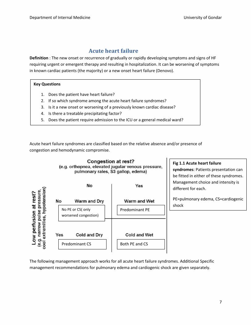
Acute heart failure Definition : The new onset or recurrence of gradually or rapidly developing symptoms and signs of HF
requiring urgent or emergent therapy and resulting in hospitalization. It can be worsening of symptoms
in known cardiac patients (the majority) or a new onset heart failure (Denovo).
Acute heart failure syndromes are classified based on the relative absence and/or presence of
congestion and hemodynamic compromise.
The following management approach works for all acute heart failure syndromes. Additional Specific
management recommendations for pulmonary edema and cardiogenic shock are given separately.
Key Questions
1. Does the patient have heart failure?
2. If so which syndrome among the acute heart failure syndromes?
3. Is it a new onset or worsening of a previously known cardiac disease?
4. Is there a treatable precipitating factor?
5. Does the patient require admission to the ICU or a general medical ward?
No PE or CS( only
worsened congestion) Predominant PE
Predominant CS Both PE and CS
Fig 1.1 Acute heart failure
syndromes: Patients presentation can
be fitted in either of these syndromes.
Management choice and intensity is
different for each.
PE=pulmonary edema, CS=cardiogenic
shock
Department of Internal Medicine University of Gondar
8
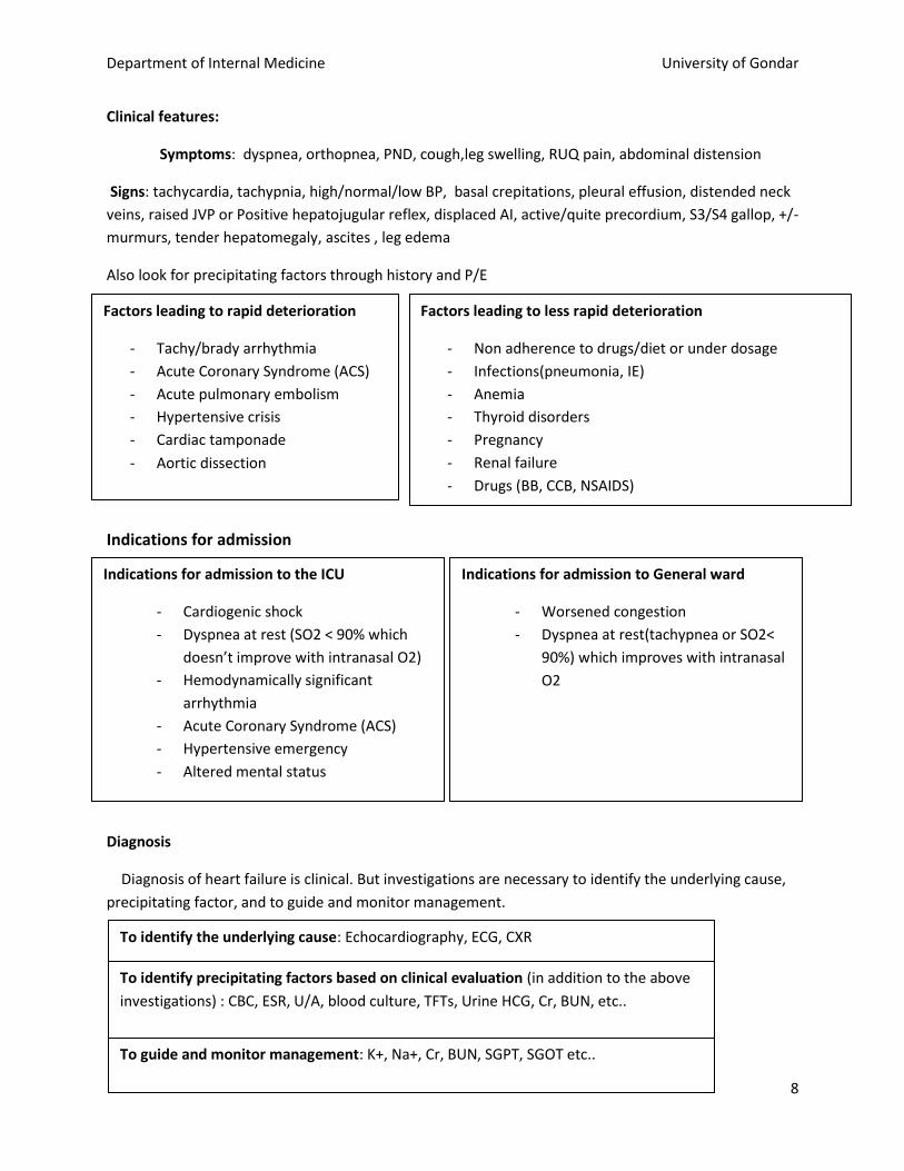
Clinical features:
Symptoms: dyspnea, orthopnea, PND, cough,leg swelling, RUQ pain, abdominal distension
Signs: tachycardia, tachypnia, high/normal/low BP, basal crepitations, pleural effusion, distended neck
veins, raised JVP or Positive hepatojugular reflex, displaced AI, active/quite precordium, S3/S4 gallop, +/-
murmurs, tender hepatomegaly, ascites , leg edema
Also look for precipitating factors through history and P/E
Indications for admission
Diagnosis
Diagnosis of heart failure is clinical. But investigations are necessary to identify the underlying cause,
precipitating factor, and to guide and monitor management.
Indications for admission to General ward
- Worsened congestion
- Dyspnea at rest(tachypnea or SO2<
90%) which improves with intranasal
O2
Indications for admission to the ICU
- Cardiogenic shock
- Dyspnea at rest (SO2 < 90% which
doesn’t improve with intranasal O2)
- Hemodynamically significant
arrhythmia
- Acute Coronary Syndrome (ACS)
- Hypertensive emergency
- Altered mental status
To identify the underlying cause: Echocardiography, ECG, CXR
Factors leading to rapid deterioration
- Tachy/brady arrhythmia
- Acute Coronary Syndrome (ACS)
- Acute pulmonary embolism
- Hypertensive crisis
- Cardiac tamponade
- Aortic dissection
Factors leading to less rapid deterioration
- Non adherence to drugs/diet or under dosage
- Infections(pneumonia, IE)
- Anemia
- Thyroid disorders
- Pregnancy
- Renal failure
- Drugs (BB, CCB, NSAIDS)
To identify precipitating factors based on clinical evaluation (in addition to the above
investigations) : CBC, ESR, U/A, blood culture, TFTs, Urine HCG, Cr, BUN, etc..
To guide and monitor management: K+, Na+, Cr, BUN, SGPT, SGOT etc..
Department of Internal Medicine University of Gondar
9
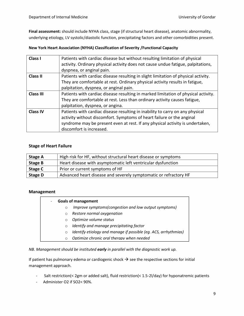
Final assessment: should include NYHA class, stage (if structural heart disease), anatomic abnormality,
underlying etiology, LV systolic/diastolic function, precipitating factors and other comorbidities present.
New York Heart Association (NYHA) Classification of Severity /Functional Capacity
Class I Patients with cardiac disease but without resulting limitation of physical activity. Ordinary physical activity does not cause undue fatigue, palpitations, dyspnea, or anginal pain.
Class II Patients with cardiac disease resulting in slight limitation of physical activity. They are comfortable at rest. Ordinary physical activity results in fatigue, palpitation, dyspnea, or anginal pain.
Class III Patients with cardiac disease resulting in marked limitation of physical activity. They are comfortable at rest. Less than ordinary activity causes fatigue, palpitation, dyspnea, or angina.
Class IV Patients with cardiac disease resulting in inability to carry on any physical activity without discomfort. Symptoms of heart failure or the anginal syndrome may be present even at rest. If any physical activity is undertaken, discomfort is increased.
Stage of Heart Failure
Stage A High risk for HF, without structural heart disease or symptoms
Stage B Heart disease with asymptomatic left ventricular dysfunction
Stage C Prior or current symptoms of HF
Stage D Advanced heart disease and severely symptomatic or refractory HF
Management
NB. Management should be instituted early in parallel with the diagnostic work up.
If patient has pulmonary edema or cardiogenic shock see the respective sections for initial
management approach.
- Salt restriction(< 2gm or added salt), fluid restriction(< 1.5-2l/day) for hyponatremic patients
- Administer O2 if SO2< 90%.
- Goals of management
o Improve symptoms(congestion and low output symptoms)
o Restore normal oxygenation
o Optimize volume status
o Identify and manage precipitating factor
o Identify etiology and manage if possible (eg. ACS, arrhythmias)
o Optimize chronic oral therapy when needed
Department of Internal Medicine University of Gondar
10
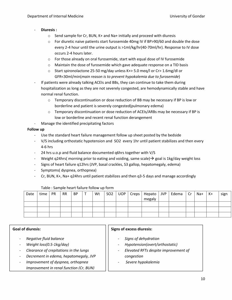
- Diuresis :
o Send sample for Cr, BUN, K+ and Na+ initially and proceed with diuresis
o For diuretic naive patients start furosemide 40mg IV if BP>90/60 and double the dose
every 2-4 hour until the urine output is >1ml/kg/hr(40-70ml/hr). Response to IV dose
occurs 2-4 hours later.
o For those already on oral furosemide, start with equal dose of IV furosemide
o Maintain the dose of furosemide which gave adequate response on a TID basis
o Start spironolactone 25-50 mg/day unless K+> 5.0 meq/l or Cr> 1.6mg/dl or
GFR<30ml/min(main reason is to prevent hypokalemia due to furosemide)
- If patients were already talking ACEIs and BBs, they can continue to take them during
hospitalization as long as they are not severely congested, are hemodynamically stable and have
normal renal function.
o Temporary discontinuation or dose reduction of BB may be necessary if BP is low or
borderline and patient is severely congested(pulmonary edema)
o Temporary discontinuation or dose reduction of ACEIs/ARBs may be necessary if BP is
low or borderline and recent renal function derangement
- Manage the identified precipitating factors
Follow up
- Use the standard heart failure management follow up sheet posted by the bedside
- V/S including orthostatic hypotension and SO2 every 1hr until patient stabilizes and then every
4-6 hrs
- 24 hrs u.o.p and fluid balance documented q6hrs together with V/S
- Weight q24hrs( morning prior to eating and voiding, same scale) goal is 1kg/day weight loss
- Signs of heart failure q12hrs (JVP, basal crackles, S3 gallop, hepatomegaly, edema)
- Symptoms( dyspnea, orthopnea)
- Cr, BUN, K+, Na+ q24hrs until patient stabilizes and then q3-5 days and manage accordingly
Table : Sample heart failure follow up form
Date time PR RR BP T Wt SO2 UOP Creps Hepatomegaly
JVP Edema Cr Na+ K+ sign
Goal of diuresis:
- Negative fluid balance
- Weight loss(0.5-1kg/day)
- Clearance of crepitations in the lungs
- Decrement in edema, hepatomegaly, JVP
- Improvement of dyspnea, orthopnea
Improvement in renal function (Cr, BUN)
-
-
Signs of excess diuresis:
- Signs of dehydration
- Hypotension(overt/orthostatic)
- Elevated RFTs despite improvement of
congestion
- Severe hypokalemia

Department of Internal Medicine University of Gondar
11
Patient not responding
- Make sure that
o Patient is taking medications as prescribed and is on salt free diet
o Precipitating factors is managed
o Patient is not getting drugs like NSAIDS, CCBs, BBs
o NB. Patients with deranged renal function and hypoalbuminemia require higher doses of
frusemide from the outset.
- Adjust the diuresis
o Increase the dose of furosemide(max. 400-600mg/day) and increase spironolactone to
50-100mg/day
o Increase the frequency of administration of furosemide 4-6 xper day. Repeated IV bolus
doses are recommended that continuous infusion.
o In ICU continuous furosemide infusion by perfuser according to protocol: ( 10-80mg/hr)
can be used if still refractory to the above measures.
o If patient not responding with the above approaches, add hydrochlorthiazide 25mg/day
in the morning 30mts before frusemide administration
- For patients with HFrEF with border line low BP(SBP<100 mmhg) the following can be
considered in addition to diuretics to improve LV function and promote natriuresis
o Dopamine infusion(5-10ug/kg/min) (see cardiogenic shock section)
o Digoxin 0.125-0.25 mg/day for positive inotropy and rate control in patients with atrial
fibrillation.
- For patients with hypertension and severe Acute MR intravenous nitroglycerin infusion can be
considered in addition to diuretics.(see pulmonary edema section)
Patient improving
- Decrease the dose of diuretics every day depending on patient condition goal is to use the
lowest possible dose and frequency to keep patient dry
- For patients in whom previous BB and ACEIs/ARBs have been discontinued consider reinitiating
the drugs as soon as possible sequentially(ACEIs/ARBs followed by B blockers)
- For HFrEF previously not taking ACEIs/ARBs or BB
o start one of the ACEIs/ARBs as soon as BP and RFTs permit and escalate until
discharge(see chronic heart failure section)
o start one of the BB following ACEIs/ARBs when BP and PR permit and escalate until
discharge(see chronic heart failure section)
o Consider Spirinolactone 12.5-25mg/d.
- Change IV furosemide to PO and observe the patient with ambulation for a day or two
(NB. Patients requiring higher dose of furosemide may require a double dose)
- Institute further management for the underlying heart disease(see elsewhere ) and comorbidities
Before Discharge
- Proper advise: salt consumption, activity, adherence to medications and follow up
- Prescribe adequate medications and give requests for further planned outpatient
investigations
- document medications with dose and further plans clearly on the discharge note
- Early appointment preferably in 1 week time to follow up clinic
Department of Internal Medicine University of Gondar
12
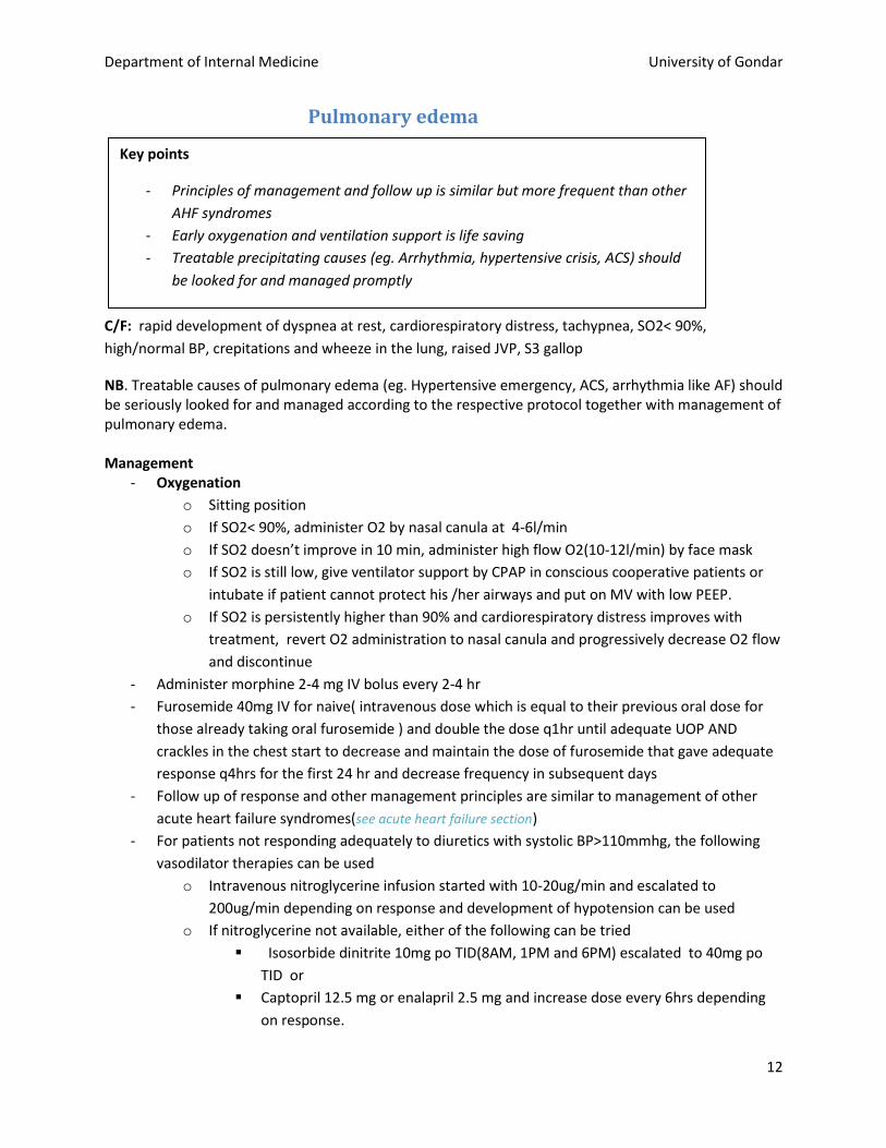
Pulmonary edema
C/F: rapid development of dyspnea at rest, cardiorespiratory distress, tachypnea, SO2< 90%,
high/normal BP, crepitations and wheeze in the lung, raised JVP, S3 gallop
NB. Treatable causes of pulmonary edema (eg. Hypertensive emergency, ACS, arrhythmia like AF) should be seriously looked for and managed according to the respective protocol together with management of pulmonary edema. Management
- Oxygenation
o Sitting position
o If SO2< 90%, administer O2 by nasal canula at 4-6l/min
o If SO2 doesn’t improve in 10 min, administer high flow O2(10-12l/min) by face mask
o If SO2 is still low, give ventilator support by CPAP in conscious cooperative patients or
intubate if patient cannot protect his /her airways and put on MV with low PEEP.
o If SO2 is persistently higher than 90% and cardiorespiratory distress improves with
treatment, revert O2 administration to nasal canula and progressively decrease O2 flow
and discontinue
- Administer morphine 2-4 mg IV bolus every 2-4 hr
- Furosemide 40mg IV for naive( intravenous dose which is equal to their previous oral dose for
those already taking oral furosemide ) and double the dose q1hr until adequate UOP AND
crackles in the chest start to decrease and maintain the dose of furosemide that gave adequate
response q4hrs for the first 24 hr and decrease frequency in subsequent days
- Follow up of response and other management principles are similar to management of other
acute heart failure syndromes(see acute heart failure section)
- For patients not responding adequately to diuretics with systolic BP>110mmhg, the following
vasodilator therapies can be used
o Intravenous nitroglycerine infusion started with 10-20ug/min and escalated to
200ug/min depending on response and development of hypotension can be used
o If nitroglycerine not available, either of the following can be tried
Isosorbide dinitrite 10mg po TID(8AM, 1PM and 6PM) escalated to 40mg po
TID or
Captopril 12.5 mg or enalapril 2.5 mg and increase dose every 6hrs depending
on response.
Key points
- Principles of management and follow up is similar but more frequent than other
AHF syndromes
- Early oxygenation and ventilation support is life saving
- Treatable precipitating causes (eg. Arrhythmia, hypertensive crisis, ACS) should
be looked for and managed promptly
Department of Internal Medicine University of Gondar
13
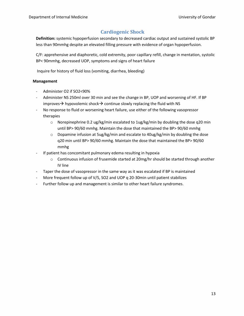
Cardiogenic Shock Definition: systemic hypoperfusion secondary to decreased cardiac output and sustained systolic BP
less than 90mmhg despite an elevated filling pressure with evidence of organ hypoperfusion.
C/F: apprehensive and diaphoretic, cold extremity, poor capillary refill, change in mentation, systolic
BP< 90mmhg, decreased UOP, symptoms and signs of heart failure
Inquire for history of fluid loss (vomiting, diarrhea, bleeding)
Management
- Administer O2 if SO2<90%
- Administer NS 250ml over 30 min and see the change in BP, UOP and worsening of HF. If BP
improves hypovolemic shock continue slowly replacing the fluid with NS
- No response to fluid or worsening heart failure, use either of the following vasopressor
therapies
o Norepinephrine 0.2 ug/kg/min escalated to 1ug/kg/min by doubling the dose q20 min
until BP> 90/60 mmhg. Maintain the dose that maintained the BP> 90/60 mmhg
o Dopamine infusion at 5ug/kg/min and escalate to 40ug/kg/min by doubling the dose
q20 min until BP> 90/60 mmhg. Maintain the dose that maintained the BP> 90/60
mmhg
- If patient has concomitant pulmonary edema resulting in hypoxia
o Continuous infusion of frusemide started at 20mg/hr should be started through another
IV line
- Taper the dose of vasopressor in the same way as it was escalated if BP is maintained
- More frequent follow up of V/S, SO2 and UOP q 20-30min until patient stabilizes
- Further follow up and management is similar to other heart failure syndromes.
Department of Internal Medicine University of Gondar
14
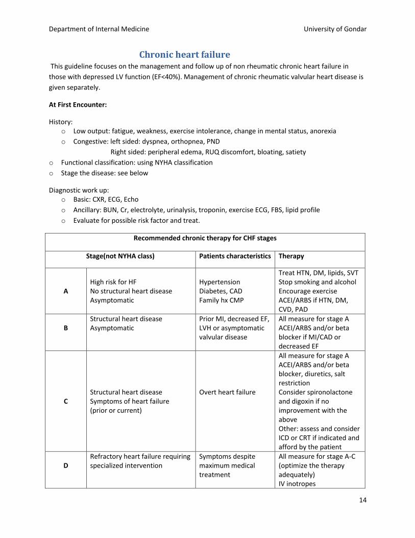
Chronic heart failure This guideline focuses on the management and follow up of non rheumatic chronic heart failure in
those with depressed LV function (EF<40%). Management of chronic rheumatic valvular heart disease is
given separately.
At First Encounter:
History: o Low output: fatigue, weakness, exercise intolerance, change in mental status, anorexia
o Congestive: left sided: dyspnea, orthopnea, PND
Right sided: peripheral edema, RUQ discomfort, bloating, satiety
o Functional classification: using NYHA classification
o Stage the disease: see below
Diagnostic work up: o Basic: CXR, ECG, Echo
o Ancillary: BUN, Cr, electrolyte, urinalysis, troponin, exercise ECG, FBS, lipid profile
o Evaluate for possible risk factor and treat.
Recommended chronic therapy for CHF stages
Stage(not NYHA class) Patients characteristics Therapy
A
High risk for HF No structural heart disease Asymptomatic
Hypertension Diabetes, CAD Family hx CMP
Treat HTN, DM, lipids, SVT Stop smoking and alcohol Encourage exercise ACEI/ARBS if HTN, DM, CVD, PAD
B
Structural heart disease Asymptomatic
Prior MI, decreased EF, LVH or asymptomatic valvular disease
All measure for stage A ACEI/ARBS and/or beta blocker if MI/CAD or decreased EF
C
Structural heart disease Symptoms of heart failure (prior or current)
Overt heart failure
All measure for stage A ACEI/ARBS and/or beta blocker, diuretics, salt restriction Consider spironolactone and digoxin if no improvement with the above Other: assess and consider ICD or CRT if indicated and afford by the patient
D
Refractory heart failure requiring specialized intervention
Symptoms despite maximum medical treatment
All measure for stage A-C (optimize the therapy adequately) IV inotropes
Department of Internal Medicine University of Gondar
15
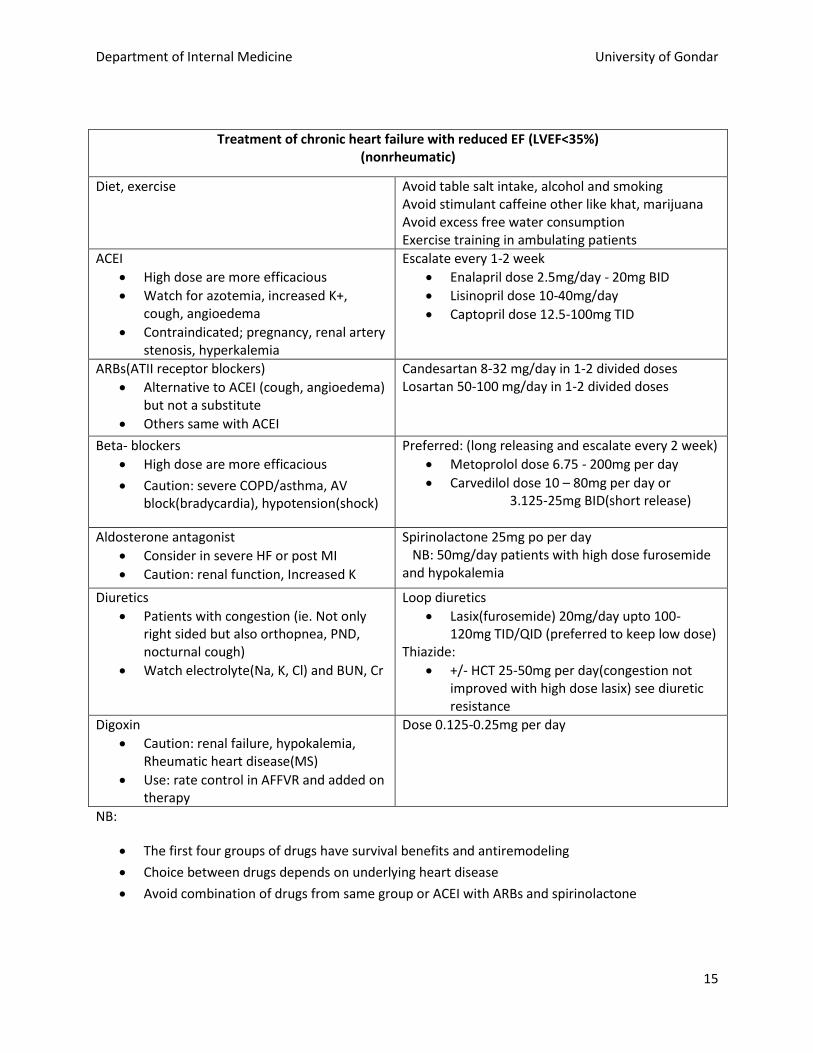
Treatment of chronic heart failure with reduced EF (LVEF<35%) (nonrheumatic)
Diet, exercise Avoid table salt intake, alcohol and smoking Avoid stimulant caffeine other like khat, marijuana Avoid excess free water consumption Exercise training in ambulating patients
ACEI
High dose are more efficacious
Watch for azotemia, increased K+, cough, angioedema
Contraindicated; pregnancy, renal artery stenosis, hyperkalemia
Escalate every 1-2 week
Enalapril dose 2.5mg/day - 20mg BID
Lisinopril dose 10-40mg/day
Captopril dose 12.5-100mg TID
ARBs(ATII receptor blockers)
Alternative to ACEI (cough, angioedema) but not a substitute
Others same with ACEI
Candesartan 8-32 mg/day in 1-2 divided doses Losartan 50-100 mg/day in 1-2 divided doses
Beta- blockers
High dose are more efficacious
Caution: severe COPD/asthma, AV block(bradycardia), hypotension(shock)
Preferred: (long releasing and escalate every 2 week)
Metoprolol dose 6.75 - 200mg per day
Carvedilol dose 10 – 80mg per day or 3.125-25mg BID(short release)
Aldosterone antagonist
Consider in severe HF or post MI
Caution: renal function, Increased K
Spirinolactone 25mg po per day NB: 50mg/day patients with high dose furosemide and hypokalemia
Diuretics
Patients with congestion (ie. Not only right sided but also orthopnea, PND, nocturnal cough)
Watch electrolyte(Na, K, Cl) and BUN, Cr
Loop diuretics
Lasix(furosemide) 20mg/day upto 100-120mg TID/QID (preferred to keep low dose)
Thiazide:
+/- HCT 25-50mg per day(congestion not improved with high dose lasix) see diuretic resistance
Digoxin
Caution: renal failure, hypokalemia, Rheumatic heart disease(MS)
Use: rate control in AFFVR and added on therapy
Dose 0.125-0.25mg per day
NB:
The first four groups of drugs have survival benefits and antiremodeling
Choice between drugs depends on underlying heart disease
Avoid combination of drugs from same group or ACEI with ARBs and spirinolactone
Department of Internal Medicine University of Gondar
16
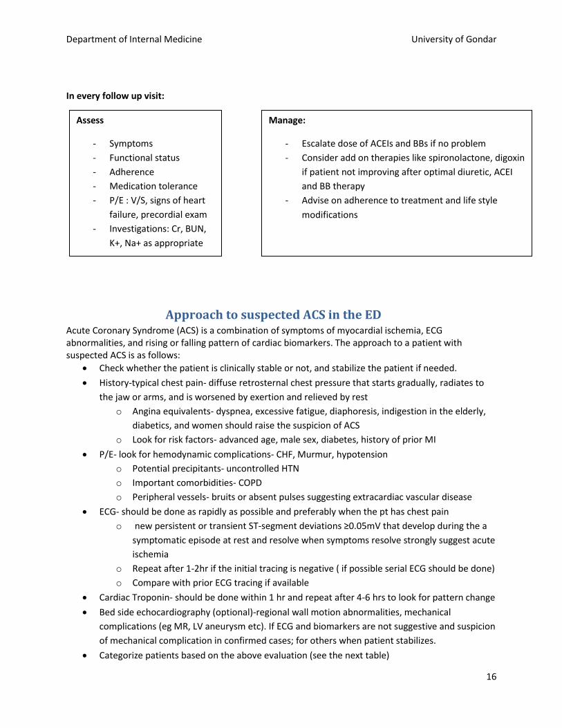
In every follow up visit:
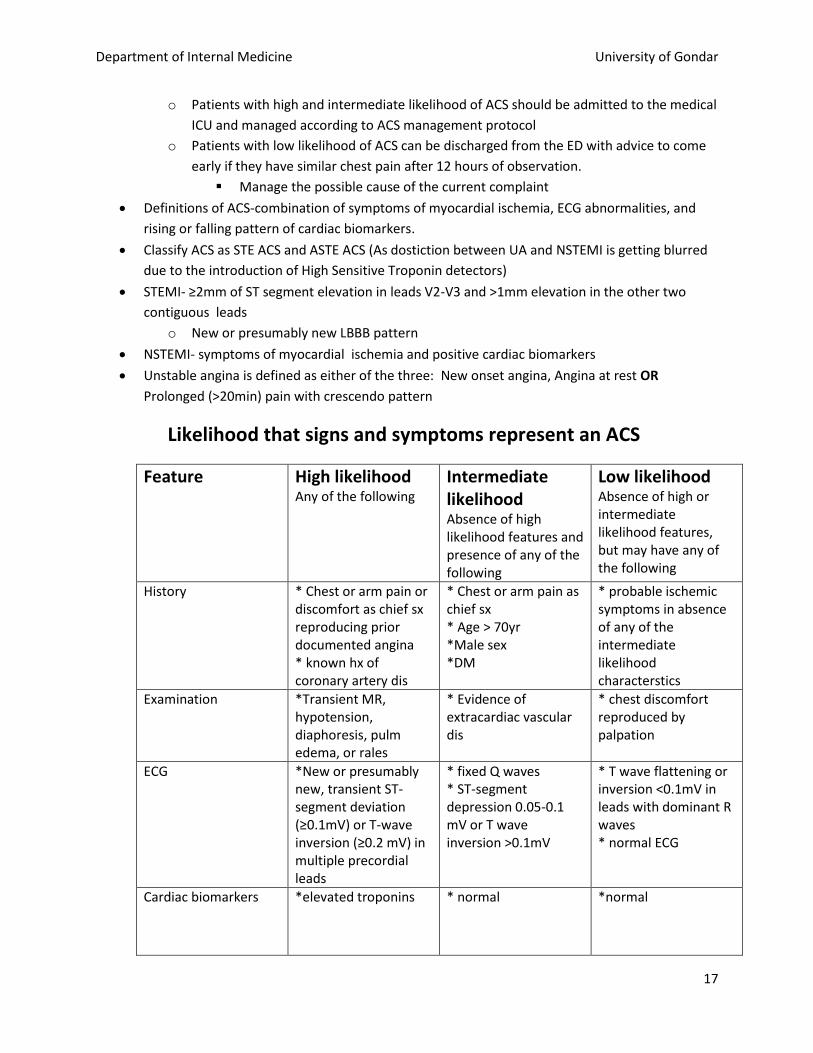
Approach to suspected ACS in the ED Acute Coronary Syndrome (ACS) is a combination of symptoms of myocardial ischemia, ECG abnormalities, and rising or falling pattern of cardiac biomarkers. The approach to a patient with suspected ACS is as follows:
Check whether the patient is clinically stable or not, and stabilize the patient if needed.
History-typical chest pain- diffuse retrosternal chest pressure that starts gradually, radiates to
the jaw or arms, and is worsened by exertion and relieved by rest
o Angina equivalents- dyspnea, excessive fatigue, diaphoresis, indigestion in the elderly,
diabetics, and women should raise the suspicion of ACS
o Look for risk factors- advanced age, male sex, diabetes, history of prior MI
P/E- look for hemodynamic complications- CHF, Murmur, hypotension
o Potential precipitants- uncontrolled HTN
o Important comorbidities- COPD
o Peripheral vessels- bruits or absent pulses suggesting extracardiac vascular disease
ECG- should be done as rapidly as possible and preferably when the pt has chest pain
o new persistent or transient ST-segment deviations ≥0.05mV that develop during the a
symptomatic episode at rest and resolve when symptoms resolve strongly suggest acute
ischemia
o Repeat after 1-2hr if the initial tracing is negative ( if possible serial ECG should be done)
o Compare with prior ECG tracing if available
Cardiac Troponin- should be done within 1 hr and repeat after 4-6 hrs to look for pattern change
Bed side echocardiography (optional)-regional wall motion abnormalities, mechanical
complications (eg MR, LV aneurysm etc). If ECG and biomarkers are not suggestive and suspicion
of mechanical complication in confirmed cases; for others when patient stabilizes.
Categorize patients based on the above evaluation (see the next table)
Assess
- Symptoms
- Functional status
- Adherence
- Medication tolerance
- P/E : V/S, signs of heart
failure, precordial exam
- Investigations: Cr, BUN,
K+, Na+ as appropriate
Manage:
- Escalate dose of ACEIs and BBs if no problem
- Consider add on therapies like spironolactone, digoxin
if patient not improving after optimal diuretic, ACEI
and BB therapy
- Advise on adherence to treatment and life style
modifications
Department of Internal Medicine University of Gondar
17
o Patients with high and intermediate likelihood of ACS should be admitted to the medical
ICU and managed according to ACS management protocol
o Patients with low likelihood of ACS can be discharged from the ED with advice to come
early if they have similar chest pain after 12 hours of observation.
Manage the possible cause of the current complaint
Definitions of ACS-combination of symptoms of myocardial ischemia, ECG abnormalities, and
rising or falling pattern of cardiac biomarkers.
Classify ACS as STE ACS and ASTE ACS (As dostiction between UA and NSTEMI is getting blurred
due to the introduction of High Sensitive Troponin detectors)
STEMI- ≥2mm of ST segment elevation in leads V2-V3 and >1mm elevation in the other two
contiguous leads
o New or presumably new LBBB pattern
NSTEMI- symptoms of myocardial ischemia and positive cardiac biomarkers
Unstable angina is defined as either of the three: New onset angina, Angina at rest OR
Prolonged (>20min) pain with crescendo pattern
Likelihood that signs and symptoms represent an ACS
Feature High likelihood Any of the following
Intermediate likelihood Absence of high likelihood features and presence of any of the following
Low likelihood Absence of high or intermediate likelihood features, but may have any of the following
History * Chest or arm pain or discomfort as chief sx reproducing prior documented angina * known hx of coronary artery dis
* Chest or arm pain as chief sx * Age > 70yr *Male sex *DM
* probable ischemic symptoms in absence of any of the intermediate likelihood characterstics
Examination *Transient MR, hypotension, diaphoresis, pulm edema, or rales
* Evidence of extracardiac vascular dis
* chest discomfort reproduced by palpation
ECG *New or presumably new, transient ST-segment deviation (≥0.1mV) or T-wave inversion (≥0.2 mV) in multiple precordial leads
* fixed Q waves * ST-segment depression 0.05-0.1 mV or T wave inversion >0.1mV
* T wave flattening or inversion <0.1mV in leads with dominant R waves * normal ECG
Cardiac biomarkers *elevated troponins * normal *normal

Department of Internal Medicine University of Gondar
18
DDX for acute chest pain in the ED
o Acute pericarditis (cardiac tamponade) * PUD
o Aortic dissection *tension pneumothorax
o Pulmonary embolism *musculoskeletal causes (eg. rib #)
o Psychiatric disorders- panic attack
Acute Coronary Syndrome 1. Bed rest in patients with chest pain with continuous ECG monitoring
2. Provide Oxygen if patient has SaO2 < 90%
3. Antiplatelets:
a. Load with 325 mg of ASA chewed right away and continue with 81mg per day
b. Clopidegrol 300mg loading and 75 mg per day
4. Perform focused history and P/E( look for hemodynamic compromise(Cardiogenic shock, heart
failure, sustained tachyarrythmia and manage according to protocol)
5. Secure IV access and obtain blood for troponin, electrolyted, coagulation profile, RFT)
6. Anticoagulants( duration of therapy is for 5-7 days)
a. Enoxaparin 1mg/ Kg SC BID ( if Cr Cl< 30ml/min, then 1mg/kg SC once daily)
b. If enoxaparin not available, UFH 60u/kg IV bolus then 12U/Kg/hr IV continuous infusion
dose adjusted to aPTT of 1.5-2 xUNL or 50-70sec)
c. If it is not possible to use enoxaparin or continuous infusion of heparin, then 80U/kg IV
loading dose of UFH followed by 250U/kg subcutaneous BID can be given
7. Anti ischemic therapy
a. Give three doses of sublingual nitroglycerine(0.4 mg) for persistent chest pain every 5
minutes if no hypotension or RV infarction
b. Give metoprolol (12.5-25mg) BID preferably or atenolol 12.5-25 BID escalated if no
heart failure, hypotension or bradycardia, or active obstructive air way disease
c. If B blockers are C/I due to obstructive airway disease use Verapamil 80-120 mg BID
d. Morphine sulfate IV 2-4 mg every 5-15 mts for persistent chest pain and anxiety( C/I:
hypotension) (pethidine 25-50 mg IV as needed is weaker alternative)
e. For patients with continuous or recurrent chest pain despite the above treatment,
intravenous nitroglycerine infusion should be given in the absence of contraindications
8. Other therapies
a. Give Atrovastatin 80mg per day( simvastatin 40mg is an alternative)
b. ACEIs( enalapril 2.5 mg BID, captopril 6.25 TID and escalated) in the absence of C/I or
ARBs(Candasartan 4mg BID escalated) in ACEI intolerant is indicated for
i. UA/NSTEMI pts with LVEF<40% or hypertension
ii. All STEMI pts
c. Aldosterone antagonists(spironolactone 25 mg/day) for those with LV EF < 40%, are
already on ACEis and either HF or DM in absence of hyperkalemia and renal failure
Department of Internal Medicine University of Gondar
19
d. Oral anticoagulation(warfarin) for those at high risk of thromboembolization
i. Atrial fibrillation with CHADS2 > 1, LV thrombus
ii. Considered in patients with poor LV function and apical hypokinesis
e. GI prophylaxis and bowel care: omeprazole 20 mg po BID routine and laxatives for
constipation
9. Glucose and electrolytes
a. keep RBS level b/n 140-180 mg/dl(use the protocol for management of blood glucose in
critical patients)
b. keep serum K+ level > 4 meq/l
10. Follow up
a. Follow for chest pain or angina equivalent symptoms
b. Take ECG every day for at least 3 days evolutionary changes( degree of ST elevation, Q
waves)
c. Echocardiography: LV function, wall motion abnormality, LV thrombus
d. Follow troponin levels
e. Watch for mechanical and electrical complications
f. Watch for bleeding in patients at high risk for bleeding
11. Preparation for discharge
a. Ambulation: hemodynamically stable and no chest pain for 12-24 hrs
b. Discharge after a minimum of 5 days of admission can be considered if
i. Pt is hemodynamically stable and no signs of mechanical or electrical
complications
ii. No sign of ongoing ischemia( no chest pain, new ECG changes and troponin level
decreasing)
iii. Pt is able to ambulate without symptoms
12. Long term medications
a. ASA, B blockers, ACEIs/ARBs, aldosterone antagonists, and statin should be continued
life long
b. Clopidegrol should be continued for a minimum of 1 month and ideally up to 1 yr
13. Risk factor modification:
a. Optimal treatment of HTN, DM, dyslipidemia, smoking cessation
14. Return to activities
a. Most patients with uncomplicated course can return to activities after 2 weeks
b. Should be advised to slowly increase the level of exertion watching for symptoms(eg.
Walking distance)
15. Finally all patients who can afford, the option of getting referred to a cardiac hospital in A.A. for
risk stratification with stress ECG and diagnostic and therapeutic coronary intervention should
be given. Hemodynamically stable patients with no ongoing or recurrent ischemia who are on
appropriate medications can fly with reasonable safety after 2 weeks.
NB. Consult the appropriate risk stratification models for prognostication
Department of Internal Medicine University of Gondar
20
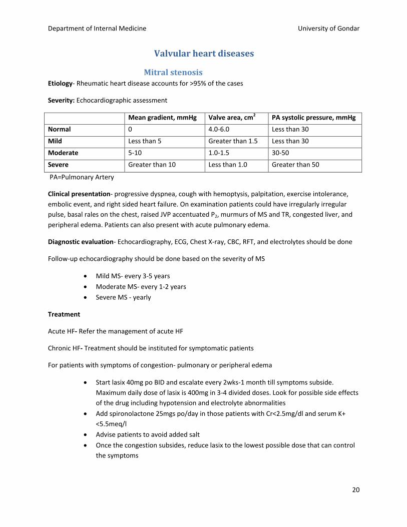
Valvular heart diseases
Mitral stenosis Etiology- Rheumatic heart disease accounts for >95% of the cases
Severity: Echocardiographic assessment
Mean gradient, mmHg Valve area, cm2 PA systolic pressure, mmHg
Normal 0 4.0-6.0 Less than 30
Mild Less than 5 Greater than 1.5 Less than 30
Moderate 5-10 1.0-1.5 30-50
Severe Greater than 10 Less than 1.0 Greater than 50
PA=Pulmonary Artery
Clinical presentation- progressive dyspnea, cough with hemoptysis, palpitation, exercise intolerance,
embolic event, and right sided heart failure. On examination patients could have irregularly irregular
pulse, basal rales on the chest, raised JVP accentuated P2, murmurs of MS and TR, congested liver, and
peripheral edema. Patients can also present with acute pulmonary edema.
Diagnostic evaluation- Echocardiography, ECG, Chest X-ray, CBC, RFT, and electrolytes should be done
Follow-up echocardiography should be done based on the severity of MS
Mild MS- every 3-5 years
Moderate MS- every 1-2 years
Severe MS - yearly
Treatment
Acute HF- Refer the management of acute HF
Chronic HF- Treatment should be instituted for symptomatic patients
For patients with symptoms of congestion- pulmonary or peripheral edema
Start lasix 40mg po BID and escalate every 2wks-1 month till symptoms subside.
Maximum daily dose of lasix is 400mg in 3-4 divided doses. Look for possible side effects
of the drug including hypotension and electrolyte abnormalities
Add spironolactone 25mgs po/day in those patients with Cr<2.5mg/dl and serum K+
<5.5meq/l
Advise patients to avoid added salt
Once the congestion subsides, reduce lasix to the lowest possible dose that can control
the symptoms
Department of Internal Medicine University of Gondar
21
Patients without congestion but still have exertional symptoms- start atenolol 25mg po/day and
escalate every 2wks-1month till PR becomes 60-80BPM. Follow for hypotension.
Anticoagulation- warfarin 2.5 – 5 mg po/day with target INR 2.0-3.0 Follow with INR every 2wks initially
till the INR stabilizes and then every 1-2months. Indications include
prior embolic event, left atrial thrombus and presence of AF
patients who cannot afford for follow-up INR or have higher risk of bleeding- start
aspirin 81mg po/day
AF- refer to the management of AF
Pregnancy- advise patients to avoid pregnancy and use dual contraception
Benzanthine penicillin- 1.2 million IU IM every month lifelong should be given for all patients with
chronic rheumatic heart disease
Endocarditis prophylaxis- not indicated unless there is prior IE episode
Indications for intervention- patients who can afford and have the following indications should be
referred as early as possible to undergo PBMV or valve replacement
moderate to severe MS with symptoms
asymptomatic patients with moderate to severe MS and either pulmonary artery
pressure at rest >50mmHg or new onset AF
Department of Internal Medicine University of Gondar
22
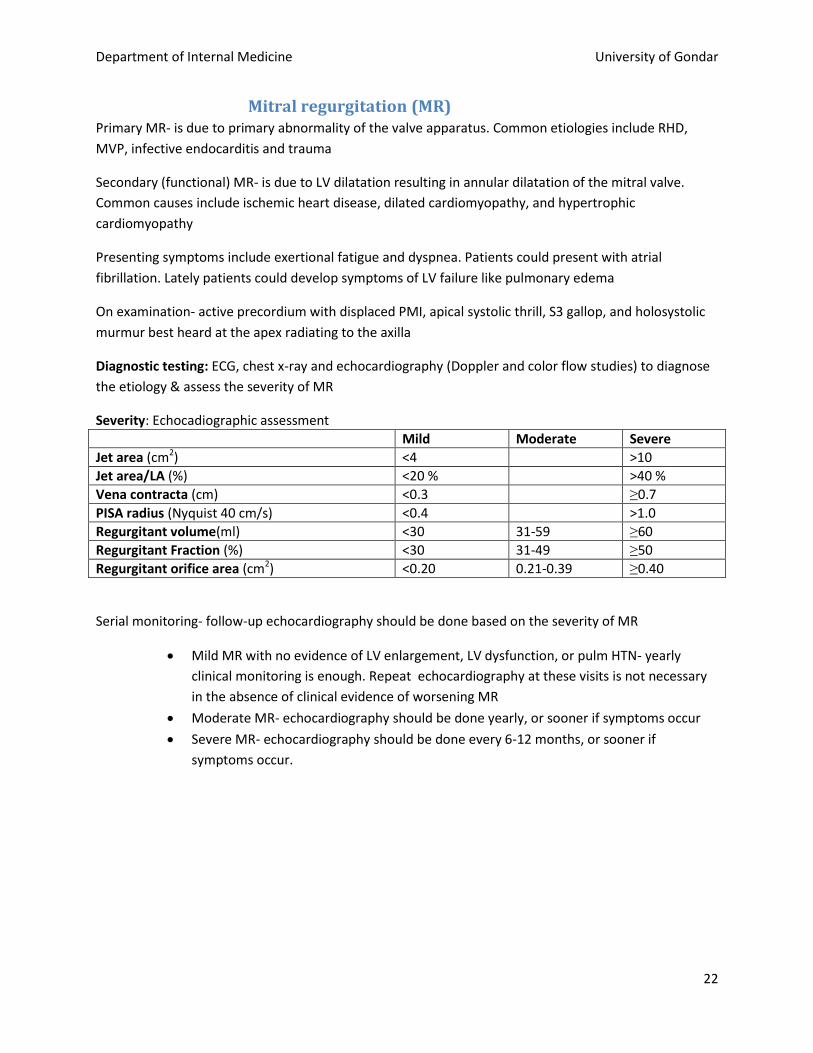
Mitral regurgitation (MR) Primary MR- is due to primary abnormality of the valve apparatus. Common etiologies include RHD,
MVP, infective endocarditis and trauma
Secondary (functional) MR- is due to LV dilatation resulting in annular dilatation of the mitral valve.
Common causes include ischemic heart disease, dilated cardiomyopathy, and hypertrophic
cardiomyopathy
Presenting symptoms include exertional fatigue and dyspnea. Patients could present with atrial
fibrillation. Lately patients could develop symptoms of LV failure like pulmonary edema
On examination- active precordium with displaced PMI, apical systolic thrill, S3 gallop, and holosystolic
murmur best heard at the apex radiating to the axilla
Diagnostic testing: ECG, chest x-ray and echocardiography (Doppler and color flow studies) to diagnose
the etiology & assess the severity of MR
Severity: Echocadiographic assessment
Mild Moderate Severe
Jet area (cm2) <4 >10
Jet area/LA (%) <20 % >40 %
Vena contracta (cm) <0.3 ≥0.7
PISA radius (Nyquist 40 cm/s) <0.4 >1.0
Regurgitant volume(ml) <30 31-59 ≥60
Regurgitant Fraction (%) <30 31-49 ≥50
Regurgitant orifice area (cm2) <0.20 0.21-0.39 ≥0.40
Serial monitoring- follow-up echocardiography should be done based on the severity of MR
Mild MR with no evidence of LV enlargement, LV dysfunction, or pulm HTN- yearly
clinical monitoring is enough. Repeat echocardiography at these visits is not necessary
in the absence of clinical evidence of worsening MR
Moderate MR- echocardiography should be done yearly, or sooner if symptoms occur
Severe MR- echocardiography should be done every 6-12 months, or sooner if
symptoms occur.

Department of Internal Medicine University of Gondar
23
Treatment- depends on the presence or absence of symptoms. Asymptomatic patients- need only close follow-up. If patients have associated hypertension, it should be
treated with enalapril or amilodipine
Symptomatic patients- need medical therapy
lasix 40mg po BID and escalate till the congestion is relieved
start enalapril 2.5mg po/day and escalate up to 20mg po/day or maximally tolerated
dose
if LVEF <40%- refer to the protocol for heart failure with reduced ejection fraction
Atrial fibrillation- refer to AF management protocol
Anticoagulation- similar to patients with mitral stenosis
Endocarditis prophylaxis- not indicated unless prior IE
Prevention of rheumatic recurrence- similar to patients with rheumatic MR
Indications for surgical intervention- patients who can afford and have the following indications should
be referred early to undergo mitral valve repair or replacement
symptomatic severe MR
asymptomatic severe MR with one of the following factors
o LVEF<60% or LVESD >40mm
o New onset AF
o Pulmonary hypertension
Aortic Stenosis Etiology: mainly caused by rheumatic heart disease, congenital bicuspid aortic valve, and degenerative calcification. Symptoms: patients remain asymptomatic till the valve area is 1 cm2. Classic symptoms include exertional angina, syncope, dyspnea, and excessive fatigue. Physical findings include active precordium with displaced PMI and ejection systolic murmur that radiates to the carotid arteries. Diagnostic evaluation: ECG, echocardiography, chest X-ray, and lipid profile Severity: Echocardiogrphic assessment
Aortic jet Velocity, m/sec Mean gradient, mmHg Valve area, cm2
Normal ≤ 1.5 <5 3.0-4.0
Mild <3.0 <25 >1.5
Moderate 3.0-4.0 25-40 1.0-1.5
Severe >4.0 >40 <1.0

Department of Internal Medicine University of Gondar
24
Follow-up echocardiography should be done based on the severity of AS
Mild AS- every 3-5 years
Moderate AS- every 2 years
Severe AS- yearly
Treatment – mainly indicated for symptomatic patients
For asymptomatic patients
Prevention of progression of rheumatic AS- monthly pencillin prophylaxis
Hypertension- should be controlled. Preferably use ACEIs or calcium channel blockers,
like amilodipine. Beware of excessive vasodilatation as these patients have already
vasodilatation as compensatory mechanism.
Concomitant coronary artery disease (CAD) is common, especially in the elderly. Thus,
look for possible evidence of CAD and treat
For symptomatic patients- goals include
Treat concurrent cardiovascular conditions, like coronary artery disease
Prevent or treat superimposed diseases that often exacerbate the effects of valve
obstruction, like HTN
Treat symptoms
o CHF- start lasix 40mg po BID/TID and escalate till the congestion improves
o Exertional angina and syncope - advice patients to avoid exertion
Patients with depressed EF (LVEF <40%)
o Refer to the management of heart failure.
Endocarditis prophylaxis- not indicated
Indications for surgical intervention- - patients who can afford and have the following indications should
be referred early to undergo aortic valve replacement
Symptomatic severe AS
Severe AS with LVEF <50%
Moderate AS with concomitant coronary artery disease requiring bypass graft surgery
Aortic regurgitation May be caused by primary valve disease or by primary aortic root disease. Causes of Primary valve disease include rheumatic heart disease, congenital bicuspid aortic valve disease, and infective endocarditis. Primary aortic root disease- widening of the aortic annulus and separation of the aortic leaflets are responsible for the AR. causes include cystic medial degeneration of the ascending aorta, idiopathic dilation of the aorta, annuloaortic ectasia, osteogenesis imperfect, severe HTN, syphilis, and ankylosing spondylitis. Symptoms: uncomfortable awareness of the heartbeat, head pounding, exertional dyspnea, orthopnea,
PND, and nocturnal angina.

Department of Internal Medicine University of Gondar
25
Physical findings: peripheral stigmas include water-hammer pulse, quicken’s pulse, “pistol shot” over the femoral arteries, wide pulse pressure etc
Active precordium with displaced PMI
High-pitched, blowing, decrescendo diastolic murmur, heard best in the third
intercostals space along LSB
Diagnostic evaluation- ECG, echocardiography, chest X-ray
Severity: Echocardiographic assessment
Mild Moderate Severe
Vena Contracta width (cm) <0.3 >0.6
Jet width/LVOT diam. (%) <25 ≥65
Regurgitant Volume (ml/beat) <30 31-59 ≥60
Regurgitant Fraction (%) <30 31-49 ≥50
Regurgitant Orifice area (cm2) <0.10 0.11-0.29 ≥0.30
VTI diastolic flow reversal (upper Dao) (cm) - 15
Pressure Half Time (ms) >500 <250
Serial monitoring
Mild chronic AR- clinical evaluation yearly and routine echocardiography every 2-3 years
Moderate AR - echocardiography every 1-2 years
Severe AR - echocardiography yearly
Treatment
Start enalapril 2.5mg po/day and escalate or amilodipine 5-10mg po/day for the following patients
Symptomatic severe AR with LV dilation
Asymptomatic severe AR with LVEF <50%
Patients with congestion- refer to acute heart failure management
Severe AR with reduced LVEF- refer to the management of heart failure with reduced ejection fraction
Endocarditis prophylaxis- not indicated
N.B. avoid the use of beta blockers in patients with severe AR except in severe Aortic dilatation like in
Marfan Syndrome.
Pregnancy – should be avoided in the following patients
NYHA class III to IV symptoms
LVEF <40%
Marfan syndrome
Indications for surgical interventions- patients who can afford and have the following indications should
be referred early to undergo aortic valve replacement or repair
Symptomatic patients with severe chronic AR
Asymptomatic patients with severe chronic AR and LVEF <50%
Severe chronic AR and severe LV dilatation- LVEDD >75mm or LVESD >55mm
Severe AR with coronary artery disease requiring bypass graft surgery

Department of Internal Medicine University of Gondar
26
Infective endocarditis Definition: an infection of the endocardial surface of the heart, which may include one or more heart
valves, the mural endocardium, or a septal defect.
Risk factors- structural heart disease (esp. rheumatic regurgitant valve lesions), prosthetic valves, prior
history of IE, and injection drug use, recent dental manipulation, GI, GU instrumentation
Clinical presentations- patients could present with one of the following patterns
Acute endocarditis is characterized by high grade fever with rapidly progressive heart failure and
metastatic infection. Common etiologies include S.aureus, pneumococci, and enterococci
Subacute endocarditis- evolves during weeks to months with only modest toxicity. It is commonly
caused by viridians streptococci, CONS, Enterococci, and HACEK group
Common findings include- persistent fever, heart murmurs, otherwise-unexplained arterial emboli, and
cutaneous and mucocutaneous lesions, like petechiae, splinter hemorrhages, janeway lesions, osler’s
node, and Roth spots.
Diagnosis – should be suspected in any patient with rheumatic VHD(esp. regurgitant) presenting with
fever and/or worsening of heart failure despite therapy.
Three sets of blood culture should be obtained prior to antibiotic therapy. A minimum of 10ml of blood
for each set, each set of cultures should be taken from separate venipuncture sites, avoid taking
samples from preexisting IV lines, and blood cultures can be taken at any time.
Acutely ill pt- take 3 blood culture samples over 1hr before beginning empiric therapy
Subacute endocarditis- take 3 samples over 12hrs
Echocardiography- look for vegetations, abscess, new valvular regurgitation, or new partial dehiscence
of prosthetic valve.
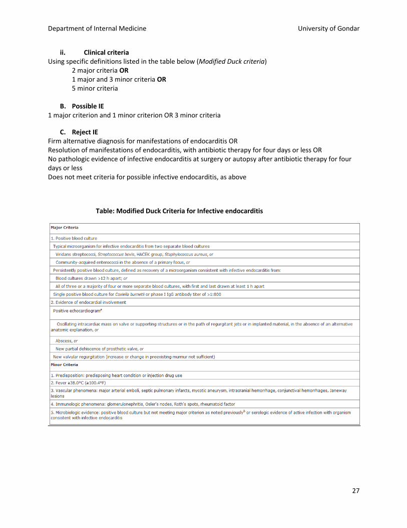
Diagnostic criteria- use Modified Duke criteria
Modified Duck criteria for diagnosis of Infective endocarditis
A. Definite IE
i. Pathological criteria Microorganism: demonstrated by culture or histology in a vegetation, or in a vegetation that has embolized, or in an intracardiac abscess OR Pathologic lesions: vegetation or intracardiac abscess, confirmed by histology showing active
endocarditis.
Department of Internal Medicine University of Gondar
27
ii. Clinical criteria Using specific definitions listed in the table below (Modified Duck criteria) 2 major criteria OR 1 major and 3 minor criteria OR 5 minor criteria
B. Possible IE 1 major criterion and 1 minor criterion OR 3 minor criteria
C. Reject IE Firm alternative diagnosis for manifestations of endocarditis OR Resolution of manifestations of endocarditis, with antibiotic therapy for four days or less OR No pathologic evidence of infective endocarditis at surgery or autopsy after antibiotic therapy for four days or less Does not meet criteria for possible infective endocarditis, as above
Table: Modified Duck Criteria for Infective endocarditis

Department of Internal Medicine University of Gondar
28
Treatment
Acute endocarditis or prosthetic valve endocarditis- start vancomycin 30mg/kg/day in two divided doses
plus gentamycin 3mg/kg/day once daily or in 2-3 divided doses immediately after blood culture is
collected over 1 hour.
If vancomycin is not available or patient cannot afford, use ceftriaxone 2gm/day with
gentamycin
Subacute endocarditis (SBE)- if patient hemodynamically stable wait for 2-3days till blood culture result.
Culture positive: treat based on the isolated organism and susceptibility pattern
Culture negative endocarditis
SBE- ceftriaxone 2gm/day + gentamycin 3mg/kg for the first 2wks and continue with
ceftriaxone only for the remaining wks of therapy
Acute endocarditis- continue vancomycin + gentamycin for 2 wks and then vancomycin
only for the remaining wks of therapy
Patients with proven or suspected enterococcal endocarditis should receive
combination of vancomycin and gentamicin for the whole duration of therapy
Duration of therapy- 4 weeks for most patients
Indications for 6 weeks of antibiotic therapy
Patients with slow clinical response than expected
Presence of cardiac or extracardiac complications
Patients with infections of long duration prior to diagnosis
Infection with highly virulent or resistant organisms
Route of antibiotic administration – intravenous throughout the course of therapy
Patient should receive the whole course of antibiotics in the hospital
Patients with complications like heart failure should get the standard management of the identified
complication.
Anticoagulants and antiplatelets are not recommended to prevent or treat embolic complications.
Monitoring during therapy
Temperature should be followed at least 4 times a day. Fever should subside after 5-7 days of
antibiotics. If fever persists beyond 7 days despite appropriate antibiotic therapy, patients should be
evaluated for paravalvular abscess, extracardiac abscess (spleen, kidney), or complications (embolic
events), resistant organisms like enterococci
Repeat blood culture can be considered in those patients with initial positive cultures and
suboptimal clinical response after 1 week of antibiotic therapy
after the completing the course of antibiotic therapy
CBC, ESR and RFT should be done at least every week to check for drug toxicities and response

Department of Internal Medicine University of Gondar
29
Arrhythmia
Atrial Fibrillation 1. Evaluation
a. Confirm AF by ECG: also look for preexcitation, myocardial ischemia
b. look for signs of unstablility (cardiogenic shock, severe pulmonary edema, myocardial
ischemia)
c. look for structural heart disease(clinical exam, echocardiography)
d. look for electrolyte abnormalities, thyrotoxicosis
2. Acute management
a. For Unstable patients due to AF
i. Start patient on anticoagulation with Heparin 5000U IV bolus and 12U/kg/hr
infusion
ii. Immediately perform synchronized direct current cardioversion with 200J
biphasic cardioverter
b. Stable patients
i. Rate control: goal HR < 80 at rest, < 110 with mild excercise
1. B blockers: PO Metoprolol 25mg BID escalated to 100mg BID or
Atenolol 25-100mg per day, ( stat dose of propranolol 40 mg can be
added for faster rate control)
2. Calcium Channel blockers: Verapamil 80-120 mg po TID or Diltiazem 30-
60 mg po QID if B blockers are contraindicated due to hyperactive
airway or if they cann’t control the rate at maximum tolerated dose
3. For patients in acute heart failure: Digoxin 0.25mg IV every 2 hrs( max.
1mg) and maintainance with 0.125-0.25mg po per day(be cautious In
patients with stenotic valve lesion)
ii. Anticoagulation:
1. AF associated with mitral valve disease warfarin (target INR 2-3)
2. Non valvular AF: CHADS2 score>= 2 warfarin
: CHADS2 score=1=> ASA 81mg/day
CHADS2 score= 0 => none
NB. Patient should afford for monthly follow up of INR to be put on
warfarin. Otherwise bleeding risk is higher and better to use ASA 81mg
c. Manage associated conditions like heart failure, thyrotoxicosis, electrolyte abnormalities
3. Long term management
a. Maintain HR under the goal by increasing doses and combining the above drugs
b. Follow INR and maintain it between 2-3
N.B. Rhythm control strategy is not a feasible option in this setup
Department of Internal Medicine University of Gondar
30
Ventricular Tachyarrythmia
Ventricular premature complexes(VPCs):
Assess patient for presence of organic heart disease
Assess patient for presence of symptoms : palpitations , syncope , quality of life
No need treatment if assymptomatic or no organic heart disease : reassure pt
If organic heart disease : drugs which are used for the primary treatment of heart disease may
also work to prevent VPCs eg. B blockers , anti hypertensives
If no organic heart disease but pt symptomatic : 1st line treatment is the lowest dose of B
blocker that controls symptoms. If pt intolerant to B blockers or symptomatic despite higher
dose of B blocker second drug is amiodarone.
Ventricular tachycardia:
Nb. All WCT with hemodynamic compromise should be treated as VT. WCT with no hemodynamic
instability can give you time to differentiate weather it is an SVT or VT before management.
Ventricular fibrillation , ventricular flutter, polymorphic ventricular tachy : patient managed according
to ACLS protocol. Immediate assynchronized defibrillation with at least 360 J monophasic or 200 J
biphasic external defibrillator
Monomorphic ventricular tachycardia with hemodynamic compromise : synchronized 100 -200 J
monophasic or 50-100 J biphasic cardiovesion
In all the above cases intravenous amiodarone or lidocaine should be administered but shouldnot delay
defibrillation:
Lidocaine : 1-1.5 mg/kg bolus over 2-3 minutes; may repeat doses of 0.5-0.75 mg/kg every 5-10
minutes up to a total of 3 mg/kg; continuous infusion: 1-4 mg/minute
Amiodarone: : initial slow Iv push 300 mg in 20-30 mL NS or D5W; if VT recurs, supplemental dose of
150 mg followed by infusion of 1 mg/minute for 6 hours, then 0.5 mg/min (maximum daily dose: 2.2 g)
Wide complex tachycardia with no hemodynamic compromise : appropriate history and P/E , revise
previous ECG to differentiate between SVT with aberrant conduction and VT
For tolerated monomorphic VT : Try with IV amiodarone or lidocaine ( response rate is < 30%) =>
synchronized cardioversion
NB. Always look for precipitating conditions and treat ischemia, electrolyte abnormalities, acidosis
etc.. and underlying structural heart disease
Torsade de Pointes: polymorphic VT due to QT prolongation. Commonly secondary to drugs(eg. SSG,
quinine) and electrolyte abnormalities (hypokalemia, hypocalcemia)

Department of Internal Medicine University of Gondar
31
Management : after defibrillation of the polymorphic VT assess the QT interval and if prolonged:
avoid drugs that cause QT prolongation(including amiodarone ), administer MgSo4(1-2gm in 5-20min
then 1g/hr), correct hypokalemia and hypocalcemia
Secondary prevention is required for those with structural heart disease and those with structurally
normal heart and cause not identified for the VT.
Symptomatic PVCs and NSVT B blockers, if refractory amiodarone
VT/VF amiodarone refer for insertion of ICD if possible

Department of Internal Medicine University of Gondar
32
Adult Tachycardia
Heart rate typically of ≥150 beats per minute unless the patient has impaired ventricular function
Determine whether the tachycardia is the primary cause of the presenting symptoms or secondary to an underlying condition that is causing both the presenting symptoms and the faster heart rate
Maintain patent airway, supplement oxygen if the patient is hypoxemic
Obtain 12 lead ECG
Monitor SaO2 continously and blood pressure as frequently as possible
Persistent tachycardia causing o Acute altered mental status, ischemic chest discomfort, acute heart failure, hypotension, or
other signs of shock suspected to be due to the tachycardia
Synchronized cardioversion
o Doses
Narrow regular:50-100 J
Narrow irregular: 200 J
Wide regular: 100 J
Wide irregular: 360 J
o Increase dose if no response
Refer to cardiovesion/ defibrillation
protocol before administering either
Wide QRS complex i.e.
QRS ≥ 0.12 seconds?
Establish IV access
Obtain 12 lead ECG
Give adenosine 6mg IV push and flush with
NS, second dose if required give 12 IV push
and flash with saline: if only regular and
monomorphic
If irregular
o Amiodarone 150 mg IV over 10
minutes: dosing should be repeated
as needed to a maximum dose of 2.2
g IV per 24 hours OR
o Lidocaine can be administered at a
dose of 1 to 1.5 mg/kg IV bolus.
Maintenance infusion is 1 to 4
mg/min (30 to 50 mcg/kg per minute)
Establish IV access
Obtain 12 lead ECG
Vagal maneuvers
Give adenosine 6mg IV push and flush
with NS: if no response within 1-2min,
give 12 IV push and flash with saline
If no response
o Propranolol 20-30 mg every 6-8
hours
Yes No
Yes No

Department of Internal Medicine University of Gondar
33
Adult Bradycardia
Bradycardia: heart rate of <50 beats per minute
Determine whether the bradycardia is the primary cause of the presenting symptoms or secondary to an underlying condition that is causing both the presenting symptoms and the slower heart rate
Maintain patent airway, supplement oxygen if the patient is hypoxemic
Obtain 12 lead ECG
Monitor SaO2 continously and blood pressure as frequently as possible
Persistent bradycardia causing o Acute altered mental status, ischemic chest discomfort, acute heart
failure, hypotension, or other signs of shock suspected to be due to a bradycardia
Type II second degree AV block?
Third degree AV block or?
Third degree AV block with new wide QRS complex?
Yes
No Monitor and observe
Yes
Dopamine infusion started at 2-10 µg/kg/min and
titrate every 20 minutes to patient’s response OR
Epinephrine infusion start at 2-10 µg/kg/min and
titrate every 20 minutes to patient’s response
Atropine IV dose
First dose: 0.5mg
Repeat every 3-5minutes
Maximum dose: 3mg
No
No response
Consider expert consultation and
urgent referral for pacemaker
implantation
No response

Department of Internal Medicine University of Gondar
34
Adult Cardiac Arrest
Shout for help!
No
12
11
10
9
8
7
6
5
4
3
2
1
No Yes
Yes
Shock
Yes
Shock
Start CPR
Give oxygen through a nasal cannula or a facemask
Attach an ECG monitor
Shockable rhythm?
VT/VF Asystole/PEA
CPR for 2 minutes Establish IV access
Shockable rhythm?
CPR for 2 minutes Epinephrine 1 mg IV every 3-5minutes
Shockable rhythm?
CPR for 2 minutes Amiodarone
First dose: 300mg IV bolus
Second dose: 150 mg IV bolus
Treat reversible causes
CPR for 2 minutes Epinephrine 1 mg IV every 3-5minutes
Shockable rhythm?
CPR for 2 minutes Treat reversible causes
Shockable rhythm?
Go to 5 or 7 If no signs of return of
spontaneous circulation
(ROSC):go to 10 and 11
Shock
No
No
Yes
CPR quality
Push hard (≥5 cm ) and
fast (≥100/min) and
allow complete chest
recoil
Minimize interruptions
Avoid excessive
ventilation
Rotate compressor
every 2 minutes
30:2 compression-
ventilation ratio
ROSC
Detection of pulse or
blood pressure
Shock energy
360J
Reversible Causes
5H’s: hypoxia, hypovolemia, hydrogen ion
(acidosis), hypo- or hyperkalemia,
hypothermia: 5T’s: tension pneumothorax,
cardiac tamponade, toxins, thrombosis
coronary, thrombosis pulmonary
Department of Internal Medicine University of Gondar
35
Cardioversion and Defibrilation
Attach ECG monitor and pulse oxymeter
Prepare
o Oxygen with preferably a facemask or nasal prong
o Endotracheal tube
o Laryngoscope
o Suction machine
o Airway adjuncts (bag-valve mask, oral and nasal airways)
o Resuscitation medications (epinephrine, atropine, amiodarone)
o A defibrillator with hand held paddle electrodes
Establish IV line
Monitoring and preoxygenation
o Blood pressure, heart rate, and respiratory rate should be measured at frequent,
regular intervals; the oxygen saturation (SpO2), and cardiac rhythm should be
monitored continuously
o Patient's level of alertness, depth of respiration, and response to painful stimuli
o 100% oxygen should be administered for 5 to 15 minutes by nasal cannula or face
mask before the procedure and continued throughout the procedure
Perform procedural sedation
o Discuss the risks, benefits, and alternatives of the procedure and the planned
sedation with the patient or caretaker and answer any questions
o Propofol
initial loading dose of 0.5 to 1 mg/kg IV, followed by doses of 0.5 mg/kg IV
every three to five minutes as necessary
o midazolam
0.5 or 1 mg at a time and titrated to effect. No single dose should exceed
2.5 mg
o Ketamine
1 to 2 mg/kg is given IV over one to two minutes. Doses of 0.25 to 0.5 mg/kg
may be repeated every 5 to 10 minutes thereafter
*Procedural sedation may be skipped in a patient who is unstable
Electrode positioning
o Pad placement
Sternal-apical (anterolateral) position, with the right pad placed on the right
superior-anterior chest below the clavicle, and the left pad placed on the
inferior-lateral left chest, lateral to the left breast
Apply pads at least 2.5 cm away from any implantable devices
Apply a wet gause at the site of electrode placement
Department of Internal Medicine University of Gondar
36
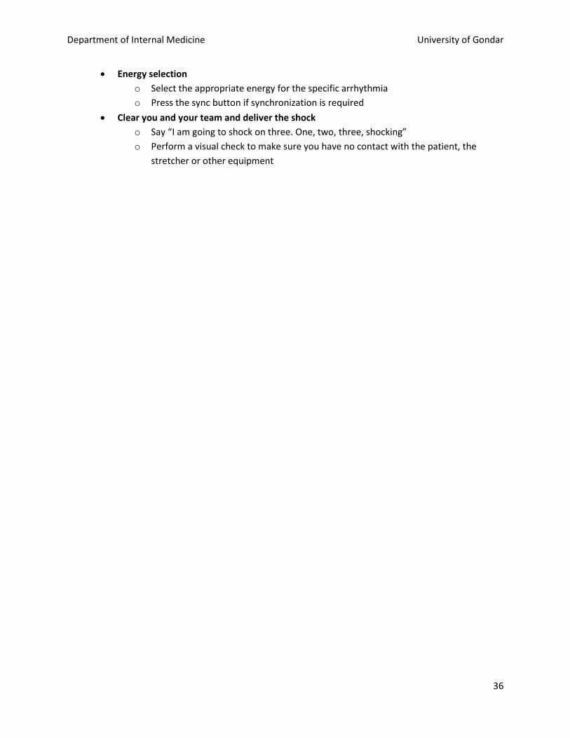
Energy selection
o Select the appropriate energy for the specific arrhythmia
o Press the sync button if synchronization is required
Clear you and your team and deliver the shock
o Say “I am going to shock on three. One, two, three, shocking”
o Perform a visual check to make sure you have no contact with the patient, the
stretcher or other equipment
Department of Internal Medicine University of Gondar
37
Hypertension
Hypertensive crises Definition:
Hypertensive emergency: increased BP with acute target organ ischemia and damage
o Neurologic damage: encephalopathy, stroke/TIA, papilledema(fundoscopy)
o Cardiac damage: acute heart failure/pulmonary edema, ACS, aortic dissection
o Renal damage: acute renal failure/proteinuria, hematuria, cast
o Microangiopathic hemolytic anemia: preeclapsia/Eclapsia
Hypertensive urgency: SBP>180or DBP>110 with minimal or no target organ damage
Precipitants:
Progression of essential hypertension +/- medical noncompliance
Progression of renovascular disease: AGN, preeclampsia
Endocrine: pheochromocytoma
Cerebral injury(low BP in acute stroke- with treatment)
Basic investigations:
ECG, BUN, Cr, electrolyte, Urinalysis, CXR(optional)
Treatment:
Tailor goal with the clinical context( eg. Rapid lowering for aortic dissection than acute ischemic
stroke)
Emergency: decrease MAP by ≈25% in minimum of 2 hours by IV agents; goal DBP<110 with in
2-6 hours, as tolerated
Urgency: decrease BP in hours/days using PO agents; goal normal BP in ≈ 1-2 days
Always watch for: UOP, creatinine, mental status; which may indicates lower BP is not tolerated
NB: basically hypertensive emergency should be treated with IV agents but in short of appropriate drugs
we can use a hydralazine with short acting PO drugs(captopril, propranolol, nifedipine) for almost all
cases of emergency
Drug selection:
Hydralazine IV: 10-20mg q20-30 min for maximum of 3-4 doses; fall in BP begins within 10 to 30
minutes and lasts 2 to 4 hours combined with one of the following;
o Propranolol 20mg Po stat and evaluate: (i.e. if no contraindication for propranolol/overt
heart failure or severe COPD start with hydralazine)
Patients with ACS and aortic dissection( avoid reflex tachycardia)
Acute renal failure/ATN
o Captopril 12.5mg PO stat and evaluate( caution in patients with acute renal failure and
better to use propranolol)
Overt heart failure/pulmonary edema
Encephalopathy and stroke(ischemic or hemorrhagic if indicated)

Department of Internal Medicine University of Gondar
38
o Nifedipine 20mg Po stat and evaluate( use if propranolol and captopril are not available
or contraindicated
Acute renal failure
Encephalopathy and stroke
NB: caution in combination of hydralazine with nifedipine( reflex tachycardia)
NB: For acute stroke(ischemic, hemorrhagic, SAH) BP should be kept a bit higher 180-185/100-
110mmHg until the acute event passed.
Chronic hypertension Definition:
Category Systolic(mmHg) Diastolic(mmHg)
Normal <120 <80
Pre-HTN 120-139 80-89
Stage 1 HTN 140-159 90-99
Stage 2 HTN ≥160 ≥100
NB: should be determined by making ≥ measurements separated by >2min. confirm stage 1 within
month; can treat stage 2 immediately
Standard workup: o Goal: (1) identify CV risk factors or other disease that would modify prognosis or treatment;
(2)consider 2nd causes(patient<20 or >50, or if sudden onset severe or refractory/labile
hypertension); (3)assess for end organ damage
o History: CAD, CHF, TIA/CVA, PAD, DM, renal insufficiency, sleep apnea; family hx; alcohol,
smoking: drugs like OCP, steroids, NSAIDs(esp. COX-2), Epo
o Physical exam: BP in both arms; fundoscopy, cardiac(LVH, murmurs/gallop),
vascular(thickening, bruits), abdominal(mass, bruits), neurologic
o Lab tests: BUN, Cr, electrolyte(k+, Ca++), glucose, Hct, U/A, lipid, TSH, ECG/Echo and if high
suspicion work up for secondary’s
Treatments: Threshold for treatment: all patients including diabetes and CKD
o Age <80 years: ≥140/90mmHg
o Age >85 years: ≥160/90mmHg
Treatment Goal: all patients including diabetes and CKD
o Age <80 years:≤140/90mmHg
o Age >85 years: ≤160/90mmHg

Department of Internal Medicine University of Gondar
39
Treatment options: Non pharmacologic: lifestyle modification
o Weight loss: goal BMI 18.5-24.9; aerobic exercise: ≥30min exercise/day, ≥5d/week
o Diet: rich in fruit and vegetables, low in saturated and total fat(DASH)
o Avoid salt consumption “table salt”
o Avoid alcohol consumption
Pharmacologic option:
o Uncomplicated HTN: no end organ damage
First line
Hydrochlorothiazide 25mg/day
Nifedipine(immediate release) 20-40mg/2-3 times /day
Amilodipine/felodipine 2.5-10 mg once daily(escalate 2.5mg every week)
Second line
Enalapril 2.5-40 mg in 2 divided dose
Lisinopril 10-40 mg/day
Candesartan 8-32 mg/day in 1-2 divided doses
Losarthan 25-200 mg/day in 1-2 divided doses
Alternative second lines
Beta blockers: selective B/α or non selective eg. Metoprolol, atenolol, propranolol, carvedilol
Central acting
methyldopa, clonidine

Department of Internal Medicine University of Gondar
40
Uncomplicated HTN plus(CKD, Diabetes)
Yes
No
No
Yes
No
No Yes
BP goal SBP <140 mm Hg DBP <90 mm Hg
Initiate HCT or CCB, alone or in combination.
Initiate ACEI or ARB, alone or in combination with other drug class
Select a drug treatment titration strategy A. Maximize first medication before adding second or B. Add second medication before reaching maximum dose of first medication or C. Start with 2 medication classes separately or as fixed-dose combination
At goal BP?
Titrate drugs to maximum tolerable level
Add another drug fron different class(2nd
or 3rd
drug)
Reinforce medication and lifestyle adherence. Add additional medication class (eg, β-blocker,
aldosterone antagonist, or consider 2nd
HTN)
At goal BP?
Continue current treatment and monitoring
At goal BP?
Department of Internal Medicine University of Gondar
41
Pregnancy and Heart Diseases
Recommendations for the management of heart disease in pregnancy appear in various guidelines. These heart diseases are valvular heart disease, atrial fibrillation, supraventricular tachycardias, and stroke, as well warfarin therapy during pregnancy.
Atrial Fibrillation
Atrial fibrillation (AF) is rare during pregnancy and is usually associated with another underlying cause, such as mitral stenosis, congenital heart disease, or hyperthyroidism. Diagnosis and treatment of the underlying condition causing the dysrhythmia are of utmost importance. Antithrombotic therapy is recommended for all pregnant women with atrial fibrillation. The type of therapy should be chosen with regard to the stage of pregnancy. Ventricular rate should be controlled with digoxin or a nondihydropyridine calcium channel antagonist to control the rate of ventricular response. Direct-current cardioversion can be performed without fetal damage in women who become hemodynamically unstable because of AF. Administration of quinidine or procainamide is a reasonable approach for cardioversion in pregnant women with AF who are hemodynamically stable.
Valvular Disease
Many women with valvular heart disease can be successfully managed throughout pregnancy, labor, and delivery with conservative medical measures. Symptomatic or severe valvular lesions should be addressed and rectified before conception and pregnancy whenever possible. Drugs should be avoided when possible.
Mitral Stenosis
Pregnant women with mild to moderate mitral stenosis can almost always be managed with judicious use of diuretics and beta blockade. A cardioselective beta blocker may prevent deleterious effects of epinephrine blockade on myometrial tissue. Women with severe mitral stenosis should be considered for percutaneous balloon mitral valvotomy before conception, if possible. Percutaneous balloon valvotomy is a reasonable option for women who develop severe symptoms during pregnancy.
Mitral Regurgitation
Mitral regurgitation can usually be managed medically with diuretics and vasodilator therapy. If surgery is required, repair is always preferred.
Aortic Stenosis
Pregnant women with mild obstruction and normal left ventricular systolic function can be managed conservatively throughout pregnancy. Those with moderate to severe obstruction or symptoms should be advised to delay conception until aortic stenosis can be corrected. Women with severe aortic stenosis who develop symptoms may require percutaneous aortic balloon valvotomy or surgery before labor and delivery.
Department of Internal Medicine University of Gondar
42
Aortic Regurgitation
Isolated aortic regurgitation can usually be managed with diuretics and vasodilator therapy when needed. Surgery during pregnancy should be contemplated only for control of refractory symptoms.
Endocarditis Prophylaxis
The guidelines do not recommend routine antibiotic prophylaxis in patients with valvular heart disease undergoing uncomplicated vaginal delivery or cesarean section unless infection is suspected. For high-risk patients, such as those with prosthetic heart valves or a prior history of endocarditis, prophylactic antibiotics are considered.
Supraventricular Tachycardias
Premature atrial beats, which are commonly observed during pregnancy, are generally benign and well tolerated. In patients with mild symptoms and structurally normal hearts, no treatment other than reassurance should be provided. Given that all commonly used antiarrhythmic drugs cross the placental barrier to some extent, antiarrhythmic drug therapy should be used only if symptoms are intolerable or if the tachycardia causes hemodynamic compromise.
Stroke
Pregnancy increases the risk for stroke and complicates the selection of acute and preventive therapies. Recommendations for stroke prevention in pregnant women focus on anticoagulation and antiplatelet strategies, which are similar to those for management of valvular heart disease during pregnancy.
Hypertrophic Cardiomyopathy
Women with hypertrophic cardiomyopathy are generally not at increased risk during pregnancy and undergo normal vaginal delivery without the necessity for cesarean section. Morbidity and mortality appear to be confined principally to women with high-risk clinical profiles, who should receive specialized preventive obstetrical care during pregnancy.
Anticoagulation
This hand book recommends Heparin or low-molecular-weight heparin in the first trimester of pregnancy, switching to warfarin in the second trimester, continuing it until about 38 weeks’ gestation, and then changing to heparin or low-molecular-weight heparin at 38 weeks with planned labor induction at about 40 weeks. Clear recommending evidences are lacking.