
Cardiac System - Nursing Ed · Cardiac System 323 CHAPTER SEVENTEEN PHYSIOLOGY OF THE CARDIAC...
32
Cardiac System 323 CHAPTER SEVENTEEN PHYSIOLOGY OF THE CARDIAC SYSTEM Structure of the Heart A. The heart is located in the mediastinal space of the thoracic cavity. B. The apex of the heart points downward and to the left; the apex comes in contact with the chest wall at about the fifth to sixth intercostal space. In the healthy indi- vidual, the point of maximum impulse (PMI) may be palpated here; this is also the area to auscultate when evaluating the apical heart rate. C. The heart is contained in a loose sac called the pericardium. 1. Fibrous pericardium: the outer surface. 2. Parietal layer: lines the fibrous pericardium. 3. Epicardial (visceral layer): vascular and adherent to the heart. 4. There is a potential space between the visceral and parietal layers of the pericardium. This area contains about 5 to 20 mL of pericardial fluid to lubricate the sac and prevent friction from cardiac movement. D. Myocardial wall. 1. Epicardium: the outer surface. 2. Myocardium: the middle layer of cardiac muscle. 3. Endocardium: the lining of the inner surface of the cardiac chambers. E. Cardiac chambers (Figure 17-1). 1. Four chambers are located within the heart; these chambers represent two pumps. 2. Both atria are the receiving chambers; both ventricles are the ejecting chambers. 3. The right side of the heart has a thinner myocardium than the left side and is a low-pressure system. 4. The left ventricle is composed of a thicker muscle, is a high-pressure system, and is capable of generating enough force to eject blood through the aortic valve and through the systemic circulation. F. Cardiac valves: maintain the directional flow of blood through the heart chambers. 1. Atrioventricular valves are controlled and supported by papillary muscle connected to the ventricular muscle and chordae tendineae extending from papil- lary muscle to valve leaflets. a. The tricuspid valve lies between the right atrium and the right ventricle. b. The mitral valve lies between the left atrium and the left ventricle. c. Both valves prevent backflow of blood from the ventricles into the atria during systole. 2. Semilunar valves (cusp valves) are controlled by the backward pressure of blood flow at the end of systole. a. Pulmonic valve: the outflow valve of the right ven- tricle into the pulmonary circulation. b. Aortic valve: the outflow valve of the left ventricle into the aorta. c. Both valves prevent the backflow of blood from the pulmonary artery and the aortic arch into the ventricle during diastole. G. Direction of blood flow through the heart structure (see Figure 17-1). 1. From the venous system, the blood enters the right atrium via the superior and inferior vena cavae; it flows through the tricuspid valve into the right ven- tricle; it is ejected through the pulmonic valve into the pulmonary artery; and it flows to the lungs for oxygenation. 2. Oxygenated blood returns to the left atrium via the pulmonary veins; it flows through the mitral valve into the left ventricle; it is ejected through the aortic valve into the aortic arch; and it flows into the sys- temic circulation. 3. The pulmonary artery is the only artery in the circula- tory system to carry deoxygenated blood; the pulmo- nary vein is the only vein in the circulatory system to carry oxygenated blood. Cardiac Function A. One complete cardiac cycle consists of contraction of the myocardium (systole) and subsequent relaxation of the myocardium (diastole). B. The amount of blood ejected with ventricular contrac- tion is the stroke volume. C. Starling’s law of the heart: the greater the cardiac muscles are stretched, the more forceful the contraction. If an increased amount of blood flows into the heart, the heart will increase the force of contraction and eject a larger amount of blood.
Transcript of Cardiac System - Nursing Ed · Cardiac System 323 CHAPTER SEVENTEEN PHYSIOLOGY OF THE CARDIAC...

L
Cardiac System
323
CHAPTER SEVENTEEN
PHYSIOLOGY OF THE CARDIAC SYSTEM
Structure of the HeartA. The heart is located in the mediastinal space of the
thoraciccavity.B. Theapexoftheheartpointsdownwardandtotheleft;
theapexcomesincontactwiththechestwallataboutthefifthtosixth intercostalspace.Inthehealthy indi-vidual, the point of maximum impulse (PMI) may bepalpated here; this is also the area to auscultate whenevaluatingtheapicalheartrate.
C. The heart is contained in a loose sac called thepericardium.1. Fibrouspericardium:theoutersurface.2. Parietallayer:linesthefibrouspericardium.3. Epicardial (visceral layer): vascular and adherent to
theheart.4. There is a potential space between the visceral
and parietal layers of the pericardium. This areacontains about 5 to 20 mL of pericardial fluid tolubricate the sac and prevent friction from cardiacmovement.
D. Myocardialwall.1. Epicardium:theoutersurface.2. Myocardium:themiddlelayerofcardiacmuscle.3. Endocardium: the liningof the innersurfaceof the
cardiacchambers.E. Cardiacchambers(Figure17-1).
1. Four chambers are located within the heart; thesechambersrepresenttwopumps.
2. Bothatriaarethereceivingchambers;bothventriclesaretheejectingchambers.
3. Therightsideofthehearthasathinnermyocardiumthantheleftsideandisalow-pressuresystem.
4. Theleftventricleiscomposedofathickermuscle,isahigh-pressuresystem,andiscapableofgeneratingenoughforcetoejectbloodthroughtheaorticvalveandthroughthesystemiccirculation.
F. Cardiac valves: maintain the directional flow of bloodthroughtheheartchambers.1. Atrioventricularvalvesarecontrolledandsupported
by papillary muscle connected to the ventricularmuscleandchordaetendineaeextendingfrompapil-larymuscletovalveleaflets.
a. Thetricuspidvalveliesbetweentherightatriumandtherightventricle.
b. Themitralvalveliesbetweentheleftatriumandtheleftventricle.
c. Both valves prevent backflow of blood from theventriclesintotheatriaduringsystole.
2. Semilunarvalves (cuspvalves) are controlledby thebackwardpressureofbloodflowattheendofsystole.a. Pulmonicvalve:theoutflowvalveoftherightven-
tricleintothepulmonarycirculation.b. Aorticvalve:theoutflowvalveoftheleftventricle
intotheaorta.c. Both valves prevent the backflow of blood from
thepulmonaryarteryandtheaorticarchintotheventricleduringdiastole.
G. Directionofbloodflowthroughtheheartstructure(seeFigure17-1).1. Fromthevenous system, thebloodenters the right
atrium via the superior and inferior vena cavae; itflowsthroughthetricuspidvalveintotherightven-tricle; it is ejected through the pulmonic valve intothe pulmonary artery; and it flows to the lungs foroxygenation.
2. Oxygenatedbloodreturns to the leftatriumvia thepulmonary veins; it flows through the mitral valveintotheleftventricle;itisejectedthroughtheaorticvalve into theaorticarch;and itflows into thesys-temiccirculation.
3. Thepulmonaryarteryistheonlyarteryinthecircula-torysystemtocarrydeoxygenatedblood;thepulmo-naryveinistheonlyveininthecirculatorysystemtocarryoxygenatedblood.
Cardiac FunctionA. Onecompletecardiaccycleconsistsofcontractionofthe
myocardium (systole) and subsequent relaxationof themyocardium(diastole).
B. Theamountofbloodejectedwithventricularcontrac-tionisthestrokevolume.
C. Starling’slawoftheheart:thegreaterthecardiacmusclesare stretched, the more forceful the contraction. If anincreasedamountofbloodflowsintotheheart,theheartwill increase the forceofcontractionandejecta largeramountofblood.

L
HHHHH5HHHHH10HHHHH15HHHHH20HHHHH25HHHHH30HHHHH35HHHHH40HHHHH45HHHHH50HHHHH55H56H57H58
324 CHAPTER 17 Cardiac System
b. Slows transmission of the impulse through theatrioventricular(AV)node.
c. Atropineblocksvagalstimulationtotheheart.2. Sympatheticstimulation.
a. Increasesheartrate.b. Increasesforceofcontraction.
F. Factorsthatincreasemyocardialoxygendemands.1. Increasedheartrate.2. Increasedforceofcontractions.3. Increasedafterload.
G. Cardiac compensatory mechanisms: when the normalcompensatory mechanisms cannot maintain cardiacoutput to meet body needs, the client is in a state ofcardiacdecompensation.1. Acute.
a. Sympatheticnervoussystemreceptors initiateanincreaseinthereleaseofepinephrineandnorepi-nephrinetoincreasethecardiacrateandmyocar-dialcontractility.
b. Increased diastolic filling (preload) increasescardiac output by increasing the stretch of themyocardialmusclefibers,thusincreasingthecon-tractility (Starling’s law). In a healthy heart, thisis themechanism that functionswhen there is aneed for increased cardiac output, as in exercise.Theincreaseinthestretchofthemyocardialfibersand the increase in the contractile force alsorequireanincreaseinoxygenconsumption.Ifthemyocardialfibershaveadecreasedoxygensupplyand/or the demand on the myocardial muscle isincreasedforaprolongedperiod,decompensationwilloccur.
2. Chronic: ventricular hypertrophy increases cardiacoutput by increasing the size of the myocardialmuscle.This also increases the myocardial need for
D. Cardiacoutput(CO=SV×HR).1. Thecardiacoutputcanbedeterminedbymultiplying
the stroke volume (SV) by the heart rate (HR) inbeatsperminute(CO=SV×HR).
2. Theheartpumpsapproximately5Lofbloodeveryminute.
3. The heart rate increases with exercise; therefore,cardiacoutputincreases.
4. Thecardiacoutputwillvaryaccordingtotheamountofvenousreturn(preload).
5. Factorsregulatingstrokevolume.a. Degree of stretch of the cardiac muscle before
contraction (Starling’s law): determined by thevolume of blood in the ventricle at the end ofdiastoleordiastolicfilling.
b. Contractility: ability of the myocardium to con-tract;contractility is increasedbycirculatingcat-echolaminesandmedicationssuchasdigitalis.
c. Preload:thefillingoftheventriclesattheendofdiastole.Themoretheventriclesfill,themorethecardiacmusclesarestretched,andthegreatertheforceofthecontractionduringsystole(Starling’slaw).Ifthereisadecreaseinthepreload,thereisalso a decrease in contractility and in cardiacoutput.
d. Afterload:thepressureintheaortathattheven-tricles must overcome to pump blood into thesystemic circulation. A decrease in the afterloadcausesadecreaseintheworkloadoftheventricles;thisinturnhelpsincreasethestrokevolumeandthecardiacoutput(Figure17-2).
E. Innervation of the myocardium (autonomic nervoussystem).1. Parasympathetic(vagusnerve)stimulation.
a. Slowsrateofimpulsegenerationatthesinoatrial(SA)node.
Pulmonaryartery
Pulmonaryvein
Left atrium
Aortic valveMitral valve
Pulmonicvalve
Leftventricle
InterventricularseptumDescending
aorta
Aortic arch
Rightventricle
Tricuspidvalve
Rightatrium
Inferiorvena cava
Superiorvena cava
Lung
FIGURE 17-1 Blood flow through the heart. (From Lewis SL et al: Medical-surgical nursing: assessment and management of clinical prob-lems, ed 7, St. Louis, 2007, Mosby.)
FIGURE 17-2 Preload and afterload. (From Zerwekh J, Claborn J: Memory notebook of nursing, vol 1, ed 4, Ingram, Texas, 2008, Nursing Education Consultants.)

L
CHAPTER 17 Cardiac System 325
System AssessmentA. Healthhistory.
1. Identifypresenceofriskfactorsforthedevelopmentofarterioscleroticdisease.
2. Copingstrategies.3. Respiratory.
a. Historyofdifficultybreathing.b. Medicationstakenforrespiratoryproblems.c. Determinenormalactivitylevel.
4. Circulation.a. Historyofchestdiscomfort(Table17-1).b. Historyofedema,weightgain.c. Historyofsyncope.d. Medicationstakenfortheheartorforhighblood
pressure.B. Physicalassessment.
1. Whatisthegeneralappearanceoftheclient:Isthereanyevidenceofdistress?Whatistheclient’sleveloforientationandabilitytothinkclearly?
2. Evaluatebloodpressure.a. Pulsepressure:thedifferencebetweensystolicand
diastolicpressure.b. Assessforposturalhypotension(decreaseinblood
pressurewhenclientstands).c. Takebloodpressuresitting,standing,andlyingif
client is having problems with pressure changes(see Chapter 16 for accurate blood pressuremeasurement).
d. Paradoxical blood pressure (paradoxical pulse): adecrease in systolic blood pressure of at least10mmHgthatoccursduringinspiration.
3. Evaluate quality and rate of pulse; assess for dys-rhythmias (see Appendix 17-7 for determiningdysrhythmias).a. Pulsedeficit:theradialpulserateislessthanthe
apicalpulserate;occursinatrialfibrillation.
oxygen,andinthediseasedmyocardium,thehyper-trophywilleventuallyleadtoadecompensatedstate.
Myocardial Blood SupplyA. Coronaryarteries.
1. Originateatthecoronarysinus,justoutsidetheaorticvalve.
2. Providetheonlysourceofoxygenatedbloodforthemyocardium.
3. Arteries fill during diastole; a diastolic pressure of60mmHgisrequiredtoadequatelyperfusethecoro-naryarteries.
B. Collateralcirculation.1. There are no direct connections between the large
coronaryarteries.2. When gradual occlusion of large coronary vessels
occurs as a result of arteriosclerotic heart disease(ASHD), the smaller vessels increase in size andprovidealternativebloodflow.
3. Becauseofthedevelopmentofcollateralcirculation,coronaryarterydiseasemaybewelladvancedbeforetheclientexperiencessymptoms.
Conduction SystemA. Controlstherateandrhythmoftheheart.B. Locatedinthemyocardiumarepathwaysforconduction
ofanelectricalimpulsethatinitiatescontractionoftheheartmuscle.
C. Characteristicsofcellsintheconductionsystem.1. Automaticity: the ability of certain conductive
pathway cells to initiate an impulse spontaneouslyandconsistently.
2. Excitability: the ability of a cell to respond to animpulse.
3. Conductivity:theabilityofacelltoconductanelec-tricalimpulse.
4. Refractoriness: the inability of a cell to respond toincomingstimuli.
D. Impulsegeneration.1. Restingstate:cellisreadytoreceiveanimpulse.2. Depolarization:flowofelectricalcurrentalongcardiac
membrane,initiatingmusclecontraction.3. Repolarization:cellsregaintheelectricalchargeand
arereturnedtoarestingstate.E. Relationshipofconductingpathwaystotheelectrocar-
diogram(ECG)(Figure17-3).1. Pwave:indicativeoftheimpulsegeneratedfromthe
sinoatrialnode;initiatesatrialdepolarization.2. PRinterval:delayoftheimpulseattheatrioventricu-
lar node and bundle of His to promote ventricularfilling.
3. QRS complex: passage of the impulse through thebundleofHis,downthebundlebranches,throughthePurkinjefibers;depolarizationoftheventricleoccurs.
4. Twave:ventricular repolarizationandreturn to therestingstate.
5. S-Tsegment:abovethebaselineincardiacinjuryandbelowthebaselinewithischemia.
FIGURE 17-3 Normal electrocardiogram (ECG). (From Hockenberry MJ, Wilson D: Wong’s nursing care of infants and children, ed 8, St. Louis, 2007, Mosby.)

L
HHHHH5HHHHH10HHHHH15HHHHH20HHHHH25HHHHH30HHHHH35HHHHH40HHHHH45HHHHH50HHHHH55H56H57H58
326 CHAPTER 17 Cardiac System
Pulmonicarea
Erb’s point
Precordium
Angle of Louis
TricuspidareaMitral area(apex)Epigastricarea
Aortic area (base)
Secondrib
SecondICS
FIGURE 17-4 Cardiac auscultatory sites. (From Lewis SL et al: Medical-surgical nursing: assessment and management of clinical problems, ed 7, St. Louis, 2007, Mosby.)
Table 17-1 ASSESSING CHEST PAIN (PQRST)
P—Precipitating Factors Q—QualityR—Region and Radiation
S—Symptoms and Signs (associated with chest pain)
T—Timing and Response to Treatment
Mayoccurwithoutprecipitators
PhysicalexertionEmotionalstressEatingalargemeal
PressureSqueezingHeavinessSmotheringBurningSeverepainIncreaseswith
movement
Substernalorretrosternal
SpreadsacrossthechestRadiatestotheinside
ofeitherorbotharms,theneck,jaw,back,upperabdomen
Diaphoresis,coldclammyskin
Nausea,vomitingDyspneaOrthopneaSyncopeApprehensionDysrhythmiasPalpitationsAuscultationofextra
heartsoundsAuscultationofcracklesWeakness
SuddenonsetConstantDuration>30minNotrelievedwith
nitratesorrestReliefwithnarcotics
b. Pulsus alternans: regular rhythm but quality ofpulsealternateswithstrongbeatsandweakbeats.
c. Threadypulse:weakandrapid;difficulttocount.4. Assess quality and pattern of respirations and evi-
denceofrespiratorydifficulty.5. Auscultationoftheheart(Figure17-4).
a. Heartsoundsheardduringthecardiaccycle.(1) S1:closureofthemitralandtricuspidvalves.(2) S2:closureoftheaorticandpulmonicvalves.(3) S3:representsrapidventricularfilling;normal
inchildrenandyoungadults; inadultsolderthan 30 years, it may be an indication ofvolumeoverload,ventriculardysfunctionsec-ondarytohypertension.
(4) S4: extra soundsheardduring atrial contrac-tion are abnormal; may indicate a forcefulatrialcontractionduetoincreasedresistance.
b. Presence ofmurmurs createdby turbulent bloodflow:gradedonascaleofloudness.
ALERT Identify common abnormal heart sounds (e.g., S3, S4).
6. Evaluate adequacyofperipheral vascular circulationandcheckforpresenceofperipheraledema.
7. Evaluateforpresenceofchestdiscomfort(seeTable17-1).a. Location.b. Intensityofpain.c. Precipitatingcauses.
ALERT Perform focused assessment or reassessment; interpret data that need to be reported immediately (see Box 17-1).
DISORDERS OF THE CARDIAC SYSTEM
Angina PectorisArteriosclerotic heart disease (ASHD), also called coro-nary artery disease (CAD), occurs as a result of the athero-sclerotic process (seeChapter 16) in the coronary arteries. Angina pectoris is caused by myocardial ischemia due to narrowed or blocked coronary arteries. The buildup of plaque or fatty material in the coronary artery causes a narrowing of the lumen of the artery and precipitates myo-cardial ischemia that causes chest pain.A. Pain (angina)occurswhen theoxygendemandsof the
heartmuscleexceedtheabilityofthecoronaryarteriestodeliverit.
(1) Abnormal flow through diseased valves: ste-nosisandinsufficiency.
(2) Abnormal flow of blood between cardiacchambers(congenitalheartdisease).
c. Presenceofa frictionrub:usuallyheardovertheapex during S1 and S2; can be heard best withclientsittingandleaningforward.

L
CHAPTER 17 Cardiac System 327
4. Client may describe pain as squeezing, choking, orconstricting or as a vague feeling of pressure andindigestion.
5. Clientwillfrequentlydenyseriousnessofthepain.6. Mostclientscorrelatepainwithactivityandincreased
cardiacdemands.7. Pain is of short duration, generally lasting about 5
minutes; may be longer if associated with anger orheavymeals.
8. Accompanying symptoms may include diaphoresis,increasedanxiety,pallor,anddyspnea.
C. Diagnostics—chronic stable angina (see Appendix17-1).
Treatment—Chronic Stable AnginaA. Primarygoaloftreatmentistorelievepainandprevent
futureattacks.B. Medication.
1. Vasodilators:nitroglycerin(Appendix17-2).2. Beta-adrenergicblockers(Appendix17-3).3. Calcium-channelblockers(Appendix17-3).4. Antiplateletmedications(seeAppendix16-4).
C. Procedures/surgicalintervention.1. Percutaneouscoronaryintervention(PCI):aballoon
is threaded from an artery in the groin to theaffectedcoronaryartery.Theballoonistheninflatedin an effort to compress the plaque and dilate thenarrowed artery reestablishing blood flow to themyocardium.
2. Laserangioplasty:a specialcatheterwitha laser tipis inserted into coronary artery. When confrontedwith a blockage, the laser emits pulsating beams oflightthatvaporizetheplaque.
3. Atherectomy:techniqueinwhichthecatheterhasarotatingshaveronthetipthatcutsawaytheplaque.
4. An intracoronary stent is an expandable wire meshthatcanbe insertedduringanyof theaboveproce-dures.Astentservesasascaffoldtomaintainpatencyofthecoronaryartery.
5. Cardiac revascularization: coronary artery bypassgraft(CABG)surgery,openheartsurgery.
D. Restrictedactivity.E. Supplementaloxygen.F. Controlofthemodifiableriskfactors(seeBox17-1).
Acute Coronary Syndrome: Unstable Angina Pectoris
According to the American Heart Association, acute cor-onary syndrome (ACS) includes the various degrees of coronary artery occlusion that can develop in individuals with coronary atherosclerosis.A. Includes unstable angina, non-ST-segment elevation
myocardial infarction (MI), and ST-segment elevationMI,allofwhichcanleadtosuddencardiacdeath.
B. Unstable angina pectoris occurs when a thrombuspartially occludes a coronary artery causing prolongedsymptomsofischemiawhichcanoccuratrest.
B. Temporaryischemiadoesnotcausepermanentdamageto themyocardium.Pain frequently subsideswhen theprecipitatingfactorisremoved.
C. Typesofangina.1. Chronicstableangina:predictablewithlevelofstress
orexertion;consistentlyrespondswelltomedication;painrarelyoccursatrest.
2. Unstableangina(acutecoronarysyndrome).a. Unstableanginapainoccursspontaneouslyatrest
(usuallybetweenmidnight and8 a.m.); progres-sive in frequency of attacks; unpredictable, notrelievedwithsublingualnitroglycerin.
b. Variant angina (Prinzmetal angina) pain thattendstobecycliccausedbycoronaryspasms.
3. New onset angina: first symptoms of angina thatmostfrequentlyoccurafterexertion.
AssessmentA. Riskfactors/etiology.
1. ASHD(Box17-1).2. Cardiacischemia.3. Aorticvalvedisease(impedesfillingofthecoronary
arteries).4. Increasedcardiacdemands.
a. Exercise,emotionalstress.b. Heavymeals,thyrotoxicosis.c. Exposuretocoldtemperature.
B. Clinicalmanifestations—chronicstableangina.1. Paininvaryinglevelsofseverity(seeTable17-1).2. Painmostoftenis locatedbehindor justtotheleft
ofthesternum.3. Painmayradiatetoneck,jaw,andshoulders.
Box 17-1 RISK FACTORS IN ARTERIOSCLEROTIC HEART DISEASE
Modifiable Risk FactorsElevatedserumcholesterollevelsHighbloodpressureCigarettesmokingSedentarylifestyleObesityType A personality (high-pressure lifestyle, driving, competi-
tive)Diabetesmellitus
Nonmodifiable Risk FactorsGeneticpredispositionPositivefamilyhistoryofheartdiseaseIncreasingageGender: occurs more often in men; increase in women after
menopause
ALERT Teach health promotion information. Know the ASHD risk factors and be able to teach the client how to effectively reduce his or her risk factors.

L
HHHHH5HHHHH10HHHHH15HHHHH20HHHHH25HHHHH30HHHHH35HHHHH40HHHHH45HHHHH50HHHHH55H56H57H58
328 CHAPTER 17 Cardiac System
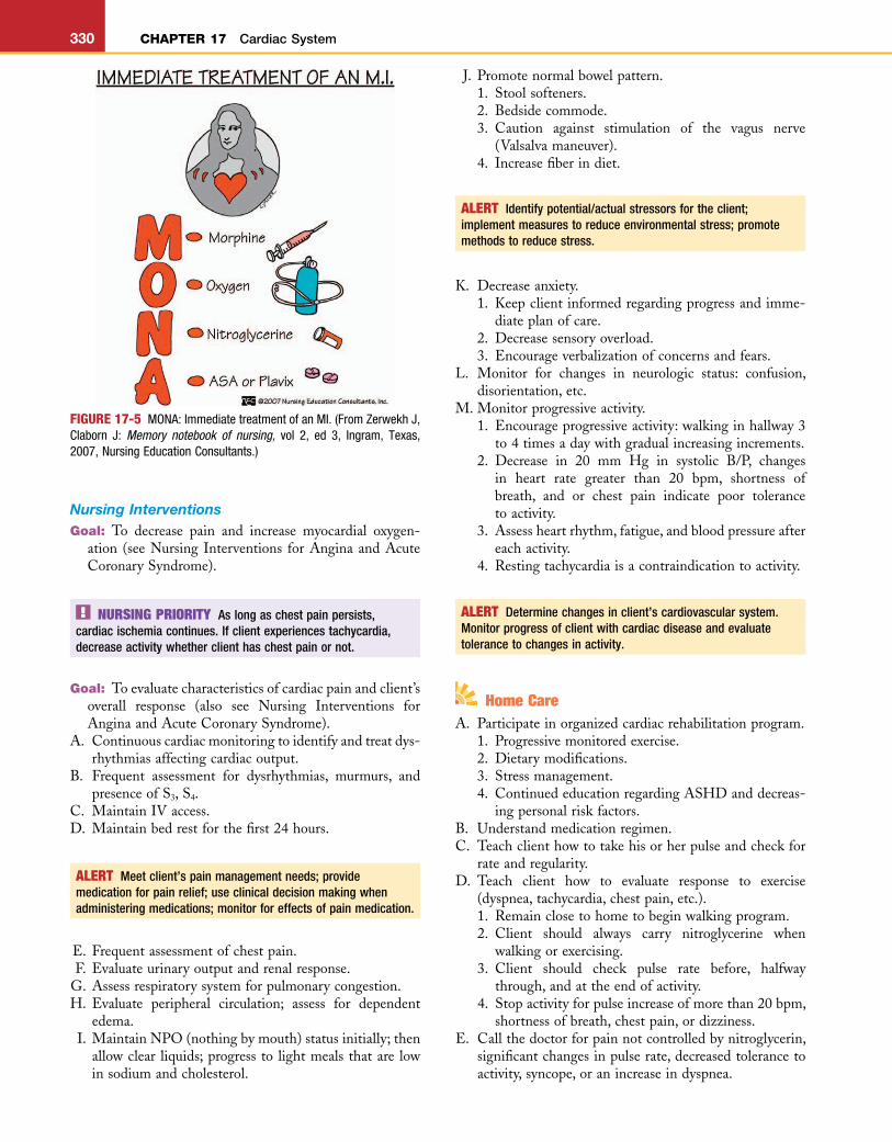
A. Beginsupplementaloxygen.B. Positionclientinrecliningpositionwithheadelevated.C. Assesscharacteristicsofpain:administermorphine for
paincontrol.D. Administermedications.
1. Administer nitroglycerin (sublingually, IV, or spray;seeAppendix17-2): evaluate client’s response; painfrom chronic angina is usually relieved; pain fromacuteanginamaynotberelieved.
2. Narcoticanalgesics(IVmorphineinsmallincrementsuntilpainsubsides).
3. Antiplateletagent(seeAppendix16-4).E. Maintaincalm,reassuringatmosphere.F. EstablishvenousaccessforfluidsandIVmedications.G. Notifyphysicianifpaindoesnotrespondtomedication
orifvitalsignsdeteriorate.
AssessmentA. Riskfactors/etiology.
1. Familyhistoryofcoronaryarterydisease.2. Hypertension,hypercholesterolemia.3. Diabetes,smoking.4. Averageagefor1stMI-menover64.5years,women
over70.5years.5. Womenareatincreasedriskaftermenopause.
B. Clinicalmanifestations.1. Twoormoreanginaleventswithinthepast24hours.2. Prolongedchestpain(greaterthan20minutes).3. Presenting symptoms in women: indigestion, pain
between the shoulders, shortness of breath, andanxiety.
4. Hypotension,bradycardia,ortachycardia.5. NewonsetS3.
C. Diagnostics(Appendix17-1).1. 12-leadECG.
a. STsegmentelevation(STEMI),traditional.b. Non-ST elevation MI (NSTEMI), common in
women.2. Elevatedcardiactroponin1.3. ElevatedCK-MB.4. Presenceofunstableangina.
D. Treatment(initial).1. Bedrest.2. Monitorvitalsigns,includingoxygensaturationlevel.3. Supplemental oxygen at 4 L/min via nasal cannula
(maintainO2satabove90%).4. Reducecoronaryreocclusionwithantiplateletmedi-
cations(Appendix16-4).5. Reduce and control ischemic pain: vasodilators
(nitroglycerin,sublingual,sprayorIV,seeAppendix17-2), narcotics (morphine sulfate IV—if pain notrelievedbythenitroglycerin).
6. Beta-adrenergic receptor blockers (Appendix 17-3)to decrease cardiac demand for oxygen; anticoagu-lants(Appendix16-3)topreventemboli.
7. Complete fibrinolytic checklist and, if appropriate,initiatefibrinolytictherapy(Appendix17-5).
8. Transmyocardiallaserrevascularization(TMR):laserprobe is inserted into the wall of the left ventricle;channelsarecreatedtopromotethedevelopmentofrevascularization.
ComplicationsA. Dysrhythmias(seeAppendix17-7).B. Myocardialinfarction(MI).
Nursing Interventions for Angina and Acute Coronary Syndrome
ALERT Intervene to improve client’s cardiovascular status; assess client for decreased cardiac output; meet client’s pain management needs; use critical thinking when addressing pain management.
Goal: To decrease pain and increase myocardial oxygen-ation.
NURSING PRIORITY To relieve chest pain and to decrease cardiac damage resulting from an inadequate blood and oxygen supply to the myocardium, there must be an immediate reduction in the workload of the heart that results in a decrease in oxygen consumption: rest, nitroglycerin, oxygen therapy.
Goal: To evaluate characteristics of anginal pain and cli-ent’soverallresponse.
A. Does pain increase with breathing? (Anginal pain isgenerally not affected by breathing or changes ofposition.)
B. Assessactivitytoleranceorprecipitatingfactor.C. Assess changes in characteristics of pain (see Table
17-1).D. Evaluateresponseofpaintotreatmentorprogressionto
moreseverelevel.E. Obtaina12-leadECG.F. Evaluatevitalsigns.
1. Presence of S3 gallop, which may indicate heartfailure.
2. Presenceofjugularveindistention;peripheraledema.3. Presenceofdyspneaorwetbreathsounds.4. Adequacyofcardiacoutput:peripheralpulses,urinary
output,levelofconsciousness.G. Continuous ECG monitoring: assess for presence of
dysrhythmiaandimpactoncardiacoutput.H. Monitortroponinlevels(Appendix17-1). I. Assessclient’spsychosocialresponse:denialiscommon;
anger, fear and depression occur in both client andfamily.
ALERT Intervene to improve client’s cardiovascular status; provide client with strategies to manage decreased cardiac output.
Goal: To provide care after percutaneous coronary inter-vention(withorwithoutstent).
A. Monitorforchestpainandhypotension;reocclusionisaprimarycomplication.
B. Assessforbleedingorhematomaformation.

L
CHAPTER 17 Cardiac System 329
C. Frequently assess status of circulation distal to area ofcannulation.
D. Asheathmaybeleftinplace;monitorareaforbleeding;ifbleedingoccurs,putmanualpressureontheareaandnotifythephysician.
E. Preventflexionofaffectedextremityandmaintainbedrestfor6to8hours.
F. Clientistoavoidheavylifting;mayreturntoworkin1to2weeks.
G. Notifythedoctorofanychestpain,syncope,orbleedingatthesite.
H. AssessECGforevidenceofST-segmentchanges.
Home CareA. EducationregardingASHD.
1. Clientwillbeabletoidentifypersonalriskfactorsandappropriatehealthpracticestodecreaseriskfactors.
2. Client will be able to identify factors precipitatingpain.
B. Determinethatclientunderstandshisorhermedicationregimen(Box17-2).
C. Assessunderstandingofdietandexerciseregimen.D. Assess understanding of seeking medical assistance if
painpersistsandisnotrelievedbymedication.E. Helpclientidentifyresourcesforcounselingtodecrease
stress.F. Advise client not to take erectile dysfunction drugs
(Appendix22-1)ifonnitratesforchestpain.
NURSING PRIORITY Danger of death from an MI is greatest during the first 2 hours.
AssessmentA. Riskfactors/etiology(seeunstableangina).B. Clinicalmanifestations.
1. Typicalpainissevere,substernal,crushing,andunre-lievedbynitroglycerin.
2. Denialoftheseriousnessofthepain;clientswithMIfrequentlywaitmorethan2hourstoseekassistance.
3. Dyspnea,nausea,vomiting,indigestion.4. Pale,duskyskin.5. Painmayradiatedownarmorupthejaw.6. Onsetisusuallysudden.7. Diaphoresis;extremeweakness.8. Decreaseinbloodpressure,tachycardia,syncope.
C. Diagnostics:seeacuteangina(seeAppendix17-1).
Treatment (Figure17-5)A. Bedrest.B. Monitorvitalsigns,oxygensaturation,andECG.C. Supplemental oxygen at 4 L/min via nasal cannula
(maintainO2satabove90%).D. Painrelief(morphinesulfateIV).E. Beta-adrenergic receptor blockers (Appendix 17-3);
antiplatelets(Appendix16-4);anticoagulants(Appendix16-3).
F. Antidysrhythmicagents(Appendix17-4).G. Reperfusion(fibrinolytic)therapy(Appendix17-5).H. Percutaneous coronary intervention (PCI): “door-to-
balloon”goalof90minutesforoptimumresponse. I. Cardiacsurgeryifappropriate.
ComplicationsA. Dysrhythmias(seeAppendix17-7).B. Cardiogenicshock.C. Heartfailure(congestiveheartfailure).
Acute Coronary Syndrome: Myocardial Infarction
A myocardial infarction (MI), also called coronary occlu-sion or heart attack, is a total occlusion of a portion of a coronary artery. After the occlusion, myocardial ischemia, injury, or death occurs.A. Infarction most often occurs in the area of the left
ventricle.B. Theseverityofthesituationdependsontheareaofthe
heartinvolved,aswellasthesizeoftheinfarction.C. Healingprocess.
1. Inthefirst24hours,theinflammatoryprocessiswellestablished; leukocytes invade the area, and cardiacenzymesarereleasedfromthedamagedcells.
2. In4to10days:necroticzoneiswelldefined.3. In10to14days:theformationofscartissuefounda-
tionbegins.D. The presence of pre-established collateral circulation
willassistindecreasingthesizeofthenecroticarea.
Box 17-2 CLIENT EDUCATION FOR NITROGLYCERIN ADMINISTRATION
1. Keepinatightlyclosed,darkglasscontainer.2. Carrysupplyatalltimes—eithersublingual(SL)tabletsor
translingualspray;donotswallowsublingualtablets.3. Fresh tablets should cause a slight burning under the
tongue.4. Dateallopenedcontainersanddiscardallmedicationthat
is24monthsold.5. Take nitroglycerin prophylactically to avoid pain—before
sexualintercourse,exercise,walking,etc.6. Takenitroglycerinwhenpainbegins;stopallactivity.7. Ifpain isnot relieved in5minutes, call911andactivate
EMS.8. While waiting for EMS response, if chest pain remains
unrelieved,takeanotherSLpillor1meteredspray.9. Remain lying down; orthostatic hypotension can be a
problem.10. Long-acting preparations should not be abruptly discon-
tinued;thismayprecipitatevasospasm.11. Todecreasedevelopmentoftoleranceinlong-actingprep-
arations, schedule an 8-hour nitro-free period each day,preferableatnight.
12. Donottakeerectiledysfunctiondrugswithnitroglycerine.
ALERT Instruct clients about self-administration of medications.
L
HHHHH5HHHHH10HHHHH15HHHHH20HHHHH25HHHHH30HHHHH35HHHHH40HHHHH45HHHHH50HHHHH55H56H57H58
330 CHAPTER 17 Cardiac System
Nursing InterventionsGoal: To decrease pain and increase myocardial oxygen-
ation (seeNursing Interventions forAnginaandAcuteCoronarySyndrome).
FIGURE 17-5 MONA: Immediate treatment of an MI. (From Zerwekh J, Claborn J: Memory notebook of nursing, vol 2, ed 3, Ingram, Texas, 2007, Nursing Education Consultants.)
NURSING PRIORITY As long as chest pain persists, cardiac ischemia continues. If client experiences tachycardia, decrease activity whether client has chest pain or not.
Goal: Toevaluatecharacteristicsofcardiacpainandclient’soverall response (also see Nursing Interventions forAnginaandAcuteCoronarySyndrome).
A. Continuouscardiacmonitoringtoidentifyandtreatdys-rhythmiasaffectingcardiacoutput.
B. Frequent assessment for dysrhythmias, murmurs, andpresenceofS3,S4.
C. MaintainIVaccess.D. Maintainbedrestforthefirst24hours.
ALERT Meet client’s pain management needs; provide medication for pain relief; use clinical decision making when administering medications; monitor for effects of pain medication.
E. Frequentassessmentofchestpain.F. Evaluateurinaryoutputandrenalresponse.G. Assessrespiratorysystemforpulmonarycongestion.H. Evaluate peripheral circulation; assess for dependent
edema. I. MaintainNPO(nothingbymouth)statusinitially;then
allowclear liquids;progressto lightmealsthatare lowinsodiumandcholesterol.
ALERT Identify potential/actual stressors for the client; implement measures to reduce environmental stress; promote methods to reduce stress.
K. Decreaseanxiety.1. Keepclientinformedregardingprogressandimme-
diateplanofcare.2. Decreasesensoryoverload.3. Encourageverbalizationofconcernsandfears.
L. Monitor for changes in neurologic status: confusion,disorientation,etc.
M.Monitorprogressiveactivity.1. Encourageprogressiveactivity:walkinginhallway3
to4timesadaywithgradualincreasingincrements.2. Decrease in 20 mm Hg in systolic B/P, changes
in heart rate greater than 20 bpm, shortness ofbreath, and or chest pain indicate poor tolerancetoactivity.
3. Assessheartrhythm,fatigue,andbloodpressureaftereachactivity.
4. Restingtachycardiaisacontraindicationtoactivity.
ALERT Determine changes in client’s cardiovascular system. Monitor progress of client with cardiac disease and evaluate tolerance to changes in activity.
J. Promotenormalbowelpattern.1. Stoolsofteners.2. Bedsidecommode.3. Caution against stimulation of the vagus nerve
(Valsalvamaneuver).4. Increasefiberindiet.
Home CareA. Participateinorganizedcardiacrehabilitationprogram.
1. Progressivemonitoredexercise.2. Dietarymodifications.3. Stressmanagement.4. ContinuededucationregardingASHDanddecreas-
ingpersonalriskfactors.B. Understandmedicationregimen.C. Teachclienthowtotakehisorherpulseandcheckfor
rateandregularity.D. Teach client how to evaluate response to exercise
(dyspnea,tachycardia,chestpain,etc.).1. Remainclosetohometobeginwalkingprogram.2. Client should always carry nitroglycerine when
walkingorexercising.3. Client should check pulse rate before, halfway
through,andattheendofactivity.4. Stopactivityforpulseincreaseofmorethan20bpm,
shortnessofbreath,chestpain,ordizziness.E. Callthedoctorforpainnotcontrolledbynitroglycerin,
significantchangesinpulserate,decreasedtolerancetoactivity,syncope,oranincreaseindyspnea.

L
CHAPTER 17 Cardiac System 331
2. Low-output failureoccurswhen themyocardium issodamagedthatitcannotmaintainadequatecardiacoutput;itisthefailureoftheheartasapump.
E. Cardiac compensatory mechanisms will attempt tomaintain the body requirements for cardiac output;when these mechanisms become ineffective, cardiacdecompensationorfailurewilloccur.
F. Edemadevelopmentinheartfailure.1. Decreased cardiac output leads to decrease in renal
perfusion, the kidneys respond by stimulating theadrenal cortex to increase the secretion of aldoste-rone, thus increasing the retention of sodium andwater.
2. With an increase in the venous pressure from theincreased circulating volume, there is an increase inthecapillarypressure;anddependent,pittingedemaoccurs.
G. In children, HF occurs most often as the result of astructural problem of the heart. Ventricular functionmay not be impaired, but symptoms occur because ofincreased pulmonary artery pressure and pulmonaryvenouscongestion.
F. Sexual intercourse can generally be resumed in 4 to 6weeksafterMIorwhentheclientcanwalkoneblockorclimbtwoflightsofstairswithoutdifficulty.1. Donotdrinkanyalcoholbeforesexualactivity.2. Takenitroglycerinbeforesexualactivity.3. Donothavesexafteraheavymeal.4. Position for intercourse does not influence cardiac
workload.5. Donottakeerectiledysfunctionmedications(Appen-
dix22-1)iftakingnitrates.
ALERT Assess client’s ability to perform self-care. Determine whether the client with cardiac disease understands the illness and whether he or she can demonstrate knowledge of care.
Heart Failure (HF)Heart failure (also referred to as congestive heart failure [CHF] and chronic heart failure) includes cardiac decom-pensation, cardiac insufficiency, ventricular failure, and ultimately results in the inability of the heart to pump adequate amounts of blood into the systemic circulation to meet tissue metabolic demands.
Physiology of Heart FailureA. Left-sidedheartfailure.
1. Resultsfromfailureoftheleftventricletomaintainadequatecardiacoutput.
2. Blood backs up into the left atrium and into thepulmonaryveins.
3. Increasing pressure in the pulmonary capillary bedcauses lungs to become congested, resulting inimpairedgasexchange.
4. Precipitatingfactors.a. MI(leftventricularinfarct).b. Hypertension.c. Aorticandmitralvaluedisease.
B. Right-sidedheartfailure.1. The right ventricle is unable to maintain adequate
output.2. Blood backs up into the systemic circulation and
causesperipheraledema.3. Precipitatingfactors.
a. Left-sidedheartfailure.b. Chronicpulmonarydisease.
C. Each side of the heart is dependent on the other foradequatefunction.1. Left-sided failure results in pulmonary congestion;
thiscausesanincreaseinpulmonarypressure,whichfurther increases the workload and pressure in therightsideoftheheartandprecipitatesfailure.
2. Themajorityof clinical situations involve failureofbothsidesoftheheart.
D. High-outputversuslow-outputfailure.1. High-outputfailureoccurswhenthebody’sneedsfor
oxygen are excessively increased; theheart increasesoutput but is still unable to meet the body’s needs(hyperthyroidandanemia).
ALERT If a test question states that a client is in heart failure, assume that both sides are in failure unless indicated otherwise.
AssessmentA. Riskfactors/etiology.
1. Myocardialdisease,valvulardisease.2. Hypertension.3. Congenitalheartdisease.4. Fluidoverload.
B. Clinicalmanifestations(Box17-3).1. Impairedcardiacfunction.
a. Tachycardiaevaluatedaccordingtoage level(seeTable15-2).
b. Cardiomegaly from dilation and hypertrophy;PMIisdisplaced.
c. S3 or S4 (heart gallop) caused by impaired ven-tricularfunction.
ALERT Reevaluate the client with abnormal heart sounds.
d. In infants, failure to thrive and failure to gainadequateweight.
e. Poorperfusion:coolextremities,weakpulses,poorcapillaryrefill.
2. Pulmonarycongestion.a. Dyspnea,dyspneaonexertion,tachypnea.b. Orthopnea:infantsandadults.c. Paroxysmalnocturnaldyspneaoccurswhileclient
isasleep.d. Symptomsofrespiratorydistressandhypoxia(see
Table15-2).e. Congestedbreathsounds,cough.f. Feedingdifficultiesininfants,causedbydyspnea.g. Increaseinpulmonaryarterypressure(PAP)and
pulmonary arterial wedge pressure (PAWP) (seeAppendix17-9).

L
HHHHH5HHHHH10HHHHH15HHHHH20HHHHH25HHHHH30HHHHH35HHHHH40HHHHH45HHHHH50HHHHH55H56H57H58
332 CHAPTER 17 Cardiac System
Box 17-3 ASSESSMENT FINDINGS OF HEART FAILURE
Right-Sided (Systemic Symptoms)NeckveindistentionGeneralizededema(anasarca)DependentpittingedemaNausea,vomitingAnorexiaAlteredGIfunctionWeightgainAscitesDecreasedurinaryoutputLiverengorgementElectrolyteimbalancesDysrhythmiasElevatedCVP
Left-Sided (Pulmonary Symptoms)DuskyskinandnailbedsChestpainTachycardiaDecreasedsystolicBPVentricularectopicbeatsPresenceofS3andS4
ShiftofthePMItotheleftIrritability,restlessnessDecreasedcardiacoutputIncreasedPAWPWheezingcracklesDyspneaProductivecough
BP, Blood pressure; CVP, central venous pressure; GI, gastrointestinal;PMI, point of maximum impulse; PAWP, pulmonary arterial wedgepressure.
3. Systemiccongestion.a. Hepatomegaly:maybeanearlysigninchildren.b. Dependentedemageneralizedininfants;evaluate
byweightgain.c. Peripheralpittingedema.d. Ascites.e. Increase in central venous pressure (CVP) (see
Appendix17-9).
ALERT Determine changes in client’s cardiovascular status as related to the client’s HF; interpret what data need to be reported immediately.
D. Activitylimitations.E. Medications:treatmentforHFincludesreductionofthe
preload and afterload, as well as improvement of thecontractilityofthemyocardium.1. Cardiacglycosides(Appendix17-6).2. Diuretics(seeAppendix16-6).3. Aldosteroneantagonists(seeAppendix16-6).4. Vasodilators(seeAppendix16-5).5. Angiotension-converting enzyme (ACE) inhibitors
(seeAppendix16-5).6. Beta blockers (adrenergic receptor antagonists)
(Appendix17-3).7. Electrolyte replacement, carefully monitor serum
potassiumlevels.F. Decreasedsodiumdiet,aswellasfluidrestrictions, for
adultsandolderchildren.G. Sodiumandfluidsmaynotberestrictedforinfantsand
children;infantsseldomneedfluidrestrictionbecauseofdifficultyfeeding.
ComplicationsA. Pulmonaryedema(Chapter15).B. Cardiogenicshock.C. Dysrhythmias.
NURSING PRIORITY The goals for therapeutic intervention in clients with CHF are to:• Improve cardiac output: digitalis and oxygen.• Decrease cardiac afterload: decrease activity, administer
vasodilator.• Decrease cardiac preload: diuretics; decrease sodium and fluid
intake; place client in semi-Fowler’s position.
Nursing InterventionsGoal: To decrease cardiac demands and improve cardiac
output.A. Assess vital signs and compare with other physical
assessmentdata.B. Conserveenergy.
1. Encouragerestalternatedwithactivity.2. Monitor pulse, respiratory rate, and dysrhythmias
duringperiodsofactivity.C. Avoid chilling; it increases oxygen consumption, espe-
ciallyininfants.D. Supplemental oxygen, especially when needed with
increasedactivity.E. Provideuninterruptedsleep,whenpossible.F. Minimizecryinginchildrenandinfants.G. Decreasestressandanxiety;encourageparentstoremain
withchild.
ALERT Check for interactions among client’s drugs, foods, and fluids. For clients receiving digitalis and diuretics, it is very important to monitor the serum potassium level. A significant decrease in the serum potassium level may affect cardiac rhythm and can precipitate digitalis toxicity.
C. Diagnostics(seeAppendix17-1).
TreatmentA. Treatmentoftheunderlyingproblem.B. Prevention.
1. Administrationofprophylacticantibiotics toclientswith rheumatic heart disease before medical proce-durestopreventmitralvalvedamage.
2. Effectiveearlytreatmentofhypertension.3. Earlytreatmentofdysrhythmia.
C. Oxygen.
Goal: Todecreasecirculatingvolume.A. Assessbreathsounds;checkfordistendedneckveinsand
peripheraledema.

L
CHAPTER 17 Cardiac System 333
B. Low-sodiumdietandfluidrestrictioninadultsandolderchildren.
C. Evaluate fluid retention by determining accurate dailyweight(1kgor2.2lbweightgain=1Loffluidlossorretention).
D. Accurateintakeandoutput;monitorelectrolytebalanceandtherapeuticeffectoffluidloss.
Goal: Toreducerespiratorydistress.A. Position.
1. Adult may be placed in Fowler’s or semi-Fowler’spositionormaysitinanarmchair.
2. When insemi-Fowler’sposition,donotelevatecli-ent’slegs;thisincreasesvenousreturn(preload).
3. Aninfantorsmallchildmaybreathebetterinaside-lyingpositionwiththekneesdrawnuptothechest.
4. Aninfantmaybeplacedinaninfantseat.5. Make sure diapers are loosely pinned and safety
restraintsdonothindermaximumexpansionof thechest.
6. Hold infant upright over the shoulder with kneesflexed(knee-chestposition).
B. Administer oxygen: use oxygen hood for infants andnasalcannulaforadultsandolderchildren;supplementaloxygentokeepsaturationlevelsatorabove90%.
C. Evaluate breath sounds, evaluate adventitious breathsoundsandpresenceofcongestion.
D. Donotallowinfantstocryforextendperiods.
PeDIATRIC NURSING PRIORITY Infants: if cyanosis decreases with crying, the problem is usually pulmonary; if cyanosis increases with crying, the problem is usually cardiac.
D. Discussuseofandsafetyfactorsforhomeoxygen.E. Contacthealthcareproviderfor:
1. Weightgainof3to5poundsoveraweekor1to2poundsovernight.
2. Increase in dyspnea or angina, especially withdecreasedactivityoratrest.
3. Decreaseinactivitytolerancethatexceeds3or4days.4. Increasedurinationatnight;presenceor increase in
peripheraledema.5. Cough or respiratory congestion that lasts longer
than3or4days.F. Providewritten instructions formedications, especially
iftheyareondigitalis.G. Assessclient’shomesituationandabilityofcaregivers.
Rheumatic Heart DiseaseRheumatic heart disease is an inflammatory disease that is usually self-limiting. The primary concern is the devel-opment of rheumatic heart disease involving the endocar-dium and cardiac valves.A. UsuallyprecededbyagroupAbeta-hemolyticstrepto-
coccalinfection.
NURSING PRIORITY Prevention and adequate treatment of streptococcal infections prevent the development of rheumatic heart disease.
B. Myocardialinvolvementischaracterizedbythedevelop-mentofvalvulitis,pericarditis,andmyocarditis.1. Valvulitisproducesscarringofthecardiacvalves.2. Rheumaticcarditisistheonlysymptomthatproduces
permanent damage, most often involves damage totheendocardiumandprimarilytothemitralvalve.
3. Rheumaticfeverusuallyoccursduringchildhood,butmanifestationsofcardiacdamagemaynotbeevidentforyears.
AssessmentA. Risk factors/etiology: previous infection by group A
beta-hemolyticstreptococcus.B. Clinical manifestations: symptoms vary; no specific
symptom or lab test is diagnostic of rheumatic fever.Criteria for the diagnosis require a combination ofsymptomstobepresent.1. Carditis.
a. Tachycardiaoutofproportiontofever.b. Longhigh-pitchedapicalsystolicmurmurbegin-
ningwithS1andcontinuingthroughoutcycle.c. Pericarditis, pericardial friction rub, and com-
plaintsofchestpain.2. Migratorypolyarthritis.3. Chorea.4. Erythemamarginatum.5. Subcutaneousnodulesoverbonyprominences.6. Historyofarecentstreptococcalinfection.
TreatmentA. Adequatetreatmentofinitialstreptococcalinfection.B. Restanddecreasedactivityuntiltachycardiasubsides.
Goal: Tomonitorfordevelopmentofhypoxia(seeChapter15).
Goal: Tomaintainnutrition.A. Providesmall,frequentmealsofeasilydigestiblefoods;
allowclientadequatetimetoeat.B. Assist client with cultural implications in dietary
management.C. Aninfantwillrequireincreasedcaloriesduetoincreased
metabolicrate.1. Mayrequiretubefeedings.2. Aninfantdoesnotgenerallyrequirefluidrestriction,
because of decreased fluid intake secondary to thedyspnea.
3. Donotpropthebottle;burptheinfantfrequently.4. Decreased calorie intake will result in decreased
strength,decreasedweightgain,andfailure tomeetdevelopmentalmotorskills.
Home CareA. Clientshouldbeginwalkingshortdistances,250to300
feet,at leastthreetofourtimesperweek;distancecanbeincreasedastolerated(noshortnessofbreath,dizzi-ness,chestpain,ortachycardia).
B. Teachclienthowtocounthisorherpulse.C. Teachclienttododailyweightseverymorning,before
breakfast and with similar clothes on (nightgown,pajamas,etc.).

L
HHHHH5HHHHH10HHHHH15HHHHH20HHHHH25HHHHH30HHHHH35HHHHH40HHHHH45HHHHH50HHHHH55H56H57H58
334 CHAPTER 17 Cardiac System
leukocytes, and microbes; the vegetation may theninvadeadjacentvalves.
D. Vegetation is fragile and may break off, resulting inemboli.
AssessmentA. Riskfactors/etiology.
1. Mostoftenbacterial,butmaybefungalorviral.2. Historyofendocarditis,prostheticvalves,oracquired
valvulardisease.3. IVdrugabuse.4. Historyofrecentinvasiveprocedures.
B. Clinicalmanifestations.1. Symptoms of systemic infection: low-grade fever,
chills,weakness,malaise.2. Murmur develops; or changes in previous murmurs
occur.3. Symptomsassociatedwithheartfailure.4. Vascularsymptoms.
a. Petechiae in conjunctiva, lips, buccalmucosa, ontheankle,andintheantecubitalandpoplitealareas.
b. Osler’snodesonfingertips.c. Janeway lesions: small,flat,painless red spotson
thepalmandsoles.5. Symptomsassociatedwithemboli.
a. Spleen:splenomegaly,upperleftquadrantpain.b. Kidney:flankpain,hematuria.c. Brain: hemiplegia, decreased level of conscious-
ness,visualchanges.C. Diagnostics.
1. Echocardiography.2. Bloodcultures,drawn30minutesapart.
TreatmentA. IVantibiotictherapyfor4to6weeks(seeAppendix6-9).B. Bed rest if high fever or evidence of heart failure is
present.C. Prophylacticantibioticsfor3to5yearsinchildrenwith
historyofrheumaticcarditis.D. Surgicalinterventionforseverevalvulardamage.
ComplicationsA. HFsecondarytovalvedamage.B. Generalsystemicemboli.
Nursing InterventionsGoal: Tohelpparentsunderstand theneed for long-term
prophylactic therapy (see Nursing Interventions underRheumaticHeartDisease).
Goal: To maintain homeostasis and prevent complica-tions.
A. IVantibioticmedications.B. Assess activity tolerance; activities may be restricted
until:1. Temperatureisnormal.2. Restingpulseisbelow100beats/min.3. ECGisstable.
C. Evaluateforoccurrenceofemboliandheartfailure.
C. Salicylatestocontrolinflammatoryprocess.D. Prophylactictreatment:clientissusceptibletoreoccur-
renceofrheumaticfever.1. Beginafterimmediatetherapyiscomplete.2. Monthly administration of penicillin over extended
period of time, depending on extent of cardiacinvolvement.
3. Administrationofadditionalprophylacticantibioticswhen invasive procedures are necessary (genitouri-naryprocedures,dentalwork,etc.).
ComplicationsSeverevalvulardamageprecipitatesthedevelopmentofHFandmayrequireopenheartsurgeryforreplacementofdis-easedvalve.
Nursing InterventionsChildisgenerallycaredforinthehomeenvironment.Goal: Toassistparents and family toprovidehomeenvi-
ronmentconducivetohealingandrecovery.A. Decreaseactivity ifpulserate is increasedor ifchild is
febrile.B. Friends may visit for short periods; child is not
contagious.C. Maintainadequatenutrition.
1. Maybeanorexicduringthefebrilephase.2. Providesoftorliquidfoodsastolerated.3. Assist child with feeding if choreic movements are
severe.4. Maintainadequatehydration.
D. Salicylatestocontrolinflammatoryprocessandasanal-gesicsforarthralgia.
E. Reassure child that chorea and joint involvement areonlytemporaryandtherewillbenoresidualdamage.
Goal: Tohelpparentsunderstandneedforlong-termpro-phylacticantibiotictherapy.
A. Importanceofpreventingrecurringinfections.B. Include child in planning, especially when numerous
injectionsareinvolved.C. Importance of prophylactic therapy before invasive
procedures.D. Continued medical follow-up for the development of
valvularproblemsaschildgrows.E. Follow-uprequiredwithfemales;cardiacproblemsmay
notbemanifesteduntilwomanispregnant.
endocarditis (Bacterial, Infective)Endocarditis is an infection of the valves and inner lining or endocardium of the heart.A. Organismstendtogrowontheendocardiuminanarea
ofincreasedturbulenceofbloodfloworinareasofprevi-ouscardiacdamage(rheumaticheartdisease,congenitalmalformations).
B. Bacteria may enter from any site of localized infec-tion.
C. Organisms grow on the endocardium and produce acharacteristic vegetation consisting of fibrin deposits,

L
CHAPTER 17 Cardiac System 335
3. Antiinflammatorymedications(NSAIDs).4. Ifpleuraleffusionandtamponadeoccur,pericardio-
centesis(aspirationoffluidfromthepericardialsac)isperformed.
Nursing InterventionsGoal: Tomaintainhomeostasisandpromotecomfort.A. Assess characteristics of pain; administer appropriate
analgesics.B. Upright position, with client leaning forward, may
relievethepain.C. Decrease anxiety, because client often associates prob-
lem with an MI; help client distinguish the differ-ence.
D. Observeforsymptomsofcardiactamponade.1. Paradoxical blood pressure: precipitous decrease in
systolicbloodpressureoninspiration.2. CVP increased; presence of jugular venous disten-
sion.3. Heartsoundsaremuffledordistant.4. Narrowingpulsepressure.5. Tachypnea,tachycardia,decreaseincardiacoutput.
E. In a client with chronic pericarditis, evaluate forsymptoms of HF and initiate appropriate nursingintervention.
Cardiac Valve DisordersA. Causesofvalvedisease.
1. Congenitalheartdisease.2. Rheumaticheartdisease.3. Bacterialendocarditis.4. IschemiacausedbyASHD.
B. Mitral valve is themost commonareaof involvement,followedbytheaorticvalve(seeFigure17-1).
C. Valvularstenosis:anarrowingofthevalveopeningandprogressiveobstructiontobloodflow;increaseinwork-loadof thecardiacchamberpumpingthroughtheste-nosedvalve.
D. Valvular insufficiency (incompetency, regurgitation):impairedclosureofthevalveallowsbloodtoflowbackintothecardiacchamber, thereby increasingthework-loadoftheheart.
E. Mitralvalve.1. Stenosis: increases workload on the left atrium
as it attempts to force blood through the narrowedvalve.
2. Mitralinsufficiency(regurgitation):witheachcardiaccontraction,theleftventricleforcesbloodbackintotheleftatrium.
3. With both conditions, the left atrium dilates andhypertrophies because of an increase in workload.ThiscausesanincreaseinpulmonarypressureandthesubsequentdevelopmentofHF.
F. Aorticvalve.1. Aorticstenosis:increased(afterload)workoftheleft
ventricleas itattempts topropelbloodthroughthenarrowedvalve.
Home CareA. Goodoralhygiene:dailycareandregulardentalvisits.B. Avoidexcessivefatigue;planrestperiodsandactivity.C. Clientandfamilymustunderstandtheneedforcontin-
uedantibiotics.D. Report temperature elevations, fever, chills, anorexia,
weightloss,andincreasedfatigue.E. Advise all health care providers of history of endocar-
ditis.
PericarditisPericarditis is an inflammation of the pericardium. The pericardial space is a cavity between the inner and the outer layers of the pericardium.A. Acute pericarditis: may be dry or may cause excessive
fluidaccumulationinthepericardialspace.B. Chronic constrictive pericarditis: results from scarring
andcausesalossofelasticity;thescarringandthicken-ingof thepericardiumpreventadequate cardiacfillingduring diastole.
AssessmentA. Riskfactors/etiology.
1. Acutepericarditis.a. Infection.b. Myocardialinfarction.
(1) Acute pericarditis may occur within 48-72hoursafteranMI.
(2) Dressler’s syndrome occurs about 4-6 weeksafteranMI.
2. Chronicconstrictivepericarditis:usuallybeginswithacuteepisode;fluid isgraduallyabsorbedwithscar-ringandthickening.
B. Clinicalmanifestations.1. Acute.
a. Pericardial friction rub caused by myocardiumrubbingagainstinflamedpericardium.
b. Pain increases with deep inspiration and lyingsupine;sittingmayrelievepain;painmayradiate,makingitdifficulttodifferentiatefromangina.
2. Chronic: symptoms are characteristic of graduallyoccurring HF and decreased cardiac output; chestpainisnotaprominentsymptom.
C. Diagnostics:acuteandchronic(seeAppendix17-1).1. Increasedleukocytes.2. Inflammation: increased C-reactive protein (CRP),
increasederythrocytesedimentationrate(ESR).
ComplicationsA. Pericardialeffusion(fluidinpericardialspace)canresult
incardiactamponade.
TreatmentA. Acuteepisode.
1. Treatunderlyingproblem.2. Bedrest.

L
HHHHH5HHHHH10HHHHH15HHHHH20HHHHH25HHHHH30HHHHH35HHHHH40HHHHH45HHHHH50HHHHH55H56H57H58
336 CHAPTER 17 Cardiac System
2. Aorticinsufficiency:increased(afterload)workoftheleftventricleasbloodleaksbackintotheleftventricleaftercontraction.
3. With both conditions, the left ventricle dilates andhypertrophies as a result of increased pressure; thisprecipitatesleftventricularfailure.
AssessmentA. Riskfactors:associatedwithhistoryofrheumaticfever,
endocarditis,cardiovasculardisease.B. Clinicalmanifestations—mitralvalvedisorders.
1. Exertionaldyspneaprogressingtoorthopnea.2. Progressive fatigue caused by decrease in cardiac
output.3. Cardiacmurmur(diastolic),palpitations.4. Systemicembolization.5. Atrialfibrillation.
C. Clinicalmanifestations—aorticvalvedisorders.1. Syncopeandvertigo.2. Angina (condition interferes with coronary artery
filling).3. Dysrhythmia,systolicmurmurinstenosis.4. Dyspneaandincreasingfatigue,heartfailure.
D. Diagnostics(seeAppendix17-1).
TreatmentA. Prevention of heart failure (HF, CHF), pulmonary
edema,thromboembolism,andendocarditis.1. Digitalis(seeAppendix17-6).2. Diuretics.3. Beta-blockerstodecreasecardiacrate.
B. Prophylactic anticoagulation to prevent thrombusformation.
C. Prophylacticantibiotics.D. Openheartsurgeryforvalvereplacementwhenthereis
evidenceofprogressivecardiacfailure.E. Percutaneous transluminal balloon valvuloplasty
(PTBV): performed in cardiac catheterization lab;balloon is threaded through the affected valve in anattempttoseparatethevalveleaflets.
Nursing InterventionsGoal: Topreventdevelopmentof rheumaticheartdisease
and provide prophylactic treatment to individuals withhistoryofrheumaticheartdisease.
Goal: Toprevent and/or to identify earlydevelopmentofHF(seeHeartFailure,NursingInterventions).
NURSING PRIORITY Primary care for the client with cardiac valve disease consists of maintaining homeostasis and preventing the development of HF. The client should be advised to avoid excessive fatigue and should be assessed according to the level of activity tolerance.
Adapted from the Criteria Committee, New York Heart Association(NYHA),Boston,2005.
Table 17-2 CLASSIFICATION OF CARDIOVASCULAR DISEASE
Class Description
I Clientswithnolimitationsonactivities;theysuffernosymptomsfromordinaryactivities
II Clientswithslight,mildlimitationofactivity;theyarecomfortableatrestorwithmildexertion
III Clientswithmarkedlimitationofactivity;theyarecomfortableonlyatrest
IV Clientswhoshouldbeatcompleterest,confinedtoabedorchair;anyphysicalactivitybringsondiscomfort,andsymptomsoccurevenatrest
NURSING PRIORITY The functional classification of the disease is determined at 3 months’ gestation and again at 7 to 8 months’ gestation. Pregnant women may progress from class I or II to III or IV during pregnancy. Women with cyanotic congenital heart disease do not fit into the New York Heart Association (NYHA) classifications because the causes of their exercise-induced symptoms are not related to heart failure.
A. Normalphysiologicalterationsofpregnancythatincreasecardiovascularstress.1. Increaseinoxygenrequirements.2. 30% to50% increase in cardiacoutput;peaks at20
to26weeks’gestation.3. Increaseinplasmavolume.4. Weightgain.5. Hemodynamicchangesduringdelivery.
B. As normal pregnancy advances, the cardiovascularsystem is unable to maintain adequate output tomeet increasingdemands.Classificationof theseverityof cardiac disease in pregnancy (including relationto prescription of physical activity) is presented inTable17-2.
AssessmentClinical manifestations indicative of cardiac decompensa-tionarethoseofimpendingCHF.A. Frequentcough,progressivedyspnea.B. Progressive general edema, jugular venous distension
( JVD).C. Palpitations.D. Excessivefatigueforlevelofactivity.E. Dysrhythmia,atrialfibrillation,andtachycardia.F. Congestedbreathsounds.G. Cardiacdecompensationincreaseswithlengthofgesta-
tion; highest incidence of HF is observed at 28 to 32weeks’gestation.
TreatmentA. Managementofthepregnantclient.
1. Balancednutritionalintake;ironsupplements.2. Limited physical activity; stop any activity that
increasesshortnessofbreath.
Cardiovascular Disease in PregnancyRheumatic heart disease (mitral valve problems) and con-genital heart defects account for the greatest incidence of cardiac disease in pregnancy.
L
CHAPTER 17 Cardiac System 337
C. Gradualprogressionofactivities(dependingoncardiacstatus)asindicatedby:1. Pulserate.2. Respiratorystatus.3. Activitytolerance.
D. Progressive ambulation as soon as tolerated topreventvenousthrombosis.
E. Assistmotherandfamilytopreparefordischarge.
Congenital Heart DiseaseA. Clinical manifestations depend on the severity of the
defectandtheadequacyofpulmonarybloodflow.B. Normalpressure in therightsideof theheart is lower
thanpressureintheleftside;thereisanincreasedbloodflow from an area of high pressure to an area of lowpressure.1. Whenthereisanopeningbetweentherightandleft
sideof theheart,oxygenatedbloodwill shunt fromtheleftsideofthehearttotherightside(right-to-leftshunt).
2. When the pressures on the right side of the heartexceed the pressure on the left side of the heart,unoxygenatedbloodfromtherightsidewillflowintothe left side andunoxygenatedbloodwillflow intothesystemiccirculation(left-to-rightshunt).
C. Physicalconsequencesofcongenitalheartdefects.1. Delayedphysicaldevelopment.
a. Failure to gain weight, caused by inability tomaintainadequatecaloricintaketomeetincreasedmetabolicdemands.
b. Tachycardiaandtachypneaprecipitateincreaseincaloricrequirements.
2. Excessivefatigue,especiallyduringfeedings.3. Frequentupperrespiratorytractinfections.4. Dyspnea,tachycardia,tachypnea.5. Hypercyanotic spells (called “blue” spells or “tet”
spells):infantsuddenlybecomesacutelycyanoticandhyperpneic;occurmostofteninchildren2monthsto1yearofage.
D. Diagnostics(seeAppendix17-1).
Defects With Increased Pulmonary Artery Blood FlowIncreased blood volume on the right side of the heartincreases the flow of blood through the pulmonary artery.Thismaydecreasethesystemicbloodflow.A. Atrial septal defect (ASD): opening in the septum
between the atrium; blood shunts from left to theright;oftencausedbyfailureof foramenovaletoclose(Figure17-6).
B. Ventricularseptaldefect(VSD):openingintheseptumbetween the ventricles; blood shunts from left side torightside(Figure17-7).
C. Patentductusarteriosis(PDA):failureofthefetalductusarteriosustocloseafterbirth;bloodisshuntedfromtheleft side to the right side; may be treated with indo-methacinoribuprofen(Advil),nonsteroidalantiinflam-matorydrugs that change theoxygenconcentrationof
3. Diuretics,digitalis,anticoagulants,andantidysrhyth-micsmaybegiven.
4. May be hospitalized at 28 to 32 weeks’ gestationbecauseofimpendingHF.
5. Ifcoagulationproblemsoccur,heparinisusuallyusedbecauseitdoesnotcrosstheplacenta.
B. Managementoftheclientduringlaboranddelivery.1. Supplementaloxygen.2. Epidural regional anesthesia is generally used for
delivery.C. Managementoftheclientduringthepostpartumperiod:
treatedsymptomaticallyaccordingtostatusofcardiovas-cular system; the first 24 to 48 hours postpartum isperiodofhighestriskforHFinthemother.
Nursing InterventionsGoal: To assist client to maintain homeostasis during
pregnancy.A. Writteninformationregardingnutritionalneeds.B. Nursingassessmentandclienteducationregardingearly
symptomsofHF.C. Frequentrestperiods;activitymaybeseverelyrestricted
duringthelasttrimester.
NURSING PRIORITY One of the most effective means of decreasing the cardiac workload is to decrease activity; therefore the pregnant client needs to avoid excessive fatigue to prevent or decrease cardiac decompensation.
D. Decreasestressbykeepingclientinformedofprogress.Goal: To help client maintain homeostasis during labor
anddelivery.A. Position client on side with head and shoulders
elevated.B. Observe for increasingdyspnea,cough,oradventitious
breathsoundsduringlabor.C. Evaluateinformationfromcontinuousfetalandmater-
nalmonitoring.D. Encourage open glottis pushing during labor; prevent
Valsalvamaneuver.E. Prepareforvaginaldelivery.F. Providepainreliefasindicated.
1. Painincreasescardiacwork.2. Evaluateeffectsofanalgesiaonfetus.
ALERT Interpret what data from a client need to be reported immediately.
Goal: Tomaintainhomeostasisinthepostpartumperiod.A. Assessmentofcardiacadaptationtochanges inhemo-
dynamics.1. Increased blood flow due to decreased abdominal
pressuremayprecipitaterefluxbradycardia.2. Assess for chest pain and adequacy of cardiac
output.B. Maintain semi-Fowler’s position or side lying position
withtheheadelevated.

L
HHHHH5HHHHH10HHHHH15HHHHH20HHHHH25HHHHH30HHHHH35HHHHH40HHHHH45HHHHH50HHHHH55H56H57H58
338 CHAPTER 17 Cardiac System
Atrialseptaldefect
FIGURE 17-6 Atrial septal defect. (From Hockenberry MJ, Wilson D: Wong’s nursing care of infants and children, ed 8, St. Louis, 2007, Mosby.)
Ventricularseptaldefect
FIGURE 17-7 Ventricular septal defect. (From Hockenberry MJ, Wilson D: Wong’s nursing care of infants and children, ed 8, St. Louis, 2007, Mosby.)
Patent ductusarteriosus
FIGURE 17-8 Patent ductus arteriosus. (From Hockenberry MJ, Wilson D: Wong’s nursing care of infants and children, ed 8, St. Louis, 2007, Mosby.)
the tissue and enhance tissue changes that close thedefect(Figure17-8).
D. Clinicalmanifestations.1. Maybeasymptomatic.2. SignsofCHFarecommon.3. Characteristicsmurmurs.
Obstructive Defects With Decreased Pulmonary Artery Blood FlowProblems occur when the normal blood flow through theheartmeetsanobstruction.Thepressureintheventricleandinthearteryprior totheobstruction is increased;pressurebeyondtheobstructionisdecreased.A. Coarctation of the aorta: a narrowing of the aorta;
specificsymptomsdependonthelocationofthecoarcta-
tion in relation to arteries coming off the aortic arch(Figure17-9).1. Clinicalmanifestations.
a. Markeddifferencesinbloodpressureintheupperandlowerextremities;areaproximaltothedefecthasahighpressureandaboundingpulse.(1) Epistaxis.(2) Headaches.(3) Boundingradialandtemporalpulses.(4) Dizzinessandfainting.
b. Areadistaltodefecthasdecreasedbloodpressureandweakerpulse.(1) Lower extremities are cooler; mottling is
present.(2) Weakperipheralandfemoralpulses.
c. MaydevelopCHF.
Coarctationof aorta
FIGURE 17-9 Coarctation of the aorta. (From Hockenberry MJ, Wilson D: Wong’s nursing care of infants and children, ed 8, St. Louis, 2007, Mosby.)

L
CHAPTER 17 Cardiac System 339
d. Posturingorsquattingintheolderchild.e. CHF does not develop, because overload of
right ventricle flows into the aorta and the leftventricle; therefore pulmonary congestion doesnotoccur.
2. Atriskforemboli,seizures,changesinlevelofcon-sciousness,orsuddendeathfromanoxia.
B. Tricuspidatresia: tricuspidvalvedoesnotdevelop,andthere isnoopening fromthe rightatriumto the rightventricle; an ASD, or patent foramen ovale, is presentand provides mixing of oxygenated and unoxygenatedblood.1. Clinicalmanifestations.
a. Cyanosisusuallyoccursduringnewbornperiod.b. Tachycardia,dyspnea.c. Older children have chronic hypoxemia and
clubbing.2. ContinuousinfusionofprostaglandinE1maybeused
tomaintainapatentductusarteriosus.
Mixed DefectsMixed defects are present when the survival of the infantdepends on the mixing of oxygenated and unoxygenatedblood for survival. Pulmonary congestion occurs as aresult of the increased flow of blood into pulmonaryvasculature.A. Transposition of great vessels: the pulmonary artery
receives blood from the left ventricle, and the aortareceivesbloodfromtherightventricle.Acommunicateddefect—ASD,VSD,orpatentductusarteriosus—mustbe present to provide for mixing of oxygenated andunoxygenatedblood(Figure17-11).1. Clinicalmanifestationsdependonsizeofassociated
defectsandmixingofblood.a. Newborns with minimal communication defects
arecyanoticatbirth.
2. Increasedriskforhypertension,rupturedaorta,aorticaneurysm,andstroke.
B. Aorticstenosis:anarrowingorconstrictionoftheaorticvalvethatincreasesresistancetobloodflowfromtheleftventricle intosystemiccirculation; increasespulmonaryvascularcongestion.1. Clinicalmanifestations.
a. Decreased cardiac output, faint pulses, hypoten-sion,tachycardia.
b. Childrenhaveexercise intoleranceandmayhavechestpain.
c. Characteristicmurmur.2. At risk for bacterial endocarditis and coronary
insufficiency.
Defects With Decreased Pulmonary Blood FlowAn anatomic defect is present between the right and leftsidesof theheart (ASDorVSD).There is increasedpul-monary artery resistance to blood flow; therefore pressureincreasesintherightsideoftheheart.Astheresistanceinthe pulmonary circulation increases, pressure in the rightventricleincreases.Thispressureincreasesuntilitisgreaterthanpressureontheleftsideoftheheartandunoxygenated(unsaturated) blood moves into the left side of the heart(right-to-leftshunt).A. TetralogyofFallot:consistsoffourdefects—ventricular
septaldefect(VSD),pulmonicstenosis,overridingaorta,andrightventricularhypertrophy(Figure17-10).1. Clinical manifestations depend on the size of the
VSDandthepressuresintheheart.a. May be acutely cyanotic at birth or may have
minimalcyanosisduetopatentductusarteriosus.b. Childmayhavemildcyanosisthatprogresses.c. Infant may experience acute episodes of hypoxia
(bluespells,tetspells),mayoccurduringagitationorcrying.
Pulmonicstenosis
Overridingaorta
Ventricularseptal defect
Right ventricularhypertrophy
FIGURE 17-10 Tetralogy of Fallot. (From Hockenberry MJ, Wilson D: Wong’s nursing care of infants and children, ed 8, St. Louis, 2007, Mosby.)
Aorta
Pulmonaryartery
FIGURE 17-11 Transposition of the great vessels. (From Hockenberry MJ, Wilson D: Wong’s nursing care of infants and children, ed 8, St. Louis, 2007, Mosby.)

L
HHHHH5HHHHH10HHHHH15HHHHH20HHHHH25HHHHH30HHHHH35HHHHH40HHHHH45HHHHH50HHHHH55H56H57H58
340 CHAPTER 17 Cardiac System
sideoftheheartanddivertsmorebloodflowthroughthepulmonaryartery.
3. Administer 100% O2 via a face mask until respira-tionsareimproved.
4. Maintain good hydration and carefully monitorhemoglobinlevels.
5. Older child may assume squatting position toincreaseperipheral resistanceanddecrease right-to-left shunting.
B. Supplemental oxygen, good pulmonary hygiene, andchestphysiotherapytomaintainoxygensaturationlevels.
C. Prevent overexertion, decrease excessive crying, andpromote rest in order to conserve energy and caloricexpenditure.
D. Maintain hydration for pulmonary hygiene and topreventembolicproblemsandstroke.
E. Infantsmayrequiregavagefeedingifrespiratorydistresslimitsoralfeeding.
Goal: Toassistparentsinadjustingtodiagnosis.A. Allowfamilytogrieveoverlossofperfectinfant.B. Evaluate parents’ level of understanding of the child’s
problem.C. Fosterearlyparent-infantattachment;encouragetouch-
ing,holding,feeding,andgeneralphysicalcontact.D. Help the family develop a relationship that fosters
optimumgrowthanddevelopmentofallfamilymembers.(See Chapter 3 for psychosocial aspects of caring forchronicallyillchildren.)
b. SymptomsofCHF.c. Cardiomegalydevelopsshortlyafterbirth.
2. Prostaglandinsmaybegiventomaintainpatencyofductusarteriosustopromotemixingofblood.
B. Truncusarteriosus:thepulmonaryarteryandaortafailtoseparate;resultsinasinglevesselthatreceivesbloodfrombothventricles(Figure17-12).1. Clinicalmanifestations.
a. CyanosisandCHFdevelopininfancy.b. Characteristicmurmur.
2. Multiplesurgicalrepairsarenecessarytoreplacecon-duitsaschildgrows.
Nursing InterventionsGoal: Toevaluateinfant’sresponsetocardiacdefect.A. Evaluateinfant’sApgarscoresatbirth.B. Evaluate adequacy of weight gain in first few
months.C. Assessfeedingproblems.
1. Poorsuckingreflex.2. Poor coordination of sucking, swallowing, and
breathing.3. Fatigueseasilyduringfeeding.
D. Frequencyofupperrespiratorytractinfections.E. Determinewhethercyanosisoccursatrestorisprecipi-
tatedbyactivity.F. Presenceandqualityofpulsesinextremities.G. Bacterial endocarditis is a primary concern before and
aftercorrectionofacongenitaldefect;allfeversshouldbereported.
Goal: Topromoteoxygenation.A. Effectively cope with hypercyanotic spells (blue spells,
tetspells).1. Occurssuddenly,oftenwhentheinfantisagitated.2. Holdtheinfantuprightinakneechestposition;this
reducesthevenousreturnandincreasessystemicvas-cularresistance,whichincreasespressureontheright
Truncusarteriosus
Type III
FIGURE 17-12 Truncus arteriosus. (From Hockenberry MJ, Wilson D: Wong’s nursing care of infants and children, ed 8, St. Louis, 2007, Mosby.)
ALERT Provide emotional support to family; assist family members in managing care of a child with a chronic illness.
Goal: Todetect,prevent,andtreatCHF.Goal: To provide appropriate nursing interventions for
theclientundergoingopenheartsurgeryforrepairofadefect.
Cardiac SurgeryA. Coronaryarterybypassgraft(CABG),aorticandmitral
valvereplacements:openheartsurgeryisperformedwiththe use of the cardiopulmonary bypass machine; thismachineallowsfor fullvisualizationof theheartwhilemaintainingperfusionandoxygenation.
B. Closedheartsurgeryisperformedwithouttheuseofthebypassmachine.1. Minimally invasive direct coronary artery bypass
graft (MIDCABG):oneof themost commonpro-cedures are grafts to the left anterior descendingartery.
2. Aorticandmitralballoonvalvuloplasty.
Nursing InterventionsForpreoperativeandpostoperativecare,seeChapter3.Goal: Topreparetheclientpsychologicallyandphysiologi-
callyforsurgery.A. Evaluate client’s history for other chronic health
problems.
L
CHAPTER 17 Cardiac System 341
F. Encourageactivityassoonaspossible;clientshouldbeoutofbedonthefirstpostoperativeday.
G. Monitorpulseoximetrylevels.Goal: Toevaluateneurologicstatusforcomplicationsafter
surgery.A. Recovery from anesthesia within appropriate time
frame.B. Abletomoveallextremitiesequally.C. Appropriateresponsetoverbalcommand.D. Assessforchangesinneurologicstatus.Goal: To prevent complications of immobility (see
Chapter3)aftersurgery.Goal: To decrease anxiety and promote comfort after
surgery.A. Frequentorientationtosurroundings.B. Carefulexplanationofallprocedures.C. Preventsensoryoverload.D. Promoteuninterruptedsleep.E. Administerpainmedicationsasappropriate.
Common Complications After Cardiac SurgeryA. Dysrhythmia(seeAppendix17-7).
1. Prematureventricularcontractions.2. Ventriculartachycardia.3. Atrial dysrhythmias are common with mitral valve
replacement.4. Conductionblocks.
B. Hypovolemia.1. Lowcardiacoutputmaybeduetohypovolemia.2. Frequently, vasoconstriction is present immediately
after surgery. Vasodilation occurs as client’s bodytemperatureincreasesandprecipitateshypovolemia.
3. Evaluate hemodynamic parameters for adequacy ofcardiacoutput.
4. Observe client closely for response tofluid replace-ment, because fluid overload occurs very rapidly,especiallyinchildren.
C. Emboli.1. Pulmonaryemboli.2. Arterialemboli.
a. Occurmostfrequentlyaftervalvularsurgery.b. Client may be receiving prophylactic antico-
agulants.c. Evaluateneurologic system for evidenceof cere-
bralemboli.D. Cardiactamponade.
1. Pressureontheheartcausedbyacollectionoffluidinpericardialsac.
2. Maybecausedbyfluidcollectingpostoperativelyorfrompericarditis.
3. Pressure prevents adequate filling of ventricles,therebydecreasingcardiacoutput.
4. Paradoxical blood pressure: precipitous decrease insystolicbloodpressureoninspiration;diagnosticfortamponade.
5. CVP is frequently increased; blood pressure isdecreased.
6. Heartsoundsaredescribedasdistant.
B. Establishbaselinedataforpostoperativecomparison.C. Provide appropriate preoperative teaching according
toage level; frequently includesavisit to the intensivecare unit (parents should accompany the child on thevisit).
D. Discussimmediatepostoperativenursingcareandantic-ipatednursingprocedures.
E. Correctmetabolicandelectrolyteimbalances.F. Establishandmaintainadequatehydration.G. Anticipate adjustments in medication schedule before
surgery.1. Digitalisdosageisusuallydecreased.2. Diuretics may be discontinued 48 hours before
surgery.3. Long-acting insulin will be changed to regular
insulin.4. Antihypertensive medication dosage may be modi-
fied.H. Eliminatepossiblesourcesofinfection.Goal: To evaluate and promote cardiovascular function
aftersurgery.A. Maintainadequatebloodpressure.B. Evaluate for dysrhythmia; document and treat accord-
ingly(seeAppendixes17-4and17-7).C. Maintainclientinsemi-Fowler’sposition.D. Evaluatefluidandelectrolytebalances.
1. Record daily weight and compare with previousweight.
2. Evaluate electrolyte balance, especially potassiumlevels.
3. Assess for adequate hemoglobin and hematocritvalues.
4. Evaluate hemodynamic levels for adequate volume(seeAppendix17-9).
5. Measureurineoutputhourlytoevaluateadequacyofrenalperfusion.
6. AdministerIVfluids,asindicated.E. Observe for hemorrhage: most often identified by
increaseinchestdrainage.F. Evaluateadequacyofperipheralcirculation.G. Mediastinalchesttubesarefrequentlyplacedintheperi-
cardialsactopreventtamponade.Thesetubesarecaredfor in the same manner as pulmonary chest tubes(Appendix15-4).
H. Bedside hemodynamic monitoring as indicated (seeAppendix17-9).
Goal: Toevaluateandpromoterespiratoryfunction.A. Mechanicalventilationviaanendotrachealtubemaybe
used for approximately 12 to 24 hours after surgery.Generally,childrenareextubatedsoonerthanadults.
B. Maintain water-sealed drainage for mediastinal/chesttubes(seeAppendix15-4).
C. Pulmonary hygiene via endotracheal tube; carefullyassesspulseoximetryduringsuctioning.
D. Promote good respiratory hygiene after extubation(fluids,incentivespirometry,activity).
E. Evaluatearterialbloodgases foradequateoxygenation,aswellasacid-basebalance.

L
HHHHH5HHHHH10HHHHH15HHHHH20HHHHH25HHHHH30HHHHH35HHHHH40HHHHH45HHHHH50HHHHH55H56H57H58
342 CHAPTER 17 Cardiac System
C-Reactive Protein (CRP), Highly Sensitive C-Reactive Protein (hs-CRP)Identificationoftheproteinthatissynthesizedbytheliverandis
notnormallypresent in theblood exceptwhen there is tissuetrauma.Theresponsetothetestassistsinevaluatingtheseverityandcourseof inflammatory conditions.Highly sensitiveCRP(hs-CRP)maybeusedtoidentifyriskfordevelopinganMI.
Normal:Lessthan1mg/Lor8mg/dL;increasinglevelissignifi-cant and may indicate some degree of inflammatory responsecausedbyplaqueformation.
Serum ElectrolytesECGchangeswithpotassiumlevels.Hyperkalemia1. Tall,peakedTwave.2. WideningoftheQRS.3. Cardiacarrest.
Serum Laboratory StudiesCardiac EnzymesCreatinine kinase (CK):CKisoenzymeMB(CK-MB).Increases
greaterthan5%oftotalcreatinekinasearehighlyindicativeofMI.Increasesoccurwithin4to6hoursafteranMI,peakin12to24hours,andreturntonormalin24-36hours.
Cardiac troponin T and cardiac troponin I: Normal level is<0.2ng/mL(forT)and<0.03ng/dL(forI).Levelsareelevatedwithin4to6hoursafteranMIandpeakwithin10to24hours.Elevatedlevelsarehighlyindicativeofmyocardialdamage.
Nursing Implications1. Enzymes must be drawn on admission and obtained in a
serial manner thereafter. There is a characteristic pattern tothe increases and decreases of enzyme levels in a client withanMI.
2. Thelargertheinfarction,thelargertheenzymeresponse.3. Evaluateseriallevelsoftheenzymes;increasedlevelsoftropo-
nin are the most significant and diagnostic of myocardialdamage.
4. Cardiac-specifictroponinhelpsdiscriminate fromother tissueinjury.
Appendix 17-1 CARDIAC DIAGNOSTICS
NURSING PRIORITY For a client with an increase in CK-MB2 and an increase in cardiac troponin levels, it is important to monitor closely for symptoms of acute coronary syndrome (ACS or MI). In a client with ACS serum levels may be checked every 6 to 8 hours during the acute phase.
Hypokalemia1. Prematureventricularcontractions(PVCs).2. Ventriculartachycardia.3. Ventricularfibrillation.
Erythrocyte Sedimentation RateUsedasagaugeforevaluatingprogressionofinflammatorycondi-
tions; inflammatoryprocessprecipitatesa rapid settlingof theredbloodcells.
Normalvaluesvarygreatly,dependingon the laboratorymethodused.
B-Type Natriuretic Peptide (BNP)Firstwholebloodmarkerforidentifyingandtreatingheartfailure.
SerialBNPvaluesmaybedeterminedtoevaluateleftventricularfunction;assiststoidentifyheartfailureversusrespiratoryfailureascauseofdyspnea.Normalislessthan100pg/mL.
Noninvasive DiagnosticsElectrocardiogram (ECG)A graphic representation of the electrical activity of the heart,
generallyconductedbyusinga12-leadformat.Testtoidentifyconductioninrhythmdisordersandtheoccurrenceofischemia,injury,ordeathofmyocardialtissue.
Holter MonitorClient is connected to a small portable ECG unit with recorder
thatrecordstheclient’sheartactivityforapproximately24to48hours.Theclientkeepsalogofactivity,pain,andpalpitations.Clientshouldnotremove leadsbut isencouragedtomaintainnormal activity; client should not shower or bathe while themonitor is in place.The recording is analyzed and comparedwiththeclient’sactivitylog.
Event Monitor (Transtelephonic Event Recorder)Client is connected to portable ECG unit with a recorder.The
clientcanactivatetherecorderwheneverheorshefeelsanytypeofdizzinessorpalpitations.Monitormaybewornforextendedperiods of time. Monitor leads and battery are removed forshoweringbutshouldbereconnectedimmediately.Recordingsaretransmittedoverthephone.
Exercise Stress TestThis test involves the client exercising, usually on a treadmill,
which increases speed and incline to increase the heart rate.ECGleadsareattachedto theclient,andthe responseof thehearttoexerciseisevaluated.
7. Adecreaseindrainagemayoccurinthemediastinaltubes.
8. Treatmentinvolvesreestablishingpatencyofmedias-tinal tube, surgical exploration of bleeding areas orpericardiocentesiswithpericarditis.
E. Psychosis(mostcommoninadults).1. Characteristics.
a. Inappropriateverbalresponse.b. Disorientationtotimeandplace.
c. Visualandauditoryhallucinations.d. Paranoiddelusions.
2. Preventionofpsychosis.a. Maintainorientationtotimeandplace.b. Maintaincontinuityinnursingstaff.c. Carefullyexplainallprocedures.d. Encourageventilationoffeelings.

L
CHAPTER 17 Cardiac System 343
tion.Cardiaccatheterizationwillprovidedataregardingstatusof the coronary arteries, as well as cardiac muscle function,valvular functions, and left ventricular function (ejectionfraction).
Nursing Implications1. Pretestpreparation.
a. MaintainNPOstatusfor6hoursbeforetest.b. Checkfordyeallergy,especiallyallergytoiodineandcon-
trastmedia.c. Recordqualityofdistalpulsesforcomparisonaftertest.d. Determinewhetheranymedicationsneedtobewithheld.e. Appropriateclienteducation(feelingofwarmthwhendye
isinjected;willbeawakeduringprocedure;reportanychestpain;needtoliestill).
2. Afterthetest.a. Evaluatecatheterizationentrysite(mostoftenfemoral)for
hematoma formation. Notify physician immediately forexcessive bleeding at the site, dramatic changes in bloodpressure.
b. Evaluate circulatory status (pulses distal to catheterizationsite, color, sensation of the extremity). Notify physicianimmediatelyforadecreaseinperipheralcirculationorneu-rovascularchangesinaffectedextremity.
c. Assessfordysrhythmias.d. Maintainbedrest;avoidflexion;keepextremitystraightfor
3to6hours.e. Keepheadofbedelevatedat30°orless.f. Encourage oral intake of fluids; IV access may be
maintained.
Positron Emission Tomography (PET)Very sensitive in identifying viable and nonviable cardiac tissue.
Theproceduretakesabout2to3hours,andaradioactivedyeisinjectedintravenously,followedbyglucose.Aclient’sglucosemustbebetween60-140mg/dLbeforethetest.
Nursing Implications1. Provideappropriatepretestpreparation;establishbaselinevital
signsandcardiacrhythm.2. Clientshould:
a. Avoid smoking, eating, or drinking any caffeine beveragesfor3hoursbeforethetest.
b. Avoidstimulants,eating,andextremetemperaturechangesimmediatelyafterthetest.
3. Cardiacmonitoringisdoneconstantlyduringtest.4. Reasonsforterminatinganexercisestresstest:chestpain,sig-
nificant increase or decrease in blood pressure, or significantECGchanges.
5. Ifanyoftheabovechangesoccur,thetestisterminatedandthestresstestresultissaidtobepositive.
EchocardiogramAn ultrasound procedure to evaluate valvular function, cardiac
chambersize,ventricularmuscle,andseptalmotion.Theultra-soundwavesaredisplayedonagraphandinterpreted.
Transesophageal EchocardiographyProvides a higher-quality picture than regular echocardiogram.
The throat is anesthetized, and a flexible endoscope is passedinto the esophagus to the level of the heart. Sedation is usedduringtheprocedure.
Nursing Implications1. MaintainNPOfor4to8hoursbeforetest.2. Check for gag and swallow reflex before client resumes oral
intakeoffluids.
Invasive DiagnosticsCardiac CatheterizationAn invasiveprocedure inwhicha catheter ispassed through the
brachial or femoral artery for left-side cardiac catheterization;thebrachialor femoralvein for right-sidecardiaccatheteriza-
Appendix 17-2 NITRATES
Nitrates Increase blood supply to the heart by dilating the coronary arteries; cardiac workload is reduced due to decrease in venous return because of peripheral vasodilation.
MEDICATIONS SIDE EFFECTS NURSING IMPLICATIONSNitroglycerin(NTG, Nitrostat):
sublingualNitroglycerin(Nitro-BID, Nitrol):
topical(patch)Nitroglycerinointment
(Nitropaste):topical,bytheinch
NitroglycerintranslingualsprayIsosorbidedinitrate(Isordil,
Sorbitrate):PO
Headaches(willdiminishwiththerapy),posturalhypotension,syncope,blurredvision,drymouth,reflextachycardia
1. Adviseclientthatalcoholwillpotentiateposturalhypotension.
2. Educateclientregardingself-medication(seeBox17-2).
3. Donottakewitherectiledysfunctiondrugs.4. Topicalortransdermalapplicationisusedfor
sustainedprotectionagainstanginalattacks.5. Avoidskincontactwithtopicalform;removeall
previousapplicationswhenapplyingtopicalform.6. Sublingualtabletsandtranslingualspraygivenfor
animmediateresponse.
Appendix 17-1 CARDIAC DIAGNOSTICS—cont’d

L
HHHHH5HHHHH10HHHHH15HHHHH20HHHHH25HHHHH30HHHHH35HHHHH40HHHHH45HHHHH50HHHHH55H56H57H58
Appendix 17-3 CALCIUM CHANNEL BLOCKERS
MEDICATIONS SIDE EFFECTS NURSING IMPLICATIONSCalcium Channel Blockers Blockade of calcium channel receptors in the heart causes decreased cardiac contractility and a decreased rate of sinus and AV node conduction. Used to treat chronic stable angina, hypertension, and supraventricular dysrhythmias.
Diltiazem(Cardizem):PO,IVNifedipine(Procardia):POVerapamil(Calan, Isoptin):IV,PO
Constipation,exacerbationofCHF,hypotension,bradycardia,peripheraledema
1. Nifedipineislesslikelytoexacerbatepreexistingcardiacconditions;isnoteffectiveintreatingdysrhythmias.
2. Intensifiescardiosuppressanteffectsofbeta-blockermedications.
Beta-Adrenergic Blocking Agents (adrenergic antagonists) Blockade of beta1 receptors in the heart causes decreased heart rate, and decreased rate of AV conduction. Used to treat hypertension as well as angina. Beta blockers should be administered to all clients experiencing unstable angina or having an MI unless contraindicated. Give with caution to clients with history of bronchospasm.
�Labetalol(Trandate):PO,IVMetoprolol(Lopressor, Toprol XL):PO,IVPropranolol(Inderal):PO,IVAtenolol(Tenormin):PO,IV�High-AlertMedications:allIV
preparations.Carvedilol(Coreg):PONote:Carvedilol,bisoprolol,andmetoprolol
sustainedreleaseareusedtotreatHF.
Bradycardia,hypotension,depression,lethargyandfatigue
1. High-alertmedications.2. Closelymonitorcardiacclient—mayprecipitateheart
failure,butisalsousedtotreatheartfailure.3. Teachclienthowtodecreaseeffectsofpostural
hypotension.4. Teachclientnottostoptakingmedicationwhenhe
orshefeelsbetter.5. Bradycardiaiscommonadverseeffect.6. Ifclienthasdiabetes,bloodglucosecontrolmaybe
impaired.
Appendix 17-4 ANTIDYSRHYTHMIC DRUGS
Antidysrhythmics Decrease cardiac excitability; delay cardiac conduction in either the atrium or ventricle. Atropine is cardiac stimulant for bradycardia.
General Nursing Implications—Assessclientforchangesincardiacrhythmandimpactoncardiacoutput.—Evaluateeffectofmedicationondysrhythmiaandresultingeffectsoncardiacoutput.—ClientshouldbeonbedrestandhaveacardiacmonitorattachedduringIVadministration.—Haveatropineavailableforcardiacdepressionresultinginsymptomaticbradycardia.—AllcardiacdepressantmedicationsarecontraindicatedinclientswithsinusnodeorAVnodeblocks.—Digitaliswillenhancecardiacdepressanteffects.—Closelymonitorfordysrhythmiasthatareprecipitatedbythetreatment.—SeeAppendix17-7:CardiacDysrhythmias.
MEDICATIONS SIDE EFFECTS NURSING IMPLICATIONSQuinidinesulfate(Quinidine Sulfate,
Quinidex):POHypotensionDiarrhea,nausea,
vomiting
1. Administerwithfood.2. MonitorforECGchangesfortoxicity—widened
QRSandprolongedQT.3. Uses:supraventriculartachycardiaandventricular
dysrhythmias.
Atropine:subQ,IV TachycardiaDrymouth,blurred
vision,dilatedpupils
1. Use:blockcardiacstimulation,primarilyforbradycardiasthatarehemodynamicallysignificantorforPVCsrelatedtoslowheartrate.
2. Assessclient’scardiacoutputinresponsetothebradycardicepisode.
Adenosine(Adenocard, Adenoscan):IV SignificanthypotensionAVheartblock
1. Use:firstdrugforstable,narrow-complexsupraventriculartachycardia(SVT).
2. Hasarapidhalf-life;thereforemustbegivenrapidlyundilutedover1to3secfollowedby20mLofnormalsaline.

L
Appendix 17-4 ANTIDYSRHYTHMIC DRUGS—cont’d
MEDICATIONS SIDE EFFECTS NURSING IMPLICATIONSAmiodaronehydrochloride(Cordarone):
PO,IV�High-AlertMedication:IVroute
AVheartblock,hypotension
Toxicity—lungandvisualproblems
1. Use:life-threateningventriculararrhythmias,atrialfibrillation.
2. DonotadministerwithotherdrugsthatprolongtheQTinterval.
3. ContinuallymonitorECGduringIVinfusion.
Lidocainehydrochloride(Xylocaine):IV�High-AlertMedication:IVroute
Drowsiness,confusion,seizures,severedepressionofcardiacconduction
1. Use:ventriculardysrhythmias.2. MustuseIVpreparatioinofLidocaineforIV
infusion.
Mexiletine(Mexitil):PO GIdisturbances—nausea/vomiting,diarrhea;CNSdisturbances—tremor,dizziness
1. Use:prevent/controlventriculardysrhythmias.
Procainamide(Pronestyl):PO,IV Abdominalpain,cramping,hypotension,prolongedQTinterval
Long-termuse:systemiclupuserythematosus-likesyndrome(painandinflammationofjoints)
Blooddyscrasias
1. Use:short-andlong-termcontrolofventricularandsupraventriculardysrhythmias.
2. Clientshouldreportanyjointpainandinflammation.
3. Closelymonitorforbradycardiaandhypotension.4. DonottakeOTCcoldpreparations.
Propranololhydrochloride(Inderal):PO,IV�High-AlertMedication:IVroute
Seepreviousdiscussionofbeta-adrenergicblockers
1. Use:long-andshort-termtreatmentandpreventionoftachycardia.
Appendix 17-5 FIBRINOLYTIC MEDICATIONS
Fibrinolytics work by different actions to initiate fibrinolysis of a clot. Medications are not clot specific; they will break up a clot on a surgical incision as well as a clot from an MI (see Fibrinolytic Therapy below).
General Nursing Implications—TherapyshouldbeginassoonastheMIisdiagnosedorwhenthereisahistoryofprolongedangina.
Forbestresults,fromadmissionintheEDuntilmedicationisadministeredis30min(door-to-needle),orwithin60minofonsetofsymptoms.
—Bleedingprecautions(seeTable9-1).
� MEDICATIONS SIDE EFFECTS NURSING IMPLICATIONSAlteplase(tPA, Activase):IVReteplase(Retavase):IVStreptokinase(Streptase, Kabikinase):IV
Bleedingandhypotension 1. Obtainbasevitalsignsandcoagulationstudies.2. Monitorforallergicreactionswithstreptokinase.3. Avoidvenipuncturesduringandafterinfusion.4. Use:MI,PE,DVT;contraindicatedinclientswith
activebleeding.
Fibrinolytic Therapy“Door-to-needlegoal”of30minformostoptimumresultsIfaclienthasanyofthefollowing,fibrinolytictherapymaybecontraindicated:• SystolicBP>180mmHgordiastolicBP>110mmHg• Significantclosedheadorfacialtraumawithinthepast3months• Historyofintracranialhemorrhage• Currentlyonanticoagulants• CPR>10min• Pregnancy• Serioussystemicdisease(liver,kidney)
�

L
HHHHH5HHHHH10HHHHH15HHHHH20HHHHH25HHHHH30HHHHH35HHHHH40HHHHH45HHHHH50HHHHH55H56H57H58
346 CHAPTER 17 Cardiac System
Appendix 17-6 CARDIAC GLYCOSIDES
� Cardiac Glycosides (e.g., digitalis) Increase myocardial contractility, thereby increasing cardiac output. Decrease heart rate by slowing conduction of impulses through the AV node. Enhance diuresis by increasing renal perfusion.
General Nursing Implications—Taketheapicalpulseforafullminute;iftherateisbelow60beats/mininanadultorbelow90to110
beats/minininfantsandyoungchildren,orbelow70inachild,holdthemedicationandnotifythephysician.
—Evaluatefortachycardia,bradycardia,andirregularpulse.Ifthereissignificantchangeinrateandrhythm,holdthemedicationandnotifythephysician.
—Evaluateserumpotassiumlevelsandresponsetodiuretics;hypokalemiapotentiatesactionofdigitalis.—Gastrointestinalsymptomsarefrequentlythefirstindicationofdigitalistoxicity.—Teachclientnottoincreaseordoubleadoseinthecaseofamisseddose;ifclientvomits,donotgive
anadditionaldose.—Quinidine,verapamil,andACEinhibitorsincreaseplasmalevelsofdigitalis.—Toachievemaximumresultsrapidly,aninitialloadingdoseisadministered;thendoseisreducedtoa
maintenancedose.—Digibindisadigitalisantagonistandmaybeusedfordigitalistoxicity;watchfordecreasedpotassium
levelsandclient’sresponsetodecreaseddigitalislevels.
MEDICATION SIDE EFFECTS NURSING IMPLICATIONS�Digoxin(Lanoxin):PO,
IVMostcommon:anorexia,nausea,vomitingMostserious:drug-induceddysrhythmiasVisualdisturbances,fatigue
Children/infants:frequentvomiting,poorfeeding,orslowheartratemayindicatetoxicity
1. Therapeuticplasmalevelsofdigoxinare0.5-2.0ng/mL.
2. FirstsignoftoxicityisusuallyGIsymptoms.3. Uses:supraventriculartachycardia,CHF.
OLDeR ADULT PRIORITY Older adults are more sensitive to digitalis and are more likely to experience toxicity.
Appendix 17-7 CARDIAC DYSRHYTHMIAS
A dysrhythmia is defined as an interruption in the normal con-duction of the heart, either in the rate or the rhythm.
3. Convert abnormal rhythm to normal sinus rhythm—medica-tionsorcardioversion.
4. Preventcomplications.
Interpretation of Electrocardiogram1. Determine heart rate. Count the P waves in a 6-sec strip to
determinetheatrialrateandcounttheRorQwavestodeter-minetheventricularrate.
2. AretheQRScomplexesoccurringatregularintervals?3. ArePwavespresentforeachQRScomplex?4. MeasuretheP-Rinterval.5. MeasuretheQRSinterval.6. IdentifytheTwaveandS-Tsegment.7. Analyzeanyabnormalities.8. Correlatefindingswithcharacteristicsofnormalsinusrhythm.
ALERT Initiate, maintain, and/or evaluate telemetry monitoring; identify abnormal cardiac rhythms; and initiate protocols to manage cardiac arrhythmias.
Characteristics of Normal Sinus RhythmRate:60to100beats/minRhythm:regularPwaves:present,precedeeachQRScomplexP-Rinterval:0.12to0.20secQRScomplex:presentandunder0.10secDysrhythmiasmaybe classified according to rate—eitherbrady-
cardiaortachycardia.Theyarealsoclassifiedaccordingtotheirorigin—atrialorventricular.Ventriculardysrhythmiasaremorelife-threatening thanatrialdysrhythmias. (SeeCommonDys-rhythmiasbelow.)
Nursing Implications1. Identifydysrhythmiaandevaluateclient’stolerance.2. Monitor adequacy of cardiac output, keep on bed rest, and
oxygen.
ALERT Identify cardiac rhythm strip abnormalities (sinus tachycardia, premature ventricular contractions, V-tach, atrial, and ventricular fibrillation); initiate, maintain, and/or evaluate telemetry monitoring; intervene to improve client cardiovascular status; initiate protocol to manage cardiac dysrhythmias (see Appendix 17-4).

L
CHAPTER 17 Cardiac System 347
Appendix 17-7 CARDIAC DYSRHYTHMIAS—cont’d
Common DysrhythmiasRHYTHM CHARACTERISTICS INTERVENTIONS/IMPLICATIONSSinusbradycardia Sinusrhythmbelowarateof
60beats/minAssessclientfortoleranceofthe
rate.IfhypotensionordecreasingLOCoccurs,therhythmistreated.
Treatment:atropine.
Sinustachycardia
P P P P P P P P PP
Regularrhythm;Pwavespresent
Rate:100to150beats/min
Assessclientfortoleranceoftherate.Mostoftencausedbycaffeine,alcohol,orphysiologicresponsetostimuli.
Treatmentbasedonunderlyingcause.
Treatment:adenosine,beta-blockers,paincontrol.
Paroxysmalatrial(supraventricular)tachycardia Rhythmisintermittentandisinitiatedsuddenly;terminatessuddenlywithorwithouttreatment
Atrialfibrillation Grosslyirregularrate;cannotidentifyPwavesorP-RintervalonECG
Controlledrate:60to100beats/min
Uncontrolledrate:140to180beats/min
Assessclientfortolerancetorapidcardiacrate.
Mostcommondysrhythmia.Evaluateclientforsystemicemboli(pulmonaryembolioranembolicstroke)fromclotsthattendtoforminfibrillatingatrium;adecreaseincardiacoutputwilloccurwithtachycardia;maintainbedrestuntilrateiscontrolled.Pulsedeficitwilloccurwithrapidrate.AnticoagulationbeforecardioversionifclientisinAFforlongerthan48hours.
Treatment:digitalis,calciumchannelblockers,betablockers,cardioversion.
AV BlockFirst-degreeblock
PRI 0.32
ProlongedP-Rinterval;allPwavesarefollowedbyaQRScomplex
Mayresultfromdigitalistoxicity,electrolyteimbalance,ormyocardialischemia
Identifyandtreattheunderlyingcause.
MusthaveanECGtracingtoidentify.
Continued

L
HHHHH5HHHHH10HHHHH15HHHHH20HHHHH25HHHHH30HHHHH35HHHHH40HHHHH45HHHHH50HHHHH55H56H57H58
348 CHAPTER 17 Cardiac System
RHYTHM CHARACTERISTICS INTERVENTIONS/IMPLICATIONSThird-degreeorcompleteblock
P P
R R R
P P P P P
PRI 0.72(false)
PRI 0.04(false)
PRI 0.44(false)
Nocorrelationbetweenatrialimpulsesandventricularresponse
Rate:atrialrate80to90beats/min,withventricularratearound30beats/min
PRinterval(PRI)isvariable
MayoccurafteranMI;symptomsdependonclient’stolerancetorhythm;veryunstableandmayleadtoasystole.
Treatment:atropine,isoproterenol,dopamineuntilpacingcanbeestablishedwithapacemaker.
Prematureventricularcontractionsorbeats(PVCs,PVBs)
AtriaAtriaAtriaAtria PVBsite 1
PVBsite 1
PVBsite 1
Prematureectopicbeats;occurwithinabasicrhythm;areofventricularorigin;noPwaves;wide,bizarreQRScomplex
Indicativeofventricularirritability;consideredtobesignificantandshouldbetreatedifthey:
1. Occurinexcessof6beats/min.
2. Occurinaconsecutivemannerorinpairs.
3. OccuronaTwaveofaprecedingcomplex.
4. Causeapulsedeficit.Treatment:oxygen,electrolyte
replacement,lidocaine,procainamide;PVCsfrequentlyprecedeventriculartachycardia.
Ventriculartachycardia
R R R
QRS 0.20
Regular R to R’s
R R R R R RRRRRRR
LookslikearowofPVCsinconfiguration,wideandbizarre;veryrapidrateof125to200beats/min
Severedecreaseincardiacoutput;potentiallylife-threateningsituation.
Mayhaveapulseorbepulseless.IfpulselessinitiateCPR.
Treatment:lidocaine,amiodarone,procainamide,cardioversion.
Ventricularfibrillation Veryrapid,disorganizedrate;cannotidentifyQRScomplexes;erraticconduction
Clientisunresponsive,withnopulse;initiateacodeandbeginCPR.
Treatment:defibrillation,lidocaine.
Asystole Noelectricalactivity“Straightline”
Nocardiacoutput.InitiateCPR.Treatment:externalpacemaker,
epinephrine,atropine.
IllustrationsfromPhippsWetal:Medical-surgical nursing: health and illness perspectives,ed8,St.Louis,2007,Mosby.AV,Atrioventricular;CPR,cardiopulmonaryresuscitation;ECG,electrocardiogram;LOC,levelofconsciousness;MI,myocardialinfarction;PVCs,prematureventricularcontractions.
Appendix 17-7 CARDIAC DYSRHYTHMIAS—cont’d

L
CHAPTER 17 Cardiac System 349
Appendix 17-8 PACEMAKERS
Temporary PacingNoninvasive:applicationoftwolargeexternalelectrodesattached
toanexternalpulsegenerator.Invasive: wire electrodes are in contact with the heart and are
attached to a battery operated pulse generator. Wires may beplacedontheepicardiumduringheartsurgery;leadsarepassedthroughthechestwall.Wiresmayalsobeplacedviaacatheterinsertedintheantecubitalveinandlodgedintherightatriumand/or ventricle. Used in emergency situations and in severecases of bradycardia. Anticipate placement of permanentpacemaker.
Permanent: An internal generator is inserted into soft tissue ofupper chest or abdomen, and electrodes are positioned in theatrium and ventricles. PM may synchronize pacing betweenatriumandventricle.(Figure17-13).Procedureisplannedandconductedunderoptimalconditions;usedinpersistentchronicheartblock,dysrhythmias,and/orseverebradycardia.
Pacing ModesSynchronous demand:Theheart isstimulatedtobeatwhenthe
client’s pulse rate falls below a set value or rate (majority ofpacemakershaveasetratebetween60and72beats/min;ifcli-ent’spulseratefallsbelowthesetvalue,thepacemakerinitiatesaheartbeat).Thepacemaker“senses”thenormalheartbeatandthe following conduction. If anormal cardiacbeat is initiatedandconducted,thepacemakerdoesnotinitiateanimpulse.
Atrioventricular:Theventricleissensed,andtheatriumispaced.Iftheventricledoesnotdepolarize,thenitisalsopaced.
Universal atrioventricular:Boththeatrialandventricularcircuitssense and pace in the respective chambers.This most closelyresemblesthenormalconductionsystem.
Pacemaker Failure A cardiac monitor or an electrocardio-gram(ECG)mustbeavailabletoverifypacemakerfailure.Failure to sense:The pacemaker fails to recognize spontaneous
heart beats and will fire inappropriately. This is dangerousbecause thepacer impulsemaybedischargedduringacriticaltimeinthecardiaccycle.
Failure to pace:Amalfunctioninthepacemakergenerator;itmayalsobeduetodislodgmentoftheleads.
Failure to capture:Mayoccurwithalowbatteryorpoorconnec-tion.Thepacemakerisdischargingtheimpulse;however,thereisnorespondingcardiaccontraction.
Nursing Implications1. Assessfordysrhythmias;rateshouldnotfallbelowpresetlevel.2. Maintainbedrestifpulseratedropsbelowpresetlevel.3. Hypotension,syncope,andbradycardiashouldnotoccur.
ALERT Connect and maintain pacing devices; monitor pacemaker and implantable cardioverter defibrillator functions.
Pacemaker electrodein right ventricle
Pulse generator(implanted in
subcutaneous pocket)
FIGURE 17-13 Permanent pacemaker placement. (From Monahan FD et al: Phipps’ medical-surgical nursing: health and illness perspectives, ed 8, St. Louis, 2007, Mosby.)
Client Education1. Notify all health care providers of pacemaker placement and
parameters;wearamedicalalertidentification.2. Avoidconstrictiveclothingthatputspressureonorirritatesthe
site;reportanysignsofinfection.3. Safeenvironment:Avoidareasofhighvoltage,magneticforce
fields(MRIs).4. Avoid activity that requires vigorous movement of arms and
shouldersoranydirectblowstoPMsite.5. Teachclienttocheckhispulseandreportifpulserateisbelow
60 and client is experiencing symptoms; if this occurs, thepacemaker is not functioning properly, and this needs to bereportedimmediately.
6. Adviseclienttoimmediatelyreportepisodesofsyncope.7. Follow-upcareandmonitoringofpacemakerareimportant.8. Clientmaytravelwithoutrestrictions.

L
HHHHH5HHHHH10HHHHH15HHHHH20HHHHH25HHHHH30HHHHH35HHHHH40HHHHH45HHHHH50HHHHH55H56H57H58
350 CHAPTER 17 Cardiac System
Port for inflatingballoon
Thermistor formeasuring CO
Port for measuringCVP (proximal lumen)
Port for measuringPAP and PCWP
FIGURE 17-15 Four-lumen thermodilution pulmonary artery catheter. (From Monahan FD et al: Phipps’ medical-surgical nursing: health and illness perspectives, ed 8, St. Louis, 2007, Mosby.)
Pulmonaryartery
Balloondeflation Pulmonary artery
Wedgepressure
Pulmonarycapillary
Pulmonaryvein
Superiorvena cava
Balloon inflationin right atrium
FIGURE 17-14 Pulmonary artery catheter. (From Monahan FD et al: Phipps’ medical-surgical nursing: health and illness perspectives, ed 8, St. Louis, 2007, Mosby.)
Central Venous Pressure MonitoringCentral venous pressure (CVP) is an indication of the pressure within the right atrium or within the large veins in the thoracic cavity. An intravenous (IV) catheter is placed in the superior vena cava or in the right atrium via the subclavian vein or jugular vein. A CVP reading may also be obtained from the proximal lumen of the pulmonary artery catheter.(Figure 17-14)
Interpretation of Pressure ReadingsNormal responsemay range from4 to10mmofH2Opressure.CVPisadynamicmeasurement;itmustbecorrelatedwithclient’soverallclinicalstatusforcorrectinterpretation.
Factors That Affect CVP Reading1. Venousreturntotheheart(venoustoneandvascularvolume)2. Intrathoracicpressure3. Functionoftheleftsideoftheheart4. Coughingorstraining(increaseCVP)
Appendix 17-9 BEDSIDE HEMODYNAMIC MONITORING
NURSING PRIORITY The CVP is an index of the ability of the right side of the heart to handle venous return. Rising CVP may indicate hypervolemia or poor cardiac contractility. Decreasing CVP may indicate a state of hypovolemia or vasodilation.
The zero level on the manometer or the transducer shouldbe placed at this level to obtain the reading. Client shouldbe supinewhen themeasurementsareobtained;headmaybeslightly elevated, but the position should be consistent for allmeasurements.
2. If the CVP catheter is patent within the thoracic cavity, thefluidinthemanometerorthedigitalreadoutwillfluctuatewithrespirationsorchangesinthoracicpressurewhenthereadingisobtained.
3. PatencyoftheCVPlineismaintainedviaIVinfusiondrip.4. Cathetershouldbestabilizedontheskinattheinsertionsite,
andanocclusivesteriledressingshouldbeapplied.
ComplicationsAiremboliandinfections
NURSING PRIORITY To obtain hemodynamic measurements, the zero point of the transducer must be at the level of the right atrium, which is the midaxillary line at the fourth or fifth intercostal space.
Nursing Considerations1. Each CVP reading should be measured from the same zero
levelpoint (levelof the rightatriumor thephlebostaticaxis).
Pulmonary Artery Pressure Monitoring (Swan-Ganz Catheter)A multilumen catheter (Figure 17-15) is inserted and advanced through the right atrium and right ventricle into the pulmonary artery. The flow of blood carries the catheter into the pulmonary artery. Heparinized saline solution is infused under pressure to maintain the patency of the line.

L
CHAPTER 17 Cardiac System 351
ALERT Evaluate invasive monitoring data obtained with a pulmonary artery pressure.
Appendix 17-10 CARDIOPULMONARY RESUSCITATION (CPR) FOR HEALTH CARE PROVIDERS
ComplicationsInfections,sepsis,ventriculardysrhythmias,pulmonaryinfarction,airemboli
The American Heart Association (AHA) has established stan-dards for cardiopulmonary resuscitation for the health care pro-vider. For further delineation of the procedure, consult the American Heart Association Cardiopulmonary Resuscitation Guidelines. For health care providers, the American Heart Association uses the term infant to refer to individuals between birth and 1 year of age; child is used to refer to those who are between 1 year of age and the onset of puberty.1. Airway.
• Identifythatthevictimisunconscious.• Activatetheemergencymedicalservices(EMS)system.• Obtainautomatedexternaldefibrillator(AED).UseAEDas
soonasit isavailableforalladultsandchildrenforsudden,witnessedcollapseandforin-hospitalclients.
• Placevictiminpositiontoopenairway.• Opentheairway:head-tilt/chin-liftmaneuver.• Check for adequate breathing (look, listen, and feel for
breaths).• Ifvictimisbreathing,placeinrecoveryposition.• Ifvictimisnotbreathing,give2rescuebreathsusingmouth
tomouth,orapocketmaskorbagmask.2. Breathing.
• Maintaintheopenairway.• Pinchnostrilsclosed.• Givetwobreathsthatmakethechestrise,at1secperbreath,
using mouth-to-mouth technique (mouth–to–nose andmouthtechniquemaybeusedforsmallchildrenandinfants).
• Donotgive“extra”breaths;donotgivelarge,forcefulbreaths.• Whenanadvancedairwayispresent,ventilateattherateof
8-10/min;donotpauseforcompressions.3. Check the pulsefornomorethan10sec.
• Adultandchild:Checkthecarotidpulse.• Infant:Checkthebrachialorfemoralpulse.• Ifthepulseisabsent,beginchestcompressions—cyclesof30
compressionsand2breathsuntilACLSorPALSstandardsareinitiated.
• Ifthepulseispresent,continuerescuebreathing,andrecheckpulseevery2min.If,despiteadequateventilation,theheartrateofaninfantorchildremainsunder60bpm,chestcom-pressionsshouldbestarted.
• Adult:1breathevery5-6sec,10-12permin.• Child and infant: 1 breath every 3-5 sec, 12-20 breaths
permin.• Advanced airway present (laryngeal mask airway, endotra-
chealtube):1breathevery6-8secwithouttryingtosynchro-nizebreathswithcompressions.
Interpretation of Pressure ReadingsPressurereadingsareobtainedviaatransducer;forreadingstobeaccurate,thezerolevelofthetransducermustbeattheleveloftherightatriumorthephlebostaticaxis(seeCVP).Right atrial (RA) pressure:Normalis1-8mmHg;adirectmea-
surementofthepressureintherightatrium.Pulmonary artery pressure (PAP):Normalis15-25mmHgsys-
tolicand5-15mmHgdiastolic,withameanpressurearound12-15mmHg.MeanPAPisincreasedinclientswithchronicpulmonarydiseasesandthosewithCHF.
Pulmonary capillary wedge pressure (PAWP or PCWP):Normalis 4-12 mm Hg. It is determined as a mean pressure and isobtainedbyinflatingorwedgingasmallballoononthecathetertipinadistalbranchofthepulmonaryartery.Thewedgepres-sure is indicative of pressure in the left cardiac chambers andreflectsthefunctionoftheleftatriumandventricle.Anupwardtrendisindicativeofincreasesinpressureand/orfluid,whereasa downward trend indicates fluid, dehydration, or severevasodilation.
Nursing Implications1. Maintainpatencyoflineviaaheparinizedflushsolutionadmin-
isteredunderpressure.2. The transducer should be at the level of the right atrium to
ensure accurate measurements. Catheter should be secured totheskinandcoveredwithanocclusivedressing.EachtimethePCWP is evaluated, it is imperative that the balloon or thewedge be deflated to prevent pulmonary infarction, which isvalidatedonthepressuremonitor.
PeDIATRIC PRIORITY Be careful not to hyperextend the infant’s head; this may block the airway. Don’t pinch the infant’s nose shut—cover the nose with your mouth instead. Breathe slowly, just enough to make the chest rise.
4. External cardiac compression.• Place thevictimonafirmsurface. If theclient is inabed,
putacardiacboardbehindhimorher.DONOTattempttoremovethevictimfromthebed.
• Locate the lower half of the sternum in the adult. For theadult,placeonehandoverthelowersternum;placetheotherhandontopoftheprevioushand.Forachild(age1yeartopuberty), use theheel of onehand, or twohandsbasedonthesizeofthechild,andpressonthecenterofthechestatthenippleline.Foraninfant,locatethenippleline;theareaforcompressionisonefinger’swidthbelowtheline.
• Depressthesternum112 to2inchesintheadult;inchildren
and infants depress approximately one-third to one-half ofdepthofchest.
• Push hard and fast; allow the chest to recoil completelybetweencompressionsandasfewinterruptionsaspossible.Adult and child:Onerescuerandtworescuers:30compressions(rateof100permin)to2ventilations.
Appendix 17-9 BEDSIDE HEMODYNAMIC MONITORING—cont’d
Continued

L
HHHHH5HHHHH10HHHHH15HHHHH20HHHHH25HHHHH30HHHHH35HHHHH40HHHHH45HHHHH50HHHHH55H56H57H58
352 CHAPTER 17 Cardiac System
8. PresstheSHOCKbutton.9. ResumeCPRimmediatelywithcyclesof30compressions
to2breaths.After 2 min, the AED will prompt you to repeat steps 6through9.
5. Termination of CPR.Arescuerwhoisnotaphysicianshouldcontinueresuscitationeffortsuntiloneofthefollowingoccurs:• Spontaneouscirculationandventilationhavebeenrestored.• Resuscitation efforts are transferred to another equally
responsiblepersonwhocontinuestheresuscitationprocedure.• Aphysician,orphysician-directedperson,assumesresponsi-
bilityforresuscitationprocedure.• The victim is transferred to an emergency medical service
(e.g.,paramedicsandambulance).• The rescuer is exhausted and unable to continue
resuscitation.
Infant:One rescuer: 30 compressions (rate of 100 per min) to 2ventilations.Tworescuers:15compressionsto2ventilations.Defibrillation:Useautomaticelectronicdefibrillator(AED)as soon as available for sudden collapse and for in-hospitalclients.Stepsforusinganautomatedexternaldefibrillator(AED):1. ProvideCPRuntiltheAEDarrives.2. Onarrival,openthecaseandturnthepoweron.3. Selectthecorrectpadsforthesizeandageofthevictim
(only use the child pads/system for children less than 8yearsold).
4. Attach the adhesive electrodes—one on the upper-rightside of the chest directly below the clavicle, the otherbelowtheleftnipplebelowtheleftarmpit.
5. AttachtheconnectingcabletotheAED.6. Compressions will need to be discontinued while the
rhythmisanalyzed.7. If shock is indicated, loudly state “Clear!” and visually
checktoensurethatnooneistouchingtheclient.
Appendix 17-10 CARDIOPULMONARY RESUSCITATION (CPR) FOR HEALTH CARE PROVIDERS—cont’d
ALERT Identify and intervene in life-threatening client situations. Notify primary health care provider about unexpected response/emergency situation of client.
Appendix 17-11 NONDRUG TREATMENT OF DYSRHYTHMIAS
Defibrillation is depolarization of the electrical impulses in the heart to allow the heart’s own intrinsic pacemaker to regain control of cardiac rhythm.
Defibrillation (Unsynchronized)Depolarization of cardiac cells in an emergency situation with alife-threateningdysrhythmia:
1. Alwaysbegincardiopulmonaryresuscitation(CPR)ifclienthas no pulse and is unconscious; do not wait for adefibrillator.
2. Makesureeveryoneisclearofthebedandtheclientisnotlyinginfluids.
3. Make sure everyone is clear from touching the bed orclient.
4. Synchronizationisturnedoff.5. Defibrillationisstartedat200Jandprogressestoahigher
energylevel(300-360J).6. Documentrhythmafterdefibrillation.7. Remainwithclientandcloselyassessforadequacyandsta-
bilityofcardiacoutputandcardiacrhythm.
Synchronized CardioversionDepolarizationof cardiac cells to allow the sinusnode to regaincontrolofcardiacrhythm:
1. An elective, planned procedure or used when client has atachycardicrhythmwithapulserategreaterthan150.
2. Requiresclientteachingandinformedconsentwhenitisanelectiveprocedure.
3. Document rhythm before synchronized cardioversion andimmediatelyafterward.
4. Thedefibrillatorisset(synchronized)todelivertheelectricaldischarge during the QRS complex; this prevents the dis-charge from occurring on theT wave. Look for the syncmarkersontheRwave.
5. Verbalize“Clear!”loudlyandvalidatethatnooneistouch-ingtheclientorbedbeforeadministeringtheshock.
6. Cardioversionisstartedat50Jto100J.7. Remainwithclientandcloselyassessforadequacyandsta-
bilityofcardiacoutputandcardiacrhythm.
Implantable Cardioverter Defibrillators (ICD)1. Usedinclientswhohavesurvivedoneormoreepisodesof
suddendeathfromventriculardysrhythmiasorfromrefrac-toryventriculardysrhythmiasnotassociatedwithamyocar-dialinfarction.
2. Implantabledevicecanalsobeusedasapacemaker.3. Inserted in a subcutaneous pocket in the subclavicular or
abdominalarea.4. Clientshouldavoidcontactsports, increaseactivitygradu-
ally,andcarryICDidentificationinformation.5. Clientshouldavoidworkingover largemotorsandshould
move away from devices if dizziness occurs; client shouldavoidmagnetsdirectlyoverareaofICD.
6. Commonresponsetodeviceisanxietyandfear.7. Assess for fluid hazards; intravenous fluids and urine may
conductvoltage.8. Clientshouldsitorliedowniftheybecomedizzyorifthey
feeltheICDdischarge.
Radiofrequency Catheter Ablation1. Used in clients who experience atrial tachycardia or AV
nodalreentranttachycardia.Electrophysiologictestingiden-tifies the small region of the myocardium generating thedysrhythmia.Aradiofrequencycatheterisplacedatthesite,and the catheter is activated to generate RF energy.Thisenergydestroysthetissueeliminatingthedysrhythmia.
2. ComplicationsareminimalbutcanincludeAVblocksandmyocardialperforation.

L
CHAPTER 17 Cardiac System 353
Study Questions Cardiac System
1. Aclientwithadiagnosisofacutecoronarysyndromeisonacardiacmonitor.Thenurseinterpretsthemonitorrhythm to be ventricular tachycardia at a rate of 160beats/min.Theclientisawakeandcoherent,andoxygenisbeingadministeredat a rateof6L/minvia anasalcannula.Whatisthefirstnursingaction?1 Immediatelydefibrillate.2 AdministerlidocaineIVpush.3 Initiatetransvenouspacing.4 AdministeradenosineIVpush.
2. Thenurseiscaringforaclientwithcorpulmonale,orright-sideheartfailure.Whatnursingassessmentinfor-mationcorrelateswithanincreaseinvenouspressure?1 Jugular vein distention with client sitting at a
45-degreeangle2 Cracklingsoundsoverthelowerlobeswithclientin
anuprightposition3 Bradycardia,restlessness,andanincreaseinrespira-
toryrate4 Decreased pulsations at the point of maximum
impulse3. Thenurse isadministeringnitroglycerin intravenously
torelievechestpain.Whatisthetherapeuticactionofthismedication?1 Increases glomerular filtration, which results in
decreasedvenousreturn2 Enhances contractility of the myocardium, which
increasesoxygendelivery3 Producesanimmediateanalgesiceffectandrelieves
chestpain4 Increases the coronary blood supply anddecreases
theafterload4. Indischargeplanningfortheclientwithheartfailure,
the nurse discusses the importance of adequate rest.Whatinformationismostimportant?1 Awarm,quietroomisnecessary.2 Bedrestpromotesvenousreturn.3 Ahospitalbedisnecessary.4 Adequaterestdecreasescardiacworkload.
5. Thenurseisassessingaclientwhoseconditionisbeingstabilized after experiencing an acute coronary syn-drome (myocardial infarction). What finding on thenursing assessment would indicate inadequate renalperfusion?1 Decreasingserumbloodureanitrogen(BUN)level2 Urinespecificgravityoflessthan1.0103 Urineoutputoflessthan30mL/hr4 Lowurineosmolarityandcreatinineclearance
6. Thenurseapplies aNitro-Durpatchona clientwhohasundergonecardiacsurgery.Whatnursingobserva-tion indicates thataNitro-Durpatch isachievingthedesiredeffect?1 Chestpainiscompletelyrelieved.2 Client performs activities of daily living without
chestpain.3 Painiscontrolledwithfrequentchangesofpatch.4 Clienttoleratesincreasedactivitywithoutpain.
7. Thenursehasbeenassignedagroupofcardiacclients.Whatwouldbethemostimportantinformationforthenursetocheckontheinitialevaluationofeachclient?Selectallthatapply:______1 Presenceofcardiacpain______2 Medicationstakenpriortohospitalization______3 Presenceofjugularveindistention______4 Heartsoundsandapicalrate______5 Presenceofdiaphoresis______6 Historyofdifficultybreathing
8. During the night, a client with a diagnosis of acutecoronarysyndrome(myocardial infraction) isfoundtobe restless and diaphoretic. What is the best nursingaction?1 Check his temperature and determine his serum
bloodglucoselevel.2 Turnthealarmslowandpromotesleepbydecreas-
ingthenumberofinterruptions.3 Checkthemonitortodeterminehiscardiacrhythm
andevaluatevitalsigns.4 Callthephysiciantoobtainanorderforsedation.
9. Thenurse is caring fora clientwhobegins toexhibitthecardiacrhythmshownintheillustrationbelow.Asthenurseobserves,therhythmremainsthesame.Whatisthebestnursingaction?1 Callanemergencycodeandbeginresuscitation.2 Turn client on his left side and take his blood
pressure.3 PreparetoadministerlidocaineIVpush.4 Taketheapicalpulseforafullminute.
More questions on companion CD!

L
HHHHH5HHHHH10HHHHH15HHHHH20HHHHH25HHHHH30HHHHH35HHHHH40HHHHH45HHHHH50HHHHH55H56H57H58
354 CHAPTER 17 Cardiac System
15. The nurse is assessing the pulse of a client in atrialfibrillation.Basedon thegraphicbelow,where shouldthe stethoscope be placed to correctly auscultate thisclient’spulse?
1
23
4
5
67
8
9
10
16. The vital signs of a client with cardiac disease are asfollows:bloodpressureof100/78mmHg,pulseof46beats/min,andrespiratoryrateof18breaths/min.Atro-pine (atropine sulfate) is administered IVpush.Whatnursingassessment indicatesa therapeutic response tothemedication?1 Pulseratehasincreasedto70beats/min.2 Systolicbloodpressurehasincreasedby20mmHg.3 Pupilsaredilated.4 Oralsecretionshavedecreased.
17. The nurse is taking the history of a client with heartfailure (congestive heart failure) caused by chronichypertension. The nurse identifies what data as sup-portiveoftheclient’smedicaldiagnosis?1 Dyspneaafterwalkingabouthalfablock2 Weightlossof15poundsoverlast3months3 Lowerextremityedemaintheevenings4 Dizzinessandfaintingwhenrisingtooquickly
18. Aclient isadmittedwithmitralvalvediseaseand leftventriculardysfunction.What is themost reliable testtodeterminecardiacstatus?1 Electrocardiogram2 Stresstest3 Cardiacangiogram4 Echocardiogram
19. Whatwouldbeanimportanthomecaregoalforaclientwhohasbacterialendocarditis?1 Tobeginanexerciseregimenassoonaspossible2 Tomonitorurinaryoutput3 Tocontinueantibiotictherapy4 Todecreaseactivityuntilpulsestabilizes
10. The nurse is providing preoperative care for clientwho is scheduled for cardiac surgery.During thepre-operative preparation, what is an important nursingaction?1 Performa thoroughnursingassessment toprovide
anaccuratebaselineforevaluationaftersurgery.2 Discusswiththeclientthestepsofmyocardialcel-
lular metabolism and the anticipated surgicalresponse.
3 Provide preoperative education regarding themechanicsofthecardiopulmonarybypassmachine.
4 Discusswith the client and family the anticipatedamountofpostoperativechesttubedrainage.
11. Anolderadultclientistakingdigitalis,0.025mgtwicea day, and Lasix (furosemide), 40 mg daily, for CHF.Shehascomplainedof increasing lethargyandnauseaover thepast 2days; however, she is still able to takehermedication.Herbloodpressureis150/98mmHg;pulseis110beats/minandirregular;respiratoryrateis18 breaths/min.What laboratory information is mostimportantforthenursetoevaluate?1 Hemoglobin, hematocrit, and white blood cell
count2 Arterialbloodgasesandacid-basebalance3 Blood urea nitrogen (BUN) and serum creatinine
levels4 Serumelectrolytelevels,particularlypotassium
12. Duringtheshifthand-offreport,thenurselearnsthatoneoftheassignedclientsisinfirst-degreeheartblock.What isanursingmeasure toassess thestatusof thisdysrhythmia?1 Counttheradialpulsefor1fullminute.2 DeterminethecardiacrateatthePMI.3 EvaluateanECGormonitorstrip.4 Hourly pulse checks and correlate with blood
pressure.13. Thenurseispreparingaclientforacardiaccatheteriza-
tion. What is the best explanation regarding thepurpose of a cardiac catheterization with coronaryangiography?1 Evaluatetheexercisetolerance.2 Studytheconductionsystem.3 Evaluatecoronaryarterybloodflow.4 Measurethemyocardialcontractility.
14. A client is admitted for evaluation of his permanentpacemaker. What data would confirm that the pace-makerisnotworkingcorrectly?1 Pulse rate of 96 beats/min with regular rate and
rhythm2 Irregular pulse rate with premature ventricular
beats3 Atrialprematurebeatsshownonthemonitor4 Pulserateof48beats/minwithprematureventricu-
larbeats
Answers and rationales to these questions are in the section at the end of the book titled Chapter Study Questions: Answers and Rationales.