Cardiac reflexes and implications in anaesthetic practice Dr. Anuradha Patel University College of...
60
Cardiac reflexes and implications in anaesthetic practice Dr. Anuradha Patel University College of Medical Sciences & GTB Hospital, Delhi
-
Upload
loren-mcdaniel -
Category
Documents
-
view
218 -
download
1
Transcript of Cardiac reflexes and implications in anaesthetic practice Dr. Anuradha Patel University College of...

Cardiac reflexes and implications in anaesthetic practice
Dr. Anuradha Patel
University College of Medical Sciences & GTB Hospital, Delhi

Cardiac Reflex• Fast acting reflex loops between the heart and CNS• Regulates cardiac function• Maintains physiologic homeostasis• Cardiac receptors are linked to CNS by myelinated /
unmyelinated afferents of vagus nerve• Cardiac receptors are present in
– Atria– Ventricles– Pericardium– Coronary arteries

Cardiac receptor
Myelinated / unmyelinated afferent of vagus nerve
Central processing of sympathetic and parasympathetic nerve input in the CNS
Efferent fibres
Heart or systemic circulation
Particular reaction

Afferent fibres of cardiac receptors (in vagus nerve)
Myelinated fibres (25%) Unmyelinated fibres (75%)
Present in walls of atria and atriocaval junction
Present in walls of all cardiac chambers

Cardiac innervation
• Sympathetic nerve – noradrenergic fiber; Parasympathetic nerve- cholinergic fiber
• Noradrenergic sympathetic nerve – to the heart increase the cardiac rate (chronotropic effect) – the force of cardiac contraction (inotropic effect).
• Cholinergic vagal cardiac fibers decrease the heart rate.


Cardiovascular Reflexes
• Baroreceptor reflex (carotid sinus reflex) (pressure receptor reflex)
• Chemoreceptor reflex
• Bainbridge atrial reflex (volume reflex) (atrial stretch reflex)
• Bezold-Jarisch reflex (cardiopulmonary reflex)
• Oculocardiac reflex (trigeminovagal reflex)
• Cushing’s reflex
• Valsalva maneuver

Baroreceptor Reflex (carotid sinus reflex) (pressor receptor reflex)
• Reflex initiated by stretch receptors, called baroreceptors or pressor receptors
• Baroreceptors are present in– Carotid sinus– Aortic arch– Walls of right atrium at the entrance of SVC and IVC– Walls of left atria at the entrance of pulmonary vein
• These receptors in low pressure part of the circulation are called as cardiopulmonary receptors
• They are stimulated by distension of the structure in which they are located
pressure in these structures are associated with discharge rate

Baroreceptor areas in the carotid sinus and aortic arch

Carotid sinus
• At the bifurcation of the common carotid arteries
• the root of internal carotid artery shows a little bulge
• has stretch receptors in the adventitia
• are sensitive to arterial pressure fluctuations
• Afferent nerves from these stretch receptors travel in the carotid sinus nerve which is a branch of the glossopharyngeal nerve (IXth cranial nerve)

Baroreceptor system for controlling BP

Aortic Arch
• baroreceptors are also present in the adventitia of the arch of aorta
• have functional characteristics similar to the carotid sinus receptors.
• their afferent nerve fibers travel in the aortic nerve,
• branch of the vagus nerve. (Xth cranial nerve)
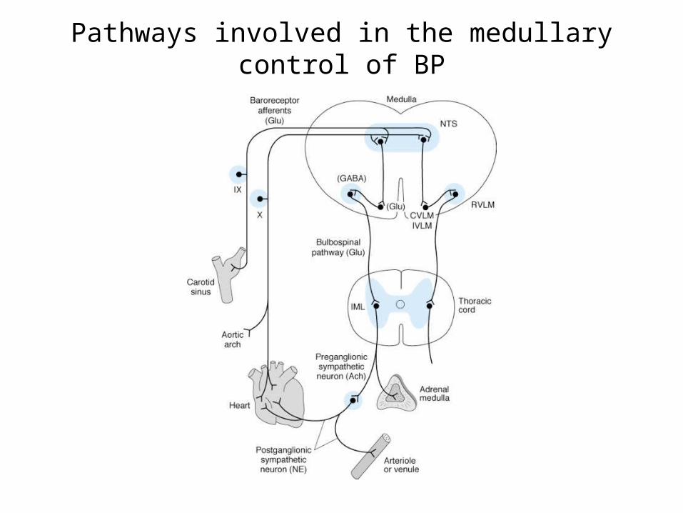
Pathways involved in the medullary control of BP

Basic pathways involved in the medullary control of heart rate by the vagus nerves

Buffer nerves activity• The carotid sinus nerves and vagal fibers from the aortic
arch are commonly called the buffer nerves• At normal blood pressure levels, the fibers of the buffer
nerve discharge at a low rate. • When the pressure in the sinus and aortic arch rises, the
discharge rate increases; • when the pressure falls, the rate declines.

Discharges (vertical lines) in a single afferent nerve fiber from the carotid sinus at various levels of mean MAP, plotted
against changes in aortic pressure with time

Fall in systemic blood pressure produced by raising the pressure in the isolated carotid sinus of a normal monkey to various values
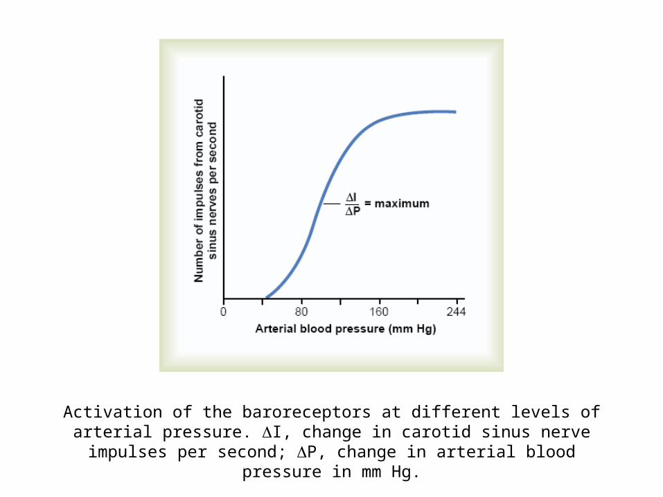
Activation of the baroreceptors at different levels of arterial pressure. I, change in carotid sinus nerve impulses per second; P, change in arterial blood pressure in mm Hg.

• Baroreceptors respond extremely rapidly to changes in BP (within fraction of seconds)
• Response much more to rapidly changing BP than to stationary pressure
• Maintains BP through a negative feedback loop

Role in acute blood loss and shock


Importance of the baroreceptor reflex
• To keep the arterial pressure relatively constant
• Short term regulation of blood pressure in the range of 70 mmHg to 150 mmHg, maintain the mean blood pressure at about 100 mmHg
• Pressure buffer system – reduce the blood fluctuation during the daily events, such as changing of the posture, respiration and excitement

Baroreceptor Resetting
• Baroreceptor will adapt to the long term change of blood pressure. – That is, if the blood pressure is elevated for a longer time, as in
chronic HTN, the set point will transfer to the elevated mean blood pressure
– So there is decrease baroreceptor response in pts with chronic HTN
• This makes the baroreceptor system unimportant for long-term regulation of arterial pressure

• CCBs, ACE-inhibitors, PDE inhibitors - cardiovascular response of increasing BP through this reflex
• Baroreceptors are compromised by diabetic neuropathy
• Volatile anaesthetics (particularly halothane) inhibit HR component of the reflex
• These reflexes are well preserved with moderate doses of fentanyl but high doses depresses this reflex

Chemoreceptor Reflexes
Mediated by
• Peripheral chemoreceptors– Carotid bodies
– Aortic bodies
• Central chemoreceptors– Medulla (associated with cardiovascular control “centers”)
• Sinus nerve of Hering (branch of 9th cranial nerve) and vagus nerve

Peripheral Chemoreceptors
• Present in carotid & aortic bodies
• 2 mm in size
• Supplied with abundant blood flow through a small nutrient artery (senses changes in BP)
• Rich sensory innveration
• Rate of response is fast

Chemoreceptor areas in carotid and aortic bodiesCAROTID
BODY
AORTIC BODY

Respiratory control by peripheral chemoreceptors in the carotid and aortic bodies
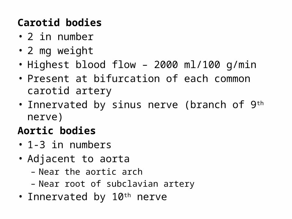
Carotid bodies• 2 in number• 2 mg weight• Highest blood flow – 2000 ml/100 g/min• Present at bifurcation of each common carotid artery• Innervated by sinus nerve (branch of 9th nerve)Aortic bodies• 1-3 in numbers• Adjacent to aorta
– Near the aortic arch– Near root of subclavian artery
• Innervated by 10th nerve

Central chemoreceptoprs – medulla (slow response)
Cardiac control centres in medulla oblongeta
Cardioaccelerator stimulatory centre (VMC)
Cardioaccelerator inhibitory centre (CIC)
Sympathetic stimulation Parasympathetic stimulation
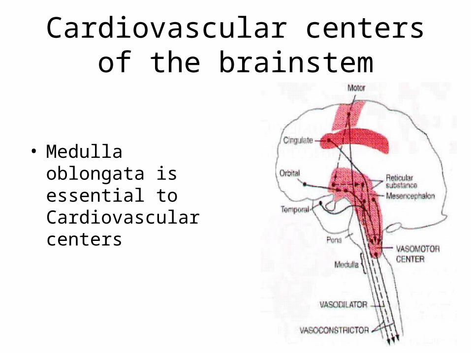
Cardiovascular centers of the brainstem
• Medulla oblongata is essential to Cardiovascular centers

• Central & peripheral chemoreceptors respond to changes in chemical composition of blood or surrounding fluid
• Central chemoreceptors respond only to acidosis
• Peripheral chemoreceptors are sensitive to changes in arterial O2 and CO2 tension and to pH
• Increasingly important when mean arterial pressure falls below 60 mmHg (i.e. when arterial baroreceptor firing rate is at minimum)

PaO2 < 50 mmHg / acidosis / PaCO2
Stimulate chemoreceptor in carotid & aortic bodies
Sinus nerve of Hering & vagus nerve
Medullary vasomotor centres
Stimulate respiratory centres
Directly stimulates sympathetic system
pulm ventilation BP
Indirectly catecholamine secretion from the adrenal medulla
BP

Bainbridge Atrial Reflex(atrial stretch reflex, volume reflex)
vagal tone right sided filling pressure
Stimulates stretch receptors present in right atrial wall & cavoatrial junction
Vagal myelinated afferent fibres
Cardiovascular center of medulla
Inhibit parasympathetic activity
HR
Directly stimulates SA node
HR
Stretching of atria
Efferents of vagus nerve
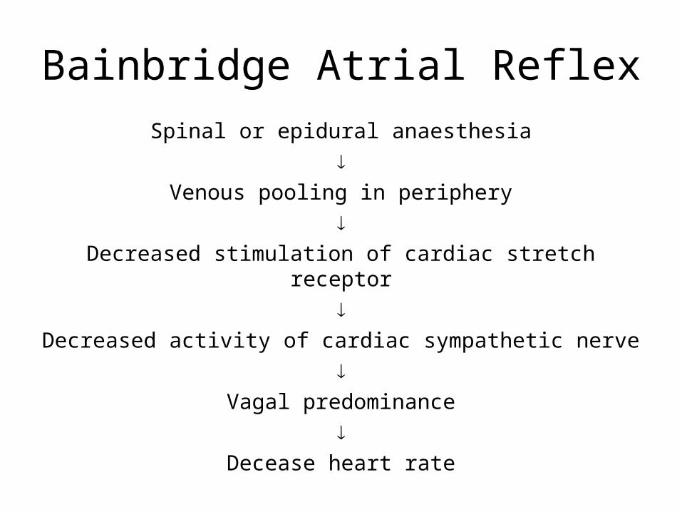
Spinal or epidural anaesthesia
Venous pooling in periphery
Decreased stimulation of cardiac stretch receptor
Decreased activity of cardiac sympathetic nerve
Vagal predominance
Decease heart rate
Bainbridge Atrial Reflex

• Reflex depends upon the preexisting heart rate• With slow heart rate, it causes progressive tachycardia• With pre-existing tachycardia, there is no effect• It helps prevent collection of blood in veins, atria and
pulmonary circulation• It inhibits ADH release and promote secretion of ANP• Denervation of vagi to heart eliminate this reflex• The Bainbridge and baroreceptor reflex acts
antagonistically to control heart rate• When blood volume is increased, the Bainbridge reflex is
dominant, when it is decreased, baroreceptor reflex is dominant

Bezold-Jarisch reflex(cardiopulmonary reflex)
Reflex triggered by• Intracoronary injection of veratrum alkaloids, serotonin,
nicotine, capsaicin, phenyldiguamide• Coronary ischemia (MI)• Bradykinin, PGI2, Arachidonic acid• Ventricular distension• Coronary angioplasty• Thrombolysis• Revascularization• Syncope

Bezold-Jarisch reflex(cardiopulmonary reflex)
Triggering factors
Stimulates cardiopulmonary chemoreceptors and mechanoreceptors of LV wall
Unmyelinated vagal afferent type C fibres
Inhibit medullary vasomotor centre
parasympathetic tone
Triad of – bradycardia, hypotension, peripheral vasodilatation

Bezold-Jarisch reflex
• Responsible for hypotension during regional anaesthesia

MI / Coronary reperfusion
Molecules generated during ischemia and reperfusion such as free radicals and PG
Stimulate cardiac inhibitory receptors(present in inferior & posterior walls of heart)
Hypotension Bradycardia and renal vasodilatation
Sudden cardiac death Decreases myocardial oxygen demand and augments renal perfusion (protective reflex)

• Cardio-protective reflex, regulates BP in conjunction with baroreceptor reflex
• Less pronounced in patients with cardiac hypertrophy & AF
• Veratrum alkaloid can be used as an antihypertensive agent
• Reflex dangerous in acute MI, coronary angiography, aortic stenosis and vaso-vagal syncope

Prevention• Prophylactic blockers
Treatment blockers & vagolytics (e.g. disopyramide)• Atropine for bradycardia

Oculocardiac Reflex(Trigemino-vagal Reflex, Aschner Phenomenon, Aschner Reflex,
Aschner Dagini Reflex)Reflex triggered by• Pressure on globe• Traction on the extraocular muscle (esp. medial rectus
muscle) as in strabismus surgery• Ocular trauma• Severe pain• Orbital compression due to hematoma or edema• Procedures under topical anaesthesia• Orbital injections• Hypercapnia or hypoxemia• Fentanyl, sufentanil and remifentanil

Pressure on the globe of the eye or traction on the extraocular muscles
Stimulates stretch receptors of extraocular muscle
Afferents of short and long ciliary nerves
Ciliary ganglion
Ophthalmic division of trigeminal nerve
Gasserian ganglion
Sensory nucleus of trigeminal in the floor of 4th ventricle
Efferents of vagus nerves (vagal cardiac depressor nerve)
Parasympathetic stimulation
Bradycardia / hypotension / asystole / AV block / ventricular ectopy

Oculocardiac reflex

Treatment• Immediate• Cessation of manipulation• IV atropine: 0.005 – 0.4 mg/kg or 7 µg/kg increments• Lignocaine infiltration – near extrinsic muscles in case of
recurrence• IV epinephrine 6-12 mg for hypotensionPrevention• Indicated in patients with h/o conduction block, vaso-vagal
responses or -blocker therapy• Premedication with anti-cholinergics (atropine or
glycopyrrolate) (block efferent pathway)• Retrobulbar block with 1-3 ml of 1-2% lidocaine

• Reponses ceases with repeated stimulation
• Reflex is more sensitive in neonates and children, especially during squint surgery
• Incidence during ophthalmic surgery: 30-90%

Cushing Reflex
Reflex activated by
• Cerebral edema
• Hematoma – subdural, epidural, contusion, ICH
• Foreign body
• Depressed skull fracture
• Hydrocephalus
• Hypoventilation
• Venous sinus thrombosis
• IC-SOL: Tumor, hematoma, abscess
• Brainstem compression
• Acute traumatic brain injury
• Craniotomy
• Neuroendoscopy

CSF pressure / ICT
Compression of cerebral arteries
Cerebral ischemia at the medullary VMC ( CO2 in blood, lactic acid in VMC)
Sympathetic stimulation
HR, BP, myocardial contractility improve cerebral perfusion
Stimulaes baroreceptors
Reflex bradycardia
Stimulates vasoconstrictor and cardioaccelerator neurons in VMC

• Triad of HTN, bradycardia and apnea
• Seen in 33% of patients with ICT
• Inhalational agents are generally associated with ICT
• Both thiopental and propofol ICT
• Occurrence of bradycardia & HTN is used as warning sign of ICT during neuroendoscopy

Treatment• Treated by measures to reduce ICP rather than pharmacological
treatment of HTN• Elevate head to 30-45°• Control HTN• Avoid hypoxia (pO2 <60 mmHg)
• Ventilate to normocarbia (pCO2 35-40 mmHg)• Sedation• Drain 3-5 ml CSF if ventriculostomy present• Hyperosmolar agents – mannitol, urea, glycerol• 3% NaCl infusion• Barbiturates – thiopentone• Systemic diuretics – furosemide, ethacrynic acid• steroids – dexamethasone

Forced expiration against a closed glottis (after full inspiration)
Valsalva Maneuver
Intrathoracic pressure ( BP initially)
VR, CO
BP
Inhibit baroreceptors
Sympathetic stimulation
HR myocardial contractility
Compression of veins ( CVP)

Opening of glottis (return of intrathoracic pressure to normal)
VR
myocardial contractility BP
Stimulates baroreceptors
Stimulates parasympathetic efferent pathways to the heart
BP, HR

Phase I• Transient rise in BP due to increased intra-thoracic and
intra-abdominal pressurePhase II• Fall of BP followed by recovery• Increased HR due to sympathetic activationPhase III• Fall in BP due to release of intrathoracic pressurePhase IV• Increase in BP due to “overshoot” of cardiac output into a
vasoconstricted peripheral circulation• Fall in HR with transient bradycardia, in the normal state,
until after the BP overshoot

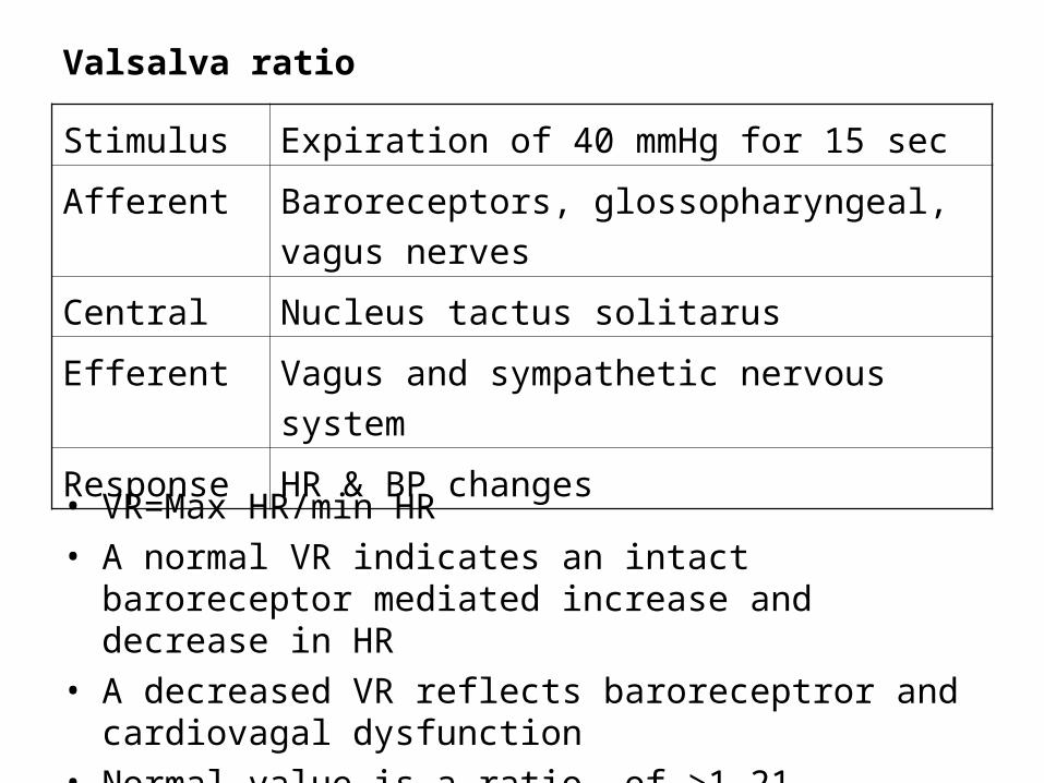
Valsalva ratio
Stimulus Expiration of 40 mmHg for 15 sec
Afferent Baroreceptors, glossopharyngeal, vagus nerves
Central Nucleus tactus solitarus
Efferent Vagus and sympathetic nervous system
Response HR & BP changes
• VR=Max HR/min HR• A normal VR indicates an intact baroreceptor mediated increase and
decrease in HR• A decreased VR reflects baroreceptror and cardiovagal dysfunction• Normal value is a ratio of >1.21

• In sympathectomized patients, HR changes occur since baroreceptors and vagi are intact
• In autonomic insufficiency HR changes does not occur

Head up tilt table test
Stimulus Decreased central blood volume
Afferent Baroreceptors, vagus, glossopharyngeal nerves
Central NTS, RVLM, hypothalamus
Efferent Sympathetic vasomotor
Response HR and BP changes
• This test is the BP and HR response to an orthostatic challenge as a measure of sympathetic function
• It is used to assess orthostatic intolerance caused by sympathetic nervous system dysfunction and to detect any predisposition to vasovagal syncope

Respiratory sinus arrhythmia
• Index of cardiac vagal function
• HR variability in synchrony with respiration
• RR interval on an ECG is shortened during inspiration and prolong during expiration

Thank Thank YouYou