CARDIAC EFFECTS OF OBESITY DURING PREGNANCY IN …
51
University of Kentucky University of Kentucky UKnowledge UKnowledge Theses and Dissertations--Dietetics and Human Nutrition Dietetics and Human Nutrition 2020 CARDIAC EFFECTS OF OBESITY DURING PREGNANCY IN C57BL/ CARDIAC EFFECTS OF OBESITY DURING PREGNANCY IN C57BL/ 6J MICE 6J MICE Kayla Lynn Dudick University of Kentucky, [email protected] Author ORCID Identifier: https://orcid.org/0000-0001-6583-192X Digital Object Identifier: https://doi.org/10.13023/etd.2020.431 Right click to open a feedback form in a new tab to let us know how this document benefits you. Right click to open a feedback form in a new tab to let us know how this document benefits you. Recommended Citation Recommended Citation Dudick, Kayla Lynn, "CARDIAC EFFECTS OF OBESITY DURING PREGNANCY IN C57BL/6J MICE" (2020). Theses and Dissertations--Dietetics and Human Nutrition. 81. https://uknowledge.uky.edu/foodsci_etds/81 This Master's Thesis is brought to you for free and open access by the Dietetics and Human Nutrition at UKnowledge. It has been accepted for inclusion in Theses and Dissertations--Dietetics and Human Nutrition by an authorized administrator of UKnowledge. For more information, please contact [email protected].
Transcript of CARDIAC EFFECTS OF OBESITY DURING PREGNANCY IN …

University of Kentucky University of Kentucky
UKnowledge UKnowledge
Theses and Dissertations--Dietetics and Human Nutrition Dietetics and Human Nutrition
2020
CARDIAC EFFECTS OF OBESITY DURING PREGNANCY IN C57BL/CARDIAC EFFECTS OF OBESITY DURING PREGNANCY IN C57BL/
6J MICE 6J MICE
Kayla Lynn Dudick University of Kentucky, [email protected] Author ORCID Identifier:
https://orcid.org/0000-0001-6583-192X Digital Object Identifier: https://doi.org/10.13023/etd.2020.431
Right click to open a feedback form in a new tab to let us know how this document benefits you. Right click to open a feedback form in a new tab to let us know how this document benefits you.
Recommended Citation Recommended Citation Dudick, Kayla Lynn, "CARDIAC EFFECTS OF OBESITY DURING PREGNANCY IN C57BL/6J MICE" (2020). Theses and Dissertations--Dietetics and Human Nutrition. 81. https://uknowledge.uky.edu/foodsci_etds/81
This Master's Thesis is brought to you for free and open access by the Dietetics and Human Nutrition at UKnowledge. It has been accepted for inclusion in Theses and Dissertations--Dietetics and Human Nutrition by an authorized administrator of UKnowledge. For more information, please contact [email protected].

STUDENT AGREEMENT: STUDENT AGREEMENT:
I represent that my thesis or dissertation and abstract are my original work. Proper attribution
has been given to all outside sources. I understand that I am solely responsible for obtaining
any needed copyright permissions. I have obtained needed written permission statement(s)
from the owner(s) of each third-party copyrighted matter to be included in my work, allowing
electronic distribution (if such use is not permitted by the fair use doctrine) which will be
submitted to UKnowledge as Additional File.
I hereby grant to The University of Kentucky and its agents the irrevocable, non-exclusive, and
royalty-free license to archive and make accessible my work in whole or in part in all forms of
media, now or hereafter known. I agree that the document mentioned above may be made
available immediately for worldwide access unless an embargo applies.
I retain all other ownership rights to the copyright of my work. I also retain the right to use in
future works (such as articles or books) all or part of my work. I understand that I am free to
register the copyright to my work.
REVIEW, APPROVAL AND ACCEPTANCE REVIEW, APPROVAL AND ACCEPTANCE
The document mentioned above has been reviewed and accepted by the student’s advisor, on
behalf of the advisory committee, and by the Director of Graduate Studies (DGS), on behalf of
the program; we verify that this is the final, approved version of the student’s thesis including all
changes required by the advisory committee. The undersigned agree to abide by the statements
above.
Kayla Lynn Dudick, Student
Dr. Robin Shoemaker, Major Professor
Dr. Dawn Brewer, Director of Graduate Studies

CARDIAC EFFECTS OF OBESITY DURING PREGNANCY IN C57BL/6J MICE
________________________________________
THESIS
________________________________________
A thesis submitted in partial fulfillment of the
requirements for the degree of Master of Science
in Nutrition and Food Systems in the College of
Agriculture, Food and Environment at the University
of Kentucky
By
Kayla Lynn Dudick
Lexington, Kentucky
Director: Dr. Robin Shoemaker, Associate Professor of Dietetics and Human Nutrition
Lexington, Kentucky
2020
Copyright © Kayla Lynn Dudick 2020
https://orcid.org/0000-0001-6583-192x

ABSTRACT OF THESIS
CARDIAC EFFECTS OF OBESITY DURING PREGNANCY IN C57BL/6J MICE
Objective: Pregnancy requires profound cardiac and metabolic adaptation. Left
ventricular (LV) mass is increased in response to pregnancy, but is not associated with
cardiac damage. In contrast, obesity-mediated cardiac hypertrophy is pathological. Data
from animal studies indicate dietary fatty acid composition may have a protective effect
during states of extreme cardiac physiological adaptation. In contrast, aberrant cardiac
metabolism is a hallmark of disease. Over a third of reproductive-age women in the
United States are obese, but there is a paucity of data describing the effect of obesity on
maternal cardiac adaptation to pregnancy. The objective of this study was to determine
the effects of high-fat feeding during pregnancy on cardiac hypertrophy and metabolism
in a mouse model of diet-induced obesity.
Methods/Results: Female C57BL/6J mice (8 weeks old) were fed a high fat (HF;
60% kcal from fat) or a control low fat (LF; 10% kcal from fat) diet for 8 weeks, then
were either crossed with male mice to become pregnant (P) or remained non-pregnant
(NP) controls. At gestational day 18, cardiac function was quantified by
echocardiography in LF- and HF-fed P and NP females. On gestational 19 day, mice
were euthanized for tissue collection. HF-fed females had significantly increased body
weight compared to LF-fed controls, and body weight was increased in P compared to
NP mice. In response to pregnancy, LF-, but not HF-fed, mice had significantly increased
LV mass (P<0.01). In contrast, HF-fed pregnant mice had increased relative wall
thickness (RWT: [2* LV posterior wall thickness/LV end-diastolic diameter]) compared
to LF-fed pregnant mice. We quantified mRNA abundance of genes regulating fatty acid
oxidation utilization in left ventricles of LF- and HF-fed pregnant and non-pregnant mice
using Nanostring nCounter Analysis system. Acaa2, Acox1,and Acadl (genes regulated
long-chain fatty acid oxidation) and Cpt1b (regulating fatty acid transport into the
mitochondria) were upregulated with both HF-feeding and pregnancy. In contrast,

Ehhadh, a gene regulating production of medium chain fatty acids during fatty acid
oxidation, was increased in pregnant mice, but only in the LF mice, and the expression
was significantly reduced in HF- compared to LF-fed pregnant mice.
Conclusions: Physiological cardiac hypertrophy in response to pregnancy was
observed in LF-fed, but not HF-fed mice. In contrast, HF-fed pregnant mice had
increased RWT compared to LF-fed pregnant mice. While fatty acid utilization was
increased with HF-feeding and pregnancy, the expression of Ehhadh was reduced in HF-
compared to LF-fed mice. Medium chain fatty acids are demonstrated in the literature to
be protective against pathological cardiac remodeling in experimental animals. Taken
together, these data suggest obesity may impair protective fatty acid utilization pathways
in pregnancy to promote adverse cardiac remodeling.
KEYWORDS: Pregnancy, CVD, Obesity, Cardiac Metabolism, Remodeling
Kayla Lynn Dudick
(Name of Student)
11/13/2020
Date

CARDIAC EFFECTS OF OBESITY DURING PREGNANCY IN C57BL/6J MICE
By
Kayla Lynn Dudick
Robin Shoemaker, PhD
Director of Thesis
Dawn Brewer, PhD, RD, LD
Director of Graduate Studies
11/13/2020
Date

iii
TABLE OF CONTENTS
LIST OF TABLES .............................................................................................................. v
LIST OF FIGURES ........................................................................................................... vi
CHAPTER 1. INTRODUCTION ...................................................................................... 1
1.1 Background ......................................................................................................... 1
1.2 Problem Statement .............................................................................................. 2
1.3 Overall hypothesis .............................................................................................. 2
1.4 Research Questions ............................................................................................. 2
1.5 Impact ................................................................................................................. 2
CHAPTER 2. LITERATURE REVIEW ........................................................................... 4
2.1 Cardiac physiology of pregnancy ....................................................................... 4
2.1.1 Hemodynamics (the flow of blood through the vessels and organs in the
body) ......................................................................................................................... 4
2.1.2 Cardiac morphology (size, shape, and geometry) ........................................... 5
2.1.3 Reverse Adaptation ......................................................................................... 6
2.2 Epidemiology of pregnancy history and maternal heart health ......................... 6
2.2.1 Sex differences in CVD .................................................................................. 6
2.2.2 Pregnancy complications are associated with maternal risk for CVD ............ 7
2.3 Obesity and pregnancy complications ................................................................ 8
2.4 Cardiac effects of obesity .................................................................................... 9
2.4.1 Obesity augments traditional risk factors ....................................................... 9
2.4.2 Obesity is an independent risk factor for CVD ............................................. 10
2.4.3 Cardiac effects of obesity during pregnancy ................................................ 10
2.5 Cardiac metabolism .......................................................................................... 11
2.5.1 Overview of cardiac metabolism .................................................................. 11
2.5.2 Altered cardiac metabolism hallmark of disease states ................................ 12
2.5.3 Cardiac metabolism during pregnancy ......................................................... 13
CHAPTER 3. METHODS ............................................................................................... 16
3.1 Experimental animals and study design ........................................................... 16
3.2 Echocardiography............................................................................................. 17
3.3 Tissue RNA extraction and gene expression analysis ....................................... 17

iv
3.4 Statistical analysis ............................................................................................ 18
CHAPTER 4. RESULTS ................................................................................................. 20
4.1 HF-feeding increases body weight and fat mass .............................................. 20
4.2 HF-feeding reduces litter size ........................................................................... 20
4.3 HF-feeding increases heart weight and LV mass, but is not augmented with
pregnancy ...................................................................................................................... 20
4.4 Wall thickness is increased in HF-compared to LF-fed pregnant mice ........... 21
4.5 Analysis of genes regulating metabolism in hearts........................................... 21
CHAPTER 5. DISCUSSION ............................................................................................ 28
5.1 Limitations and Future Studies ......................................................................... 32
5.2 Public Health/Clinical Significance ................................................................. 33
CHAPTER 6. CONCLUSION.......................................................................................... 35
REFERENCES ................................................................................................................. 36
VITA ................................................................................................................................. 41

v
LIST OF TABLES
Table 1: Litter size and pup/placental weights from LF- or HF-fed pregnant female mice.
........................................................................................................................................... 23
Table 2: Heart weights of LF- and HF-fed pregnant and non-pregnant female mice....... 23

vi
LIST OF FIGURES
Figure 1: “Pregnancy is a “stress test” that can reveal subclinical trajectories and identify
new opportunities for chronic disease prevention”. .......................................................... 15
Figure 2: Overview of cardiac metabolism ....................................................................... 15
Figure 3. Experimental Design ......................................................................................... 19
Figure 4: Weight gain from baseline to gestational day 19 .............................................. 24
Figure 5: LV mass and RWT ............................................................................................ 25
Figure 6: Flow chart of cardiac genes measured using Nanostring significantly changed
using 2-way ANOVA by either diet or pregnancy. .......................................................... 26
Figure 7: mRNA abundance of fatty acid utilization genes in pregnant and non-pregnant
mice fed a LF or HF diet. .................................................................................................. 27

1
CHAPTER 1. INTRODUCTION
1.1 Background
Increasing evidence suggests that pregnancy is a sensitive window where adverse
effects on cardiovascular function may permanently alter maternal risk for cardiovascular
diseases (CVD) 2. Epidemiology studies demonstrate that women with a history of
pregnancy complications, such as gestational hypertension or diabetes, preterm delivery,
or intrauterine growth restriction are at greater risk for mortality from CVD 3. Rodent
studies support that the profound cardiovascular adaptations during pregnancy may serve
as a type of “stress test”, to unmask cardiovascular vulnerabilities 4. These data suggest
pregnancy complications are a sex-specific risk factor for CVD. Obesity is strongly
associated with pregnancy complications, with a nearly stepwise increase in the incidence
of preterm birth, gestational diabetes, hypertensive disorders, and other high-risk
conditions with increasing category of body mass index (BMI) 5. This is concerning as
the prevalence of both obesity and CVD are rising in women of reproductive age in the
United States 6.
Pregnancy induces many structural and functional changes in the cardiovascular
system to accommodate the growing uteroplacental unit. One such change is cardiac
hypertrophy, or enlargement of the heart in response to increased metabolic demands.
Cardiac hypertrophy with pregnancy is assumed to be transient, and is not associated with
cardiac damage 7. In contrast, cardiac hypertrophy occurring in response to obesity is
pathological, and a prognostic indicator for CVD 8. Despite the known associations
between obesity, pregnancy complications, and maternal CVD, there is limited research

2
describing how obesity modulates cardiac hypertrophy of pregnancy. This is important,
as adverse cardiac effects during pregnancy may drive future maternal risk for CVD.
1.2 Problem Statement
Women with a history of obesity-mediated pregnancy complications are at a
greater risk of CVD. However, mechanisms linking pregnancy complications and CVD
are unknown. Both obesity and pregnancy independently promote cardiac hypertrophy
and are associated with changes in cardiac metabolism. It is not known how obesity
during pregnancy modulates cardiac hypertrophy and energy metabolism.
1.3 Overall hypothesis
The overall hypothesis of this research project is that obesity during pregnancy
promotes adverse cardiac remodeling associated with altered cardiac metabolism.
1.4 Research Questions
1. Does obesity promote altered cardiac hypertrophy during pregnancy in mice?
2. What is the effect of obesity during pregnancy on the expression of genes that
regulate cardiac metabolism in mice?
1.5 Impact
If a woman is going to make a lifestyle change, she is more likely to do so while
pregnant. This is a crucial time when healthcare professionals need evidence-based

3
nutritional strategies that can be tailored to accommodate individual nutritional needs and
provide sustainable modifications. By providing individualized, sustainable nutritional
therapies, women may reduce their risk or progression of obesity-mediated pregnancy
complications and/or CVD.

4
CHAPTER 2. LITERATURE REVIEW
2.1 Cardiac physiology of pregnancy
Pregnancy is a dynamic process, requiring immense physiological adaptation of
the cardiovascular system. This includes changes in blood volume, heart function, and
changes in properties and function of blood vessels. These changes are necessary for
accommodating the growing fetoplacental unit, and to meet the increased metabolic
demands of the mother. Insufficient ability of the mother’s body to adapt to these
cardiovascular changes can adversely affect the health of both the mother and the fetus.
In fact, the cardiovascular demands of pregnancy can sometimes reveal otherwise silent
cardiovascular pathologies. This is why pregnancy is often referred to as “nature’s stress
test” 2.
2.1.1 Hemodynamics (the flow of blood through the vessels and organs in the body)
One of the biggest changes during pregnancy is the increase in blood volume.
Blood volume increases early in the first trimester, and steadily increases throughout
gestation. The total increase in blood volume varies widely, between 20% to an
astonishing 100% of pre-pregnancy volume, usually approximated to be about a 45%
increase 7. The vascular system accommodates this via vasodilation (widening of vessels)
and an overall decrease of about 35% to 40% in systemic vascular resistance (resistance
of vessels to blood flow). Vascular resistance is lowest in the first trimester,
accommodating development of the placenta, followed by a slight increase in the end of
the second trimester, and remains steady for the remainder of pregnancy. Blood pressure
5
decreases accordingly, being the lowest in the first trimester, then returning to near pre-
pregnancy levels by the third trimester 7.
The increase in blood volume translates to increased stroke volume (the volume
of blood pumped through the heart with each beat). Accordingly, cardiac output (the
volume of blood pumped through the heart each minute) also increases up to 45% during
pregnancy, where cardiac output is determined by multiplying the stroke volume by the
heart rate. In summary, Cardiac output is increased by an astounding 30-50%, and
peripheral vascular resistance and blood pressure decrease by roughly 20% during
pregnancy7.
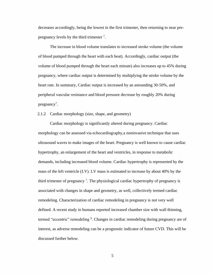
2.1.2 Cardiac morphology (size, shape, and geometry)
Cardiac morphology is significantly altered during pregnancy. Cardiac
morphology can be assessed via echocardiography,a noninvasive technique that uses
ultrasound waves to make images of the heart. Pregnancy is well known to cause cardiac
hypertrophy, an enlargement of the heart and ventricles, in response to metabolic
demands, including increased blood volume. Cardiac hypertrophy is represented by the
mass of the left ventricle (LV). LV mass is estimated to increase by about 40% by the
third trimester of pregnancy 7. The physiological cardiac hypertrophy of pregnancy is
associated with changes in shape and geometry, as well, collectively termed cardiac
remodeling. Characterization of cardiac remodeling in pregnancy is not very well
defined. A recent study in humans reported increased chamber size with wall thinning,
termed “eccentric” remodeling 9. Changes in cardiac remodeling during pregnancy are of
interest, as adverse remodeling can be a prognostic indicator of future CVD. This will be
discussed further below.

6
2.1.3 Reverse Adaptation
Literature in humans suggests cardiac changes during pregnancy return to pre-
pregnancy values, within a year 7. However, these studies are historical, and few studies
have characterized cardiac function and structure postpartum using modern technology.
Studies in healthy animals suggest that hemodynamics and cardiac morphology return to
pre-pregnancy values about 7 days postpartum 10. However, recent rodent studies
demonstrate that factors such as age and health status (i.e. obesity) may be detrimental to
reverse cardiac adaptation postpartum 1112. This is important, as a growing body of
literature suggests that factors impacting cardiovascular function during pregnancy have
a lasting impact on maternal cardiovascular function.
2.2 Epidemiology of pregnancy history and maternal heart health
2.2.1 Sex differences in CVD
Cardiovascular disease is the number one cause of death in both men and women
in the United States, but the disease manifests differently in women compared to men 13.
For example, women are more likely to develop certain types of heart disease, such as
stroke, left ventricular (LV) diastolic dysfunction, and heart failure with preserved
ejection fraction (HFpEF) 14. Women also have a more steeply increasing risk with age
compared to men 6. Only in the last decade have mechanisms contributing to sex
differences in CVD become a major research focus. Contributing to the lack of
understanding of CVD in women is the fact that females have traditionally been
marginalized in human and animal studies 15.

7
Recent studies defining the role of sex hormones on cardiovascular function have
contributed to significant advancements in explaining sex differences in type, timing, and
mortality of CVD13. Research examining sex disparities between men and women in
terms of pathology and physiology has revealed that women are more likely to
experience endothelial and microvascular dysfunctions related to coronary artery disease
compared to men 16. These differences might be attributed to estrogen, which is reported
to have protective effects on vascular and endothelial function, as well as metabolism
and adiposity 17. However, discrepancies exist in findings from randomized-controlled
trials designed to test protective effects of female sex hormones against CVDs18. In the
Women’s Health Initiative (WHI), hormone replacement therapy in postmenopausal
women actually contributed to increased risk for CVD in some women 19, suggesting that
sex hormones alone cannot be solely responsible for protective effects against CVD in
women. In addition to sex hormones, sex chromosome compliment also contributes to
development of CVD, such as aneurysms 13. These findings indicate that other sex-
specific factors also contribute to differential cardiovascular function between males and
females. The experience of pregnancy is one of the most profound physiological
differences between males and females, requiring extensive cardiovascular adaptation.
To improve heart health in women, it is essential to define how cardiovascular
stress during pregnancy affects future maternal cardiovascular function.
2.2.2 Pregnancy complications are associated with maternal risk for CVD
Pregnancy complications, such as gestational hypertension or diabetes, preterm
delivery, or intrauterine growth restriction (IUGR) are associated with mortality from
cardiovascular disease (CVD) 3. Results from several large cohort studies demonstrate

8
that women with a history of gestational hypertension and related conditions, such as
preeclampsia, have an increased risk for developing CVDs like hypertension, ischemic
heart disease, stroke, and death due to CVD even decades after delivery 20. As a group,
complicated pregnancies are characterized by inflammation, vascular dysfunction,
thrombosis, and insulin resistance; physiologic pathways which are common to CVD.
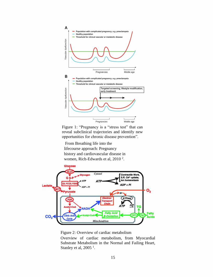
These data suggest that adverse physiology of a woman’s reproductive history may serve
as a predictor for chronic disease (see Figure 1) 2. It is not clear whether pregnancy is a
“stress test” that reveals latent cardiovascular abnormalities, or if adverse effects on the
cardiovascular system during pregnancy can permanently alter the trajectory of maternal
heart health (or both).
2.3 Obesity and pregnancy complications
Obesity is the most common medical condition during pregnancy that affects
maternal and fetal health 5. According to the CDC, the prevalence of obesity in the
United States is increasing in women of reproductive age, with approximately 55% of
women aged 20-39 years old having a body mass index (BMI) of greater than 25, and
approximately 31% having a BMI over 30 21. Maternal obesity has both short-term and
long-term health consequences for mother and offspring. Obesity during pregnancy is
associated with fertility problems, metabolic derangements, hypertensive disorders (such
as preeclampsia), premature delivery, cesarean delivery, impaired fetal growth or
macrosomia, and stillbirth 22.

9
Epidemiological data worldwide has linked maternal obesity to increased risk for
metabolic and cardiovascular disease in offspring 23. However, mechanisms directly
linking maternal obesity to subsequent cardiovascular disease are not known.
2.4 Cardiac effects of obesity
2.4.1 Obesity augments traditional risk factors
It is well established that obesity augments cardiovascular risk factors, such as
blood pressure, blood lipid levels, and the development of metabolic syndrome. In men
and women, obesity is a primary contributor to the development of hypertension 24. High
blood pressure is a significant predictor for CV events 25. While the overall worldwide
prevalence of hypertension is greater in men, with increasing age (and menopause), the
incidence of hypertension in women rises 6. Given that the prevalence of obesity is
greater in women (at all ages) 21, the overall burden of obesity-associated hypertension
may actually be greater in women than men.
Obesity contributes to dyslipidemia by increasing blood triglyceride and Free
Fatty Acids (FFA) levels 26. Consequences of this include perturbed lipoprotein
metabolism, causing the formation of atherogenic lipoprotein particles, as well as free
fatty acid-mediated insulin resistance 27. Dyslipidemia increases the risk for CVD, such
as atherosclerosis and coronary heart disease 28.
Excess adipose tissue with obesity, especially visceral or abdominal adipose
stores, also indirectly contributes to increased cardiovascular risk through release of
inflammatory mediators. Adipose tissue secretes a number of adipokines that contribute
to immune function and metabolism, and a pathological consequence of excess adipose

10
mass is increased secretion of adipokines and other pro-inflammatory factors, such as
TNF-a, IL-6, and MCP-1 29. These factors can have diverse adverse effects including
impaired endothelial function and increased insulin resistance 30.
2.4.2 Obesity is an independent risk factor for CVD
In addition to augmenting cardiovascular risk factors, such as hypertension and
type 2 diabetes, obesity is an independent risk factor for cardiovascular disease 31.
Obesity is associated with global longitudinal strain, and both impaired systolic and
diastolic function 32. In response to increased blood volume and stroke volume (SV),
mean arterial pressure and ventricular filling pressures can become elevated 33 34.
Obesity and excess adiposity are directly associated with cardiac hypertrophy and
remodeling 35, and geometric changes are further augmented with increasing blood
pressure 36. Interestingly, although absolute LV mass is greater in men, when normalized
for body size, the effect of obesity to increase LV mass is reported to be greater in
women versus men 37. Further, a direct association is reported between LV mass and
body fat percentage, especially visceral adipose tissue 38. The proportion of body fat is
generally greater in women compared to men. Taken together, these data suggest that
women may be particularly susceptible to obesity-mediated alterations in cardiac
morphology.
2.4.3 Cardiac effects of obesity during pregnancy
It is fairly well-documented that obese women have increased blood pressure
compared to non-obese women during pregnancy 39 40. Further, obesity is a risk factor for
hypertensive disorders of pregnancy, such as preeclampsia 41. What is less clear is the
direct effect of obesity on cardiac structure and function during pregnancy, as well as, the

11
long-term implications. In a study comparing obese versus non-obese women at 36
weeks gestation, blood pressure and LV mass were increased with obesity, but no
differences were observed with respect to CO or ventricular function 40. In contrast, a
serial study over trimesters in 16 obese and 17 non-obese pregnancies reported
differences in SV and contractility in obese women 42. Interestingly, the difference was
most striking with respect to the change in function throughout gestation. SV was
greater in obese versus non-obese women in the first trimester, however non-obese
women exhibited an increase in SV and CO throughout gestation, which was not
observed in obese women. A similar trend was observed in indices of contractility, with
some measures of contractility actually decreasing in obese women in the third trimester.
The authors concluded that obesity was associated with impaired LV contractile
response. Using speckle tracking in conjunction with echocardiography, Buddeberg et al
recently reported diastolic dysfunction and LV global longitudinal strain in obese
pregnant women at term compared with non-obese controls 43. These studies provide
evidence that obesity during pregnancy can adversely modulate cardiac function.
However, the long-term consequences of these alterations on cardiovascular health are
not known.
2.5 Cardiac metabolism
2.5.1 Overview of cardiac metabolism
The heart is a biological pump that converts chemical substrates into mechanical
energy 44. The heart primarily consumes carbohydrates (10-30%) and fats (60-90%) while
using oxygen to drive oxidative phosphorylation to generate ATP from ADP 45. ATP is

12
then used to drive contractile function (beating), to perfuse the body with blood and
nutrients. Energy consumption of the human heart is about 10% of whole-body fuel
consumption. The heart is described as a metabolic omnivore and has incredible
flexibility to utilize a variety of substrates in the presence of oxygen to generate ATP.
Energy producing substrates include triacylglycerols, fatty acids, glucose, glycogen,
lactate, pyruvate, the ketone bodies acetoacetate and β-hydroxybutyrate, and amino acids,
especially leucine, isoleucine, and valine (branched-chain amino acids) 45 . These
substrates enter the Krebs cycle primarily as acetyl-CoA, or other intermediates of the
Kreb’s cycle, for production of reducing equivalents (e.g., NADH and FADH2 are a
couple), which deliver electrons to the electron-transport chain in the mitochondria. The
resulting generation of a proton gradient drives the formation of ATP. In an anerobic
state, the heart can also utilize lactate (via degradation of glucose) and succinate (via
degradation of some amino acids). See Figure 2 for schematic of cardiac metabolism.
2.5.2 Altered cardiac metabolism hallmark of disease states
The heart has the capacity to adapt to an altered metabolic state by selecting
available substrates for the most effective generation of ATP. This metabolic flexibility is
lost during heart failure, and pathologic states directly influencing substrate availability
(e.g. obesity, diabetes) can further contribute to impaired cardiac function 46. It is well-
known that diabetes is associated with increased fatty acid utilization and decreased
glucose utilization 47. Likewise, obesity results in increased fatty acid utilization and
reduced glucose oxidation rates 48. This is likely due to substrate availability, as obesity is
associated with increased levels of circulating fatty acids and triglycerides. Conversely, the

13
reduction in glucose utilization is likely due to the Randle effect, where increased
utilization of fatty acids has an inhibitory effect on glucose metabolism 47.
Changes in substrate metabolism precede changes in function and may be the
first indication of functional abnormalities. Studies in humans and animals demonstrate
that prolonged increase in fatty acid utilization results in increased myocardial oxygen
consumption, i.e. increased oxidative phosphorylation, and reduced cardiac efficiency 49 .
Despite obesity being associated with an increase in the number of mitochondria, studies
in ob/ob mice (a genetic mouse model of obesity) reveal that mitochondria from obese
hearts have deficits in oxidative capacity 50. This dysfunctional condition is called
oxidative uncoupling, where oxygen consumption is increased, but a proportional
increase in ATP production is not observed 51. This is a potential mechanism by which
obesity promotes impaired cardiac function. Increased fatty acid uptake and oxidation
promote the formation of damaging reactive oxygen species (ROS) from mitochondrial
complexes, but energy production declines 51. The potential consequence for reduced
cardiac efficiency is a limitation in cardiac reserve, which may be worsened in the face
of hemodynamic stressors, such as cardiac hypertrophy and increased blood pressure.
2.5.3 Cardiac metabolism during pregnancy
As described above, pregnancy is a form of cardiac stress. Notably, LV mass
increases by up to 50%, which requires energy. Interestingly, while cardiac work
increases by 20-30% during pregnancy, cardiac oxygen consumption is only increased by
approximately 15% 52. Thus, cardiac efficiency is increased during pregnancy. There is
not much known about cardiac metabolism during pregnancy. Studies from rats indicate a
progressive decline in glucose utilization, with up to a 70% reduction by late pregnancy

14
53. Studies in dogs demonstrate a large increase in fatty acid oxidation, indicating that the
ATP production in the heart during late pregnancy is almost exclusively due to increased
utilization of fats 54.
There are no studies in humans or animals describing the combined effects of
obesity and pregnancy on cardiac metabolic function. The present study will directly fill
that gap.
15
From Breathing life into the
lifecourse approach: Pregnancy
history and cardiovascular disease in
women, Rich-Edwards et al, 2010 2.
Figure 2: Overview of cardiac metabolism
Overview of cardiac metabolism, from Myocardial
Substrate Metabolism in the Normal and Failing Heart,
Stanley et al, 2005 1.
Figure 1: “Pregnancy is a “stress test” that can
reveal subclinical trajectories and identify new
opportunities for chronic disease prevention”.

16
CHAPTER 3. METHODS
3.1 Experimental animals and study design
All studies using mice were approved by an Institutional Animal Care and Use
Committee (IACUC) at the University of Kentucky and were conducted in accordance
with the National Institutes of Health (NIH) Guide for the Care and Use of Laboratory
Animals. Female C57BL/6J mice (8 weeks of age; Jackson Laboratory, Bar Harbor,
ME, stock # 000664) were randomly assigned to receive, ad libitum, either a high fat
(HF; 60% kcal from fat; D12492, Research Diets, New Brunswick, NJ) or a control low
fat (LF, 10% kcal from fat; D12450B, Research Diets Inc) diet for 8 weeks (n=30
mice/diet group) (see Figure 3 for experimental design). The control LF diet was
purified and ingredient-matched to the HF diet, and the fat source for both diets was
soybean oil and lard (where lard comprises the excess fat in the HF diet). The energy
densities of the LF and HF diet are 3.82 and 5.21 kcal/g, respectively. Body weight was
quantified weekly throughout the study using an Ohaus portable digital scale.
At 8 weeks of diet feeding, all female mice were placed in a cage with male mice
of the same strain and diet. After 2 days, females were removed from the males, and
placed in single housing for the duration of the study.
Echocardiography was performed on LF- and HF-fed female pregnant and non-
pregnant mice 16 days following removal from male cage. The following day, mice were
anesthetized with ketamine/xylazine (100/10 mg/kg, i.p.) for exsanguination and tissue
harvest. Fetuses and placentas were dissected and weighed. Tissues were snap frozen in
liquid nitrogen and stored at -80ºC until analysis. Tissues taken included: heart, liver,
kidney, spleen, para-uterine fat, and subcutaneous fat.

17
3.2 Echocardiography
Echocardiography was performed on isoflurane-anesthetized mice as described
previously 12. Day 16 was chosen, because normal mouse gestation is 20 days or the
equivalent of the third trimester in humans. Briefly, mice were anesthetized using 2-4%
isoflurane (at effect) according to their size and then transferred to a heated platform
(37ºC) with 1-2% isoflurane supplied via nose cone. Hair on the chest region was
shaved and removed, and electrode cream was applied on the front and hind limbs before
being secured with electrical tape to electrodes on the platform. Respiration rate (RR)
and heart rate (HR) were monitored and adjusted to a certain range across all mice by
titrating isoflurane levels. An RR of 100 times/min and HR of 400 beats/min were
targeted. Images of the cross-sectional view of the left ventricle (LV) at the papillary
muscle-level in parasternal short-axis (PSAX) view were obtained in M-mode using an
M550 transducer under the cardiology package on a Vevo 3100. Images were analyzed
using VevoLab software using LV trace methodology.
3.3 Tissue RNA extraction and gene expression analysis
Approximately 20 mg of the left ventricle was used to extract total RNA using the
Maxwell RSC (Promega, Madison, WI). RNA concentrations and quality were
determined using a Nanodrop 2000. All samples had a 260/280 and 260/230 ratios > 2.0.
The NanoString nCounter Metabolic Pathways Panel and nCounter Analysis System
(NanoString Technologies, Seattle, WA) was used to quantify mRNA abundance of 768
genes regulating metabolism. As previously described 12, the Nanostring nCounter gene
expression system is a multiplexed assay that uses a combination of unique capture

18
probes and color-coded reporter probes to capture and count individual mRNA
transcripts with high sensitivity and tight correlation to real-time PCR 55. Fifty
nanograms of RNA of each sample was hybridized to the target-specific capture and
reporter probes in the CodeSet according to the manufacturer’s instructions. Samples
were cooled to 4ºC, loaded into nCounter SPRINT cartridges, then analyzed using the
nCounter Gene Expression Assay. Raw data were normalized by creating scaling factors
for the sum of the positive controls and the geometric mean of the four housekeeping
genes. Data represent the mean of normalized counts.
3.4 Statistical analysis
Data are presented as mean ± SEM. Statistical analyses were performed using
SigmaPlot version 12.3. All data passed normality or equal variance tests or logarithmic
transformation was used to achieve normality. Two-tailed Student’s t-tests were used for
analysis of data between two groups. For 2-factor analysis, a two-way ANOVA was used
to analyze end-point measurements with between-group factors of pregnancy and diet,
followed by Holm-Sidak for post hoc pairwise analyses. Values of P < 0.05 were
considered to be statistically significant.

19
Figure 3. Experimental Design

20
CHAPTER 4. RESULTS
4.1 HF-feeding increases body weight and fat mass
After 8 weeks of diet feeding, HF-fed females had significantly increased body
weight compared to LF-fed controls (P<0.001; 14A). Within HF-fed mice, the average
body weight of females who eventually became pregnant was lower than those who did
not become pregnant, but this was not significant (p=0.06). Further, HF-fed mice had
increased fat mass and decreased lean mass (as percent body weight), compared to LF-
fed mice P<0.001; Figure 4B). At study endpoint (day 18 of gestation), body weight was
increased in pregnant compared to non-pregnant animals, independent of diet (P<0.001;
and HF-fed pregnant mice had increased body weight compared to LF-fed mice (P<0.01,
Figure 4C).
4.2 HF-feeding reduces litter size
Compared to LF-fed dams, HF-fed dams had smaller average litter size (P<0.01),
and a greater number of resorbed pups (Table 1). There was no difference in pup body
weight or placental weight in LF- compared to HF dams.
4.3 HF-feeding increases heart weight and LV mass, but is not augmented with
pregnancy
In non-pregnant mice, excised heart weight was greater in HF- fed mice compared
to LF-fed mice (P<0.001; Table 2). In LF-fed mice, heart weight was increased with
pregnancy (P<0.05, Table 2), but HF-fed pregnant mice did not have significantly

21
increased heart weight compared to LF pregnant mice. Similarly, LV mass was increased
with HF-feeding in non-pregnant mice (P<0.05, Figure 5A). However, only LF-fed mice
exhibited cardiac hypertrophy with pregnancy (P<0.001); LV mass was not augmented
with pregnancy in HF-fed mice (p=0.221, Figure 5A).
4.4 Wall thickness is increased in HF-compared to LF-fed pregnant mice
The increase in LV mass in NP HF-fed mice was associated with increased LV
posterior wall thickness (P<0.01, Figure 5B). In contrast, increased LV mass with
pregnancy in LF-fed mice was associated with an increase in the ventricle chamber
(P<0.001), with no change to the wall thickness, and the diameter of the LV ventricle was
significantly larger in LF- versus HF-fed pregnant mice (P<0.05). Thus, the relative wall
thickness, a measure of LV geometry, was significantly increased in HF- compared to
LF-fed mice during pregnancy (P<0.05, Figure 5B). Data summarizing cardiac
morphology depicted in a schematic in Figure 5C.
4.5 Analysis of genes regulating metabolism in hearts
To determine the gene profile associated with changes in the cardiac structure of
HF- versus LF-fed pregnant mice, we quantified mRNA abundance of 794 genes
regulating metabolism involved in 34 pathways using NanoString nCounter gene
expression analysis in the left ventricles of LF- and HF-fed pregnant and non-pregnant
mice. Using 2-way ANOVA with pairwise comparisons, we determined that there were
47 genes with a significant effect of either gene or pregnancy with P<0.01. Using a p-
value <0.05, there were 26 genes with a significant effect of diet, 35 with a significant

22
effect of pregnancy, and 16 with a significant interaction between diet and pregnancy
(Figure 6).
Since the largest number of genes with significance differences were related to
fatty acid utilization, we focused our analysis on these genes. These genes are: Acaa2,
Acadl, Acox1, Cpt1b, Fabp3, and Ehhadh. For Fabp3, there was an overall effect of diet
to increase the abundance of cardiac mRNA, but no effect of pregnancy (P<0.01, Figure
7F). Similarly, Acaa2, Acadl, Acox1, and Cpt1b were upregulated with HF-feeding
(P<0.05, Figure 7A-D). There was also a significant of pregnancy, but this was only
significant in the LF-fed group (P<0.05). Meaning, gene expression of these genes was
increased in HF-fed mice (pregnant or non-pregnant), and increased in LF-fed pregnant
mice, but there was no additional effect of pregnancy to increase gene expression in HF-
fed pregnant mice. The expression pattern of Ehhadh was different, where Ehhadh was
elevated only in the LF-fed pregnant mice (P<0.01, Figure 7E). Further, the expression
level of Ehhadh in HF-fed pregnant mice was significantly lower than that of LF-fed
pregnant mice (P<0.05).

23
Table 1: Litter size and pup/placental weights from LF- or HF-fed pregnant female mice.
Diet Pups per
litter
Pup weight
(g)
Total number
of resorbed
pups
Number of
dams with
resorbed pups
Placenta weight
(g)
LF 8.6 + 0.3 1.01 + 0.11 4 4 0.111 + 0.005
HF 6.4 + 0.5** 0.93 + 0.11 21 10 0.111 + 0.005
**, P<0.01 effect of diet
Table 2: Heart weights of LF- and HF-fed pregnant and non-pregnant female mice.
***, P<0.001 effect of diet ###, P<0.001 effect of pregnancy #, P<0.05 effect of pregnancy
Parameter LF HF
NP (n=20) P (n=10) NP (n=18) P (n=12)
Mean + SEM
Heart weight (g) 0.118 + 0.003 0.128 +
0.004#
0.132 +
0.003***
0.136 + 0.004
Heart/body
weight (%)
0.529 + 0.012 0.374 +
0.017###
0.412 +
0.013***
0.348 +
0.016###

24
Figure 4: Weight gain from baseline to gestational day 19
A) Body weight and (B) lean and fat mass of mice fed a LF or HF diet for 8 weeks
(before pregnancy). (C) Body weight of LF- and HF-fed pregnant (gestational day
19) and non-pregnant mice at study endpoint. Data are mean + SEM in n=10-20 mice
per group. *, P<0.001 effect of diet; #, P<0.001 effect of pregnancy by 2-way
ANOVA.

25
Figure 5: LV mass and RWT
A) LV mass and (B) relative wall thickness (RTW; 2 * posterior wall
thickness divided by LV diastolic diameter) in pregnant (P) and (NP)
female mice fed a low-fat (LF) or high-fat (HF) diet. Data are mean + SEM
from n=20 LF NP, n=10 LF P, n=18 HF NP, and n=12 HF P. *, P<0.05
compared to LF; #, P<0.05 compared to NP analyzed by 2-way ANOVA
followed by Holm-Sidak pairwise analysis. (C) Schematic representation
depicting changes in LV chamber and wall thickness in response to diet
and pregnancy. Notably, LV chamber diameter is increased with pregnancy
in LF-fed mice; in contrast, HF-fed mice exhibit increased RWT with
pregnancy compared to LF-fed mice.

26
794 genes
overall
p-value<0.01
47 genes
Diet effect
pval<0.05
26 genes
Group effect
pval<0.05
35 genes
Group*Diet effect
pval<0.05
16 genes
overall
p-value>0.01
747 genes
Figure 6: Flow chart of cardiac genes measured using Nanostring
significantly changed using 2-way ANOVA by either diet or pregnancy.

27
Data are expressed as counts of mRNA transcripts, normalized to the geometric mean of counts
of four housekeeping genes. Data are mean + SEM from n= 8 (LF NP), n=9 (LF P), n=9 (HF NP),
and n=10 (HF, P). *, P<0.05 effect of diet; **, P<0.001 effect of diet; #, P<0.05 effect of
pregnancy, ##, P<0.01 effect of pregnancy by 2-way ANOVA followed by Holm-Sidak pairwise
analysis.
Figure 7: mRNA abundance of fatty acid utilization genes in pregnant and non-pregnant mice fed a
LF or HF diet.

28
CHAPTER 5. DISCUSSION
The present study examined the effects of obesity on cardiac hypertrophy of
pregnancy in mice by quantifying left ventricular (LV) mass and the expression of genes
that regulate metabolism in the hearts of pregnant obese versus lean mice. The major
findings of this study are (1) HF-feeding increased body weight and fat mass in pregnant
compared to non-pregnant controls, independent of diet, (2) HF-fed dams had smaller
average litter sizes and a greater number of reabsorbed pups compared to LF-fed dams,
(3) LF-fed mice exhibited cardiac hypertrophy in response to pregnancy; while LV mass
was increased with HF-feeding, it was not further augmented with pregnancy, (4),
relative wall thickness was decreased with pregnancy in LF-fed dams, and HF-fed dams
had increased wall thickness compared to LF-fed dams, and (5) cardiac fatty acid
utilization is increased with HF-feeding and with pregnancy, but HF- compared to LF-
fed pregnant mice had decreased expression of Ehhadh, a gene responsible for producing
medium-chain fatty acids (MCFA). These results indicate that obesity during pregnancy
promotes adverse cardiac remodeling and may impair oxidation of MCFA.
This research project addresses the important concept that cardiovascular health
during pregnancy may have a profound impact on lifetime maternal cardiovascular risk.
We previously demonstrated that mice given a HF diet during pregnancy had augmented
cardiac hypertrophy postpartum, suggesting that obesity during pregnancy is associated
with elevated risk for CVD 12. Mechanisms for obesity-mediated cardiac dysfunction are
not known.

29
Obesity and dyslipidemia are risk factors for cardiac hypertrophy, a predictor of
adverse outcomes 56. Pregnancy, a condition of rapid weight gain and elevated serum
lipid status, also induces cardiac hypertrophy, but this is not associated with CVD. We
demonstrated that mice fed a LF diet during pregnancy exhibited cardiac hypertrophy.
Similar to existing literature in humans 9 and rodents 10, this was associated with a
change in cardiac geometry, where the ventricle chamber was larger and the cardiac wall
thinner compared to LF non-pregnant mice. This reduction in relative wall thickness
(RWT) is termed “eccentric” remodeling. In contrast, HF-fed pregnant mice did not
exhibit an increase in LV mass compared to HF-fed nonpregnant mice. Further, HF-fed
pregnant mice exhibited increased RWT (termed “concentric” remodeling) compared to
LF-fed pregnant mice. Increased RWT and concentric remodeling, is an indicator of
impaired cardiac function 16. These data suggest that HF-feeding during pregnancy
promotes adverse cardiac remodeling.
Aberrant cardiac metabolism is a hallmark of disease in patients with heart failure
and diabetes mellitus; the pattern of substrate utilization under these conditions is
different compared to a healthy heart. Similarly, cardiac metabolism is altered under
conditions of physiologic hypertrophy. The Burmese python is an animal model of
extreme metabolism. Consumption of a large meal in these infrequent eaters induces a
robust and striking metabolic posprandial shift. Studies by Leinwand et al reveal that the
python heart grows in mass by an astonishing 40% 2-3 days following a large meal, and
that this is physiological, not pathological, cardiac hypertrophy 57. The researchers
determined that cardiac metabolism following a meal was associated with markedly
increased fatty acid utilization pathways, and that a composition of fatty acids in python

30
plasma promotes physiological cardiac growth. We likened this concept to physiologic
cardiac hypertrophy during pregnancy, and wondered if changes in cardiac fatty acid
utilization during pregnancy could have a protective effect against pathological cardiac
growth.
Consistent with studies in ob/ob mice, a mouse model of genetic obesity, we
report that HF-feeding increases expression of genes regulating fatty acid transport into
the mitochondria (Fabp3, Cpt1b) and fatty acid β-oxidation (Acaa2, Acadl, Acox1). With
the exception of Fabp3, these genes were increased with pregnancy in LF-fed mice.
These data indicate that both pregnancy and obesity promote increased fatty acid
utilization as a substrate in the heart, with obesity having the larger influence. However,
these effects were not additive in the HF-fed pregnant mice; the mRNA count of these
genes in HF-fed pregnant mice were roughly equivalent to those in the HF-fed
nonpregnant mice. This suggests that obesity may “max out” fatty acid utilization
through upregulation of transporters and oxidation of long- and very long-chain fatty
acids. In contrast, there was one gene, Ehhadh, that did not follow this trend. This gene
was not upregulated with HF-feeding, but was significantly upregulated with pregnancy.
Notably, the expression level in the HF- pregnant mice was significantly reduced
compared to that of the LF-pregnant mice. This suggests an important role for this gene
in cardiac metabolism of pregnancy that was impaired with obesity. We wondered if
there could be a relationship between function of Ehhadh and cardiac hypertrophy, and if
reduction of Ehhadh could be associated with the adverse cardiac remodeling (i.e.
thickened wall thickness) observed in the HF-fed pregnant mice.

31
Ehhadh encodes a protein that is part of the classical peroxisomal fatty acid β-
oxidation pathway, with an essential role in the production of medium-chain dicarboxylic
acids (MCDA) 58. The purpose of MCDA production in cardiac metabolism is not
known. Medium chain fatty acids (MCFA) serve as a rapid energy source because they
are metabolized quickly, and it can be speculated that MCFA are an important substrate
for the heart during pregnancy, when the heart’s energy needs are dramatically increased.
Literature reports that supplemental MCFA into the diet can improve weight loss and
energy expenditure, since MCFA are preferentially utilized as fuel (versus storage in
adipose tissue) 16. Studies in pregnant rats demonstrate that supplementation with
MCFA, compared to LCFA, into the diet during pregnancy prevented obesity and
improved lipid metabolism of offspring who were given a HF diet 59. Thus, MCFAs have
a beneficial effect on metabolism and health status.
Data from limited studies suggest a positive effect of MCFA on heart function in
pathological conditions. Supplementation of MCFA improved cardiac function in rats
under conditions where oxidation of fatty acids was impaired 60. Further, MCT
supplementation in the diets of rats with left ventricular hypertrophy reduced
hypertrophy and cardiac oxidative stress 61 62. This is an important piece of evidence, as
it links MCFA supplementation with reversal of adverse cardiac remodeling. There are
no studies of MCT supplementation on the heart in pregnancy. However, findings from
our current study extend those of published literature by suggesting that production of
MCFA is associated with normal cardiac hypertrophy of pregnancy. Taken together with
the findings from python hearts that certain FA are protective in the face of extreme
physiologic hypertrophy, our study describes a potential mechanism by which obesity

32
during pregnancy impairs a cardiac fatty acid oxidation pathway normally associated
with healthy pregnancy.
In addition to aberrant cardiac hypertrophy of pregnancy, we observed other
adverse effects of high fat feeding in pregnant mice. HF-fed mice had smaller litter sizes
and more resorbed pups compared to LF counterparts. The last point was particularly
evident during our experiment. Only 4 out of 10 pregnant LF mice had a fetal resorption,
and we observed only 1 resorption per dam. In striking contrast, 10 of 12 HF dams had
resorptions, and multiple resorbed fetuses were evident in many dams. We observed 21
versus only 4 total resorptions in HF versus LF mice. This is consistent with studies in
humans demonstrating overweight women are more likely to have a higher incidence of
infertility, miscarriage, and pregnancy complications 63. For example, the rate of
miscarriage in obese women is 38.1% compared to 13.3% in with normal BMI 64 .
Further, Lo et al reported that as many as 78% of recurrent miscarriages are associated
with obesity 65. Taken with findings from our study, obese pregnancies carry a higher
risk of pregnancy complications, adverse impacts on fetal development, and higher rate
of miscarriage.
5.1 Limitations and Future Studies
There were several limitations to our study and to our data analysis. We did not
characterize the cellular composition of the cardiac wall in the LF vs HF animals.
Although increased RWT is nearly always associated with pathology, that was not
confirmed in our study. Future studies will stain sections of the LV for fibrosis, which is
a hallmark of adverse cardiac remodeling. In addition, we focused our gene analysis only

33
on fatty acid utilization. There were other genes pathways that were significantly altered,
including genes regulating amino acid metabolism, glycolysis, and mitochondrial
respiration. Impairment of these pathways are demonstrated in the literature as impaired
with cardiac dysfunction. Future studies will consider interaction of multiple metabolic
pathways together with fatty acid utilization as contributors to the observed pathology in
the HF-fed pregnant mice.
Further, our analysis of genes regulating fatty acid utilization was global, and we
did not assess other genes outside of the Nanostring CodeSet regulating MCFA
oxidation. Nor did we measure fatty acid components in serum or cardiac tissue extracts.
Findings from our study generated the hypothesis that production of MCFA has a
protective effect on cardiac hypertrophy in pregnant mice fed a HF diet. To test this
hypothesis, future studies might supplement the HF diet with a source of MFCA, such as
coconut oil to see if this prevents adverse remodeling during obese pregnancy.
5.2 Public Health/Clinical Significance
Over two-thirds of the reproductive age women in the US are obese. Adverse
cardiac effects during pregnancy may contribute to increased risk for CVD postpartum.
Therapeutics to protect heart function during pregnancy are needed. Our results indicate
that MCFA may have a protective effect on cardiac adaptation during pregnancy, which
is impaired in obesity. Therefore, MCFA supplementation in the diet during pregnancy
may be a preventative therapeutic to protect the heart in women with obesity. However,
more research is needed to determine the fatty acid profile of hearts during pregnancy,
and whether supplementation with MCFA is safe. In the meantime, women who are

34
obese during pregnancy should focus on consuming a well-balanced diet containing
adequate lean protein sources, a variety of plant-based fats, and plenty of fresh fruits and
vegetables. Additionally, the inclusion of more long chain fatty acids, such as poly-
unsaturated (PUFA) and mono-unsaturated fats (MUFAs), which include
docosahexaenoic acid (DHA) and eicosapentaenoic acid (EPA) should be consumed to
meet linolenic acid (𝜔-6; LA) and alpha linolenic acid (𝜔-3; ALA) requirements (LA –
11-13g/day and ALA –1.1-1.4g/day for women ages 14 – 70 years old). 66 67 All of
which play an important role in human health and fetal development, such as reducing
cardiovascular disease, inflammatory responses, and brain development. 68 69 Sources of
these essential fatty acids should be consumed from natural food sources (flax seed, chia
seed, walnuts, fatty fish, seaweeds, etc.) and through a dietary supplement (i.e. DHA for
vegetarians/vegans).

35
CHAPTER 6. CONCLUSION
In conclusion, these results demonstrate that obesity during pregnancy did not
promote altered cardiac hypertrophy, however it did promote adverse cardiac remodeling
in mice. While LV mass was increased with HF-feeding, there was not further
augmentation with pregnancy. In contrast, HF-fed dams had increased wall thickness
compared to LF-dams. Furthermore, the results also demonstrated that obesity during
pregnancy decreases the expression of Ehhadh – a gene responsible for producing
medium chain fatty acids in cardiac tissues in mice. Suggesting that the combined effects
of pregnancy and obesity promote adverse remodeling and altered fatty acid utilization in
the heart. The clinical significance of this study is that this study provides more evidence
towards the effects of obesity during pregnancy modulating cardiac hypertrophy and
altered metabolism.

36
REFERENCES
1. Stanley WC, Recchia FA and Lopaschuk GD. Myocardial substrate metabolism
in the normal and failing heart. Physiol Rev. 2005;85:1093-129.
2. Rich-Edwards JW, McElrath TF, Karumanchi SA and Seely EW. Breathing life
into the lifecourse approach: pregnancy history and cardiovascular disease in
women. Hypertension. 2010;56:331-4.
3. Cirillo PM and Cohn BA. Pregnancy complications and cardiovascular disease
death: 50-year follow-up of the Child Health and Development Studies
pregnancy cohort. Circulation. 2015;132:1234-42.
4. Chung E and Leinwand LA. Pregnancy as a cardiac stress model. Cardiovasc Res. 2014;101:561-70.
5. Chu SY, Bachman DJ, Callaghan WM, Whitlock EP, Dietz PM, Berg CJ,
O'Keeffe-Rosetti M, Bruce FC and Hornbrook MC. Association between Obesity
during Pregnancy and Increased Use of Health Care. New England Journal of
Medicine. 2008;358:1444-1453.
6. Virani SS, Alonso A, Benjamin EJ, Bittencourt MS, Callaway CW, Carson AP,
Chamberlain AM, Chang AR, Cheng S, Delling FN, Djousse L, Elkind MSV,
Ferguson JF, Fornage M, Khan SS, Kissela BM, Knutson KL, Kwan TW,
Lackland DT, Lewis TT, Lichtman JH, Longenecker CT, Loop MS, Lutsey PL,
Martin SS, Matsushita K, Moran AE, Mussolino ME, Perak AM, Rosamond
WD, Roth GA, Sampson UKA, Satou GM, Schroeder EB, Shah SH, Shay CM,
Spartano NL, Stokes A, Tirschwell DL, Vanwagner LB and Tsao CW. Heart
Disease and Stroke Statistics—2020 Update: A Report From the American Heart
Association. Circulation. 2020;141.
7. Sanghavi M and Rutherford JD. Cardiovascular physiology of pregnancy.
Circulation. 2014;130:1003-8.
8. Brown DW, Giles WH and Croft JB. Left ventricular hypertrophy as a predictor
of coronary heart disease mortality and the effect of hypertension. American
Heart Journal. 2000;140:848-856.
9. Melchiorre K, Sharma R, Khalil A and Thilaganathan B. Maternal
Cardiovascular Function in Normal Pregnancy. Hypertension. 2016;67:754-762.
10. Umar S, Nadadur R, Iorga A, Amjedi M, Matori H and Eghbali M. Cardiac
structural and hemodynamic changes associated with physiological heart
hypertrophy of pregnancy are reversed postpartum. J Appl Physiol (1985). 2012;113:1253-9.
11. Chung E, Haizlip KM and Leinwand LA. Pregnancy late in rodent life has
detrimental effects on the heart. Am J Physiol-Heart Circul Physiol. 2018;315:H482-H491.
12. Che C, Dudick K and Shoemaker R. Cardiac hypertrophy with obesity is
augmented after pregnancy in C57BL/6 mice. Biol Sex Differ. 2019;10:59.
13. Arnold AP, Cassis LA, Eghbali M, Reue K and Sandberg K. Sex Hormones and
Sex Chromosomes Cause Sex Differences in the Development of Cardiovascular
Diseases. Arterioscler Thromb Vasc Biol. 2017;37:746-756.
14. Gori M, Lam CSP, Gupta DK, Santos ABS, Cheng S, Shah AM, Claggett B, Zile
MR, Kraigher-Krainer E, Pieske B, Voors AA, Packer M, Bransford T,
Lefkowitz M, McMurray JJV and Solomon SD. Sex-specific cardiovascular
structure and function in heart failure with preserved ejection fraction. European Journal of Heart Failure. 2014;16:535-542.

37
15. Nichols FH. History of the Women’s Health Movement in the 20th Century.
Journal of Obstetric, Gynecologic & Neonatal Nursing. 2000;29:56-64.
16. Chaudhari S, Cushen SC, Osikoya O, Jaini PA, Posey R, Mathis KW and
Goulopoulou S. Mechanisms of Sex Disparities in Cardiovascular Function and
Remodeling. Compr Physiol. 2018;9:375-411.
17. Mauvais-Jarvis F, Clegg DJ and Hevener AL. The Role of Estrogens in Control
of Energy Balance and Glucose Homeostasis. Endocrine Reviews. 2013;34:309-
338.
18. Miller VM and Harman SM. An update on hormone therapy in postmenopausal
women: mini-review for the basic scientist. Am J Physiol Heart Circ Physiol.
2017;313:H1013-H1021.
19. Miller VM and Manson JE. Women’s Health Initiative Hormone Therapy Trials:
New Insights on Cardiovascular Disease from Additional Years of Follow up.
2013;7:196-202.
20. Lykke JA, Langhoff-Roos J, Sibai BM, Funai EF, Triche EW and Paidas MJ.
Hypertensive Pregnancy Disorders and Subsequent Cardiovascular Morbidity
and Type 2 Diabetes Mellitus in the Mother. Hypertension. 2009;53:944-951.
21. Hales CM, Carroll MD, Fryar CD and Ogden CL. Prevalence of Obesity Among
Adults and Youth: United States, 2015-2016. NCHS Data Brief. 2017:1-8.
22. Catalano PM and Shankar K. Obesity and pregnancy: mechanisms of short term
and long term adverse consequences for mother and child. BMJ. 2017;356:j1.
23. Rkhzay-Jaf J, O'Dowd JF and Stocker CJ. Maternal Obesity and the Fetal Origins
of the Metabolic Syndrome. Curr Cardiovasc Risk Rep. 2012;6:487-495.
24. Wilson PWF, D'Agostino RB, Sullivan L, Parise H and Kannel WB. Overweight
and Obesity as Determinants of Cardiovascular Risk. Archives of Internal Medicine. 2002;162:1867.
25. Palatini P, Reboldi G, Beilin LJ, Casiglia E, Eguchi K, Imai Y, Kario K, Ohkubo
T, Pierdomenico SD, Schwartz JE, Wing L and Verdecchia P. Added predictive
value of night-time blood pressure variability for cardiovascular events and
mortality: the Ambulatory Blood Pressure-International Study. Hypertension.
2014;64:487-93.
26. Klop B, Elte JW and Cabezas MC. Dyslipidemia in obesity: mechanisms and
potential targets. Nutrients. 2013;5:1218-40.
27. Ormazabal V, Nair S, Elfeky O, Aguayo C, Salomon C and Zuñiga FA.
Association between insulin resistance and the development of cardiovascular
disease. Cardiovascular Diabetology. 2018;17.
28. Ference BA, Ginsberg HN, Graham I, Ray KK, Packard CJ, Bruckert E, Hegele
RA, Krauss RM, Raal FJ, Schunkert H, Watts GF, Borén J, Fazio S, Horton JD,
Masana L, Nicholls SJ, Nordestgaard BG, Van De Sluis B, Taskinen M-R,
Tokgözoğlu L, Landmesser U, Laufs U, Wiklund O, Stock JK, Chapman MJ and
Catapano AL. Low-density lipoproteins cause atherosclerotic cardiovascular
disease. 1. Evidence from genetic, epidemiologic, and clinical studies. A
consensus statement from the European Atherosclerosis Society Consensus
Panel. European Heart Journal. 2017;38:2459-2472.
29. Ouchi N, Parker JL, Lugus JJ and Walsh K. Adipokines in inflammation and
metabolic disease. Nature Reviews Immunology. 2011;11:85-97.
30. Bastien M, Poirier P, Lemieux I and Despres JP. Overview of epidemiology and
contribution of obesity to cardiovascular disease. Prog Cardiovasc Dis.
2014;56:369-81.
31. Ndumele CE, Matsushita K, Lazo M, Bello N, Blumenthal RS, Gerstenblith G,
Nambi V, Ballantyne CM, Solomon SD, Selvin E, Folsom AR and Coresh J.

38
Obesity and Subtypes of Incident Cardiovascular Disease. J Am Heart Assoc.
2016;5.
32. Blomstrand P, Sjoblom P, Nilsson M, Wijkman M, Engvall M, Lanne T,
Nystrom FH, Ostgren CJ and Engvall J. Overweight and obesity impair left
ventricular systolic function as measured by left ventricular ejection fraction and
global longitudinal strain. Cardiovasc Diabetol. 2018;17:113.
33. Alpert MA, Omran J and Bostick BP. Effects of Obesity on Cardiovascular
Hemodynamics, Cardiac Morphology, and Ventricular Function. Curr Obes Rep.
2016;5:424-434.
34. Lavie CJ, Alpert MA, Arena R, Mehra MR, Milani RV and Ventura HO. Impact
of obesity and the obesity paradox on prevalence and prognosis in heart failure.
JACC Heart Fail. 2013;1:93-102.
35. Woodiwiss AJ, Libhaber CD, Majane OH, Libhaber E, Maseko M and Norton
GR. Obesity promotes left ventricular concentric rather than eccentric geometric
remodeling and hypertrophy independent of blood pressure. Am J Hypertens.
2008;21:1144-51.
36. Avelar E, Cloward TV, Walker JM, Farney RJ, Strong M, Pendleton RC,
Segerson N, Adams TD, Gress RE, Hunt SC and Litwin SE. Left ventricular
hypertrophy in severe obesity: interactions among blood pressure, nocturnal
hypoxemia, and body mass. Hypertension. 2007;49:34-9.
37. De Simone G, Devereux RB, Chinali M, Roman MJ, Barac A, Panza JA, Lee ET
and Howard BV. Sex differences in obesity-related changes in left ventricular
morphology: the Strong Heart Study. J Hypertens. 2011;29:1431-8.
38. Neeland IJ, Gupta S, Ayers CR, Turer AT, Rame JE, Das SR, Berry JD, Khera
A, McGuire DK, Vega GL, Grundy SM, de Lemos JA and Drazner MH. Relation
of regional fat distribution to left ventricular structure and function. Circ
Cardiovasc Imaging. 2013;6:800-7.
39. Nama V, Antonios TF, Onwude J and Manyonda IT. Mid-trimester blood
pressure drop in normal pregnancy: myth or reality? J Hypertens. 2011;29:763-8.
40. Dennis AT, Castro JM, Ong M and Carr C. Haemodynamics in obese pregnant
women. Int J Obstet Anesth. 2012;21:129-34.
41. Spradley FT, Palei AC and Granger JP. Increased risk for the development of
preeclampsia in obese pregnancies: weighing in on the mechanisms. Am J Physiol Regul Integr Comp Physiol. 2015;309:R1326-43.
42. Abdullah A, Hoq S, Choudhary R, Laifer S and Zarich S. Cardiac performance is
impaired in morbidly obese pregnant females. J Obstet Gynaecol Res.
2012;38:258-65.
43. Buddeberg BS, Sharma R, O'Driscoll JM, Kaelin Agten A, Khalil A and
Thilaganathan B. Cardiac maladaptation in obese pregnancy at term. Ultrasound
Obstet Gynecol. 2018.
44. Bing RJ. The metabolism of the heart. Harvey Lect. 1954;50:27-70.
45. Taegtmeyer H, Young ME, Lopaschuk GD, Abel ED, Brunengraber H, Darley-
Usmar V, Des Rosiers C, Gerszten R, Glatz JF, Griffin JL, Gropler RJ,
Holzhuetter H-G, Kizer JR, Lewandowski ED, Malloy CR, Neubauer S, Peterson
LR, Portman MA, Recchia FA, Van Eyk JE and Wang TJ. Assessing Cardiac
Metabolism. Circulation Research. 2016;118:1659-1701.
46. Harmancey R, Wilson CR and Taegtmeyer H. Adaptation and maladaptation of
the heart in obesity. Hypertension. 2008;52:181-7.
47. Fukushima A and Lopaschuk GD. Cardiac fatty acid oxidation in heart failure
associated with obesity and diabetes. 2016;1861:1525-1534.

39
48. Abel ED, Litwin SE and Sweeney G. Cardiac remodeling in obesity. Physiol Rev.
2008;88:389-419.
49. Peterson LR, Soto PF, Herrero P, Mohammed BS, Avidan MS, Schechtman KB,
Dence C and Gropler RJ. Impact of Gender on the Myocardial Metabolic
Response to Obesity. 2008;1:424-433.
50. Buchanan J, Mazumder PK, Hu P, Chakrabarti G, Roberts MW, Yun UJ,
Cooksey RC, Litwin SE and Abel ED. Reduced cardiac efficiency and altered
substrate metabolism precedes the onset of hyperglycemia and contractile
dysfunction in two mouse models of insulin resistance and obesity.
Endocrinology. 2005;146:5341-9.
51. Boudina S, Sena S, Theobald H, Sheng X, Wright JJ, Hu XX, Aziz S, Johnson JI,
Bugger H, Zaha VG and Abel ED. Mitochondrial Energetics in the Heart in
Obesity-Related Diabetes: Direct Evidence for Increased Uncoupled Respiration
and Activation of Uncoupling Proteins. Diabetes. 2007;56:2457-2466.
52. Liu LX and Arany Z. Maternal cardiac metabolism in pregnancy. Cardiovasc
Res. 2014;101:545-53.
53. Sugden MC, Changani KK, Bentley J and Holness MJ. Cardiac glucose
metabolism during pregnancy. Biochem Soc Trans. 1992;20:195S.
54. Williams JG, Ojaimi C, Qanud K, Zhang S, Xu X, Recchia FA and Hintze TH.
Coronary nitric oxide production controls cardiac substrate metabolism during
pregnancy in the dog. Am J Physiol Heart Circ Physiol. 2008;294:H2516-23.
55. Geiss GK, Bumgarner RE, Birditt B, Dahl T, Dowidar N, Dunaway DL, Fell HP,
Ferree S, George RD, Grogan T, James JJ, Maysuria M, Mitton JD, Oliveri P,
Osborn JL, Peng T, Ratcliffe AL, Webster PJ, Davidson EH, Hood L and
Dimitrov K. Direct multiplexed measurement of gene expression with color-
coded probe pairs. Nat Biotechnol. 2008;26:317-25.
56. SundströM J, Lind L, Vessby B, AndreN B, Aro A and Lithell HO. Dyslipidemia
and an Unfavorable Fatty Acid Profile Predict Left Ventricular Hypertrophy 20
Years Later. Circulation. 2001;103:836-841.
57. Riquelme CA, Magida JA, Harrison BC, Wall CE, Marr TG, Secor SM and
Leinwand LA. Fatty acids identified in the Burmese python promote beneficial
cardiac growth. Science. 2011;334:528-31.
58. Houten SM, Denis S, Argmann CA, Jia Y, Ferdinandusse S, Reddy JK and
Wanders RJ. Peroxisomal L-bifunctional enzyme (Ehhadh) is essential for the
production of medium-chain dicarboxylic acids. J Lipid Res. 2012;53:1296-303.
59. Dong YM, Li Y, Ning H, Wang C, Liu JR and Sun CH. High dietary intake of
medium-chain fatty acids during pregnancy in rats prevents later-life obesity in
their offspring. J Nutr Biochem. 2011;22:791-7.
60. Shimojo N, Miyauchi T, lemitsu M, Irukayama-Tomobe Y, Maeda S, Ohkubo T,
Tanaka Y, Goto K and Yamaguchin I. Effects of Medium-chain Triglycerides
(MCT) Application to SHR on Cardiac Function, Hypertrophy and Expression of
Endothelin-1 mRNA and other Genes. J Cardiovasc Pharmacol. 2004;44:S181 -
S185.
61. Iemitsu M, Shimojo N, Maeda S, Irukayama-Tomobe Y, Sakai S, Ohkubo T,
Tanaka Y and Miyauchi T. The benefit of medium-chain triglyceride therapy on
the cardiac function of SHRs is associated with a reversal of metabolic and
signaling alterations. Am J Physiol Heart Circ Physiol. 2008;295:H136-44.
62. Saifudeen I, Subhadra L, Konnottil R and Nair RR. Metabolic Modulation by
Medium-Chain Triglycerides Reduces Oxidative Stress and Ameliorates CD36-
Mediated Cardiac Remodeling in Spontaneously Hypertensive Rat in the Initial
and Established Stages of Hypertrophy. 2016.

40
63. Dag ZO and Dilbaz B. Impact of obesity on infertility in women. J Turk Ger Gynecol Assoc. 2015;16:111-7.
64. Metwally M, Tuckerman E, Laird S, Ledger W and Li T. Impact of high body
mass index on endometrial morphology and function in the peri-implantation
period in women with recurrent miscarriage. Reprod Biomed Online.
2007;14:328 - 334.
65. Lo W, Rai R, Hameed A, Brailsford SR, Al-Ghamdi AA and Regan L. The effect
of body mass index on the outcome of pregnancy in women with recurrent
miscarriage. J Family Community Med. 2012;19:167-71.
66. Omega-3 Fatty Acids. 2020;2020.
67. Engel P and Yurko-Mauro K. Essential Fatty Acids - Intake Recommendations.
2010;2020.
68. Wadhwani N, Patil V and Joshi S. Maternal long chain polyunsaturated fatty acid
status and pregnancy complications. Prostaglandins Leukot Essent Fatty Acids.
2018;136:143-152.
69. Lee HS, Barraza-Villarreal A, Biessy C, Duarte-Salles T, Sly PD, Ramakrishnan
U, Rivera J, Herceg Z and Romieu I. Dietary supplementation with
polyunsaturated fatty acid during pregnancy modulates DNA methylation at
IGF2/H19 imprinted genes and growth of infants. Physiol Genomics.
2014;46:851-7.

41
VITA
Place of birth: Fort Wayne, Indiana
Educational institutions attended and degrees award: University of Kentucky,
Bachelor of Science
Publications: Che C, Dudick K and Shoemaker R. Cardiac hypertrophy with
obesity is augmented after pregnancy in C57BL/6 mice. Biol Sex Differ.
2019;10:59.
Kayla Lynn Dudick