Cardiac disease - imperial.edu disease • Cardiac disease ... Nursing care Heart Failure- H.F ... A...
18
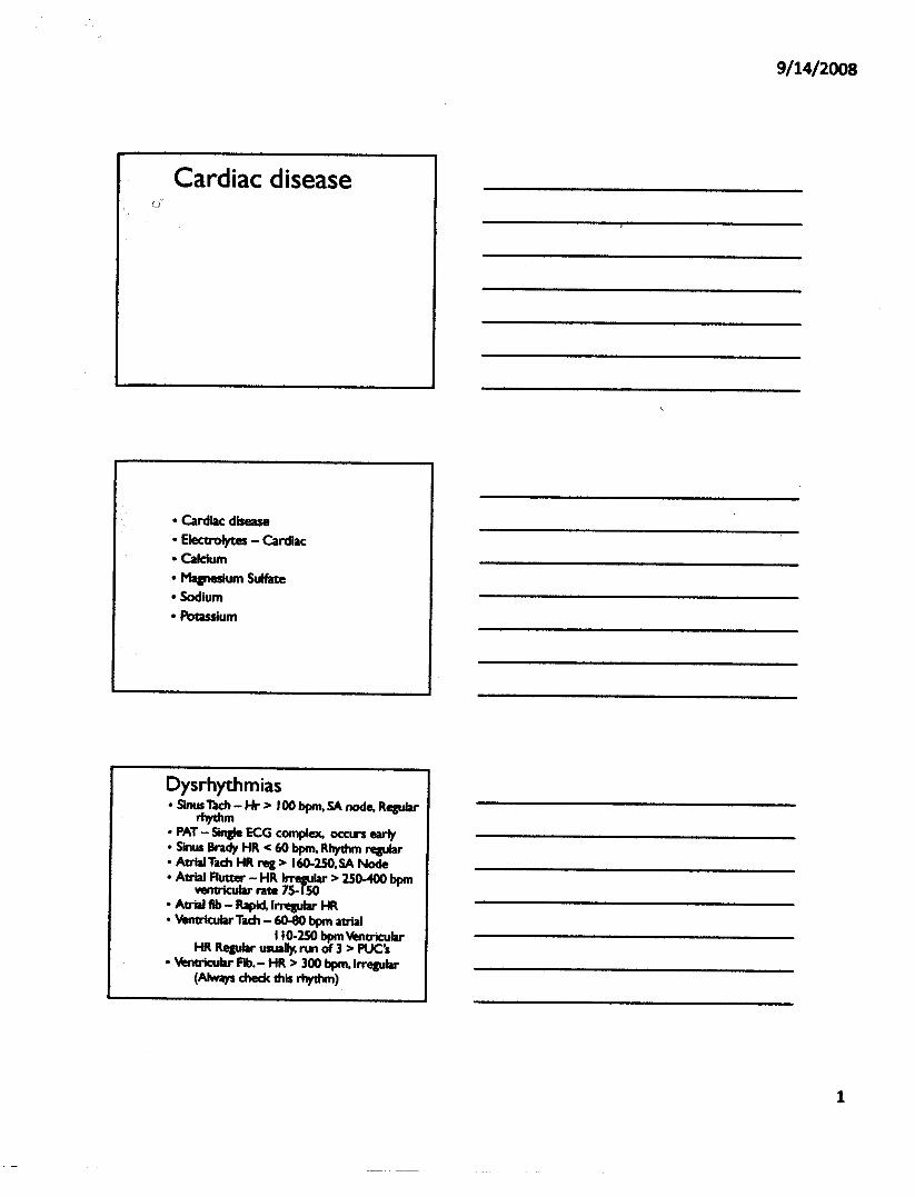
Cardiac disease • Cardiac disease • Electrolytes - Cardiac • Calcium • Magnesium Sulfate • Sodium • Potassium Dysrhythmias • Sinus Tach - Hr > 100 bpm, SA node, Regular rhythm • PAT - SIngle ECG complex. occun early • Sinus Brady HR < 60 bpm, Rhythm re,ular • Atrial Tach HR rez > 160-250. SA Node • Atrial Rutter - HR Irregular> 2S0-400 bpm ventricular rate 75- ISO • Atrial flb - Rapid, Irregular HR • VentricularTach - 60-80 bpm atrial 110-250 bpm Ventricular HR ReauJar usually. run of 3 > PUC's • Ventricular Fib.- HR > 300 bpm.lrregular (Always check this rhythm)
-
Upload
duongduong -
Category
Documents
-
view
232 -
download
2
Transcript of Cardiac disease - imperial.edu disease • Cardiac disease ... Nursing care Heart Failure- H.F ... A...
Cardiac disease
• Cardiac disease• Electrolytes - Cardiac• Calcium• Magnesium Sulfate• Sodium• Potassium
Dysrhythmias• Sinus Tach - Hr > 100 bpm, SA node, Regular
rhythm• PAT - SIngle ECG complex. occun early• Sinus Brady HR < 60 bpm, Rhythm re,ular• Atrial Tach HR rez > 160-250. SA Node• Atrial Rutter - HR Irregular> 2S0-400 bpm
ventricular rate 75- ISO• Atrial flb - Rapid, Irregular HR• VentricularTach - 60-80 bpm atrial
110-250 bpm VentricularHR ReauJar usually. run of 3 > PUC's
• Ventricular Fib. - HR > 300 bpm.lrregular(Always check this rhythm)
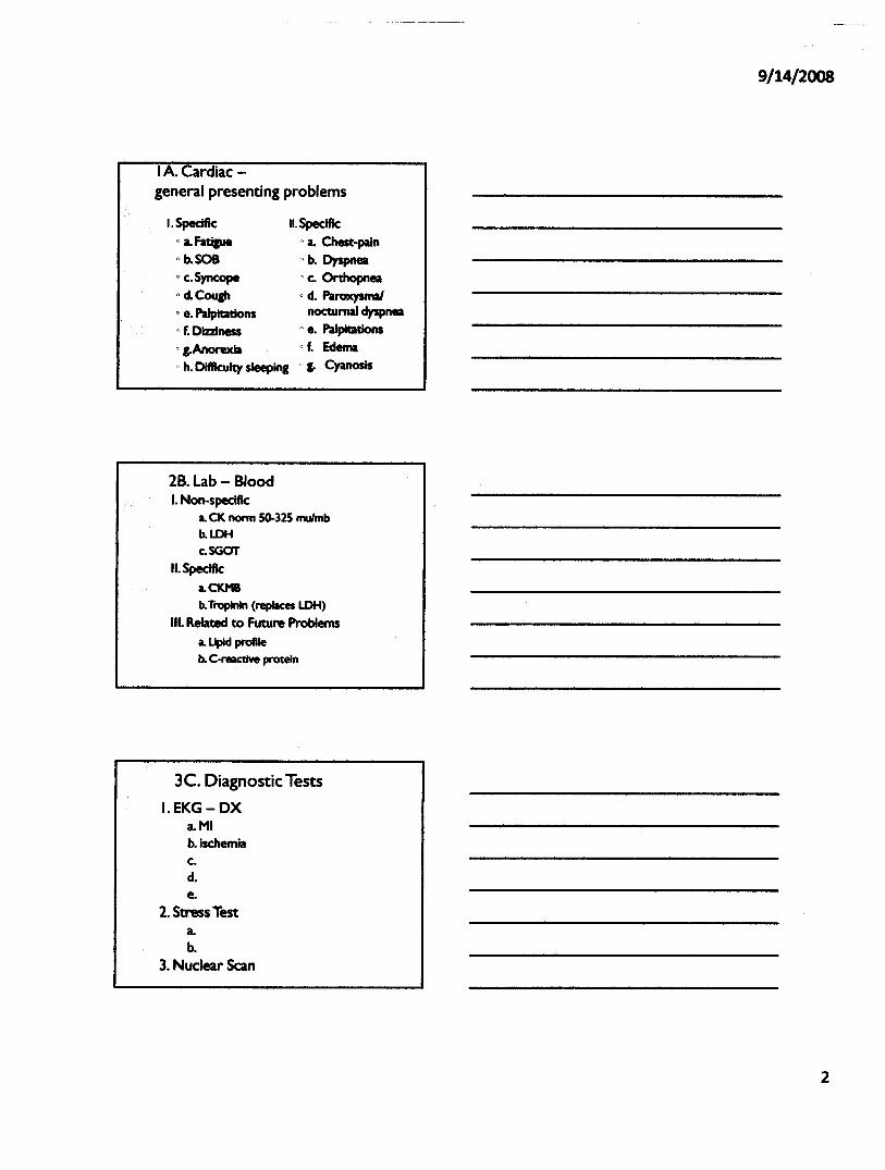
IA. Cardiac -general presenting problems
I.Specific" a. fatilueo b.SOBo c.Syncopeo d.Cougho e. Palpitationso f. Dizziness" g.Anorexia"h. DifIlculty sleeping
II.Specifico a. Chest-pain"b. Dyspneao c. Orthopneao d. Paroxysmal
nocturnal dyspneao e. PalpItations"f. Edemao g. Cyanosis
2B.lab - BloodI.Non-speclfic
a. CK nonn 50-32S mulmbb.LDHc.SGOT
II.Specifica.CKMB
b.TropInin (replaces LDH)III. Related to Future Problems
a. Upld profileb. C-reactive protein
3C. Diagnostic TestsI.EKG-DX
a.MIb.ischemiac.d.e.
2. Stress Testa.b.
3. Nuclear Scan
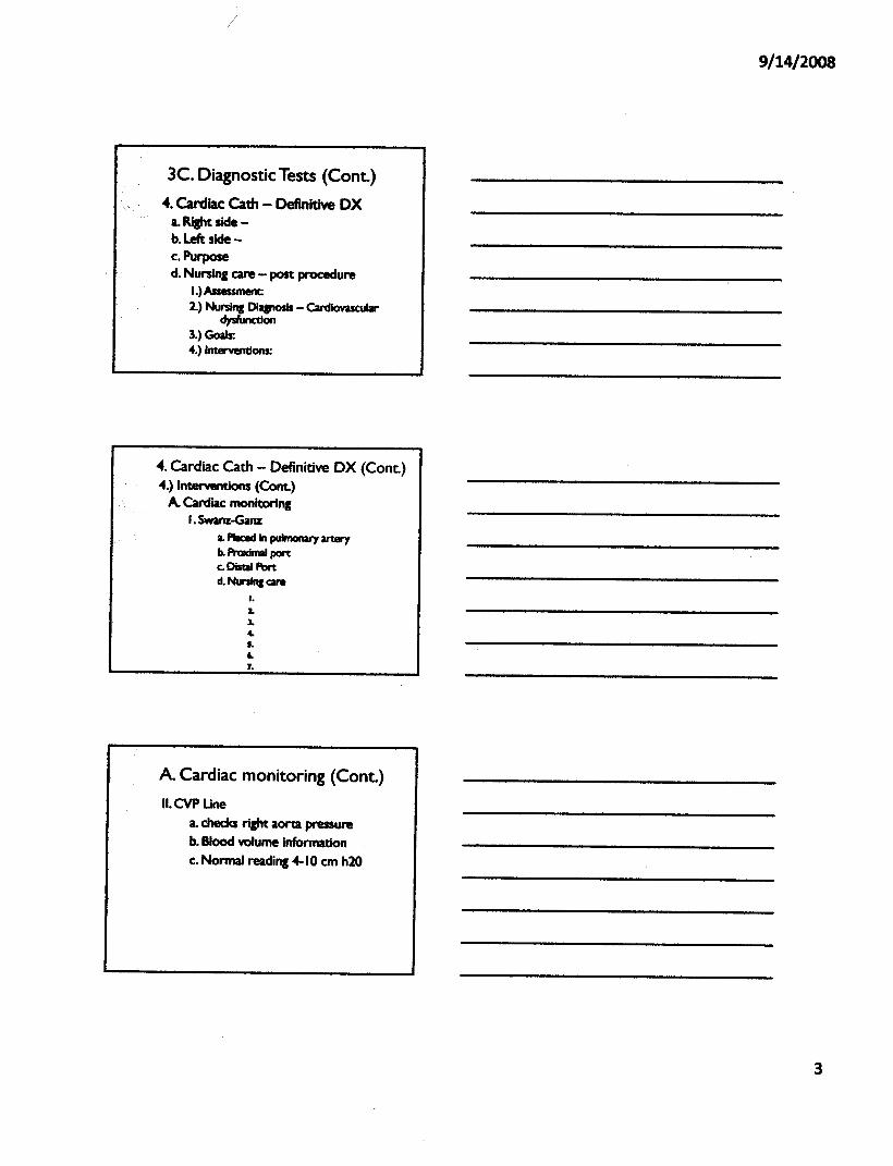
3C. Diagnostic Tests (Cont.)••.Cardiac Cath - Definitive OX
a. Right side -b. Left side -c.Purposed. Nursing care - post procedure
I.) Assessment2.) NursinI Dta&nosis - CardIovascular
clysfuncdon3.) Goals:4.) Interventions:
4. Cardiac Cath - Definitive OX (Cont.)4.)lnterYentions (Cont.)
A. Cardiac monitoringI. Swanz-Ganz
a. P!IaId in pulmonary arwyb. Proximal portc. DistaJ Portd. Nuninl care
I.2-So.•.S.
"7.
A Cardiac monitoring (Cont.)II.CVPUne
a. checks right aorta pressureb. Blood volume informationc. Normal reading 4-10 em h20
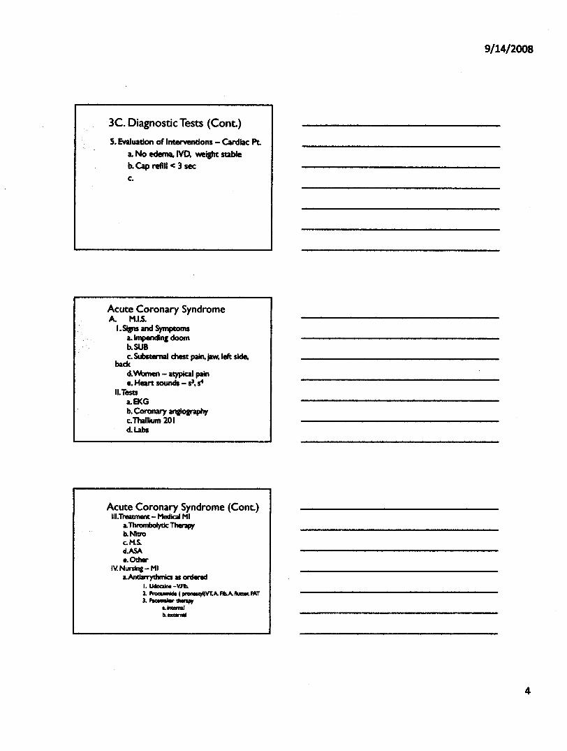
3C. Diagnostic Tests (Cant.)5. Evaluation of InterYentlons - Cardiac Pt.
a. No edema. lvo. weight stableb. Cap refill < 3 seec.
Acute Coronary SyndromeA. M.I.s.
I. Si&ns and Symptomsa.lmpendq doomb.SUBc. SubstemaI chest pain, jaw,left side,
backcI.Women - atypical paine. Heart sounds - 53, s4
II.Testsa.EKGb. Coronary aflIlo&raphyc. Thallium 20 1d.Labs
Acute Coronary Syndrome (Cont.)III.Treatment- Medic:aI MI
LThrombolytic: Therapyb.Nitroc.M.S.d.ASA•• Other
IV. Nursin& - MILAntiarTydlmic:s as ordered
I. I.ldocIiM-VA2. •••.•••••••• ( pnlllIStJI)YT.A. Fib.A. ~ MTJ. I'IcelnIIw thenpy
L •••••b. __
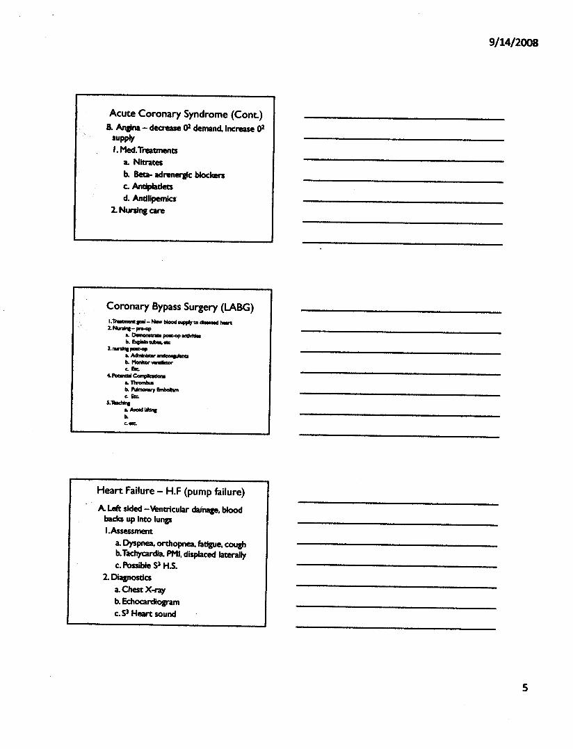
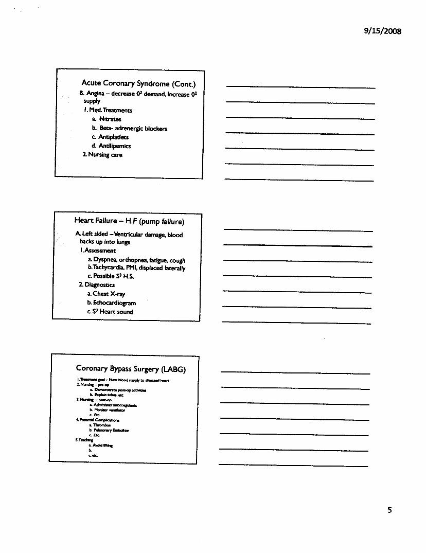
Acute Coronary Syndrome (Cant.)B. Anaina - decrease ()1 demand. Increase ()1
supplyI. MeetTreatments
a. Nitratesb. Beta- adrenergic blockersc. Antipladetsd. Andllpernlcs'
2. Nursina care
I.n--p1 - thwbIoad...", •••••••• "-t2.NInfrw- preoop
L 0...0.-..-..._b..1bcploin-'.
3. ....-- ..-...L ~ •••••••••••••
b.. 1'Ionllior ••••••••••.c.1!IIc.
•• ~ CompIIc:IldonIL TIlromlMab..~fmbolIsmc.1!IIc.
5.""'-"LAWIld~b..c."
Heart Failure - H.F (pump failure)A left sided -Ventricular dainage, blood
backs up into lungsI.Assessment
a. Dyspnea, orthopnea. fatigue, coughb.Tachycardia, PMI,displaced laterallyc. Possible 53 H.5.
2. Diagnosticsa.Chest X-rayb. Echocardiogramc. 53 Heart sound
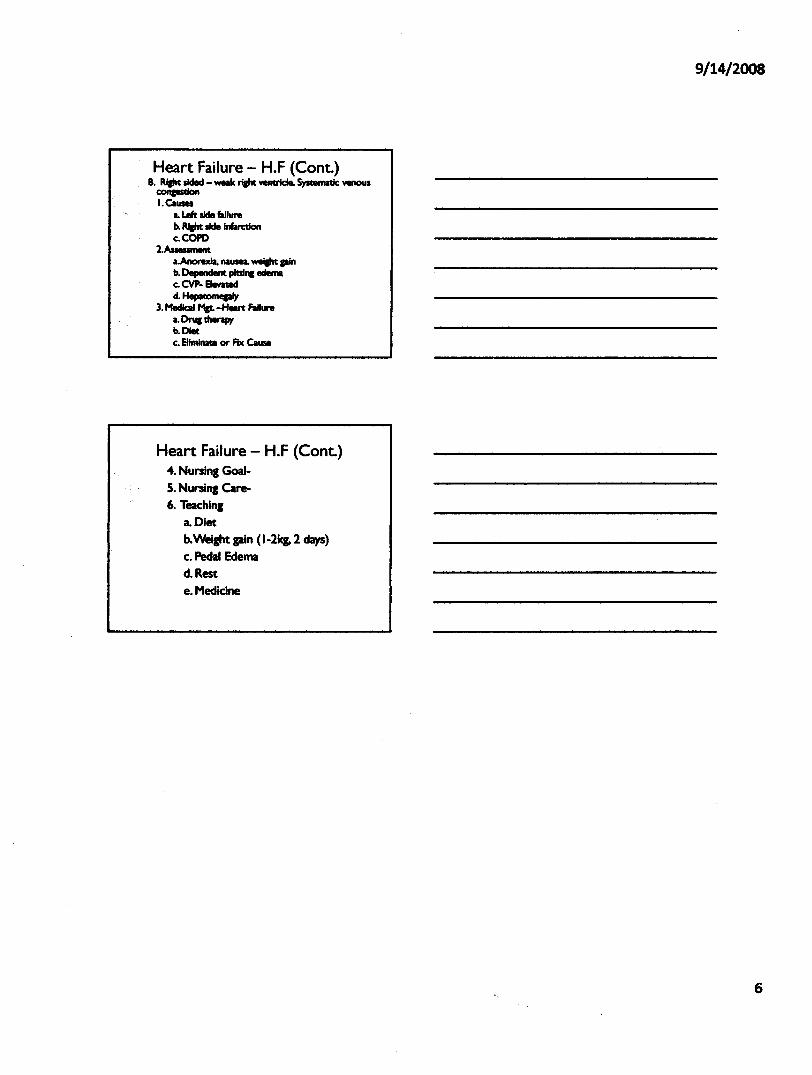
Heart Failure - H.F (Cent.)B. Rf&ht slded - WMk rl&ht -erlde, Systematic: 'IeIIOU1
COlIpldonI. Causes
a.Left side&lI_b. RJaht side InWctionc.COPD
2.~a.AnorexIa, -.-. wei&ht pinb. Dependent pltd"l edemac. CVP- Elevatedd.llepa1ICIIIlepIy
3. Medic:aI Mat -HeII't Failurea. Druc therapyb.Dietc. ElimiIllUl or Fix Cause
Heart Failure - H.F (Cent.)-4.Nursing Goal-S. Nursing Care-6. Teaching
a. Dietb.Weight pin (1-21et. 2 days)c. Pedal Edemad.Reste. Medicine
Acute Coronary Syndrome (Cont.)8. Angina - decrease ()2 demand. Increase ()2
supplyI. Med. Treatments
a. Nitratesb. Beta- adrenergic blockersc. Antipladetsd. Antilipemlcs
2. Nursing care
Heart Failure - H.F (pump failure)
A left sided -Ventricular damage. blood.... backs up Into lungs
I.Assessmenta. Dyspnea, orthopnea, fatigue. coughb.Tachycardia. PMI. displaced laterallyc. Possible 53 H.S.
2. OJaanosticsa. Chest X-rayb. Echocardiogramc.53 Heart sound
I.T_pl- NewbloodsupplytD dIseuedhart2.NunInc- pre-op
L Dornor-.te post-op __b. !xpIIIn tulles, ecc
J. NunI,. - post-opL AclrMaw 0lldc0t&Ufantsb. Mot"---.orc.&c.
4.1'otondaI Compllc:atlonsL Thrombusb. ~ EmbolIsmc.&c.
5.1OachIncLA_1lt'dncb.c.ecc.
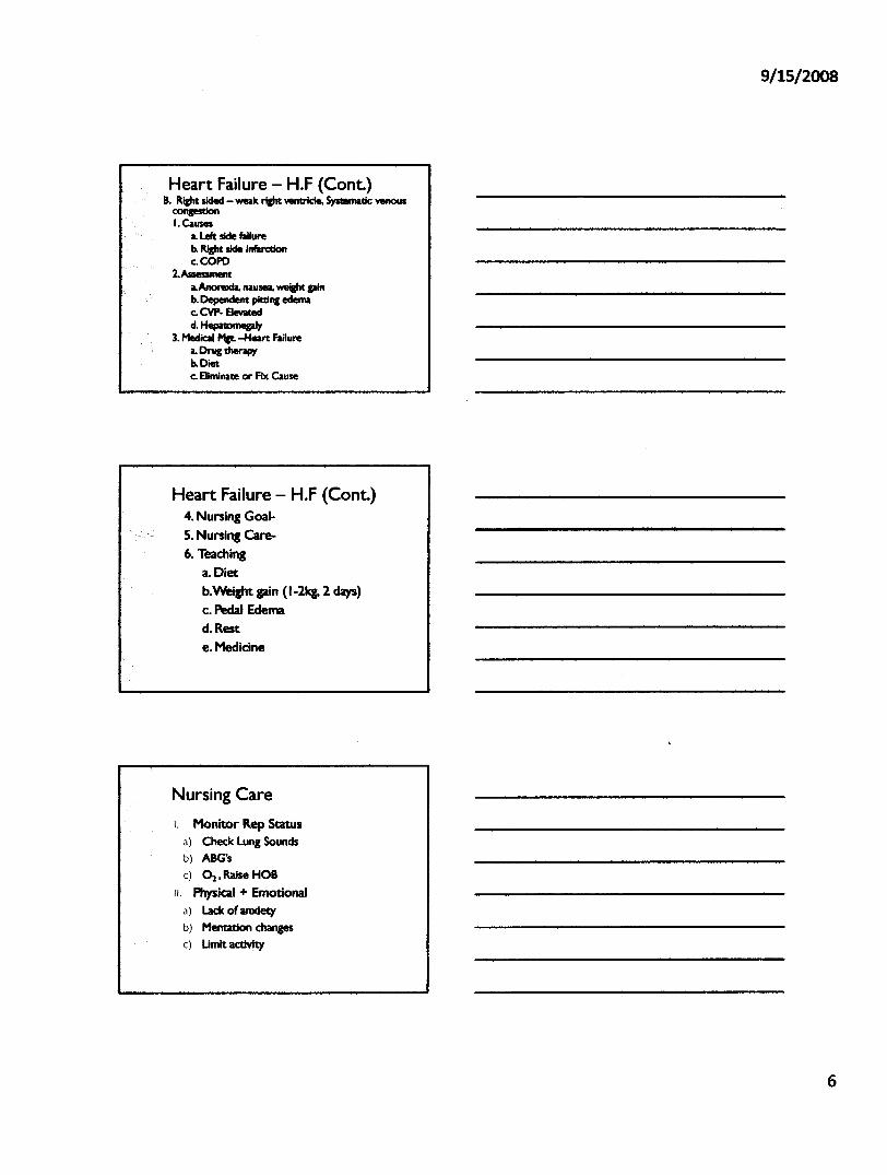
Heart Failure - H.F (Cont.)B. Right sided - weak riJht vemricIe, Systematic wnous
conpstionI.Causes
LLeft side failureb. Right side Infarctlonc.COPD
2.AssessmentLAnorexia. nausea. wei&ht gainb. 0epencIent plains edemac. evp. Elevatedd. HepnomepIy
3. Medial M&t- -Heart FailureL DI'UI therapyb.Dietc. Eliminate or Fix Cause
Heart Failure - H.F (Cont.)4. Nursing Goal-S. Nursing Care-6. Teaching
a. Dietb.Weicht gain (1-21<1.2 days)c. Pedal Edemad.Reste.Medicine
I. Monitor Rep Statusa) Check Lung Soundsb) ABG'sc) ~.RaIseHOB
II. Physical + Emotionala) Lack of anxietyb) Mentation changes
c) Umit aetIvIty
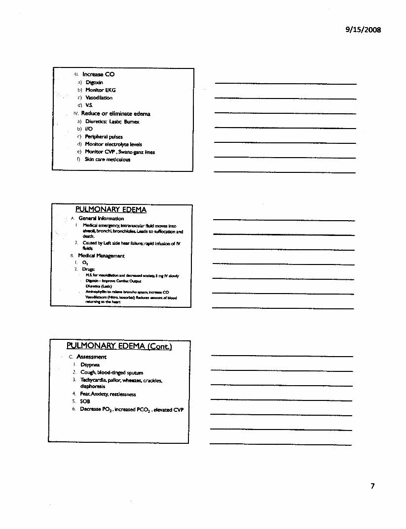
III. Increase COa) Digoxinb) Monitor EKGc) Vasodilationd) v.s.
IV, Reduce or eliminate edemaa) Diuretics; Lube; Bumexb) VOc) Peripheral pulsesd) Monitor electrolyte levelse) Monitor CVP •Swanz-ganz linesf) Skin care meticulous
PULMONARY EDEMAA Generallnfonnadon
I , Medical emergency. IntraVaSCUlarfluid moves intoalveoli. bronch~ bronchtoIes.leads to su«ocatlon anddeath.
2, Caused by left side hear faUure; rapid Infusion of IVfluids
B, Medical ManagementI. 012, Drugs:
M.S.••••.••••••••••••••••• cIea-m 11DC1eq15 '"I r'I slowlyDIpIn - •••••••••••CordIocOucpuc01..- (lMIx)AmInoPJ1IlInlD ~ brancho _1naaH coYuodIIoIacrs (NItro, IsoIofilkI) Recluces amount 0( blood•.••••••••• lD 1he hoort
PULMONARY EDEMA (Cont.)c, Assessment
I, Osypnea
2, Cough. b100d-dnged sputUm3. Tac:hycardIa, pallor; wheezes, crackles.
diaphoresiS4. Fear;Anxiety, restlessnessS, SOB6, Decrease P02 • Increased PC~ •elevated CVP
Nursing InterventionsI. Assist with intubation and monitor
ventilator2. 02. mask at high concentrations (40-60%)3. Semi-Fowlers position4. Meds5. Hemodynamic monitoring6. Pt TeachirlJ, meets. activity7. Monitor for pedal edema. restlessness
EndocarditisA. General Information
I. Inflammation of endocardium, platelets andfibrin deposit on mitral anellor aortic valves.cause stenosls or insufficiency
2. Bacteria, 5.Aureous most common3. Risk factors: Rheumatic heart disease, open
heart sUf'lP!l'Y,dent:aI extractions, invasivemonitoring
B. Medical ManagementI. Antibiotics, arItipyretiCS
c. AssessmentI. Fever, Malaise. dyspnea2. Pedchlae, murmors, cough If extensive vaive
damage3. Elevated WBC4. Ox. Tests, blood cultures
D. Nursing CareI. Administer Meds2. ControiTemperature3. Assess for complications4. alent teaching and discharge plan
RIsk for recurrences dental procedures, urinary eathor scope proceduresAntibiotic therapyAvoid Infectious people
PericarditisA Genera/Information
I. Intlammatlon of the pericardium2. Causes: bacterial or viral Infection. fungal
growth, trauma. M.I.• radiation therapy. meds :procainamlde. hydraluJne. doxorubicln(Adl'iamydn)
B. Medical managementI. Dla&nosls and control cause2. Drug therapy
a) AnaJaeslcsb) Cortlcosterolds.ASA,Antl-inflammatory meclsc) AntibIotlcs
c. AssessmentI. Chest pain,deep inspiration relie¥ed by sitting up.
couch. hemoptysis2. Tachycardia,pleural friction rub, accem:uated Sz,
JVD3. Ele¥atedWBC
D. DiagnosticsI . CXR - Heart effusion2. ECG - ST elevation
E. NursinJ CareI. Bedrest, HOB elevated2. Hemodynamic monitoring3. Meds4. Teaching:recurrence. chest pain increases with
inspirations or position change.
Cardiac Tamponade - EmergencyA. Generallnformadon
I. Au/ell Blood In pericardium preventSventricular ftlling
2. Causes: chest-trauma. malignant pericardialeffusion, complications of cardiac surpry
B. Med Management• Pericardiocentesis
c. AssessmentI. Chest-pain2. Hypotenslon.JVQ tachycardia, muflIed. or
distant heart sounds. perlcardlal frlction rub3. Elevated CVP4. Bevated CVP
D. Nursing careI. O22. CVP monitoring3. IV4. Assist with perlcardlocentesls
ECG monltor;VSCollect fluid, send to lab
Valve surgery• required by patients with stenosis or cardiacinsufficiencyI. Valves
MechanicalProsthetIc
2. Symptoms -Aortic InsufficiencyPalpitationsDizzinessDizzinessAngInaMIJI1IU'S
CXR and ECG = Lert ~ Hypertrophy
Valve surgery (Cont.)3. Aortic Sterosis - may be asymptOmatic4. Mitral Sterosis - surgery required
J. Fatl&ue. dyspneab. HemoptySIsc. Arrhythmiad. Pulmonary hypertensione. Right ventricular hypertrophy
5. Mitral Insufficiency - surgeryJ. If symptoms Interfere with ADl's
Valve surgery (Cont.)• Nursing Care- After valve replacement or
balloon valvuloplastyI. Monitor for seYer hypotensIon. decreased C.O .•
Shock2. v.s.q IS mins until stabile. Report distant heart
sounds. new murmurs3. MonItor ECG. check for bradycardia,
Ventricular taehyc:ardla4. Frequently check peripheral pulses, cap. Refill.
lung sounds5. May have chest-tube. be on vent6. Meds7. Labs
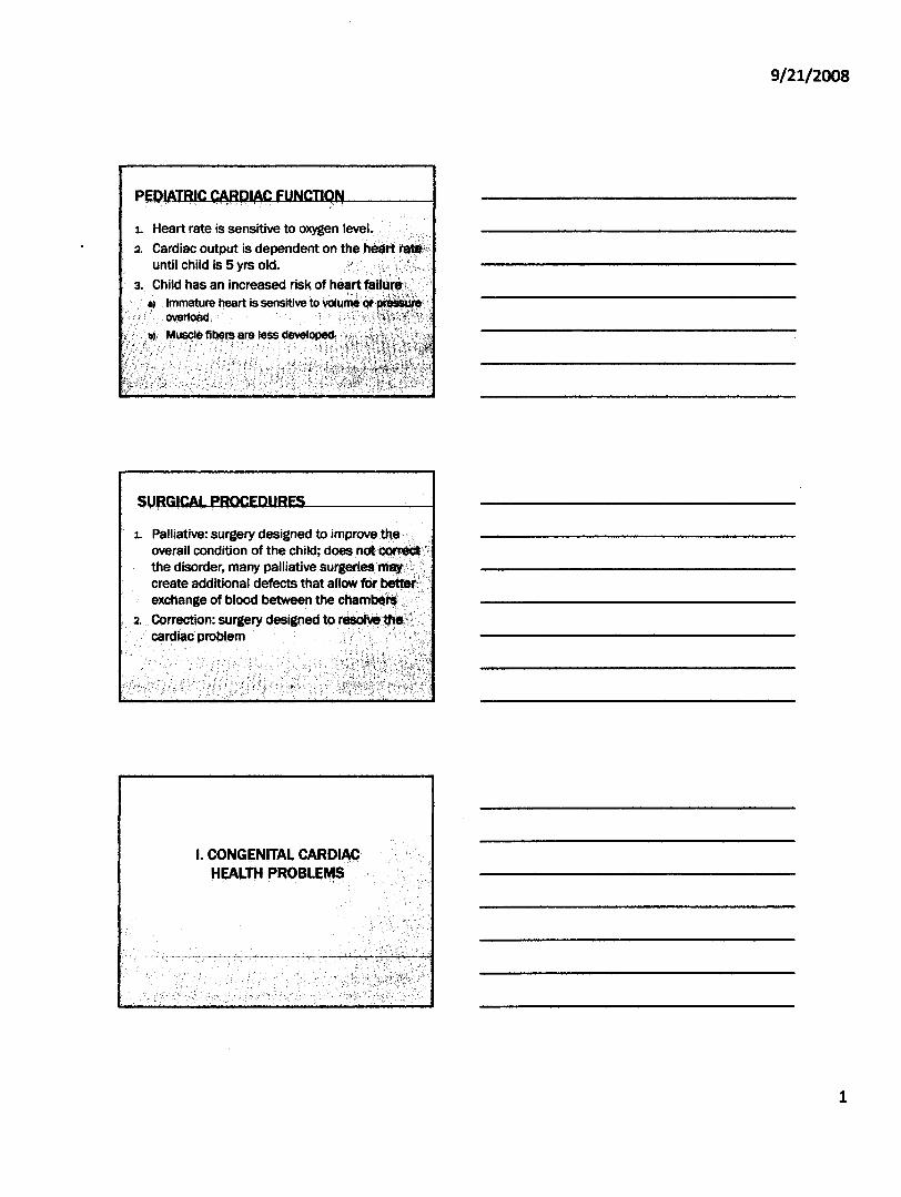
1. Heart rate is sensitive to oxygen level~
2. Cardiac output is dependent on the hedrl~luntil child is 5 yrs old. /' ..............•........
3. Child has an increased risk ofhe~ hlif(lr~!;., Immature heart is sensitive to votume~
overload
1. Palliative: surgery designed to imJ>rO\tetheoverall condition of the child;the disorder, many pallia1:ivecreate additional defects thatexchange of blood between the
2. Correction: surg~ry designed tocardi.c problem
I.CONGENITAl.. CARDIACHEAI..TH PROBLEMS
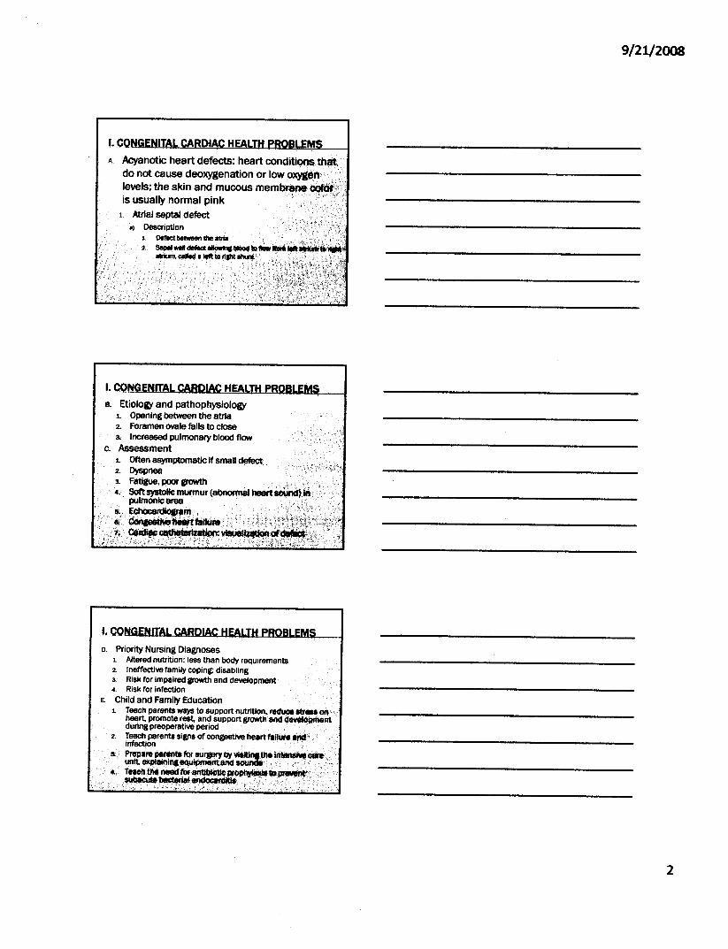
I. CON ITA I HA Acyanotic heart defects: heart corlditions ttl$,
do not cause deoxygenation or lowlevels; the skin and mucousis usually normal pink
1. Atrial septal defect0) Description
1. Defilctbe_1he IlrIa2.
B. Etiology and pathophysiologyL Opening be~ the atria2. Foramen ovale falls to closea Increased pulmonary blood flow
c. AssessmentL Often asymptomatic if small defect2. Dyspnea3. Fatigue, poCJI' growth . . '.. ,.'.,'';., 'y 'C4. Softsystollc murmur (abnom\al heertsoiJnd)~'
pulmonicartlE! .It. . Echi:lea~m ••......, ...,..,./ ~~"~f),;:;ti~~~-.u.
,· ....·.1;':, j F·:; ..•.";,". \;','-:.S''--''',.?' , ;'<'-:"-;:~ .;-
D. Priority Nursing Diagnoses1. Altered nutrition: less than body requirements2. Ineffective family coping: disabling3. Risk for impaired growth and development.•. Risk for Infection
E. Child and Family Education1. Teach parents ways to support nutrition, red\lC8 .-'01'1"
heart. promote rest, and support growth anddlMllOpmelltdurina preoperative period
2. Teach perents signs of congestive heart taRurel!td'infection . .'.:r~a::~l;,.~:.v=~e.~~y,,·'
•. Tem~neecffor~~ .•',.'.'...itD.••.~.-...;.••.....~..••....r.'.l!llQlIIcUtt blICferlal ~itle\ ..•.•.- ',' .".':!.",:
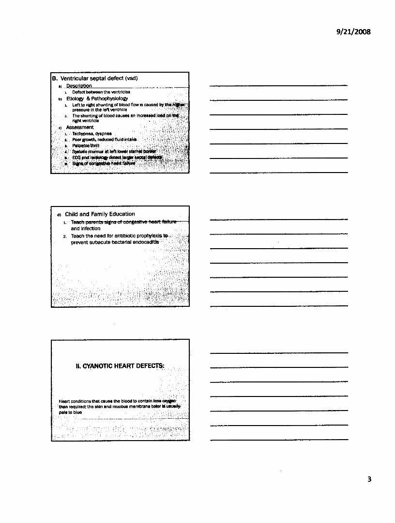
B. Ventricular septal defect (vsd)a) ,Pescri n
L Defect betWeen the ventricles
b) Etiology & PathophysiologyL Left to right shunting of ~Iood flow is caused by tIIe.lliM>
pressure," the leftventncle " ",":""'-",,-2, The shunting of blood causes an increased l<l!id' on'
right ventricle
c) Assessment1. TaChypnea,dyspnea
, ",' ~ Iieor growth, redUced fluid intake , "
, ,';,:s, Palpablethril!, " " ',,~" :"\' '?'" ~) 9l!afoIlo murlnur lit left 1<JWfr'~ lid_l
"i:' E.,.~=-:,d) Child and Family Education
1:'"
and infection2, Teach the need for antibiotic
prevent subaCute bacterial enc:locadlitls,
Heart conditions that cause the blood tothan required; the skin and mucouspale to blue
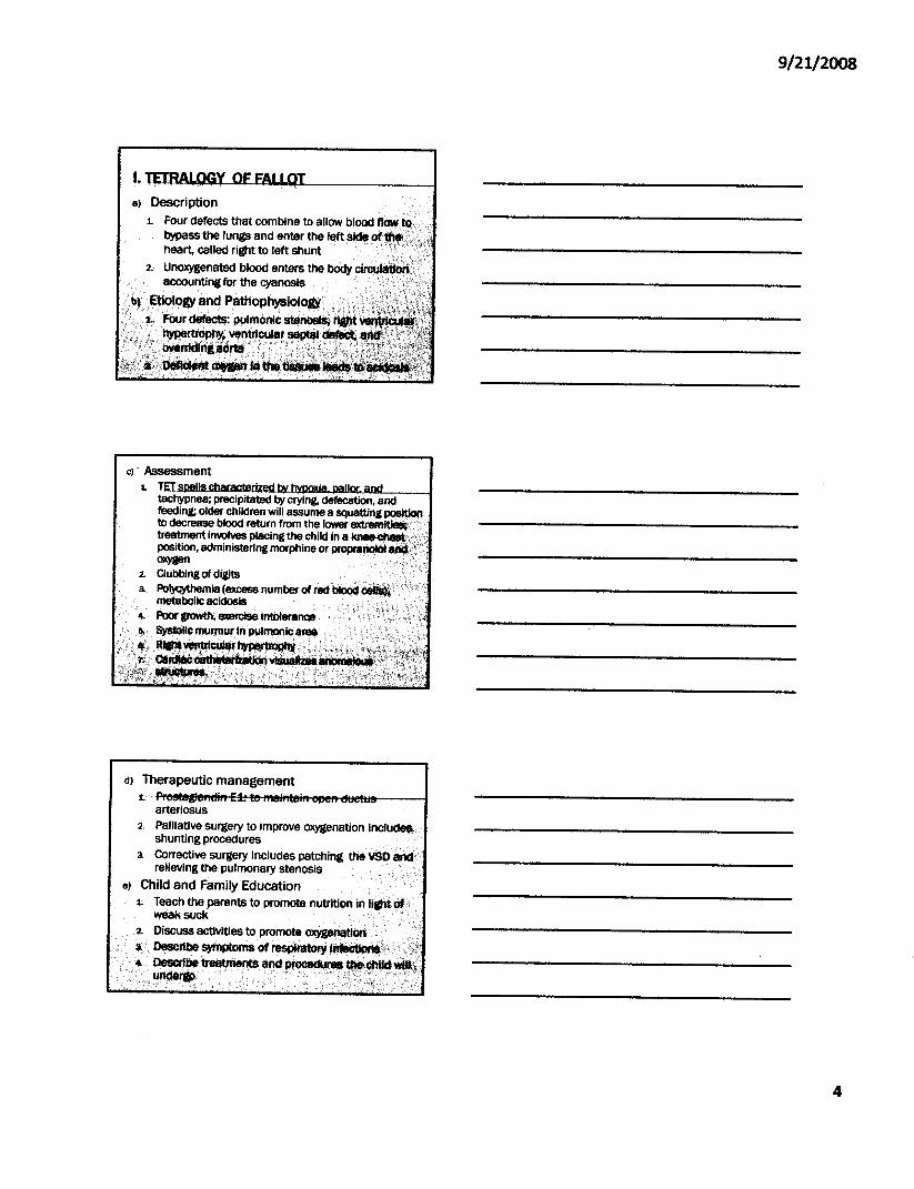
8) Description
1. Four defects that combine to allow blood flow to, '.bypass the lungs and enter the left side ot.".~'.~;heart, called right to left shunt '.' ., "'~'
2. Unoxygenated blood enters the body clfcu.accounting for the cyanosiS
~. Etlologv and Pathophysiology.. .':i 1 Four defectS: pulmoniC stenoelsi
~/;:'/;' .~~"ven~~~~; '., .
~I=f.~.·;·i .....'. ".',;;' -',:',; _";,;,~:,:' ..' (:'-' --,','''''.>:','- _i" ,,' ,:.' ),t'", , - "
c)' Assessment1.
tachypnea; precipitated by crying. defecation. andfeeding; older children will assume a squatting ~to decrease blood return from the Iower~: .treatment involves placing the child in a .position, administering morphine or proprallCllQloxygen
2. Clubbing of digits .~ Polycythemia (excess number of redtllcloddt_,'
metabolic acidosis4. Poor growth, 8llIBl'Cise intolerlln08~. SysI.o\k:muf/l\urin pUllJ1Olllea,... "
·.f'~~~~'~::'·';!~··:·••.."#iCl_c8tt_~Vfl!IU8 •• '··,"';:!ti!!1~~,r~:~:i,;~,j\,;~:l-:,-,;,,:-:.it{ '£':;'_'i,,"':'J~kL'~
d) Therapeutic management.t:··~lst!tglelftdjft-E4:-lto-1'l~'lt8ifH'_J'Mrt-dt:lettlS----I
arteriosus2, Palliative surgery to improve oxygenation include$.
shunting procedures3. Corrective surgery includes patching the VSQtlrlct\
relieving the pulmonary stenosis
e) Child and Family Education .1. Teach the parents to promote nutrition in liatti ~i
weak suck
2. Discuss activities to promote .~IllttI~ .....•..•..'"'•."'. De$criPe symPtOms of resPiratorY~x".·,:~;
'.. ~Pe treEttments $'ld pI'oc.$!r•••• Ohildwul::.,u~, ...............................•........ ;:;';,.
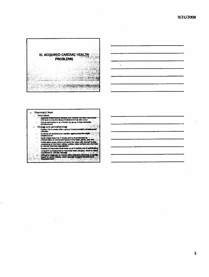
III·AcqVI~~PC,A~PIAC.PRQB"'~M$
., Rheumatic fever1 Description