Cardiac Amyloidosis – Evolving Options for Evaluation … Newsletter... · Cardiac Amyloidosis...
12
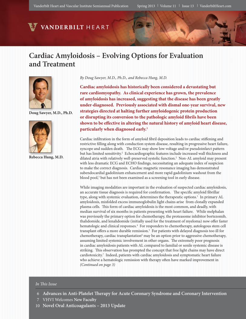
Cardiac Amyloidosis – Evolving Options for Evaluation and Treatment Vanderbilt Heart and Vascular Institute Semiannual Publication Spring 2013 Volume 11 Issue 13 VanderbiltHeart.com In is Issue 6 Advances in Anti-Platelet erapy for Acute Coronary Syndrome and Coronary Intervention 7 VHVI Welcomes New Faculty 10 Novel Oral Anticoagulants – 2013 Update Doug Sawyer, M.D., Ph.D. By Doug Sawyer, M.D., Ph.D., and Rebecca Hung, M.D. Cardiac amyloidosis has historically been considered a devastating but rare cardiomyopathy. As clinical experience has grown, the prevalence of amyloidosis has increased, suggesting that the disease has been greatly under-diagnosed. Previously associated with dismal one-year survival, new strategies directed at halting further amyloidogenic protein production or disrupting its conversion to the pathologic amyloid fibrils have been shown to be effective in altering the natural history of amyloid heart disease, particularly when diagnosed early. 1 Cardiac infiltration in the form of amyloid fibril deposition leads to cardiac stiffening and restrictive filling along with conduction system disease, resulting in progressive heart failure, syncope and sudden death. e ECG may show low voltage and/or pseudoinfarct pattern but has limited sensitivity. 2 Echocardiographic features include increased wall thickness and dilated atria with relatively well-preserved systolic function. 2 Non-AL amyloid may present with less dramatic ECG and ECHO findings, necessitating an adequate index of suspicion to make the correct diagnosis. Cardiac magnetic resonance imaging has demonstrated subendocardial gadolinium enhancement and more rapid gadolinium washout from the blood pool, 3 but has not been examined as a screening tool in early disease. While imaging modalities are important in the evaluation of suspected cardiac amyloidosis, an accurate tissue diagnosis is required for confirmation. e specific amyloid fibrillar type, along with systemic evaluation, determines the therapeutic options. 1 In primary AL amyloidosis, misfolded excess immunoglobulin light chains arise from clonally expanded plasma cells. is form of cardiac amyloidosis is the most common, and deadly, with median survival of six months in patients presenting with heart failure. While melphalan was previously the primary option for chemotherapy, the proteasome inhibitor bortezomib, thalidomide, and lenalidomide (initially used for the treatment of myeloma) now offer faster hematologic and clinical responses. 4 For responders to chemotherapy, autologous stem cell transplant offers a more durable remission. 5 For patients with delayed diagnosis too ill for chemotherapy, cardiac transplantation 6 may be an option prior to aggressive chemotherapy, assuming limited systemic involvement in other organs. e extremely poor prognosis in cardiac amyloidosis patients with AL compared to familial or senile systemic disease is striking. is observation has prompted the concept that free light chains may have direct cardiotoxicity. 7 Indeed, patients with cardiac amyloidosis and symptomatic heart failure who achieve a hematologic remission with therapy oſten have marked improvement in (Continued on page 3) Rebecca Hung, M.D.
Transcript of Cardiac Amyloidosis – Evolving Options for Evaluation … Newsletter... · Cardiac Amyloidosis...
Cardiac Amyloidosis – Evolving Options for Evaluation and Treatment
Vanderbilt Heart and Vascular Institute Semiannual Publication Spring 2013 Volume 11 Issue 13 VanderbiltHeart.com
In This Issue
6 Advances in Anti-Platelet Therapy for Acute Coronary Syndrome and Coronary Intervention 7 VHVI Welcomes New Faculty 10 Novel Oral Anticoagulants – 2013 Update
Doug Sawyer, M.D., Ph.D.
By Doug Sawyer, M.D., Ph.D., and Rebecca Hung, M.D.
Cardiac amyloidosis has historically been considered a devastating but rare cardiomyopathy. As clinical experience has grown, the prevalence of amyloidosis has increased, suggesting that the disease has been greatly under-diagnosed. Previously associated with dismal one-year survival, new strategies directed at halting further amyloidogenic protein production or disrupting its conversion to the pathologic amyloid fibrils have been shown to be effective in altering the natural history of amyloid heart disease, particularly when diagnosed early.1
Cardiac infiltration in the form of amyloid fibril deposition leads to cardiac stiffening and restrictive filling along with conduction system disease, resulting in progressive heart failure, syncope and sudden death. The ECG may show low voltage and/or pseudoinfarct pattern but has limited sensitivity.2 Echocardiographic features include increased wall thickness and dilated atria with relatively well-preserved systolic function.2 Non-AL amyloid may present with less dramatic ECG and ECHO findings, necessitating an adequate index of suspicion to make the correct diagnosis. Cardiac magnetic resonance imaging has demonstrated subendocardial gadolinium enhancement and more rapid gadolinium washout from the blood pool,3 but has not been examined as a screening tool in early disease.
While imaging modalities are important in the evaluation of suspected cardiac amyloidosis, an accurate tissue diagnosis is required for confirmation. The specific amyloid fibrillar type, along with systemic evaluation, determines the therapeutic options.1 In primary AL amyloidosis, misfolded excess immunoglobulin light chains arise from clonally expanded plasma cells. This form of cardiac amyloidosis is the most common, and deadly, with median survival of six months in patients presenting with heart failure. While melphalan was previously the primary option for chemotherapy, the proteasome inhibitor bortezomib, thalidomide, and lenalidomide (initially used for the treatment of myeloma) now offer faster hematologic and clinical responses.4 For responders to chemotherapy, autologous stem cell transplant offers a more durable remission.5 For patients with delayed diagnosis too ill for chemotherapy, cardiac transplantation6 may be an option prior to aggressive chemotherapy, assuming limited systemic involvement in other organs. The extremely poor prognosis in cardiac amyloidosis patients with AL compared to familial or senile systemic disease is striking. This observation has prompted the concept that free light chains may have direct cardiotoxicity.7 Indeed, patients with cardiac amyloidosis and symptomatic heart failure who achieve a hematologic remission with therapy often have marked improvement in(Continued on page 3)
Rebecca Hung, M.D.

By Robert N. Piana, M.D., Editor, Vanderbilt Heart, and Quinn Wells, M.D., Associate Editor
Vanderbilt Heart and Vascular Institute (VHVI) embarks on a new era in 2013 as we welcome Thomas Wang, M.D., as the new chief of Cardiovascular Medicine and Michael Petracek, M.D., as the interim chair of Cardiac Surgery.
Dr. Wang graduated from Harvard Medical School, and completed internal medicine residency and cardiology fellowship training at the Massachusetts General Hospital (MGH) in Boston. He then completed a post-doctoral research fellowship at the Framingham Heart Study prior to joining faculty at the MGH.
Dr. Wang’s clinical focus is in the field of heart failure and cardiac transplantation. At MGH he served as director of the Heart Failure Disease Management Program and associate director of the Heart Failure/Transplantation Section. Since 2011 he has also directed the Program in Human Cardiovascular Physiology and Metabolism. His research explores the role of the natriuretic peptide system in cardiovascular health, the identification of novel cardiometabolic biomarkers, mechanisms of obesity-related cardiac dysfunction, and the effects of vitamin D on the heart.
Dr. Wang’s vision for VHVI will continue to build on existing traditions of excellence in interdisciplinary care and personalized medicine while ensuring that Vanderbilt continues to grow as one of the country’s premier institutions for cardiovascular care, basic science, and patient-oriented cardiovascular research. Summarizing his attraction to Vanderbilt, Dr. Wang stated: “One appeal was the strength of the clinical enterprise in cardiology. VHVI has been a very successful model for the development of a robust cardiovascular service line. Also, Vanderbilt has been a very forward-thinking institution when it comes to personalized medicine in both research and clinical implementation. It’s one of the premier places in the country for doing patient-oriented research, which is my area of investigation.”
VHVI is also invigorated by our new interim chair of Cardiac Surgery, Michael Petracek, M.D. Dr. Petracek is a product of Johns Hopkins Medical School, and completed residency and fellowship at Vanderbilt. He has been a force in cardiac surgery for decades, and his clinical presence has helped catapult VHVI’s program into national prominence. His particular focus has been on minimally invasive complex mitral valve replacement and repair. In addition, his collaborative approach has helped foster continued expansion of dynamic programs in off-pump coronary bypass surgery and transcatheter aortic valve replacement.
We look forward to the next era with Dr. Wang and Dr. Petracek at the helm. Join us through Vanderbilt Heart to learn more about the continuing advances at VHVI under this new and dynamic leadership team.
Robert N. Piana, M.D.
Vanderbilt Heart Spring 2013
2
Editorial
Quinn Wells, M.D.
Thomas Wang, M.D.
Michael Petracek, M.D.

3
symptoms despite no obvious change in the degree of cardiac amyloid infiltration. The unique cardiotoxicity of light chain amyloid may offer a specific target for intervention in the future. The other common form of cardiac amyloidosis is due to deposition of mutant (familial) or wild-type (senile systemic) transthyreitin (TTR).1 A valine to isoleucine mutation at position 122 occurs in up to 4% of the African-American population and presents as heart failure in patients in their late 60s. The penetrance is unknown because of the overlap with coronary artery disease and hypertension in this age group. Familial amyloidosis can also occur due to variants of other proteins. Senile amyloidosis is a disease predominantly affecting older men, usually > 70 years of age, often misdiagnosed as hypertensive heart disease. TTR (also known as prealbumin) is secreted by the liver and circulates as a tetramer. For TTR to form amyloid fibrils, the tetramer must first dissociate and then refold improperly. Tafamidis, a stabilizer of correctly folded TTR, slows progression in familial amyloid polyneuropathy, and is approved for use in Europe.8 The value of tafamidis in amyloid cardiomyopathy remains under investigation. It is conceivable that in the future a pharmacologic strategy may be discovered to help patients with advanced amyloidosis clear their tissues of amyloid. The antibiotic doxycycline, shown to disrupt amyloid fibrillogenesis in vitro and in mouse models, holds promise and has advanced to human trials.9
Unfortunately, the diagnosis of cardiac amyloidosis is often delayed. It is not uncommon for cardiac evaluation to lead to plausible explanations for symptoms attributed to common conditions including hypertrophic cardiomyopathy, hypertensive heart disease, or coronary disease, further delaying consideration of cardiac amyloidosis. Because an early diagnosis of cardiac amyloidosis is a critical determinant of prognosis, particularly with AL amyloid, timely integration of the data collected in routine cardiovascular evaluation with the clinical picture is essential. Elevations in biomarkers including naturietic peptides and/or cardiac troponins confer an additive risk for mortality, even when cardiac symptoms are minimal.10 (Continued on page 4)
Cardiac Amyloidosis – Evolving Options for Evaluation and Treatment (Continued from page 1)
Figure 1.Parasternal and apical views of ATTR cardiac amyloidosis. Note the increase in right ventricular wall thickness (*) in addition to that of the left ventricle.
Figure 2.Endomyocardial biopsy of the same patient, stained with Congo Red. The extracellular pink amyloid deposits seen under white light demonstrate typical apple green birefringence under polarized light. (Courtesy of Robert Hoffman, M.D., Department of Pathology).
RV
RV
*
*
4
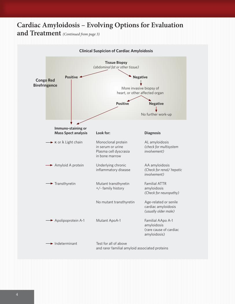
Cardiac Amyloidosis – Evolving Options for Evaluation and Treatment (Continued from page 3)
Clinical Suspicion of Cardiac Amyloidosis
Tissue Biopsy(abdominal fat or other tissue)
Congo RedBirefringence
Positive Negative
More invasive biopsy of heart, or other affected organ
Positive Negative
No further work-up
Immuno-staining orMass Spect analysis Look for: Diagnosis
κ or λ Light chain Monoclonal protein AL amyloidosis in serum or urine (check for multisystem Plasma cell dyscrasia involvement) in bone marrow
Amyloid A protein Underlying chronic AA amyloidosis inflammatory disease (Check for renal/ hepatic involvement)
Transthyretin Mutant transthyretin Familial ATTR +/- family history amyloidosis (Check for neuropathy) No mutant transthyretin Age-related or senile cardiac amyloidosis (usually older male)
Apolipoprotein A-1 Mutant ApoA-1 Familial AApo A-1 amyloidosis (rare cause of cardiac amyloidosis)
Indeterminant Test for all of above and rarer familial amyloid associated proteins

Vanderbilt Heart Spring 2013
5
The response to standard heart failure and arrhythmia therapy is unpredictable in patients with cardiac amyloidosis.1 Beta blockers and ACE inhibitors and angiotensin receptor blockers are often poorly tolerated with worsening fatigue, orthostatic hypotension, aggravated by amyloid-related autonomic dysfunction and syncope and have not proven to improve survival. Calcium channel blockers have been reported to precipitate hemodynamic collapse. With atrial involvement, even in the absence of atrial fibrillation, intracardiac thrombi may develop and broader indications for anticoagulation are needed to minimize embolic events.11 Currently, the mainstay of heart failure therapy remains diuretics. Because amyloid infiltration often affects the conduction system, pacemakers play a role in patients with advanced conduction system disease. Implantable cardiac defibrillators (ICD), particularly for primary prevention of sudden cardiac death, are controversial. While there are select patients who develop ventricular arrhythmias that are responsive to ICD therapy, sudden death is often due to pulseless electrical activity. For this reason, ICDs are usually reserved for patients with non-AL amyloid who require secondary prevention of ventricular arrhythmias.
Heart failure cardiologists at VHVI have a specific interest in cardiac amyloidosis and work in concert with the Vanderbilt Amyloid Multidisciplinary Program (VAMP) to evaluate and treat patients. We are also involved in clinical research investigating novel approaches to treatment.
To learn more about our program, please see the VAMP website: http://www.vanderbilthealth.com/hematology/37821 To refer a patient to our program, please contact our referral line at (615) 322-2318.
R E F E R E N C E S1. Falk RH. Diagnosis and management of the cardiac amyloidoses. Circulation. 2005 112(13):2047-60. Falk RH. Senile systemic
amyloidosis: are regional differences real or do they reflect different diagnostic suspicion and use of techniques? Amyloid. 2012 19 Suppl 1:68-70. Falk RH, Dubrey SW. Progress in Cardiovascular Diseases 2010 52:347-361. Falk RH. Cardiac Amyloidosis: A Treatable Disease, Often Overlooked. Circulation 2011 124:1079-1085.
2. Rahman JE, Helou EF, Gelzer-Bell R, Thompson RE, Kuo C, Rodriguez ER, Hare JM, Baughman KL, Kasper EK. Noninvasive diagnosis of biopsy-proven cardiac amyloidosis. J Am Coll Cardiol. 2004; 43: 410–415.
3. Maceira AM, Joshi J, Prasad SK, Moon JC, Perugini E, Harding I, Sheppard MN, Poole-Wilson PA, Hawkins PN, Pennell DJ. Cardiovascular magnetic resonance in cardiac amyloidosis. Circulation. 2005 111(2):186-93.
4. Gertz MA. Immunoglobulin light chain amyloidosis: 2013 update on diagnosis, prognosis, and treatment. Am J Hematol. 2013 88(5):416-25.
5. Fitzgerald BT, Bashford J, Scalia GM The Return of the Normal Heart: Resolution of Cardiac Amyloidosis After Chemotherapy and Bone Marrow Transplantation Heart Lung Circ. 2013 pii: S1443-9506.
6. DePasquale EC, Nasir K, Jacoby DL. Outcomes of adults with restrictive cardiomyopathy after heart transplantation. J Heart Lung Transplant. 2012 12:1269-75. Sack FU, Kristen A, Goldschmidt H, Schnabel PA, Dengler T, Koch A, Karck M. Treatment options for severe cardiac amyloidosis: heart transplantation combined with chemotherapy and stem cell transplantation for patients with AL-amyloidosis and heart and liver transplantation for patients with ATTR-amyloidosis. Eur J Cardiothorac Surg. 2008 33(2):257-62.
7. Mishra S, Guan J, Plovie E, Seldin DC, Connors LH, Merlini G, Falk RH, Macrae CA, Liao R. Human Amyloidogenic Light Chain Proteins Result In Cardiac Dysfunction, Cell Death and Early Mortality in Zebrafish. Am J Physiol Heart Circ Physiol. 2013 Apr 26.
8. Coelho T, Maia LF, Martins da Silva A, Waddington Cruz M, Planté-Bordeneuve V, Lozeron P, Suhr OB, Campistol JM, Conceição IM, Schmidt HH, Trigo P, Kelly JW, Labaudinière R, Chan J, Packman J, Wilson A, Grogan DR. Tafamidis for transthyretin familial amyloid polyneuropathy: a randomized, controlled trial. Neurology. 2012 79:785-92
9. Cardoso I, Saraiva MJ. Doxycycline disrupts transthyretin amyloid: evidence from studies in a FAP transgenic mice model. FASEB J. 2006 Feb;20(2):234-9. Obici L, Cortese A, Lozza A, Lucchetti J, Gobbi M, Palladini G, Perlini S, Saraiva MJ, Merlini G. Doxycycline plus tauroursodeoxycholic acid for transthyretin amyloidosis: a phase II study. Amyloid. 2012 19 Suppl 1:34-6.
10. Dispenzieri A, Gertz MA, Kyle RA, Lacy MQ, Burritt MF, Therneau TM, Greipp PR, Witzig TE, Lust JA, Rajkumar SV, Fonseca R, Zeldenrust SR, McGregor CG, Jaffe AS. Serum cardiac troponins and N-terminal pro-brain natriuretic peptide: a staging system for primary systemic amyloidosis. J Clin Oncol. 2004 22(18):3751-7.
11. Feng D, Syed IS, Martinez M, Oh JK, Jaffe AS, Grogan M, Edwards WD, Gertz MA, Klarich KW. Intracardiac thrombosis and anticoagulation therapy in cardiac amyloidosis. Circulation. 2009 119(18):2490-7. Dubrey S, Pollak A, Skinner M, Falk RH. Atrial thrombi occurring during sinus rhythm in cardiac amyloidosis: evidence for atrial electromechanical dissociation. Br Heart J. 1995 74(5):541-4.
Heart failure cardiologists at VHVI have a specific interest in cardiac amyloidosis and work in concert with the Vanderbilt Amyloid Multidisciplinary Program (VAMP) to evaluate and treat patients. We are also involved in clinical research investigating novel approaches to treatment.

Vanderbilt Heart Spring 2013
6
Advances in Anti-Platelet Therapy for Acute Coronary Syndrome and Coronary Intervention
By David H. Kim, M.D., and Jayant Bagai, M.D.
Acute coronary syndromes (ACS), which include unstable angina and non ST elevation MI (NSTEMI), account for 1.5 million hospital discharges and an estimated cost of $150 billion annually.1 ACS is usually caused by disruption of lipid rich atherosclerotic plaque, which is a stimulus for thrombogenesis. Platelets play a key role in the initiation and propagation of thrombus by crosstalk with the coagulation cascade and multiplier effects. Therefore, inhibition of platelet activation and aggregation is a major target to reduce thrombosis and improve outcomes in ACS.
The key platelet activators are thromboxane, adenosine diphosphate (ADP) and thrombin. ADP activates platelets via the P2Y12 receptor, and irreversible P2Y12 receptor inhibitors, such as clopidogrel and ticlopidine, are used in conjunction with aspirin to reduce major adverse cardiovascular events (MACE) after percutaneous coronary intervention (PCI) and ACS.2,3 Due to a small risk of serious hematologic side effects, ticlopidine is now only rarely used.
Clopidogrel has been the default P2Y12 inhibitor with a well-established safety and efficacy profile, but multiple studies have exposed several important limitations. Up to 85% of clopidogrel is inactivated after ingestion and the remaining 15% undergoes a two-step, bioactivation in the liver, largely dependent on the CYP2C19 enzyme. The CYP2C19 gene is polymorphic (exists in different forms or alleles), and some alleles, such as *2 or *3 (pronounced “star two” and “star three”), have reduced enzymatic activity. Carriers of such alleles have a 32% reduction in active clopidogrel metabolite and a 53% relative increase in MACE compared to non-carriers.4 In addition, clopidogrel has a slow onset of action with modest inhibition of platelet aggregation. Interactions with other drugs also utilizing CYP2C19 for metabolism – most notably proton pump inhibitors – are also of potential clinical importance.
To overcome the limitations of clopidogrel, newer antiplatelet drugs have been developed and studied. These include P2Y12 receptor inhibitors (prasugrel, ticagrelor and cangrelor) and protease activated receptor (PAR) inhibitors. Table 1 outlines major differences.
Prasugrel requires only a one-step bioactivation, independent of CYP2C19 activity. In the pivotal TRITON-TIMI 38 trial of moderate- to high-risk ACS patients scheduled for PCI, prasugrel resulted in an 18% relative reduction in adverse cardiovascular events compared to clopidogrel, a benefit that was magnified among diabetics.5 However, an increased risk of major and fatal bleeding was noted in the total prasugrel cohort. Because of potential bleeding, prasugrel is not recommended in patients older than 75 years (unless the benefit is expected to outweigh the risk) or with body weight < 60 kg. Due to net harm, prasugrel is contraindicated in patients with a prior history of stroke or TIA.
Ticagrelor is a reversible P2Y12 receptor inhibitor. In the PLATO trial, ticagrelor reduced vascular death, MI and stroke by 16.2% compared to clopidogrel in ACS patients without an increase in overall major bleeding.6 This translated into a significant 1.4% absolute reduction in all cause mortality. However, two subcategories of bleeding – intra cranial hemorrhage and non-CABG related major bleeding – occurred more often with ticagrelor.
David H. Kim, M.D.
Jayant Bagai, M.D.
Unique issues with ticagrelor include dyspnea, ventricular pauses and need for twice daily dosing, which may affect compliance. In the PLATO trial, no net benefit was noted in North American patients, which was postulated to be possibly due to a higher aspirin dose in those patients. Thus, a lower dose aspirin (<100 mg) is recommended for patients treated with ticagrelor.
Cangrelor is an intravenous reversible P2Y12 receptor inhibitor. Onset of action is immediate with full recovery of platelet function within 60 minutes, due to a half-life of three to six minutes. The CHAMPION PHOENIX trial showed a consistent benefit of cangrelor in prevention of ischemic complications such as death, MI and stent thrombosis, compared with clopidogrel given just before or after PCI.7 Cangrelor has also been used as a “bridge” to prevent adverse ischemic outcomes in patients who need to discontinue oral anti platelet therapy before cardiac surgery.8
PAR-1 inhibitors or thrombin receptor antagonists (TRA) block thrombin mediated platelet activation without disrupting thrombin-mediated cleavage of fibrinogen to fibrin. This property allows preservation of physiological hemostasis. The oral TRA, vorapaxar, when combined with aspirin and clopidogrel did not increase bleeding compared with placebo in patients undergoing elective PCI in the TRA-PCI study.9 However, the same drug showed a higher incidence of major bleeding and intracranial hemorrhage when combined with aspirin and a thienopyridine in ACS patients in the TRACER trial.10 Further studies are therefore required.
Table 2 outlines some of the factors which guide the choice of a specific drug for a given clinical situation and patient. In addition, platelet function and genetic testing can be considered for decision making. The VerifyNow (Accumetrics, San Diego, Calif.) point-of-care test expresses the level of P2Y12 receptor inhibitor activity as platelet reaction units or PRUs. Patients with high on-treatment platelet reactivity (HTPR) defined as > 230-240 PRU have a higher incidence of cardiovascular death, MI and stent thrombosis.11-14 Genetic testing identifies carriers of *2 CYP2C19 alleles, who can then be prescribed an alternate antiplatelet drug which does not depend on CYP2C19 for activation. This innovative approach to prospective genotyping is being studied as part of the ongoing PREDICT (Pharmacogenomic Resource for Enhanced Decisions in Care and Treatment) trial at Vanderbilt.15 (Continued on page 8)
7
VHVI Welcomes New Faculty
Kimberli Taylor Clarke, M.D.Specialty: CardiologyMedical School: Meharry Medical College School of Medicine Residency: University of Maryland School of MedicineFellowship: Vanderbilt University School of MedicinePractice Location: Vanderbilt Heart & Vascular Institute at One Hundred Oaks Appointment Phone Number: (615) 322-2318
Richard Gumina, M.D., Ph.D.Specialty: Interventional CardiologyMedical School: Medical College of Wisconsin Residency: Mayo ClinicFellowship: Medical College of Wisconsin; Mayo ClinicPractice Location: Vanderbilt Heart & Vascular Institute Appointment Phone Number: (615) 322-2318
Michael Kelley, M.D.Specialty: CardiologyMedical School: University of Vermont College of Medicine Residency: University of Vermont Fellowship: University of Vermont College of Medicine; Vanderbilt University School of MedicinePractice Location: Vanderbilt Heart & Vascular Institute at Maury Regional Medical Center, Vanderbilt Heart & Vascular Institute Lawrenceburg, and Vanderbilt Heart & Vascular Institute PulaskiAppointment Phone Number: (931) 388-8622
Thomas J.Wang, M.D.Specialty: CardiologyMedical School: Harvard Medical School Residency: Massachusetts General HospitalResearch Fellowship: Framingham Heart StudyPractice Location: Vanderbilt Heart & Vascular Institute Phone Number: (615) 322-2318

8
Advances in Anti-Platelet Therapy for Acute Coronary Syndrome and Coronary Intervention (Continued from page 7)
Drug Prodrug Onset of action Inhibition of platelet Affected by CYP2C19 aggregation (IPA) polymorphism
Clopidogrel Yes 4-6 hours after 32+ 21% (6 hours) Yes 600 mg LD 46 + 21% (chronic therapy)
Prasugrel Yes 30 minutes 75 + 13% (6 hours) No after 60 mg LD 62 + 18% (chronic therapy)
Ticagrelor No 1.5-2 hours 85-95% (2 hours and No after 180 mg LD chronic therapy)
Cangrelor No Minutes after IV >80% (within minutes) No bolus/ infusion
Table 1- Salient differences between clopidogrel and other P2Y12 inhibitors. LD-loading dose.
Drug Pro Con Specific situation Specific situation ACC/AHA to use not to use recommendation
Clopidogrel Cost, Variable Aspirin allergy, *2/*3 carrier ACS, pre PCI familiarity, effect ACS, post PCI extensive Stent thrombosis Continue for 12 data Non while on drug months post ACS responders (medically managed or with PCI) Prasugrel Reliable Bleeding STEMI and Prior stroke/TIA ACS, only after response (including diabetics (post PCI PCI planned fatal) only) Age > 75 Continue for 12 Clopidogrel failure Body weight months post < 60 kg PCI only Ticagrelor Reliable Bradycardia, ACS managed Prior stroke/ICH ACS, pre PCI response, dyspnea, BID medically or benefit regimen, with PCI Severe hepatic Continue for 12 without risk of ICH dysfunction months post ACS increased Clopidogrel (medically overall failure managed or bleeding with PCI)
Table 2- Practical considerations to assist in choice of specific P2Y12 inhibitor for ACS. ICH-intra cranial hemorrhage

9
In summary, there have been significant advances in the development and clinical testing of new antiplatelet agents. We are entering an exciting era when antiplatelet therapy will potentially be personalized based on an individual’s genetic makeup and risk profile. This will provide the best balance between improved outcomes post ACS and PCI and drug tolerability.
R E F E R E N C E S 1. Go AS, Mozaffarian D, Roger VL, Benjamin EJ, Berry JD, Borden WB, Bravata DM, Dai S, Ford ES, Fox CS, Franco S, Fullerton
HJ, Gillespie C, Hailpern SM, Heit JA, Howard VJ, Huffman MD, Kissela BM, Kittner SJ, Lackland DT, Lichtman JH, Lisabeth LD, Magid D, Marcus GM, Marelli A, Matchar DB, McGuire DK, Mohler ER, Moy CS, Mussolino ME, Nichol G, Paynter NP, Schreiner PJ, Sorlie PD, Stein J, Turan TN, Virani SS, Wong ND, Woo D, Turner MB. Executive Summary: Heart Disease and Stroke Statistics—2013 Update. circ.ahajournals.org.
2. Goods CM, al-Shaibi KF, Liu MW, Yadav JS, Mathur A, Jain SP, Dean LS, Iyer SS, Parks JM, Roubin GS. Comparison of aspirin alone versus aspirin plus ticlopidine after coronary artery stenting. Am J Cardiol. 1996 Nov 1;78(9):1042-4.
3. Yusuf S, Zhao F, Mehta SR, Chrolavicius S, Tognoni G, Fox KK, Clopidogrel in Unstable Angina to Prevent Recurrent Events Trial Investigators. Effects of clopidogrel in addition to aspirin in patients with acute coronary syndromes without ST-segment elevation. N. Engl. J. Med. 2001; 345:494–502.
4. Mega JL, Close SL, Wiviott SD, Shen L, Hockett RD, Brandt JT, Walker JR, Antman EM, Macias W, Braunwald E, Sabatine MS. Cytochrome p-450 polymorphisms and response to clopidogrel. N. Engl. J. Med. 2009; 360:354–362.
5. Wiviott SD, Braunwald E, McCabe CH, Montalescot G, Ruzyllo W, Gottlieb S, Neumann F-J, Ardissino D, De Servi S, Murphy SA, Riesmeyer J, Weerakkody G, Gibson CM, Antman EM, TRITON-TIMI 38 Investigators. Prasugrel versus clopidogrel in patients with acute coronary syndromes. N. Engl. J. Med. 2007; 357:2001–2015.
6. Wallentin L, Becker RC, Budaj A, Cannon CP, Emanuelsson H, Held C, Horrow J, Husted S, James S, Katus H, Mahaffey KW, Scirica BM, Skene A, Steg PG, Storey RF, Harrington RA, PLATO Investigators, Freij A, Thorsén M. Ticagrelor versus clopidogrel in patients with acute coronary syndromes. N. Engl. J. Med. 2009; 361:1045–1057.
7. Bhatt DL, Stone GW, Mahaffey KW, Gibson CM, Steg PG, Hamm CW, Price MJ, Leonardi S, Gallup D, Bramucci E, Radke PW, Widimský P, Tousek F, Tauth J, Spriggs D, McLaurin BT, Angiolillo DJ, Généreux P, Liu T, Prats J, Todd M, Skerjanec S, White HD, Harrington RA, CHAMPION PHOENIX Investigators. Effect of platelet inhibition with cangrelor during PCI on ischemic events. N. Engl. J. Med. 2013; 368:1303–1313.
8 Angiolillo DJ, Firstenberg MS, Price MJ, Tummala PE, Hutyra M, Welsby IJ,Voeltz MD, Chandna H, Ramaiah C, Brtko M, Cannon L, Dyke C, Liu T, Montalescot G, Manoukian SV, Prats J, Topol EJ; BRIDGE Investigators. Bridging antiplatelet therapy with cangrelor in patients undergoing cardiac surgery: a randomized controlled trial. JAMA. 2012 Jan 18;307(3):265-74.
9. Becker RC, Moliterno DJ, Jennings LK, Pieper KS, Pei J, Niederman A, Ziada KM, Berman G, Strony J, Joseph D, Mahaffey KW, Van de Werf F, Veltri E, Harrington RA, TRA-PCI Investigators. Safety and tolerability of SCH 530348 in patients undergoing non-urgent percutaneous coronary intervention: a randomised, double-blind, placebo-controlled phase II study. Lancet. 2009; 373:919–928.
10. Tricoci P, Huang Z, Held C, Moliterno DJ, Armstrong PW, Van de Werf F, White HD, Aylward PE, Wallentin L, Chen E, Lokhnygina Y, Pei J, Leonardi S, Rorick TL, Kilian AM, Jennings LHK, Ambrosio G, Bode C, Cequier A, Cornel JH, Diaz R, Erkan A, Huber K, Hudson MP, Jiang L, Jukema JW, Lewis BS, Lincoff AM, Montalescot G, Nicolau JC, Ogawa H, Pfisterer M, Prieto JC, Ruzyllo W, Sinnaeve PR, Storey RF, Valgimigli M, Whellan DJ, Widimský P, Strony J, Harrington RA, Mahaffey KW, TRACER Investigators. Thrombin-receptor antagonist vorapaxar in acute coronary syndromes. N. Engl. J. Med. 2012; 366 (1):20–33.
11. Price MJ, Endemann S, Gollapudi RR, et al. Prognostic significance of post- clopidogrel platelet reactivity assessed by a point-of-care assay on thrombotic events after drug-eluting stent implantation. Eur Heart J 2008; 29: 992-1000.
12.. Breet NJ, Van Werkum JW, Bouman HJ, et al. Comparison of platelet function tests in predicting clinical outcome in patients undergoing coronary stent implantation. JAMA 2010;303:754 – 62.
13. Brar SS, ten Berg J, Marcucci R, et al. Impact of platelet reactivity on clinical out- comes after percutaneous coronary intervention: A collaborative meta-analysis of individual participant data. J Am Coll Cardiol 2011; 58 (19): 1945-1954.
14. Ahn SG, Lee SH, Yoon JH, et al. Different Prognostic Significance of High On- Treatment Platelet Reactivity as Assessed by the VerifyNow P2Y12 Assay After Coronary Stenting in Patients With and Without Acute Myocardial Infarction. JACC Cardiovasc Interv 2012; 5: 259-267.
15. http://www.mydruggenome.org/overview.php
Vanderbilt Heart Spring 2013
In summary, there have been significant advances in the development and clinical testing of new antiplatelet agents. We are entering an exciting era when antiplatelet therapy will potentially be personalized based on an individual’s genetic makeup and risk profile.

10
Novel Oral Anticoagulants – 2013 Update
Vanderbilt Heart Spring 2013
By James Muldowney, M.D.
Several Novel Oral Anticoagulants (NOACs) are now available as alternatives to warfarin for the treatment of atrial fibrillation or venous thromboembolism (VTE). Overall, NOACs are more convenient than warfarin as there is no need for laboratory monitoring of INRs, and NOACs achieve superior or non-inferior efficacy to warfarin for multiple indications. However, NOACs are far more expensive than warfarin, there is no antidote for bleeding with these agents, and interruption of therapy can be associated with an increased risk of thombotic events. An understanding of this new class of agents is important for managing this large patient population in current practice.
Update on Dabigatran Dabigatran (Pradaxa™) is a direct thrombin inhibitor that interrupts the clotting cascade by preventing the conversion of fibrinogen to fibrin. Recommended dosing is 150 mg twice daily, with a reduction to 75 mg twice daily for creatinine clearance 15-30 mL/min. There is no recommendation for lower creatinine clearance.
Atrial Fibrillation: In the pivotal Re-Ly trial the risk of stroke and systemic embolization was significantly reduced with dabigatran (150 mg twice daily) compared to warfarin (1.1 vs 1.7%; P<0.001) in patients with non-valvular atrial fibrillation (NVAF). Major bleeding complication rates were similar (3.3 vs 3.6 %; P=NS). Dabigatran has been FDA-approved since 2010 and is now commonly used for this indication.
Treatment of Venous Thromboembolism (VTE): In the RE-COVER and RE-COVER II trials, patients with VTE were randomized to dabigatran (150 mg twice daily) or warfarin (dosage adjusted to an INR of 2 to 3) for six months, after initial parenteral UFH or LMWH for 5–10 days. Dabigatran was non-inferior to warfarin in both studies (recurrent VTE or fatal pulmonary embolism of 2.4% vs 2.1% in RE-COVER). Extending the duration of VTE therapy with dabigatran was studied in the RE-MEDY trial. Patients already anticoagulated for ≥3 months after VTE were randomized to an additional 3-36 months of dabigatran vs warfarin. Dabigatran was non-inferior to warfarin for recurrent VTE (1.8 vs 1.3%) with less major bleeding (0.9 vs. 1.8%). The FDA is currently reviewing dabigatran for a VTE indication.
Prevention of Deep Venous Thrombosis (DVT): A series of trials (RE-MODEL, RE-NOVATE, RE-MOBILIZE, RE-NOVATE II) demonstrated non-inferiority of dabigatran to warfarin. Dabigatran has been approved in Europe for DVT prophylaxis and is under review by the FDA for U.S. approval.
RivaroxabanMechanical Heart Valves: The RE-ALIGN trial was a European phase II study examining the safety and efficacy of dabigatran vs. warfarin for stroke prophylaxis in patients with mechanical heart valves. The trial was stopped early due to an increased adverse event rate compared to warfarin. Mechanical valves requiring anticoagulation are currently an absolute contraindication to dabigatran. Bioprosthetic valves have never been studied.
Rivaroxaban (Xarelto™) is an oral Factor Xa inhibitor. The standard dose for stroke prevention in NVAF is 20mg daily with dinner with a dose reduction to 15mg for a creatinine clearance of 15-50 ml/min. Once daily dosing is a distinguishing feature from other NOACs.
James Muldowney, M.D.

11
Atrial Fibrillation: In the ROCKET-AF trial, rivaroxaban was non-inferior to warfarin for stroke prevention in NVAF patients (2.1% vs 2.4 %) with similar rates of major bleeding. Rivaroxaban is FDA-approved for this indication.
Treatment of Venous Thromboembolism (VTE): The EINSTEIN trial randomized patients with DVT to rivaroxaban vs enoxaparin followed by warfarin. Patients in the rivaroxaban group received 15 mg BID for three weeks followed by 20 mg daily for the remainder of their treatment (3-12 months). The control arm received standard of care. Rivaroxaban was non-inferior to warfarin with respect to the primary endpoint of recurrent VTE (2.1 vs 3.0%) and bleeding complications were similar for both groups. The investigators claimed a net clinical benefit when recurrent VTE and major bleeding were considered in aggregate (2.9% vs. 4.2%, P = 0.03). In a subsequent continued treatment study comparing rivaroxaban to placebo, continued treatment with rivaroxaban yielded an absolute risk reduction of 5.8%, accompanied by a 4.8% increase in bleeding risk. Rivaroxaban offers a safe and effective alternative to enoxaparin/warfarin for the treatment of VTE. The risk benefit of extending therapy for secondary prevention should be weighed before proceeding.
Apixaban, the New Kid On the BlockApixaban (Eliquis™) is also an oral Factor Xa inhibitor. The recommended dosing is 5mg twice with a dose reduction to 2.5mg bid in patients with at least two of the following; age ≥ 80, weight ≤ 60 kg, creatinine ≥ 1.5. Alternately patients who do not require a dose reduction for clinical reasons, but are taking a dual strong cytochrome P450 3A4/ P-glycoprotein inhibitor (e.g. Clarithromycin or ritonavir) should also enjoy a dose reduction. The medication should be avoided in patients requiring a dose reduction for clinical reasons and are also taking a dual inhibitor. Strong CYP450 3A4/P-gp inducers (e.g. Rifampin, phenytoin, carbamazepine) should also be avoided as they reduce efficacy.
Atrial Fibrillation: The ARISTOTLE trial compared apixaban to warfarin in a patient population very similar to RE-LY. In ARISTOTLE, there was a statistically significant reduction in both stroke and systemic embolism (1.3 vs 1.6%) and major bleeding (2.1 vs 3.1%). Furthermore, there was a statistically significant reduction in all cause mortality (3.5 vs 3.9%).
Treatment of Venous Thromboembolism (VTE): The AMPLIFY study is currently randomizing acute VTE patients to apixaban vs. warfarin. The extended treatment study, AMPLIFY-EXT, has been completed. Two doses of apixaban (2.5 mg and 5 mg BID) were compared to placebo. The absolute recurrent VTE rate was reduced 7.1% for 2.5 mg and 7.0% with 5 mg. Major and clinically relevant minor bleeding rates were 2.8% in the placebo group, 3.2% in the 2.5 mg group and 4.3% in the 5 mg group, and were largely driven by clinically relevant minor bleeding.
Conclusions Anticoagulation, like all elements of clinical care must be tailored to the individual patients. Well-controlled warfarin appears to be as efficacious as the NOACs for stroke prevention in NVAF. Warfarin remains one of two choices for the treatment of VTE/PE, and it is the exclusive choice for LV thrombi and mechanical valves. The largest benefit for NOACs appears to be in the patients whose INR is difficult to regulate.
Twice daily dabigatran and apixaban offer greater efficacy than warfarin with regard to stroke reduction in NVAF. Apixaban also demonstrates a superior safety profile. Their twice daily dosing offers a short “off-time” when anticoagulation needs be stopped. Dabigatran, due to its low protein binding in plasma makes it dialyzable in the urgent preoperative setting; however, dialysis may need to be repeated in the post-op setting as drug may return to the plasma from the ‘third-space.’ While apixaban is not dialyzable, its “off-time” kinetics are not readily affected by underlying renal impairment.(Continued on page 12)
Anticoagulation, like all elements of clinical care, must be tailored to the individual patients.

VA N D E R B I LT U N I V E R S I T YVanderbilt Heart CommunicationsMCE, 5th floor, Ste. 51401215 21st Ave. S.Nashville, TN 37232-8802
Visit VanderbiltHeart.com for more information on Vanderbilt Heart programs
Novel Oral Anticoagulants – 2013 Update(Continued from page 11)
12
Rivaroxaban is once daily and at least equally efficacious as warfarin in NVAF. For patients with medication adherence challenges, once daily represents a significant advantage over twice daily dosing. Unlike dabigatran, neither rivaroxaban nor apixaban seems to cause dyspepsia. Rivaroxiban is presently the only NOAC that is FDA approved for VTE prophylaxis and treatment in the U.S.
With regard to reversal of these agents, time is the most powerful antidote. Dabigatran, as mentioned above, can be dialyzed. Activated charcoal can reduce absorption of apixaban into the blood stream. As these agents are reversible inhibitors of thrombin (dabigatran) or Xa (rivaroxaban, apixaban), replacement of clotting factors with fresh-frozen plasma has no utility. Factor VIII Inhibitor Bypassing Activity (FEIBA) and prothrombin complex concentrate (PCC) have potential as reversal agents. The medical evidence is limited, but growing. Use of these reversal agents is best done in consultation with a hematologist. An anti-Factor-X-inhibitor antibody that would act much like Digibind for apixaban and rivaroxaban is in development.
Dr. Muldowney is the Medical Director of the Anticoagulation Clinic at the Nashville VA. He is an investigator for the NIH-sponsored Clarification of Optimal Anticoagulation Through Genetics (COAG) Trial. He is on the speakers bureau for Boehringer Ingelheim and Bristol Meyers Squibb/Pfizer.
B I B L I O G R A P H Y1. RE-COVER – Schulmann S, et al., N Engl J Med 2009; 361: 2342-52.2. RE-MEDY/RE-SONATE – Schulman S, et al., N Engl J Med 2013; 368: 709-18.3. ROCKET-AF – Patel MR, et al., N Engl J Med 2011; 365: 883-91.4. EINSTEIN DVT – EINSTEIN investigators, N Engl J Med 2010; 363: 2499-510.5. AVERROES – Connolly SJ, et al., N Engl J Med 2011; 364: 806-17.6. ARISTOTLE – Granger CB, et al., N Engl J Med 2011; 365: 981-92.7. AMPLIFY-EXT – Agnelli G, et al,, N Engl J Med 2013; 368:699-708.
NONPROFIT ORG.U.S. POSTAGE
PAIDNASHVILLE, TN
PERMIT NO. 3432