Carbon utilization and growth-inhibition of citrus ...
11
Carbon utilization and growth-inhibition of citrus-colonizing Phyllosticta species Valerie A. Buijs a, c , Xander C.L. Zuijdgeest a , Johannes Z. Groenewald a , Pedro W. Crous a, c , Ronald P. de Vries b, * a Evolutionary Phytopathology, Westerdijk Fungal Biodiversity Institute, Uppsalalaan 8, 3584, CT, Utrecht, the Netherlands b Fungal Physiology, Westerdijk Fungal Biodiversity Institute & Fungal Molecular Physiology, Utrecht University, Uppsalalaan 8, 3584 CT, Utrecht, the Netherlands c Laboratory of Phytopathology, Wageningen University and Research, Droevendaalsesteeg 1, 6708 PB, Wageningen, the Netherlands article info Article history: Received 29 October 2020 Received in revised form 9 May 2021 Accepted 21 May 2021 Available online xxx Keywords: CAZymes Citrus black spot Fungal plant pathogens Sugar beet pulp abstract The genus Phyllosticta includes both endophytic and phytopathogenic species that occur on a broad range of plant hosts, including Citrus. Some pathogenic species cause severe disease, such as Phyllosticta cit- ricarpa, the causal agent of Citrus Black Spot (CBS). In contrast, other species, such as Phyllosticta capi- talensis, have an endophytic lifestyle in numerous plant hosts. Carbon utilization capabilities are hypothesized to influence both host range and lifestyle, and are in part determined by the set of Car- bohydrate Active Enzyme (CAZyme) encoding genes of a species. In this study, carbon utilization capa- bilities of five Phyllosticta species were determined, as well as the CAZyme repertoire (CAZome) encoded in their genomes. Little variation was found among species in terms of carbon utilization capabilities and CAZome. However, one of the tested carbon sources, sugar beet pulp (SBP), inhibited growth of the plant pathogens, also when combined with another carbon source, while endophytic species remained unaffected. © 2021 The Author(s). Published by Elsevier Ltd on behalf of British Mycological Society. This is an open access article under the CC BY license (http://creativecommons.org/licenses/by/4.0/). 1. Introduction Fungal plant pathogens form an increasing threat to the agricultural sector and to our society, as they are responsible for enormous global crop losses and drive the dependency on envi- ronmentally harmful fungicides (Savary et al., 2019). In order to reduce the impact of these destructive diseases, an increased understanding of the underlying mechanisms of pathogenicity and the basic biology of the responsible species is of vital importance. One such disease is Citrus Black Spot caused by the fungus Phyllosticta citricarpa, which results in various disease symptoms including leaf and fruit lesions, and leads to economic losses and in severe cases crop loss due to early fruit drop (Brentu et al., 2012; Kotz e, 1981 , 2000). Disease control includes leaf litter removal which can be highly effective but is a very labour-intensive and costly process (Truter, 2010). Successful management using fungicides is highly dependent on proper timing, the latter of which can reduce crop loss up to 77% (Eust aquio Lanza et al., 2018). However, neither of these ap- proaches is fully effective, and the use of fungicides increases the risk of antifungal resistance developing. The genus Phyllosticta includes many other species colonizing a broad range of host plants globally (Glienke et al., 2011; Guarnaccia et al., 2017; Wang et al., 2012; Wikee et al, 2013a, 2013b; Wulandari et al., 2009). Several Phyllosticta spp. have often been found to co-occur on a single Citrus host plant, with some being endophytic, while others are pathogenic. A species often found together with P. citricarpa is Phyllosticta capitalensis, an endophyte with a very broad host range (Wikee et al., 2013a). The co-occurrence and high morphological similarity of these species have led to some confusion in the past as to whether P. capitalensis is also a pathogen (Baayen et al., 2002; Glienke et al., 2011). Another endophytic species is Phyllosticta citri- braziliensis, which was relatively recently described from Citrus hosts in Brazil (Glienke et al., 2011). Other pathogenic species include Phyllosticta citriasiana, causing tan spot on pomelos (Cit- rus maxima), and the recently described Phyllosticta para- citricarpa, which is both morphologically and genetically very * Corresponding author. Westerdijk Fungal Biodiversity Institute, Uppsalalaan 8, 3584 CT, Utrecht, the Netherlands. E-mail address: [email protected] (R.P. de Vries). Contents lists available at ScienceDirect Fungal Biology journal homepage: www.elsevier.com/locate/funbio https://doi.org/10.1016/j.funbio.2021.05.003 1878-6146/© 2021 The Author(s). Published by Elsevier Ltd on behalf of British Mycological Society. This is an open access article under the CC BY license (http:// creativecommons.org/licenses/by/4.0/). Fungal Biology xxx (xxxx) xxx Please cite this article as: V.A. Buijs, X.C.L. Zuijdgeest, J.Z. Groenewald et al., Carbon utilization and growth-inhibition of citrus-colonizing Phyllosticta species, Fungal Biology, https://doi.org/10.1016/j.funbio.2021.05.003
Transcript of Carbon utilization and growth-inhibition of citrus ...
ts available at ScienceDirect
Fungal Biology xxx (xxxx) xxx
Contents lis
Fungal Biology
journal homepage: www.elsevier .com/locate/ funbio
Carbon utilization and growth-inhibition of citrus-colonizingPhyllosticta species
Valerie A. Buijs a, c, Xander C.L. Zuijdgeest a, Johannes Z. Groenewald a, Pedro W. Crous a, c,Ronald P. de Vries b, *
a Evolutionary Phytopathology, Westerdijk Fungal Biodiversity Institute, Uppsalalaan 8, 3584, CT, Utrecht, the Netherlandsb Fungal Physiology, Westerdijk Fungal Biodiversity Institute & Fungal Molecular Physiology, Utrecht University, Uppsalalaan 8, 3584 CT, Utrecht, theNetherlandsc Laboratory of Phytopathology, Wageningen University and Research, Droevendaalsesteeg 1, 6708 PB, Wageningen, the Netherlands
a r t i c l e i n f o
Article history:Received 29 October 2020Received in revised form9 May 2021Accepted 21 May 2021Available online xxx
Keywords:CAZymesCitrus black spotFungal plant pathogensSugar beet pulp
* Corresponding author. Westerdijk Fungal Biodive3584 CT, Utrecht, the Netherlands.
E-mail address: [email protected] (R.P. de Vrie
https://doi.org/10.1016/j.funbio.2021.05.0031878-6146/© 2021 The Author(s). Published by Elsecreativecommons.org/licenses/by/4.0/).
Please cite this article as: V.A. Buijs, X.C.L.Phyllosticta species, Fungal Biology, https://d
a b s t r a c t
The genus Phyllosticta includes both endophytic and phytopathogenic species that occur on a broad rangeof plant hosts, including Citrus. Some pathogenic species cause severe disease, such as Phyllosticta cit-ricarpa, the causal agent of Citrus Black Spot (CBS). In contrast, other species, such as Phyllosticta capi-talensis, have an endophytic lifestyle in numerous plant hosts. Carbon utilization capabilities arehypothesized to influence both host range and lifestyle, and are in part determined by the set of Car-bohydrate Active Enzyme (CAZyme) encoding genes of a species. In this study, carbon utilization capa-bilities of five Phyllosticta species were determined, as well as the CAZyme repertoire (CAZome) encodedin their genomes. Little variation was found among species in terms of carbon utilization capabilities andCAZome. However, one of the tested carbon sources, sugar beet pulp (SBP), inhibited growth of the plantpathogens, also when combined with another carbon source, while endophytic species remainedunaffected.© 2021 The Author(s). Published by Elsevier Ltd on behalf of British Mycological Society. This is an open
access article under the CC BY license (http://creativecommons.org/licenses/by/4.0/).
1. Introduction
Fungal plant pathogens form an increasing threat to theagricultural sector and to our society, as they are responsible forenormous global crop losses and drive the dependency on envi-ronmentally harmful fungicides (Savary et al., 2019). In order toreduce the impact of these destructive diseases, an increasedunderstanding of the underlying mechanisms of pathogenicityand the basic biology of the responsible species is of vitalimportance. One such disease is Citrus Black Spot caused by thefungus Phyllosticta citricarpa, which results in various diseasesymptoms including leaf and fruit lesions, and leads to economiclosses and in severe cases crop loss due to early fruit drop(Brentu et al., 2012; Kotz�e, 1981, 2000). Disease control includesleaf litter removal which can be highly effective but is a verylabour-intensive and costly process (Truter, 2010). Successfulmanagement using fungicides is highly dependent on proper
rsity Institute, Uppsalalaan 8,
s).
vier Ltd on behalf of British Myc
Zuijdgeest, J.Z. Groenewaldoi.org/10.1016/j.funbio.2021.
timing, the latter of which can reduce crop loss up to 77%(Eust�aquio Lanza et al., 2018). However, neither of these ap-proaches is fully effective, and the use of fungicides increases therisk of antifungal resistance developing.
The genus Phyllosticta includes many other species colonizinga broad range of host plants globally (Glienke et al., 2011;Guarnaccia et al., 2017; Wang et al., 2012; Wikee et al, 2013a,2013b; Wulandari et al., 2009). Several Phyllosticta spp. haveoften been found to co-occur on a single Citrus host plant, withsome being endophytic, while others are pathogenic. A speciesoften found together with P. citricarpa is Phyllosticta capitalensis,an endophyte with a very broad host range (Wikee et al., 2013a).The co-occurrence and high morphological similarity of thesespecies have led to some confusion in the past as to whetherP. capitalensis is also a pathogen (Baayen et al., 2002; Glienkeet al., 2011). Another endophytic species is Phyllosticta citri-braziliensis, which was relatively recently described from Citrushosts in Brazil (Glienke et al., 2011). Other pathogenic speciesinclude Phyllosticta citriasiana, causing tan spot on pomelos (Cit-rus maxima), and the recently described Phyllosticta para-citricarpa, which is both morphologically and genetically very
ological Society. This is an open access article under the CC BY license (http://
et al., Carbon utilization and growth-inhibition of citrus-colonizing05.003
V.A. Buijs, X.C.L. Zuijdgeest, J.Z. Groenewald et al. Fungal Biology xxx (xxxx) xxx
similar to P. citricarpa (Guarnaccia et al., 2017). Their close geneticrelationship combined with their varying lifestyles make thesespecies ideal candidates to study whether the ability to utilizecarbon sources differentiates pathogens from endophytes. Theaforementioned species were chosen based on their phylogeneticrelationship and distinct lifestyles; other species of Phyllosticta oncitrus not used in this study include P. citrichinaensis andP. citrimaxima.
An important process during fungal colonization of a plant is thedegradation of the plant cell wall, which consists largely of poly-meric compounds (polysaccharides, lignin, protein) (M€akel€a et al.,2014). The enzymes involved in this process are referred to asplant cell wall degrading enzymes (PCWDEs), which are secreted bythe fungus and often involved in pathogenicity (Douaiher et al.,2007; King et al., 2011; ten Have et al., 2002, for a review seeKubicek et al., 2014). Differences occur between the PCWDE arse-nals of plant-pathogenic and endophytic species, supporting theirrole in pathogenicity. For instance, Lo Presti et al. (2015) reported areduction of PCDWEs in symbionts and biotrophs (both facultativeand obligate) compared to saprotrophs, necrotrophs and hemi-biotrophs. Choi et al. (2013) also reported a higher number ofPCWDE-encoding genes in pathogens as compared to saprotrophs,as well a difference in the type of genes that are present. Accordingto their study, plant pathogens generally contain several genesencoding pectin lyases (PL) and polygalacturonases (PG), whereasmost or even all of these genes are missing in wood-decaying fungi(Choi et al., 2013).
PCWDEs are a subset of the so-called Carbohydrate Active En-zymes (CAZymes), which have been extensively described for manyfungal species, and perform the breakdown of the polymeric andoligomeric plant carbohydrates (Lombard et al., 2014). Plant cellwall differs in composition based on species and tissue, and itsdegradation therefore necessitates tailored sets of enzymes (vanden Brink and de Vries, 2011; Houston et al., 2016). Fungi thatcolonize distinct hosts or employ dissimilar lifestyles possessvarying CAZyme repertoires, which have even been proposed as abasis to classify phytopathogenic fungi into different lifestyles(Hane et al., 2020). Haridas et al. (2020) found that within the classDothideomycetes, saprobes generally had a higher number ofCAZyme encoding genes compared to pathogens.
Studies into the presence of CAZyme encoding genes in Phyl-losticta have been restricted to a limited number of species andtypically detected highly variable and relatively low numbers ofCAZyme encoding genes (Rodrigues et al., 2019; Wang et al.,2020). For instance, Rodrigues et al. (2019) found 44 and 23CAZyme encoding genes in P. capitalensis and P. citricarpa,respectively. Wang et al. (2020) studied the CAZome inP. citriasiana and found 267 genes, including many different CAZyfamilies. Although this number is much higher than those in thestudy by Rodrigues et al. (2019), this number is still lowcompared to other Dothideomycetes, which on average containbetween 300 and 400 CAZyme encoding genes (Ohm et al., 2012;Haridas et al., 2020). For Phyllosticta, Wang et al. (2020) hy-pothesized a correlation of the low number of CAZyme encodinggenes with a relatively long infection time. However, an under-estimation of gene numbers as a result of incompleteness of agenome assembly or the method used to detect CAZyme encod-ing genes cannot be excluded.
We hypothesize that there may be a difference in carbon utili-zation capabilities and CAZome content between pathogens andendophytes of the genus Phyllosticta. In this study, carbon utiliza-tion of five Phyllosticta species known to occur on Citrus, includingpathogens and endophytes, are studied, and their CAZomes arecompared to determine whether either or both of these factors playa role in determining lifestyle of Phyllosticta species on Citrus.
2
2. Materials and methods
2.1. Species and culture conditions
All Phyllosticta cultures (Table 1) were obtained from theWesterdijk Fungal Biodiversity Institute culture collection (CBS) orthe working collection of Pedro Crous (CPC). Fungi were grown onmalt extract agar (MEA) (Crous et al., 2019) at room temperature for14 d before a plug was taken to inoculate test media. Initial testconditions used Aspergillus niger minimal medium (MM) contain-ing 6 g/L NaNO3, 1.5 g/L KH2PO4, 0.5 g/L KCl, 0.5 g/L MgSO4*7H2Oand 0.2 ml/LVishniac solutionwith a standard pH¼ 6.0 at 25 �C (deVries et al., 2004). For testing the effect of pH on growth, the pH ofmedia was lowered to 4.9e5.1 (low) and increased to 7 (high);Bromocresol purple (Merck Netherlands) was added to monitor pHlevels. For testing the requirement of supplements for growth,1 mg/L thiamine (Merck Netherlands) or 12 mg/mL inositol (MerckNetherlands) was added to the media.
2.2. General carbon utilization studies
Assays for carbon utilization essentially followed those of theFungal Growth Database (fung-growth.org), with the exceptionthat a modified A. niger MM (mMM) was used (with lowered pHand added thiamine as described in the previous section). Allmono-, di- and oligosaccharides were added to mMM at a finalconcentration of 25 mM, all controls and polysaccharides wereadded to a final concentration of 1%, and all crude plant biomasssubstrates were added to a final concentration of 2% (the completeset of carbon sources is shown in Suppl. Table S1). All experimentswere performed in duplicate. A 1-mm-diameter plug was takenfrom the edge of a colony grown for 14 d on MEA. Cultures wereincubated at 25 �C and grown until the largest colony of a speciesreached the edge of the plate (plate diameter ¼ 35 mm). Colonydiameters were measured at 2e3 d intervals by taking twoperpendicular measurements of the colony. For non-circular andirregularly shaped colonies, both the longstest and the shortestwidth of the colony were measured, and the average of both wasused. When the biggest colony of a species reached the edge of theplate, the experiment was concluded for that particular species andphotos were taken of this species on all 35 sources using a standardcamera setup. This means that the experiment durationwas shorterfor species with higher growth rates.
2.3. Sugar beet pulp growth study
Modified minimal medium, mMM þ Glucose (25 mM) andmMMþ 1.5%WB (wheat bran) were used as control media. A rangeof SBP concentrations (0.25%, 0.5%, 1%, 2%, 3%) was added tomMM þ 1.5% WB to test whether SBP inhibits growth on othercarbon sources (plate diameter ¼ 35 mm). Acidity of all media wasadjusted to pH 5. A 1-mm-diameter plug was taken from the edgeof a colony grown for 14e21 d, inoculated on the media andincubated at 25 �C. All species from Table 1 were inoculated intriplicate. Culture diameters were measured after 3, 6, 8, and 10 d.All cultures were photographed after 10 d using a standard camerasetup.
2.4. Statistical analysis sugar beet pulp tests
Measurements from d 8 were chosen for statistical analysis asnone of the species had reached their maximum colony diameteron this d, but all had reached their maximum growth rate. Atransformationwas applied to all data to be able to compare specieswith different growth rates using the following method. For each

Table 1Phyllosticta species and strains used in this study, and their most commonly reported lifestyle. Strains in bold were used for establishing minimalgrowth requirements and for these strains genome assemblies were available and used for studying CAZyme genes.
Species CBS collection number Pathogen (P) or endophyte (E)
Phyllosticta capitalensis CBS 128856 (CPC, 18848) ECBS 356.52 ECBS 111638 ECBS 117118 ECBS 123374 ECBS 173.77 ECBS 226.77 E
Phyllosticta citriasiana CBS 120486 PCBS 120426 PCBS 123371 P
Phyllosticta citribraziliensis CBS 100098 ECPC 17464 E
Phyllosticta citricarpa CBS 141350 PCBS 127454 PCBS 102373 PCBS 122482 (CPC 14848) PCBS 122670 PCBS 131864 PCBS 141352 PCPC 16586 PCBS 111.20 P
Phyllosticta paracitricarpa CBS 141357 (CPC 27169) PCBS 141358 (CPC 27170) P
V.A. Buijs, X.C.L. Zuijdgeest, J.Z. Groenewald et al. Fungal Biology xxx (xxxx) xxx
species, the average of all colony diameter measurements on WBwas calculated. Species with a higher growth speed will have ahigher average colony diameter on WB (Suppl. Fig. S1A). Theaverage of one species was then subtracted from all measurementstaken for that species. The effect of this data transformation can beseen in Suppl. Fig. S1B. This method allows for comparison ofspecies with different growth rates while leaving any other varia-tion, whether they be biological or technical, intact. The sametransformation was applied to measurements taken on theWB þ SBP combined source.
To determine any statistically significant differences betweenspecies of different lifestyles, species were assigned to one of twogroups: endophytes or pathogens. P. capitalensis andP. citribraziliensis were assigned to endophytes, while P. citriasiana,P. citricarpa, and P. paracitricarpa were assigned to pathogens. All(transformed) measurements on WB were combined to use asreference. Colony sizes of the (transformed) lifestyle-specificmeasurements on WB þ SBP and the (grouped) WB referencewere then compared to determine any statistically significant dif-ference using a t-test.
2.5. SBP fractionation studies
Differential centrifugations: 1.25 g SBP was added to 25 mLdemineralized water, which was centrifuged at 100 g for 5 min. Thesupernatant was collected and centrifuged at 600 g for 10 min Thesupernatant was again collected and centrifuged at 15,000 g for20min. All pellets created by the centrifugation steps, as well as thesupernatant of the last centrifugation, were added to mMM þ1.5%WB to check for inhibitory properties. mMM, mMM þ glucose(25 mM), mMM þ 1.5% WB and mMM þ 0.5% SBP were used ascontrol media.
Autoclavation followed by centrifugation: 1.0 g of SBP was addedto 80 mL of water, autoclaved, and centrifuged at 100 g for 5 min.The pellet and supernatant were separated and added tomMM þ1.5% WB individually to check for inhibitory properties.mMM þ1.5% WB and mMM þ0.5% SBP were used as control media.
Ethyl acetate extraction: 1.0 g SBPwas added to 20mL demiwaterand 20 mL ethyl acetate (VWR chemicals), mixed for 2 h at room
3
temperature, followed by a centrifugation at 2500 rpm for 5 min.The upper ethyl acetate fraction, lower water fraction, and pelletwere separated, and added to mMMþ1.5%WB to test for inhibitoryproperties. The ethyl acetate in the upper fraction was vaporizedunder constant N2-flow prior to addition to the medium.mMM þ1.5% WB and mMM þ0.5% SBP were used as control media.
Hexane extraction: 1.0 g SBP was added to 20 mL demiwater and20 mL n-hexane (Merck), mixed for 2 h at room temperature, fol-lowed by centrifugation at 2500 rpm for 5 min. The upper hexanefraction, lower water fraction, and the pellet were separated, andadded to mMM þ1.5% WB to test for inhibitory properties. N-hex-ane in the upper fraction was vaporized under constant N2-flowprior to addition to the medium. mMMþ1.5%WB and mMM þ0.5%SBP were used as control media.
Proteinase K digestion: 1.0 g SBP was added to 20 mL Tris EDTA(TE, pH 8.0) containing 4 mg proteinase K (Roche), incubated at37 �C and 200 rpm for 3 h, and added to mMM þ 1.5 WB to checkfor inhibitory properties. mMM þ1.5% WB, mMM þ0.5% SBP, andmMM þ1.5% WB þ 20 mL TE (in 200 mL) were used as controlmedia.
2.6. CAZyme analysis
Carbohydrate Active Enzyme annotation was performed as partof the JGI annotation pipeline by the Henrissat lab (Lombard et al.,2014). To compare specific CAZyme genes across species, Ortho-finder was used to make Ortholog Groups (OG), using all annotatedgenes (Emms and Kelly, 2019). All Phyllosticta gene sequences usedwere downloaded from the genomes available on the JGI website(Guarnaccia et al., 2019). The number of CAZyme genes (as anno-tated by JGI) present in each OG from each species was thendetermined. CAZyme genes involved in biomass degradation wereselected manually. To determine which CAZymes are involved indegradation of specific types of biomass, the same classification asVesth et al. (2018) and de Vries et al. (2017) was used. CAZyme-containing OGs comprising a different number of genes in patho-gens compared to endophytes were assessed further. If genesdistinctive to one lifestyle formed a separate clade in the phylo-genetic trees automatically generated by Orthofinder, their
V.A. Buijs, X.C.L. Zuijdgeest, J.Z. Groenewald et al. Fungal Biology xxx (xxxx) xxx
nucleotide sequences were collected and blasted against both theassembled genomes as well as the raw sequence data of the speciesof the opposite lifestyle, to exclude the possibility that these geneswere merely missing because of an assembly error.
3. Results
3.1. Minimum growth requirements
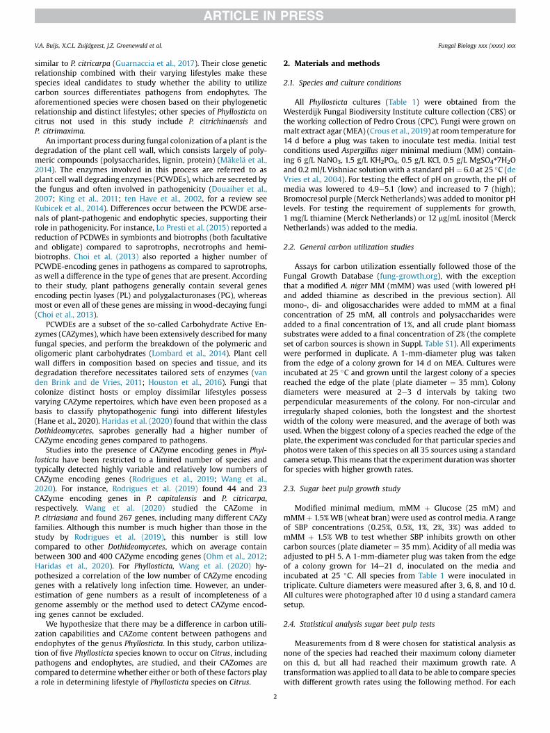
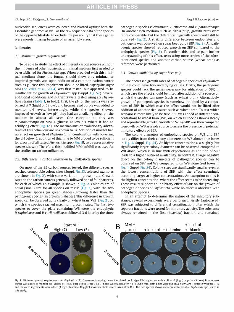
To be able to study the effect of different carbon sources withoutthe influence of other nutrients, a minimal medium first needed tobe established for Phyllosticta spp. When provided with this mini-mal medium alone, the fungus should show only minimal orimpaired growth, and upon addition of a common carbon sourcesuch as glucose this impairment should be lifted. Aspergillus nigerMM (de Vries et al., 2004) was first tested, but appeared to beinsufficient for growth of Phyllosticta spp (Suppl. Fig. S2). Severaladditional conditions and nutrients were tested using six Phyllos-ticta strains (Table 1, in bold). First, the pH of the media was sta-bilized at 7 (high) or 5 (low), and bromocresol purple was added tomonitor pH levels. Interestingly, while the species showedimproved growth at low pH, they had an alkalizing effect on themedium in almost all cases. One exception to this wasP. paracitricarpa on MM þ glucose at low pH, where it had anacidifying effect (Fig. 1A). The mechanisms or evolutionary advan-tages of this behaviour are unknown to us. Addition of inositol hadno effect on growth of Phyllosticta. In combination with loweringthe pH below 5, addition of thiamine to MM proved to be sufficientfor growth of all tested Phyllosticta spp. (Fig. 1B, two representativespecies shown). Therefore, this modified MM (mMM) was used forthe studies on carbon utilization.
3.2. Differences in carbon utilization by Phyllosticta species
On most of the 35 carbon sources tested, the different speciesreached comparable colony sizes (Suppl. Fig. S3, selected examplesare shown in Fig. 2), with some variation in growth rate. Growthrates on the carbon sources generally followed one of four patterns,for each of which an example is shown in Fig. 2. Colonies are ofequal (small) size for all species on mMM (Fig. 2), with the twoendophytic species (in green shades) growing faster than thepathogenic species (in brownish shades). This difference in growthspeed can be observed quite clearly onwheat bran (WB) (Fig. 2), onwhich the species reached maximum growth rates. The first twospecies to cover the plate containing WB were the endophyticP. capitalensis and P. citribraziliensis, followed 3 d later by the three
Fig. 1. Minimum growth requirements for Phyllosticta (A). One-mm-diam plugs were inocupurple was added to monitor pH (yellow pH < 5.5, purple/blue ¼ pH > 6.6). Photos were takand indicated ingredients were added (1 mg/L thiamine, 12 mg/mL inositol). Photos were takthis study.
4
pathogenic species P. citriasiana, P. citricarpa and P. paracitricarpa.On another rich medium such as citrus pulp, growth rates weremore comparable, but the difference in growth speed could still beobserved (Fig. 2). A striking difference between endophytes andpathogens was observed on sugar beet pulp (SBP, Fig. 2). All path-ogenic species showed reduced growth on SBP compared to theendophytic species (Fig. 3). To confirm this, and to gain furtherunderstanding of this effect, tests using more strains of the afore-mentioned species and another carbon source (wheat bran) asreference were performed.
3.3. Growth inhibition by sugar beet pulp
The decreased growth rates of pathogenic species of Phyllostictaon SBP could have two underlying causes. Firstly, the pathogenicspecies could lack the genes necessary for utilization of SBP, inwhich case the effect should be lifted after addition of a source onwhich the species can grow (such as wheat bran). Alternatively,growth of pathogenic species is somehow inhibited by a compo-nent of SBP, in which case the effect would not be lifted afteraddition of another rich source such as wheat bran. To test whichscenario is more likely to be true, SBP was added at different con-centrations to wheat bran (WB) on which all species show a steadyand reproducible growth. Growth onWBþ SBP was then comparedto growth onWB as a sole source to assess the presence of potentialinhibitory effects of SBP.
The colony diameters of endophytic species on WB and SBPhardly differ from their colony diameters on WB alone (blue boxesin Fig. 4, Suppl. Fig. S4). At higher concentrations, a slightly butsignificantly larger colony diameter can be observed compared toWB alone, which is in line with expectations as addition of SBPleads to a higher nutrient availability. In contrast, a large negativeeffect on the colony diameters of pathogenic species can beobserved on SBP and WB compared to on WB alone (red boxes inFig. 4, Suppl. Fig. S4). Colony sizes are significantly smaller even atthe lowest concentrations of SBP, with the effect seeminglybecoming larger at higher concentrations. An exception to this isthe highest concentration, where the effect seems to be decreasing.These results support an inhibitory effect of SBP on the growth ofpathogenic species of Phyllosticta, while no effect is observed withendophytic species.
In an attempt to determine the nature of the inhibitory sub-stance, several experiments were performed. Firstly (autoclaved)SBP was subjected to differential centrifugation, after which theseparate fractions were tested for inhibitory activity. The substancealways remained in the first (heaviest) fraction, and remained
lated on A. niger MM þ glucose with a pH ¼ ~7 (high) or pH ¼ ~5 (low). Bromocresolen after 7 d (B). One-mm-diam plugs were put on A. niger MM þ glucose with pH ¼ ~5,en after 11 d. The two species shown are representative of all Phyllosticta spp. tested in

Fig. 2. Colony diameters of Phyllosticta spp. illustrated on four carbon sources. When the biggest colony of a species reached the edge of the plate, the experiment was concluded forthat particular species (on all carbon sources). As a result, because endophytes grew faster, their experiments were concluded earlier. All measurements were done in duplicate.However, one of the P. paracitricarpa cultures on citrus pulp failed to grow. As we considered this to be due to technical reasons, P. paracitricarpa measurements on this source wereomitted from the figure. Lines show averages with variance indicated with bars. All sources shown in the figure were added to an end concentration of 3%.
Fig. 3. Variable growth of Phyllosticta spp. on sugar beet pulp. Photos were taken after the number of d indicated below the figure. Species are ordered by lifestyle: endophytic onthe left (grey background), pathogenic on the right (black background). Plate diameter ¼ 35 mm.
V.A. Buijs, X.C.L. Zuijdgeest, J.Z. Groenewald et al. Fungal Biology xxx (xxxx) xxx
active, indicating that it is either heat-tolerant or somehow pro-tected. Secondly, two methods commonly used for extraction ofbio-active compounds, namely ethyl-acetate extraction and n-
5
hexane extraction, were used in an attempt to isolate the inhibitorysubstance. Again, the separate fractions were tested for inhibitoryactivity, which was detected for the pellet only, indicating that
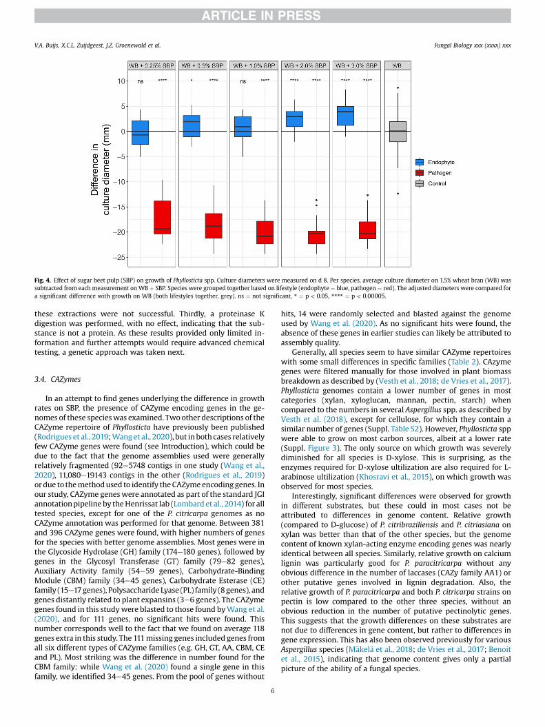
Fig. 4. Effect of sugar beet pulp (SBP) on growth of Phyllosticta spp. Culture diameters were measured on d 8. Per species, average culture diameter on 1.5% wheat bran (WB) wassubtracted from each measurement on WB þ SBP. Species were grouped together based on lifestyle (endophyte ¼ blue, pathogen ¼ red). The adjusted diameters were compared fora significant difference with growth on WB (both lifestyles together, grey). ns ¼ not significant, * ¼ p < 0.05, **** ¼ p < 0.00005.
V.A. Buijs, X.C.L. Zuijdgeest, J.Z. Groenewald et al. Fungal Biology xxx (xxxx) xxx
these extractions were not successful. Thirdly, a proteinase Kdigestion was performed, with no effect, indicating that the sub-stance is not a protein. As these results provided only limited in-formation and further attempts would require advanced chemicaltesting, a genetic approach was taken next.
3.4. CAZymes
In an attempt to find genes underlying the difference in growthrates on SBP, the presence of CAZyme encoding genes in the ge-nomes of these specieswas examined. Twoother descriptions of theCAZyme repertoire of Phyllosticta have previously been published(Rodrigues et al., 2019;Wanget al., 2020), but in both cases relativelyfew CAZyme genes were found (see Introduction), which could bedue to the fact that the genome assemblies used were generallyrelatively fragmented (92e5748 contigs in one study (Wang et al.,2020), 11,080e19143 contigs in the other (Rodrigues et al., 2019)ordue to themethodused to identify theCAZymeencodinggenes. Inour study, CAZyme geneswere annotated as part of the standard JGIannotationpipeline by theHenrissat lab (Lombard et al., 2014) for alltested species, except for one of the P. citricarpa genomes as noCAZyme annotation was performed for that genome. Between 381and 396 CAZyme genes were found, with higher numbers of genesfor the species with better genome assemblies. Most genes were inthe Glycoside Hydrolase (GH) family (174e180 genes), followed bygenes in the Glycosyl Transferase (GT) family (79e82 genes),Auxiliary Activity family (54e59 genes), Carbohydrate-BindingModule (CBM) family (34e45 genes), Carbohydrate Esterase (CE)family (15e17genes), Polysaccharide Lyase (PL) family (8genes), andgenes distantly related to plant expansins (3e6 genes). The CAZymegenes found in this studywere blasted to those found byWang et al.(2020), and for 111 genes, no significant hits were found. Thisnumber corresponds well to the fact that we found on average 118genes extra in this study. The 111missing genes included genes fromall six different types of CAZyme families (e.g. GH, GT, AA, CBM, CEand PL). Most striking was the difference in number found for theCBM family: while Wang et al. (2020) found a single gene in thisfamily, we identified 34e45 genes. From the pool of genes without
6
hits, 14 were randomly selected and blasted against the genomeused by Wang et al. (2020). As no significant hits were found, theabsence of these genes in earlier studies can likely be attributed toassembly quality.
Generally, all species seem to have similar CAZyme repertoireswith some small differences in specific families (Table 2). CAzymegenes were filtered manually for those involved in plant biomassbreakdown as described by (Vesth et al., 2018; de Vries et al., 2017).Phyllosticta genomes contain a lower number of genes in mostcategories (xylan, xyloglucan, mannan, pectin, starch) whencompared to the numbers in several Aspergillus spp. as described byVesth et al. (2018), except for cellulose, for which they contain asimilar number of genes (Suppl. Table S2). However, Phyllosticta sppwere able to grow on most carbon sources, albeit at a lower rate(Suppl. Figure 3). The only source on which growth was severelydiminished for all species is D-xylose. This is surprising, as theenzymes required for D-xylose ultilization are also required for L-arabinose ultilization (Khosravi et al., 2015), on which growth wasobserved for most species.
Interestingly, significant differences were observed for growthin different substrates, but these could in most cases not beattributed to differences in genome content. Relative growth(compared to D-glucose) of P. citribraziliensis and P. citriasiana onxylan was better than that of the other species, but the genomecontent of known xylan-acting enzyme encoding genes was nearlyidentical between all species. Similarly, relative growth on calciumlignin was particularly good for P. paracitricarpa without anyobvious difference in the number of laccases (CAZy family AA1) orother putative genes involved in lignin degradation. Also, therelative growth of P. paracitricarpa and both P. citricarpa strains onpectin is low compared to the other three species, without anobvious reduction in the number of putative pectinolytic genes.This suggests that the growth differences on these substrates arenot due to differences in gene content, but rather to differences ingene expression. This has also been observed previously for variousAspergillus species (M€akel€a et al., 2018; de Vries et al., 2017; Benoitet al., 2015), indicating that genome content gives only a partialpicture of the ability of a fungal species.

Table 2Number of genes containing specific CAZy family modules in Phyllosticta (obtained from JGI Mycocosm).
Phyllosticta capitalensis Phyllosticta citribraziliensis Phyllosticta citriasiana Phyllosticta paracitricarpa Phyllosticta citricarpa
Total 396 382 382 382 381Auxiliary Activity 58 59 55 54 54Carbohydrate-Binding Module 45 34 39 42 42Carbohydrate Esterase 15 17 16 16 15Distantly related to plant expansins 6 5 3 4 3Glycosyl Hydrolase 180 178 177 174 175Glycosyl Transferase 82 79 82 82 82Polysaccharide Lyase 8 8 8 8 8
V.A. Buijs, X.C.L. Zuijdgeest, J.Z. Groenewald et al. Fungal Biology xxx (xxxx) xxx
To gain a more precise understanding of the CAZyme repertoireof these species and to assess whether there are differences be-tween endophytes and pathogens, all gene annotations from theavailable Phyllosticta genomes were grouped into Ortholog Groups(OGs) using orthofinder. Ortholog Groups containing annotatedCAZyme genes were selected, and the number of genes annotatedas CAZyme in each OG was counted for each species (for details seemethods). Nearly all OGs contained similar or even the exact samenumber of CAZyme genes for each species (Suppl. Table S2). In sixcases, an OG contained consistently more CAZyme genes in path-ogens than in endophytes, or vice versa. However, as CAZyme geneswere annotated for only one of the two P. citricarpa genomes, butboth P. citricarpa genomes were included in the OG analysis, it wasnecessary to check whether the second P. citricarpa genome con-tained a similar number of genes in these OGs. In two OGs, this wasnot the case; the other four OGs and the putative function of thegenes contained in them are listed in Table 3. These OGs includedgenes from the CBM18 family, which contain a chitin-bindingdomain, the GT31 family potentially involved in cell wall remod-eling and the GH3 family which might play a role in cellulosedegradation. The genes specific to one lifestyle formed a distinctclade from the other genes of the OG when assembled into aphylogenetic tree (Suppl. Fig. S5). The missing genes were notfound when blasted against both the assembled genomes as well asthe raw sequence data, indicating they are not missing due to anassembly error.
4. Discussion and conclusion
In this study, carbon utilization of Phyllosticta spp. occurring onCitrus was examined with particular attention to differences be-tween endophytic and pathogenic species.
Firstly, the minimum growth requirements were established;Phyllosticta spp. have a thiamine dependency and only growproperly on a minimal medium supplemented with thiamine andat a low pH (~5). Wang et al. (2019) studied the effect of combi-nations of several nutrients on conidial germination and appres-sorium formation of Phyllosticta citricarpa. Their results suggestthat absence of thiamine reduces appressorium formation withapproximately 20%, an effect that becomes much stronger in theabsence of salts (40%). They found no significant effect of solely
Table 3CAZyme genes in OGs with consistent differences between pathogens and endophytes.
OG number Predicted function G
OG2 GH3a 9OG3 AA1/3, CBM18b 8OG36 GT31c 3OG468 CBM18d 1
a Glycoside Hydrolase family 3.b Auxiliary activity family 1, subfamily 3 and Carbohydrate Binding Module family 18c Glycosyl Transferase family 31.d Carbohydrate Binding Module family 18.
7
thiamine on conidial germination, but in absence of citric acid theaddition of thiamine had a positive effect on both conidial germi-nation and appressorium formation. These results suggest thatthiamine is involved in several complex processes in Phyllosticta,but what role it exactly plays in mycelial growth is unknown.Ustilago esculenta, a phytopathogenic species inducing edible gallsin its host Zizania latifolia, has been reported to exhibit an obligaterequirement for thiamine (Chung and Zheng, 2009), but furtherreports on the requirement of phytopathogenic species are verylimited. A thiamine dependency of Phyllosticta spp. is not entirelysurprising; many citrus fruits including oranges contain high levelsof thiamine (Bailey and Thomas, 1942). We therefore hypothesizethat the fungus has thiamine at its disposal in its natural sur-roundings and has lost the need and ability to produce it. The mostimportant genes in thiamine biosynthesis seem to be present in thegenomes of Phyllosticta spp. The inability to produce thiamine istherefore likely either the result of an unidentified mutation or dueto altered transcriptional regulation.
All the species show improved growth at low pH (pH 5), whilethey are able to grow at a broader range. All species have beenisolated from multiple host tissues, e.g. citrus leaves and citrusfruits. Citrus fruits are known to be acidic as they contain both citricacid and ascorbic acid. Therefore, it is quite possible that the pref-erence for a low pH is an adaptation to growing in the acidic hostfruit. These results are also in line with those of Wang et al. (2019),who observed that a pH between 3 and 5 is optimal for conidialgermination and appressorium formation. Interestingly, despite agrowth preference for lowpH, Phyllosticta spp. generally alkalinizedthe surrounding media. Many phytopathogenic fungi are known toeither acidify or alkalize their host environment (Alkan et al., 2013;Vylkova, 2017). Some species, such as Botrytis cinerea, releasespecialized sets of pathogenicity factors depending on the pH of thesurroundings (Manteau et al., 2003). Furthermore, studies haveshown that for certain species, such as Plenodomus lingam (Lep-tosphaeria maculans) and Sclerotinia sclerotiorum, the observedalkalinisation or acidification is directly related to canker length(Bousset et al., 2019). Several species of Colletotrichum are alsoknown for preferring an environment with low pH, but have analkalising effect on the surrounding host tissue (Alkan et al., 2013;Shnaiderman et al., 2013). For some of these, this alkalising effecthas been shown to be directly correlated to pathogenicity (Alkan
ene number in endophytes Gene number in pathogens
8740
.

V.A. Buijs, X.C.L. Zuijdgeest, J.Z. Groenewald et al. Fungal Biology xxx (xxxx) xxx
et al., 2008; Miyara et al., 2010). Whether a similar mechanismoccurs in planta in Phyllosticta spp. needs to be addressed in futurestudies.
In general, all species show similar growth on 35 carbon sources,although a few differences were observed. Endophytic speciesappear to have higher growth rates than pathogenic species on allcarbon sources, which is in line with the earlier finding thatP. capitalensis grows faster than P. citricarpa (Baayen et al., 2002;Glienke et al., 2011). Small differences in growth, which wereobserved on several carbon sources, could be due to strain variationrather than species variation. A much broader sampling using amuch larger number of globally distributed strains is needed to testwhether these small differences are significant. The results of thegrowth profiles generally correspond well to what the specieswould be expected to rely on in their natural habitat. The speciesare often found on both the citrus fruit peel as well as citrus leaves,and sometimes twigs. On rich sources, such as citrus pulp, wheatbran, and cotton seed pulp, all species grew well, as expected. Anexception is sugar beet pulp, which is discussed extensively in alater paragraph. Citrus peel contains large amounts of pectin,hemicellulose and cellulose, the latter two of which are present insmaller amounts than the first (Ting and Deszyck, 1961; Shan,2016). All species grew well on pectin, and better on citrus pectincompared to apple pectin, which is to be expected for species withcitrus as host. On cellulose as a sole source, most species grewpoorly. Citrus leaves contain large amounts of starch, as well assucrose, glucose, and fructose (the latter three to a lesser extentthan the first) (Arbona et al., 2005; Iglesias et al., 2002, 2006). Allspecies grew well on starch, with pathogens showing a slightlydecreased growth rate compared to endophytes, and medium towell on glucose, fructose and sucrose.
The most closely related species for which a similar growthprofiling has been performed is Sphaeropsis sapinea (www.fung-growth.org). Interestingly, similarly to Phyllosticta, this speciesshows reduced growth on D-xylose, while growth on L-arabinosewas unaffected. This is surprising as utilization of both sourcesdepends on nearly the same set of enzymes. The first step of theconversion of L-arabinose and D-xylose is mediated by a set of threepentose reductases in A. niger (Chroumpi et al., 2021), whichhowever have different affinities for the two pentose sugars (Ter-ebieniec et al., unpublished data). Possibly the reductase in Phyl-losticta has a low affinity for D-xylose, causing the reduced growthon D-xylose. Alternatively, Phyllosticta may have a poor uptakeability for D-xylose. It could be that the molecular basis of thereduced D-xylose growth is shared within the Bortyosphaerialesand is not specific to the genus Phyllosticta. However, to be able todetermine the extent of this effect, wider and more extensivesampling within the fungal order is required.
An inhibitory effect of SBP on pathogenic species was observed,also in presence of another carbon source (WB) and at low con-centrations (0.25%). Which compound is responsible for this effectis unknown. Although a slight decrease in growth rate is observedfor pathogens on starch, we consider this substance an unlikelycandidate for causing the inhibitory effect, as SBP generally con-tains less starch compared to wheat bran (Onipe et al., 2015;Palmgren Karlsson et al., 2002) and a strong inhibitory effect is evenobserved on 0.25% SBP compared to 1% starch (data not shown). Asthe inhibitory effect was not observed on citrus pulp, it is not likelyto be caused by pectin or cellulose, but rather a trace component ofSBP. Trace elements such as iron (Fe), zinc (Zn), copper (Cu) andmanganese (Mn) can have inhibitory effects when present in higherconcentrations than optimum for the tested fungi (Thind andMandahar, 1968). Sugar beet pulp has been shown to containrelatively high levels of Fe as well as several other trace elements(�Skrbi�c et al., 2010; Organisation for Economic Co-operation and
8
Development, 2002). However, the aforementioned trace elementshave also been detected in wheat bran (Yu et al., 2018), whichimplies that the effect would be dose-dependent, and it is also verylikely that the set and concentration of trace elements is stronglybatch-dependent. To determine whether these or other elementsare responsible for the inhibitory effect would therefore require anexact determination of the levels of the complete set of trace ele-ments in both substances, and a detailed comparison to identifycandidate compounds that may be responsible for the growth in-hibition. This is a challenging task that requires specific expertise,but it could prove very worthwhile in future studies. Furthermore,optimum concentrations of the trace elements differ widely fordifferent fungi and have not been determined for Phyllosticta spp.
Several experiments were performed in an attempt to identifythe inhibitory substance, but with only limited results. Proteinase Kdigestion had no effect on the activity of the substance, indicatingthat it is not a protein. As the substance remains active afterautoclaving, it is likely either thermotolerant, or protected by, forinstance, embedment in rigorous cell wall components. Ethyl ace-tate and hexane extractions were not effective at extracting theinhibiting substance. After differential centrifugation, the sub-stance consistently remains in the first fraction, even after spinningdown at only 100 g. This indicates that the substance is likely eitherbound to something of substantial mass, or highly hydrophobic;perhaps it is associated with the cell wall or cell membrane. Todetermine the exact nature of the inhibitory substance, a preciseanalysis of the components present in SBP should first be per-formed in the future, after which the different components couldbe tested separately and in varying combinations.
At the highest concentration tested (3%), the inhibitory effectseemed to be decreasing. It is possible that the large increase innutrient availability starts compensating for the inhibitory effect atthis concentration. Interestingly, sugar cane residue was also re-ported to have an application in diseasemanagement of P. citricarpa(van Bruggen et al., 2017). Whether this is due to a similar mech-anism is unclear.
Furthermore, we studied the presence of genes related to thecarbon utilization capabilities of Phyllosticta spp. by analysing theCAZome of these species. Previous studies reported CAZymeencoding gene counts in Phyllosticta between 23 and 267 genes,which is low compared to other Dothideomycetes, which generallycontain between 300 and 400 CAZyme genes (Haridas et al., 2020;Ohm et al., 2012; Rodrigues et al., 2019; Wang et al., 2020). In thisstudy, we found much higher numbers of CAZyme encoding genes(381e396), which is in line with what is reported for other Dothi-deomycetes. The genome assemblies used in this study seem to bemore complete than those used in previous studies. Several of thegenes found in this study were blasted to the genome used byWang et al. (2020), and no significant hits were found.We thereforethink these data on CAZyme genes provide amore accurate accountof what is present in these species.
Some notes on the method of generating gene counts in thisstudy should be taken into account when comparing to gene countsgenerated by other pipelines. Genes are annotated as CAZymewhen they possess a certain signature; as genes can possess morethan one signature, some genes are included in more than oneannotation. In fact, the numbers on the JGI Mycocosm table reflectthe number of genes carrying a specific CAZy module, not theunique number of genes that have any CAZy module, which maycause differences with other approaches for CAZyme annotation.For instance, in the Auxiliary Activity family of P. capitalensis CBS128856, two genes received a double annotation: 299,199 and255,817, as they each contain the modules belonging to twodifferent families. Functional research into these genes could pro-vide further knowledge about their functions.
V.A. Buijs, X.C.L. Zuijdgeest, J.Z. Groenewald et al. Fungal Biology xxx (xxxx) xxx
In contrast with earlier studies, we did not find a difference inthe number of putative PCWDE genes between species withdifferent lifestyles within this genus. The different Phyllostictaspecies assessed in this study possess similar CAZyme gene reper-toires, with only small differences. Phyllosticta genomes containedfewer CAZyme genes for breakdown of plant biomass categorieswhen compared to the numbers in several Aspergillus spp. asdescribed by Vesth et al. (2018), with the exception of cellulose, forwhich they contain a similar number of genes. The species ofAspergillus studied by Vesth et al. (2018) are very diverse, includingstrains isolated from several different kinds of biomass, but Asper-gillus species typically have a broad substrate range, which likelyexplains their larger set of CAZymes. This is not clearly reflected inthe ability of Phyllosticta spp. to grow on a range of carbon sources,as the only source onwhich growth was severely diminished for allspecies was xylose. Other small but significant differences ingrowth on several sources were observed, but none could beattributed to a change in gene number. The ability to degradebiomass is therefore likely transcriptionally regulated.
In four cases, an OG was found which consistently contained adifferent number of genes in endophytes as compared to pathogens(Table 3). The lifestyle-specific genes formed a separate clade fromother genes in the OG when assembled into a phylogenetic tree,indicating they are different to some extent from the other genes inthe OGs (Suppl. Fig. S5). Although further research is necessary toconfirm a role in pathogenicity for these genes, it is interesting tospeculate about their putative functions.
Two of these OGs contain genes belonging to the CBM18 family(OG3 and OG468). CBM18 consists of enzymes containing a chitin-binding domain. Genes from this family have been hypothesized toprevent the activation of a host defence response by disguisingfungal chitin in the two pathogenic fungal species Batrachochytriumdendrobatidis and Verticillium nonalfalfae (Liu and Stajich, 2015;Volk et al., 2019). A similar mechanism has been described for anendophytic Pestalotiopsis sp. by Landwehr et al. (2016). Deter-mining whether such a mechanism is also present in Phyllostictawould be an interesting subject for future studies. Genes in OG36belong to the GT31 family, and are likely important for cell wallremodelling. However, nothing is known about a potential role inpathogenicity for this gene family. Genes in OG2 belong to the GH3family and are predicted to be b-glucosidases, important in cellu-lose degradation.
Overall, very few differences were found in the CAZome ofPhyllosticta species with different lifestyles. Only four familiesshowed consistent differences between pathogens and endophytesin this study, which are therefore interesting subjects for futurestudies. In general, Phyllosticta species with different lifestyles alsoshowed only small variations in their ability to utilize differentcarbon sources, with two exceptions. Firstly, all endophytes grewfaster than all pathogens. Secondly, growth of all pathogens wassignificantly inhibited by addition of sugar beet pulp to the me-dium. Despite attempts to identify the specific substance that isresponsible for the inhibitory effect of sugar beet pulp, the sub-stance and the mechanism associated with its inhibitory effectremain unknown. It is important to note that, while few differencesin gene repertoire were found, variations could very well occur onthe transcriptional level. Future studies on transcriptomic data ofPhyllosticta spp. could be of great value in creating a better un-derstanding the difference in lifestyles between these species, andcould provide important clues into further understanding theirpathogenicity.
9
Acknowledgement
This work was funded by the Dutch Applied Science division(TTW) of NWO and the Technology Program of the Ministry ofInfrastructure and Water Management under project 15807 of theResearch Programme I&W Biotechnology and Safety.
Appendix A. Supplementary data
Supplementary data to this article can be found online athttps://doi.org/10.1016/j.funbio.2021.05.003.
References
Alkan, N., Fluhr, R., Sherman, A., Prusky, D., 2008. Role of ammonia secretion and pHmodulation on pathogenicity of Colletotrichum coccodes on tomato fruit. Mol.Plant Microbe Interact. 21, 1058e1066.
Alkan, N., Espeso, E.A., Prusky, D., 2013. Virulence regulation of phytopathogenicfungi by pH. Antioxidants Redox Signal. 19, 1012e1025.
Arbona, V., Marco, A.J., Iglesias, D.J., L�opez-Climent, M.F., Talon, M., G�omez-Cadenas, A., 2005. Carbohydrate depletion in roots and leaves of salt-stressedpotted Citrus clementina L. Plant Growth Regul. 46, 153e160.
Baayen, R.P., Bonants, P.J.M., Verkley, G., Carroll, G.C., van der Aa, H.A., deWeerdt, M., van Brouwershaven, I.R., Schutte, G.C., Maccheroni, W., deBlanco, C.G., Azevedo, J.L., 2002. Non-pathogenic isolates of the Citrus BlackSpot fungus, Guignardia citricarpa, identified as a cosmopolitan endophyte ofwoody plants, G. mangiferae (Phyllosticta capitalensis). Phytopathology 92,464e477.
Bailey, M.I., Thomas, A.W., 1942. The thiamine and riboflavin contents of citrusfruits. J. Nutr. 24, 85e92.
Benoit, I., Culleton, H., Zhou, M., DiFalco, M., Aguilar-Osorio, G., Battaglia, E.,Bouzid, O., Brouwer, C.P.J.M., El-Bushari, H.B.O., Coutinho, P.M., Gruben, B.S.,Hild�en, K.S., Houbraken, J., Jim�enez Barboza, L.A., Levasseur, A., Majoor, E.,M€akel€a, M.R., Narang, H.M., Trejo-Aguilar, B., van den Brink, J., vanKuyk, P.A.,Wiebenga, A., McKie, V., McCleary, B., Tsang, A., Henrissat, B., de Vries, R.P.,2015. Closely related fungi employ diverse enzymatic strategies to degradeplant biomass. Biotechnol. Biofuels 8, 107.
Bousset, L., Ermel, M., Soglonou, B., Husson, O., 2019. A method to measure redoxpotential (Eh) and pH in agar media and plants shows that fungal growth isaffected by and affects pH and Eh. Fungal Biology 123, 117e124.
Brentu, F.C., Oduro, K.A., Offei, S.K., Odamtten, G.T., Vicent, A., Peres, N.A.,Timmer, L.W., 2012. Crop loss, aetiology, and epidemiology of citrus black spotin Ghana. Eur. J. Plant Pathol. 133, 657e670.
Choi, J., Kim, K.-T., Jeon, J., Lee, Y.-H., 2013. Fungal plant cell wall-degrading enzymedatabase: a platform for comparative and evolutionary genomics in fungi andOomycetes. BMC Genom. 14, S7.
Chroumpi, T., Peng, M., Aguilar Pontes, M.V., Müller, A., Wang, M., Yan, J., Lipzen, A.,Ng, V., Grigoriev, I.V., M€akel€a, M.R., de Vries, R.P., 2021. Revisiting a ‘simple’fungal metabolic pathway reveals redundancy, complexity and diversity. Mi-crobial Biotechnology in press.
Chung, K., Tzeng, D., 2009. Thiamine (vitamin B1) plays a critical role on sugarutilization by the phytopathogenic fungus, Ustilago esculenta. Res. J. Microbiol.4, 178e185.
Cord-Landwehr, S., Melcher, R.L.J., Kolkenbrock, S., Moerschbacher, B.M., 2016.A chitin deacetylase from the endophytic fungus Pestalotiopsis sp. efficientlyinactivates the elicitor activity of chitin oligomers in rice cells. Sci. Rep. 6, 1e11.
Crous, P.W., Verkley, G.J.M., Groenewald, J.Z., Samson, R.A. (Eds.), 2019. WesterdijkLaboratory Manual Series 1: Fungal Biodiversity, second ed. Westerdijk FungalBiodiversity Institute, Utrecht, The Netherlands.
de Vries, R.P., Burgers, K., van de Vondervoort PJI, Frisvad, J.C., Samson, R.A.,Visser, J., 2004. A new black Aspergillus species, A. vadensis, is a promising hostfor homologous and heterologous protein production. Appl. Environ. Microbiol.70, 3954e3959.
de Vries, R.P., Riley, R., Wiebenga, A., Aguilar-Osorio, G., Amillis, S., AkemiUchima, C., Anderluh, G., Asadollahi, M., Askin, M., Barry, K., Battaglia, E.,€O, Bayram, Benocci, T., Braus-Stromeyer, S.A., Caldana, C., C�anovas, D.,Cerqueira, G.C., Chen, F., Chen, W., Choi, C., Clum, A., Correa dos Santos, R.A., deLima Dam�asio, A.R., Diallinas, G., Emri, T., Fekete, E., Flipphi, M., Freyberg, S.,Gallo, A., Gournas, C., Habgood, R., Haimaut, M., Harispe, L., Henrissat, B.,Hild�en, K.S., Hope, R., Hossain, A., Karabika, E., Karaffa, L., Kar�anyi, Z.,Kra�sevec, N., Kuo, A., Kusch, H., LaButti, K., Lagendijk, E.L., Lapidus, A.,Levasseur, A., Lindquist, E., Lipzen, A., Logrieco, A.F., MacCabe, A., M€akel€a, M.R.,Malavazi, I., Melin, P., Meyer, V., Mielnichuk, N., Miskei, M., Moln�ar, A.P.,Mul�e, G., Ngan, C.Y., Orejas, M., Orosz, E., Ouedraogo, J.P., Overkamp, K.M.,Park, H.-S., Perrone, G., Piumi, F., Punt, P., Ram, A.F.J., Ram�on, A., Rauscher, S.,Record, E., Ria~no-Pach�on, D.M., Robert, V., R€ohrig, Ruller R., Salamov, A.,

V.A. Buijs, X.C.L. Zuijdgeest, J.Z. Groenewald et al. Fungal Biology xxx (xxxx) xxx
Salih, N.S., Samson, R.A., S�andor, E., Sanguinetti, M., Schütze, T., Sep�ci�c, K.,Shelest, E., Sherlock, G., Sophianopoulou, V., Squina, F.M., Sun, H., Susca, A.,Todd, R.B., Tsang, A., Unkles, S.E., van de Wiele, N., van Rossen-Uffink, D.,Velasco de Castro Oliveira, J., Vesth, T.C., Visser, J., Yu, J.-H., Zhou, M.,Andersen, M.R., Archer, D.B., Baker, S.E., Benoit, I., Brakhage, A.A., Braus, G.H.,Fischer, R., Frisvad, J.C., Goldman, G.H., Houbraken, J., Oakley, B., P�ocsi, I.,Scazzocchio, C., Seiboth, vanKuyk PA., Wortman, J., Dyer, P.S., Grigoriev, I.V.,2017. Comparative genomics reveals high biological diversity and specific ad-aptations in the industrially and medically important fungal genus Aspergillus.Genome Biol. 18, 28.
Douaiher, M.-N., Nowak, E., Durand, R., Halama, P., Reignault, P., 2007. Correlativeanalysis of Mycosphaerella graminicola pathogenicity and cell wall-degradingenzymes produced in vitro: the importance of xylanase and poly-galacturonase. Plant Pathol. 56, 79e86.
Emms, D., Kelly, S., 2019. OrthoFinder: phylogenetic orthology inference forcomparative genomics. Genome Biol. 20, 238.
Eust�aquio Lanza, F., Germano Metzker, T., Vinhas, T., Behlau, F., Jos�e Silva Junior, G.,2018. Critical fungicide spray period for citrus black spot control in s~ao Paulostate, Brazil. Plant Dis. 102, 334e340.
Glienke, C., Pereira, O.L., Stringari, D., Fabris, J., Kava-Cordeiro, V., Galli-Terasawa, L.,Cunnington, J., Shivas, R.G., Groenewald, J.Z., Crous, P.W., 2011. Endophytic andpathogenic Phyllosticta species, with reference to those associated with CitrusBlack Spot. Persoonia 26, 47e56.
Guarnaccia, V., Groenewald, J.Z., Li, H., Glienke, C., Carstens, E., Hattingh, V.,Fourie, P.H., Crous, P.W., 2017. First report of Phyllosticta citricarpa anddescription of two new species, P. paracapitalensis and P. paracitricarpa, fromcitrus in Europe. Stud. Mycol. 87, 161e185.
Guarnaccia, V., Gehrmann, T., Silva-Junior, G.J., Fourie, P.H., Haridas, S., Vu, D.,Spatafora, J., Martin, F.M., Robert, V., Grigoriev, I.V., Groenewald, J.Z., Crous, P.W.,2019. Phyllosticta citricarpa and sister species of global importance to Citrus.Mol. Plant Pathol. 20, 1619e1635.
Haridas, S., Albert, R., Binder, M., Bloem, J., LaButti, K., Salamov, A., Andreopoulos, B.,Baker, S.E., Barry, K., Bills, G., Bluhm, B.H., Cannon, C., Castanera, R., Culley, D.E.,Daum, C., Ezra, D., Gonz�alez, J.B., Henrissat, B., Kuo, A., Liang, C., Lipzen, A.,Lutzoni, F., Magnuson, J., Mondo, S.J., Nolan, M., Ohm, R.A., Pangilinan, J.,Park, H.-J., Ramírez, L., Alfaro, M., Sun, H., Tritt, A., Yoshinaga, Y., Zwiers, L.-H.,Turgeon, B.G., Goodwin, S.B., Spatafora, J.W., Crous, P.W., Grigoriev, I.V., 2020.101 Dothideomycetes genomes: a test case for predicting lifestyles and emer-gence of pathogens. Stud. Mycol. 96, 141e153.
Houston, K., Tucker, M.R., Chowdhury, J., Shirley, N., Little, A., 2016. The plant cellwall: a complex and dynamic structure as revealed by the responses of genesunder stress conditions. Front. Plant Sci. 7, 984.
Iglesias, D.J., Lliso, I., Tadeo, F.R., Talon, M., 2002. Regulation of photosynthesisthrough source: sink imbalance in citrus is mediated by carbohydrate contentin leaves. Physiol. Plantarum 116, 563e572.
Iglesias, D.J., �A, Calatayud, Barreno, E., Primo-Millo, E., Talon, M., 2006. Responses ofcitrus plants to ozone: leaf biochemistry, antioxidant mechanisms and lipidperoxidation. Plant Physiol. Biochem. 44, 125e131.
Khosravi, C., Benocci, T., Battaglia, E., Benoit, I., de Vries, R.P., 2015. Sugar catabolismin Aspergillus and other fungi related to the utilization of plant biomass. Adv.Appl. Microbiol. 90, 1e28.
King, B.C., Waxman, K.D., Nenni, N.V., Walker, L.P., Bergstrom, G.C., Gibson, D.M.,2011. Arsenal of plant cell wall degrading enzymes reflects host preferenceamong plant pathogenic fungi. Biotechnol. Biofuels 4, 1e14.
Kotz�e, J.M., 1981. Epidemiology and control of citrus black spot in South Africa. PlantDis. 65, 945.
Kotz�e, J.M., 2000. Black spot. In: Timmer, L.W., Garnsey, S.M., Graham, J.H. (Eds.),Compendium of Citrus Diseases. American Phytopathological Society Press Inc,The American Phytopathological Society, pp. 23e25.
Kubicek, C.P., Starr, T.L., Glass, N.L., 2014. Plant cell wall-degrading enzymes andtheir secretion in plant-pathogenic fungi. Annu. Rev. Phytopathol. 52, 427e451.
Liu, P., Stajich, J., 2015. Characterization of the carbohydrate binding module 18 genefamily in the amphibian pathogen Batrachochytrium dendrobatidis. FungalGenet. Biol. 77, 31e39.
Lo Presti, L., Lanver, D., Schweizer, G., Tanaka, S., Liang, L., Tollot, M., Zuccaro, A.,Reissmann, S., Kahmann, R., 2015. Fungal effectors and plant susceptibility.Annu. Rev. Plant Biol. 66, 513e554.
Lombard, V., Golaconda Ramulu, H., Drula, E., Coutinho, P.M., Henrissat, B., 2014.The carbohydrate-active enzymes database (CAZy) in 2013. Nucleic Acids Res.42, D490eD495.
M€akel€a, M.R., Donofrio, N., de Vries, R.P., 2014. Plant biomass degradation by fungi.Fungal Genet. Biol. 72, 2e9.
M€akel€a, M.R., DiFalco, M., McDonnell, E., Nguyen, T.T.M., Wiebenga, A., Hild�en, K.,Peng, M., Grigoriev, I.V., Tsang, A., de Vries, R.P., 2018. Genomic and exopro-teomic diversity in plant biomass degradation approaches among Aspergilli.Stud. Mycol. 91, 79e99.
Manteau, S., Abouna, S., Lambert, B., Legendre, L., 2003. Differential regulation byambient pH of putative virulence factor secretion by the phytopathogenicfungus Botrytis cinerea. FEMS (Fed. Eur. Microbiol. Soc.) Microbiol. Ecol. 43,359e366.
10
Miyara, I., Shafran, H., Davidzon, M., Sherman, A., Prusky, D., 2010. pH regulation ofammonia secretion by Colletotrichum gloeosporioides and its effect on appres-sorium formation and pathogenicity. Mol. Plant Microbe Interact. 23, 304e316.
Ohm, R.A., Feau, N., Henrissat, B., Schoch, C.L., Horwitz, B.A., Barry, K.W.,Condon, B.J., Copeland, A.C., Dhillon, B., Glaser, F., Hesse, C.N., Kosti, I.,LaButti, K., Lindquist, E.A., Lucas, S., Salamov, A.A., Bradshaw, R.E., Ciuffetti, L.,Hamelin, R.C., Kema, G.H.J., Lawrence, C., Scott, J.A., Spatafora, J.W.,Turgeon, B.G., de Wit, P.J.G.M., Zhong, S., Goodwin, S.B., Grigoriev, I.V., 2012.Diverse lifestyles and strategies of plant pathogenesis encoded in the genomesof eighteen dothideomycetes fungi. PLoS Pathog. 8, e1003037.
Onipe, O.O., Jideani, A.I.O., Beswa, D., 2015. Composition and functionality of wheatbran and its application in some cereal food products. Int. J. Food Sci. Technol.50, 2509e2518.
Organisation for Economic Co-operation and Development, 2002. Series on theSafety of Novel Foods and Feeds No. 3. Consensus Document on CompositionalConsiderations for New Varieties of Sugar Beet: Key Food and Feed Nutrientsand Anti-nutrients. OECD Environmental Health and Safety Publications, Paris,France.
Palmgren Karlsson, C., Jansson, A., Ess�en-Gustavsson, B., Lindberg, J.E., 2002. Effectof molassed sugar beet pulp on nutrient utilisation and metabolic parametersduring exercise. Equine Vet. J. 34, 44e49.
Rodrigues, C.M., Takita, M.A., Silva, N.V., Ribeiro-Alves, M., Machado, M.A., 2019.Comparative genome analysis of Phyllosticta citricarpa and Phyllosticta capital-ensis, two fungi species that share the same host. BMC Genom. 20, 554.
Savary, S., Willocquet, L., Pethybridge, S.J., Esker, P., McRoberts, N., Nelson, A., 2019.The global burden of pathogens and pests on major food crops. Nature Ecologyand Evolution 3, 430e439.
Shan, Y., 2016. Functional components of citrus peel contents. ComprehensiveUtilization of Citrus By-Products, pp. 1e13.
Shnaiderman, C., Miyara, I., Kobiler, I., Sherman, A., Prusky, D., 2013. Differentialactivation of ammonium transporters during the accumulation of ammonia byColletotrichum gloeosporioides and its effect on appressoria formation andpathogenicity. Mol. Plant Microbe Interact. 26, 345e355.
�Skrbi�c, B., Duri�si�c-Mladenovi�c, N., Ma�cvanin, N., 2010. Determination of metalcontents in sugar beet (Beta vulgaris) and its products: empirical and chemo-metrical approach. Food Sci. Technol. Res. 16, 123e134.
ten Have, A., Tenberge, K.B., Benen, J.A.E., Tudzynski, P., Visser, J., van Kan, J.A.L.,2002. The contribution of cell wall degrading enzymes to pathogenesis offungal plant pathogens. In: Kempken, F. (Ed.), Agricultural Applications, first ed.,vol. 11. Springer Berlin Heidelberg, Berlin, Heidelberg, pp. 341e358. The Mycota(A Comprehensive Treatise on Fungi as Experimental Systems for Basic andApplied Research).
Thind, K.S., Mandahar, C.L., 1968. Trace element studies on some pathogenic fungi.Proc. Indian Acad. Sci. B 68, 37e51.
Ting, S.V., Deszyck, E.J., 1961. The carbohydrates in the peel of oranges and grape-fruit. J. Food Sci. 26, 146e152.
Truter, M., 2010. Epidemiology of Citrus Black Spot Disease in South Africa and itsImpact on Phytosanitary Trade Restrictions. Doctoral Dissertation, University ofPretoria.
van Bruggen, A.H.C., Sharma, K., Shin, K., 2017. Sugar cane processing residue,bagasse, enhances decomposition of citrus leaves and could contribute to citrusblack spot management. Crop Protect. 93, 89e97.
van Den Brink, J., de Vries, R.P., 2011. Fungal enzyme sets for plant polysaccharidedegradation. Appl. Microbiol. Biotechnol. 91, 1477e1492.
Vesth, T.C., Nybo, J.L., Theobald, S., Frisvad, J.C., Larsen, T.O., Nielsen, K.F., Hoof, J.B.,Brandl, J., Salamov, A., Riley, R., Gladden, J.M., Phatale, P., Nielsen, M.T.,Lyhne, E.K., Kogle, M.E., Strasser, K., McDonnell, E., Barry, K., Clum, A., Chen, C.,LaButti, K., Haridas, S., Nolan, M., Sandor, L., Kuo, A., Lipzen, A., Hainaut, M.,Drula, E., Tsang, A., Magnuson, J.K., Henrissat, B., Wiebenga, A., Simmons, B.A.,M€akel€a, M.R., de Vries, R.P., Grigoriev, I.V., Mortensen, U.H., Baker, S.E.,Andersen, M.R., 2018. Investigation of inter- and intraspecies variation throughgenome sequencing of Aspergillus section Nigri. Nat. Genet. 50, 1688e1695.
Volk, H., Marton, K., Flaj�sman, M., Radi�sek, S., Tian, H., Hein, I., �C, Podlipnik,Thomma, B.P.H.J., Ko�smelj, K., Javornik, B., Berne, S., 2019. Chitin-binding pro-tein of Verticillium nonalfalfae disguises fungus from plant chitinases and sup-presses chitin-triggered host immunity. Mol. Plant Microbe Interact. 32,1378e1390.
Vylkova, S., 2017. Environmental pH modulation by pathogenic fungi as a strategy toconquer the host. PLoS Pathog. 13, e1006149.
Wang, N.-Y., Dewdney, M.M., 2019. The effects of nutrition and environmentalfactors on conidial germination and appressorium formation of Phyllostictacitricarpa, the causal agent of Citrus Black Spot. Phytopathology 109, 650e658.
Wang, X., Chen, G., Huang, F., Zhang, J., Hyde, K.D., Li, H., 2012. Phyllosticta speciesassociated with citrus diseases in China. Fungal Divers. 52, 209e224.
Wang, M., Liu, B., Ruan, R., Zeng, Y., Luo, J., Li, H., 2020. Genomic sequencing ofPhyllosticta citriasiana provides insight into its conservation and diversificationwith two closely related Phyllosticta species associated with citrus. Front.Microbiol. 10, 2979.

V.A. Buijs, X.C.L. Zuijdgeest, J.Z. Groenewald et al. Fungal Biology xxx (xxxx) xxx
Wikee, S., Lombard, L., Crous, P.W., Nakashima, C., Motohashi, K., Chukeatirote, E.,Alias, S.A., Mckenzie, E.H.C., Hyde, K.D., 2013a. Phyllosticta capitalensis, awidespread endophyte of plants. Fungal Divers. 60, 91e105.
Wikee, S., Lombard, L., Nakashima, C., Motohashi, K., Chukeatirote, E.,Cheewangkoon, R., McKenzie, E.H.C., Hyde, K.D., Crous, P.W., 2013b.A phylogenetic re-evaluation of Phyllosticta (Botryosphaeriales). Stud. Mycol. 76,1e29.
11
Wulandari, N.F., To-anun, C., Hyde, K.D., Duong, L.M., de Gruyter, J., Meffert, J.P.,Groenewald, J.Z., Crous, P.W., 2009. Phyllosticta citriasiana sp. nov., the cause ofCitrus tan spot of Citrus maxima in Asia. Fungal Divers. 34, 23e39.
Yu, X., Han, J., Li, H., Zhang, Y., Feng, J., 2018. The effect of enzymes on release oftrace elements in feedstuffs based on in vitro digestion model for monogastriclivestock. J. Anim. Sci. Biotechnol. 9, 1e8.