Carbon Nanotube Composites …sol.rutgers.edu/~aneimark/PDFs/Landers_CNF_StemCells_AHM_2014.pdf ·...
8
© 2014 WILEY-VCH Verlag GmbH & Co. KGaA, Weinheim www.advhealthmat.de www.MaterialsViews.com wileyonlinelibrary.com 1 COMMUNICATION Carbon Nanotube Composites as Multifunctional Substrates for In Situ Actuation of Differentiation of Human Neural Stem Cells John Landers, Jeffrey T. Turner, Greg Heden, Aaron L. Carlson, Neal K. Bennett, Prabhas V. Moghe,* and Alexander V. Neimark* tin oxide (ITO) glass [13] and conductive polymers [1,14–19] like polypyrrole. [17] However, the use of conductive polymers alone is impeded by their poor processability, electroactive stability, and mechanical properties after doping. [20,21] Another alter- native has emerged involving nanocarbon materials such as carbon nanotubes (CNT) [22,23] and graphene. [24] In particular, single-walled carbon nanotubes (SWNT) have been employed due to their inherently high conductivity and the ability to regulate neuronal behavior both structurally and function- ally. [25] Along with their biocompatibility at low concentrations, SWNT are ideal candidates for biomedical composites. [26,27] It has been shown that SWNT interfaced with neural cells can promote neuron growth [28–30] and enhance differentiation of NSCs into neurons. [31,32] This is likely a result of a combina- tion of topographical cues, enhanced signal transmission from the tight contacts formed between the SWNTs and the neuron membranes, and differential production of ECM proteins that modulate synaptic stability. [7,32–34] While multi-walled carbon nanotubes (MWNTs) have been incorporated into electrospun fibers, [35] the incorporation of SWNTs into fibrous composite substrates that mimic the ECM has proved challenging. [36] The use of SWNTs during the electrospinning process could be cir- cumvented altogether if the SWNT incorporation or deposition can be designed post facto, thus avoiding any bulk modification of the substrate properties and thereby retaining the SWNT biointerfacial features. Doing so yields the additional benefit of using insulating polymers, already extensively used for differ- entiation studies, and thereby providing grounds for the use of highly biocompatible substrates. Other methods to do so have included spraying SWNT onto substrates, [37] layer-by-layer (LbL) deposition [31,38] and the attachment of SWNT to self assembled monolayers (SAM). [39] In the first method, the growth of SWNT on the substrates can leave behind unwanted catalyst particles detrimental to cell viability. Regarding the latter two methods, enhancement of the differentiation kinetics has been reported with mouse embryonic stem cells by the LbL [31] approach and with immortalized hNSCs via the SAM [40] approach, neither of which makes use of electrical stimulation. This study is the first demonstration of electrically actuated SWNT-based composites for differentiation of hNSCs. We fab- ricated ECM-mimetic, composites by vacuum impregnation of electrospun poly(lactic- co-glycolic acid) (PLGA) membranes with SWNTs and investigated the ability of these substrates to enhance differentiation of induced pluripotent stem cell (iPSC)- derived NSCs. The SWNT–polymer substrates are electrically conductive, mechanically robust, and highly biocompatible Despite the promise of human neural stem cells (hNSCs) as an emerging cell source for neural tissue engineering, hNSC applications are hindered by the lack of advanced functional biomaterials that can promote cell adhesion, survival, and dif- ferentiation while also integrating neuronal stimulatory cues, specifically electrical stimulation. Electrospun fibrous sub- strates with controlled fiber architectures provide topographical cues to cells by presenting 3-D geometries that are representa- tive of the extracellular matrix (ECM), [1] defined by a high sur- face-to-volume ratio and porosity, and are thus better suited for differentiation studies of neural stem cells than standard 2-D substrates. The architecture of ECM is of special importance because it supports 3-D cellular networks together to form a tissue, allows for the proliferation and growth of cells, and regu- lates cellular processes capable of enhancing neurite outgrowth and neuronal differentiation of several cell types, including embryonic stem cells and hESC-derived NSCs. [2–6] Further- more the inherently high surface to volume ratio of electrospun polymer substrates can facilitate mass transfer of nutrients and waste, promote cell attachment, and enable drug loading, prop- erties that are inherent to bioactive matrix microniches. For stem cells, conductive substrates can promote neuronal matu- ration by providing electrical shortcuts between developing cells, while also permitting application of electrical stimuli that can mimic the electrophysiological environment experienced by cells in a variety of biological processes, including muscle contraction, wound healing, and synaptic transmission. [7–12] In addition to enhancing neurite outgrowth and neuronal matu- ration, applied electrical stimulation may also direct neural stem cell migration, opening the possibility to guide these cells towards injured sites. [8] Various methods of delivering electrical stimulation to cells in culture include 2-D substrates such as etched indium DOI: 10.1002/adhm.201400042 Dr. J. Landers, J. T. Turner, G. Heden, A. L. Carlson, N. K. Bennett, Prof. P. V. Moghe, Prof. A. V. Neimark Department of Chemical and Biochemical Engineering Rutgers University 98 Brett Rd, Piscataway, NJ 08854, USA E-mail: [email protected]; [email protected] J. T. Turner, A. L. Carlson, N. K. Bennett, Prof. P. V. Moghe Department of Biomedical Engineering Rutgers University 599 Taylor Road, Piscataway, NJ 08854, USA Adv. Healthcare Mater. 2014, DOI: 10.1002/adhm.201400042
Transcript of Carbon Nanotube Composites …sol.rutgers.edu/~aneimark/PDFs/Landers_CNF_StemCells_AHM_2014.pdf ·...
© 2014 WILEY-VCH Verlag GmbH & Co. KGaA, Weinheim
www.advhealthmat.dewww.MaterialsViews.com
wileyonlinelibrary.com 1
CO
MM
UN
ICATIO
N
Carbon Nanotube Composites as Multifunctional Substrates for In Situ Actuation of Differentiation of Human Neural Stem Cells
John Landers , Jeffrey T. Turner , Greg Heden , Aaron L. Carlson , Neal K. Bennett , Prabhas V. Moghe , * and Alexander V. Neimark *
tin oxide (ITO) glass [ 13 ] and conductive polymers [ 1,14–19 ] like polypyrrole. [ 17 ] However, the use of conductive polymers alone is impeded by their poor processability, electroactive stability, and mechanical properties after doping. [ 20,21 ] Another alter-native has emerged involving nanocarbon materials such as carbon nanotubes (CNT) [ 22,23 ] and graphene. [ 24 ] In particular, single-walled carbon nanotubes (SWNT) have been employed due to their inherently high conductivity and the ability to regulate neuronal behavior both structurally and function-ally. [ 25 ] Along with their biocompatibility at low concentrations, SWNT are ideal candidates for biomedical composites. [ 26,27 ] It has been shown that SWNT interfaced with neural cells can promote neuron growth [ 28–30 ] and enhance differentiation of NSCs into neurons. [ 31,32 ] This is likely a result of a combina-tion of topographical cues, enhanced signal transmission from the tight contacts formed between the SWNTs and the neuron membranes, and differential production of ECM proteins that modulate synaptic stability. [ 7,32–34 ] While multi-walled carbon nanotubes (MWNTs) have been incorporated into electrospun fi bers, [ 35 ] the incorporation of SWNTs into fi brous composite substrates that mimic the ECM has proved challenging. [ 36 ] The use of SWNTs during the electrospinning process could be cir-cumvented altogether if the SWNT incorporation or deposition can be designed post facto, thus avoiding any bulk modifi cation of the substrate properties and thereby retaining the SWNT biointerfacial features. Doing so yields the additional benefi t of using insulating polymers, already extensively used for differ-entiation studies, and thereby providing grounds for the use of highly biocompatible substrates. Other methods to do so have included spraying SWNT onto substrates, [ 37 ] layer-by-layer (LbL) deposition [ 31,38 ] and the attachment of SWNT to self assembled monolayers (SAM). [ 39 ] In the fi rst method, the growth of SWNT on the substrates can leave behind unwanted catalyst particles detrimental to cell viability. Regarding the latter two methods, enhancement of the differentiation kinetics has been reported with mouse embryonic stem cells by the LbL [ 31 ] approach and with immortalized hNSCs via the SAM [ 40 ] approach, neither of which makes use of electrical stimulation.
This study is the fi rst demonstration of electrically actuated SWNT-based composites for differentiation of hNSCs. We fab-ricated ECM-mimetic, composites by vacuum impregnation of electrospun poly(lactic- co -glycolic acid) (PLGA) membranes with SWNTs and investigated the ability of these substrates to enhance differentiation of induced pluripotent stem cell (iPSC)-derived NSCs. The SWNT–polymer substrates are electrically conductive, mechanically robust, and highly biocompatible
Despite the promise of human neural stem cells (hNSCs) as an emerging cell source for neural tissue engineering, hNSC applications are hindered by the lack of advanced functional biomaterials that can promote cell adhesion, survival, and dif-ferentiation while also integrating neuronal stimulatory cues, specifi cally electrical stimulation. Electrospun fi brous sub-strates with controlled fi ber architectures provide topographical cues to cells by presenting 3-D geometries that are representa-tive of the extracellular matrix (ECM), [ 1 ] defi ned by a high sur-face-to-volume ratio and porosity, and are thus better suited for differentiation studies of neural stem cells than standard 2-D substrates. The architecture of ECM is of special importance because it supports 3-D cellular networks together to form a tissue, allows for the proliferation and growth of cells, and regu-lates cellular processes capable of enhancing neurite outgrowth and neuronal differentiation of several cell types, including embryonic stem cells and hESC-derived NSCs. [ 2–6 ] Further-more the inherently high surface to volume ratio of electrospun polymer substrates can facilitate mass transfer of nutrients and waste, promote cell attachment, and enable drug loading, prop-erties that are inherent to bioactive matrix microniches. For stem cells, conductive substrates can promote neuronal matu-ration by providing electrical shortcuts between developing cells, while also permitting application of electrical stimuli that can mimic the electrophysiological environment experienced by cells in a variety of biological processes, including muscle contraction, wound healing, and synaptic transmission. [ 7–12 ] In addition to enhancing neurite outgrowth and neuronal matu-ration, applied electrical stimulation may also direct neural stem cell migration, opening the possibility to guide these cells towards injured sites. [ 8 ]
Various methods of delivering electrical stimulation to cells in culture include 2-D substrates such as etched indium
DOI: 10.1002/adhm.201400042
Dr. J. Landers, J. T. Turner, G. Heden, A. L. Carlson, N. K. Bennett, Prof. P. V. Moghe, Prof. A. V. Neimark Department of Chemical and Biochemical Engineering Rutgers University 98 Brett Rd , Piscataway , NJ 08854 , USA E-mail: [email protected]; [email protected] J. T. Turner, A. L. Carlson, N. K. Bennett, Prof. P. V. Moghe Department of Biomedical Engineering Rutgers University 599 Taylor Road , Piscataway , NJ 08854 , USA
Adv. Healthcare Mater. 2014, DOI: 10.1002/adhm.201400042

www.MaterialsViews.com
© 2014 WILEY-VCH Verlag GmbH & Co. KGaA, Weinheimwileyonlinelibrary.com2
CO
MM
UN
ICATI
ON
www.advhealthmat.de
with human NSC cultures in vitro and showed enhanced levels of electrically responsive cells. Notably, changes in the expres-sion of two major neuronal markers, Neurofi lament M (NFM) and microtubule-associated protein-2 (MAP2) of 14-d cultures showed that the composite enhanced neuronal differentiation of NSCs compared to PLGA controls without SWNT. To further utilize the multifunctional nature of SWNT–PLGA to affect neurogenesis, early NSC cultures on SWNT–PLGA were sub-jected to a 10 min, 30 µA direct current regimen of electrical stimulation. The electrical stimulation markedly increased neuronal differentiation after 14 d. These results highlight the multifunctionality of SWNT–PLGA, which afford a fi brous topography with high surface area to volume ratios to help to organize neuronal networks, along with the ability to exploit electro-conductivity to stimulate neuronal induction and neu-ronal maturation.
An aqueous dispersion was prepared with SWNTs, bovine serum albumin (BSA), and ascorbate, and was ultrasonicated to disperse the aggregated SWNTs. Fibrous PLGA substrates approximately 10 µm thick were fabricated by electrospinning and characterized visually by scanning electron microscopy (SEM), revealing a uniform fi brous architecture ( Figure 1 a). SWNTs were deposited onto 5 cm × 5 cm substrate fi bers by vacuum-driven impregnation (Figure 1 d). The SWNT had infi l-trated completely through the substrate, appearing as a visible black circle on both sides of the PLGA substrate (Figure 1 b). SEM images show that the fi ber morphology changes upon impregnation, going from what appears to be smooth overlap-ping fi bers to those with a rougher morphology (Figure 1 a,c), indicating the presence of aggregated SWNT strongly adsorbed onto the fi bers of the substrate via van der Waals forces. [ 41 ] Although van der Waals forces are typically weak, it is well understood that for SWNT these weak forces arise from the
p z orbitals along the longitudinal axis of the SWNT. Given the high aspect ratio of the SWNT (1000:1), the summation of these typically weak forces results in an overall force that is extremely strong, thus explaining why SWNT are so diffi cult to debundle without the need for chemical modifi cation or the use of surfactants. From Figure 1 c, a change in contrast, from light to dark, is associated with the presence of SWNT in the sub-strates, and demonstrates that the SWNTs remain adsorbed to the substrate surface after drying. The lighter contrast is attrib-uted to the charging associated with the polymeric substrates during SEM imaging, a phenomenon consistent with noncon-ductive samples. Furthermore, a change in fi ber diameter is evi-dent that results from the formation of liquid bridges and sub-sequential coalescence during drying. We have characterized the change in fi ber diameter as a function of SWNT dispersion concentration and observe an increase in fi ber diameter with an increase in concentration (Figure S1, Supporting Information). Cross sections of the substrates show that an external fi brillar layer located on the polymeric fi bers, which based on previous work, [ 27,42 ] we have identifi ed as SWNT bundles (Figure S2, Supporting Information). The presence of the SWNTs was con-fi rmed using Raman spectroscopy, which shows characteristic peaks located at 1590 and 2700 corresponding to the G and G′ peaks inherent to SWNT (Figure 1 e). The sharpness and lack of broadening of these peaks indicates that the surface is com-pletely coated with SWNT. An additional peak is also promi-nent at 1350 relating to the characteristic D peak of SWNTs. [ 43 ] The splitting of the G band is the result of a mixture of both metallic and semiconducting SWNT, which is to be expected given that the SWNT were not separated, and the peaks below 500 cm −1 arise from coupling modes between the SWNT and polymer. The conductivity of the substrates was determined to be 227 ± 15 S m −1 , which is comparable to previous fi ber
Adv. Healthcare Mater. 2014, DOI: 10.1002/adhm.201400042
Figure 1. Fabrication and characterization of SWNT-PLGA-Composites. a) SEM image of the electrospun PLGA substrate prior to incorporation with SWNT. b) Optical image of the impregnated area. c) SEM image of the substrate after impregnation (Bundles of SWNT can be identifi ed in Figure S2, Supporting Information). d) Vacuum impregnation method used to forcibly drive an aqueous SWNT dispersion into the hydrophobic substrate. e) Raman spectroscopy results confi rm the presence of SWNT in substrate after wetting.
www.MaterialsViews.com
© 2014 WILEY-VCH Verlag GmbH & Co. KGaA, Weinheim wileyonlinelibrary.com 3
CO
MM
UN
ICATIO
N
www.advhealthmat.de
composites fabricated with agarose [ 27 ] or polyvinyl alcohol, [ 26 ] and boron-doped silicon, a material commonly used in neural prosthetic device. [ 44 ] In order to verify that our composite mate-rials are indeed safe, [ 45 ] we evaluated the stability of SWNT-coated scaffolds using a SWNT release study (Figure S3, Sup-porting Information). Utilizing SWNT–PLGA substrates left in PBS for 3 months, we determined that the SWNT are in fact strongly absorbed within the composite material, indicating little to no desorption from the substrate.
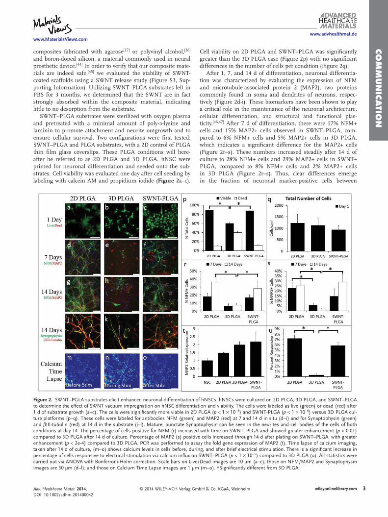
SWNT–PLGA substrates were sterilized with oxygen plasma and pretreated with a minimal amount of poly- D -lysine and laminin to promote attachment and neurite outgrowth and to ensure cellular survival. Two confi gurations were fi rst tested: SWNT–PLGA and PLGA substrates, with a 2D control of PLGA thin fi lm glass coverslips. These PLGA conditions will here-after be referred to as 2D PLGA and 3D PLGA. hNSC were primed for neuronal differentiation and seeded onto the sub-strates. Cell viability was evaluated one day after cell seeding by labeling with calcein AM and propidium iodide ( Figure 2 a–c).
Cell viability on 2D PLGA and SWNT–PLGA was signifi cantly greater than the 3D PLGA case (Figure 2 p) with no signifi cant differences in the number of cells per condition (Figure 2 q).
After 1, 7, and 14 d of differentiation, neuronal differentia-tion was characterized by evaluating the expression of NFM and microtubule-associated protein 2 (MAP2), two proteins commonly found in soma and dendrites of neurons, respec-tively (Figure 2 d-i). These biomarkers have been shown to play a critical role in the maintenance of the neuronal architecture, cellular differentiation, and structural and functional plas-ticity. [ 46,47 ] After 7 d of differentiation, there were 17% NFM+ cells and 15% MAP2+ cells observed in SWNT–PLGA, com-pared to 6% NFM+ cells and 5% MAP2+ cells in 3D PLGA, which indicates a signifi cant difference for the MAP2+ cells (Figure 2 r–s). These numbers increased steadily after 14 d of culture to 28% NFM+ cells and 29% MAP2+ cells in SWNT–PLGA, compared to 8% NFM+ cells and 2% MAP2+ cells in 3D PLGA (Figure 2 r–s). Thus, clear differences emerge in the fraction of neuronal marker-positive cells between
Adv. Healthcare Mater. 2014, DOI: 10.1002/adhm.201400042
Figure 2. SWNT–PGLA substrates elicit enhanced neuronal differentiation of hNSCs. hNSCs were cultured on 2D PLGA, 3D PLGA, and SWNT–PLGA to determine the effect of SWNT vacuum impregnation on hNSC differentiation and viability. The cells were labeled as live (green) or dead (red) after 1 d of substrate growth (a–c). The cells were signifi cantly more viable in 2D PLGA ( p < 1 × 10 −6 ) and SWNT-PLGA ( p < 1 × 10 −6 ) versus 3D PLGA cul-ture platforms (p–q). These cells were labeled for antibodies NFM (green) and MAP2 (red) at 7 and 14 d in situ (d–i) and for Synaptophysin (green) and βIII-tubulin (red) at 14 d in the substrate (j–l). Mature, punctate Synaptophysin can be seen in the neurites and cell bodies of the cells of both conditions at day 14. The percentage of cells positive for NFM (r) increased with time on SWNT–PLGA and showed greater enhancement ( p < 0.01) compared to 3D PLGA after 14 d of culture. Percentage of MAP2 (s) positive cells increased through 14 d after plating on SWNT–PLGA, with greater enhancement ( p < 2e-4) compared to 3D PLGA. PCR was performed to assay the fold gene expression of MAP2 (t). Time lapse of calcium imaging, taken after 14 d of culture, (m–o) shows calcium levels in cells before, during, and after brief electrical stimulation. There is a signifi cant increase in percentage of cells responsive to electrical stimulation via calcium infl ux on SWNT–PLGA ( p < 1 × 10 −5 ) compared to 3D PLGA (u). All statistics were carried out via ANOVA with Bonferroni-Holm correction. Scale bars on Live/Dead images are 10 µm (a–c); those on NFM/MAP2 and Synaptophysin images are 50 µm (d–l); and those on Calcium Time Lapse images are 1 µm (m–o). *Signifi cantly different from 3D PLGA.
www.MaterialsViews.com
© 2014 WILEY-VCH Verlag GmbH & Co. KGaA, Weinheimwileyonlinelibrary.com4
CO
MM
UN
ICATI
ON
www.advhealthmat.de
SWNT-containing and SWNT-lacking substrates for both markers. Further, after 14 d, hNSCs in both 3D conditions expressed mature neuronal marker synaptophysin, a synaptic vesicle protein (Figure 2 j-l). Quantitative real-time polymerase chain reaction (qRT-PCR) also revealed increased expression of the MAP2 gene at day 14 of differentiation in SWNT–PLGA relative to PLGA controls (Figure 2 t). Additionally, neuronal functionality was assessed by calcium imaging, where electri-cally active cells are defi ned as those that show an increase in fl uorescence due to calcium infl ux in response to an external electrical stimulation. We observed a signifi cant increase in the number of electrically responsive cells in SWNT–PLGA versus 3D PLGA conditions, 0.3%–5.9% (Figure 2 u). Treatment with 1 × 10 −6 M tetrodotoxin (TTX), a voltage-gated sodium channel blocker, abolished most calcium activity, suggesting that these temporal increases in intracellular calcium are due to elicited action potentials. [ 48 ]
The enhancement of neuronal differentiation kinetics within SWNT–PLGA relative to 3D PLGA can be attributed to several factors including a change in electrical conductivity, substrate topography, and substrate surface chemistry. The individual and concerted contributions of these factors remain to be elu-cidated. The increase in conductivity is reported to facilitate communication between neuronal cells, by allowing the cells to form “electrical shortcuts,” or transient electrical connections without being synapsed with one another. [ 34 ] Enhanced neu-ronal network connectivity has been proposed as the primary mechanism by which SWNT increase the frequency of postsyn-aptic currents. [ 7,34,49 ] In addition, cell behavior has been shown to be markedly altered by substrate topography. [ 50 ] In our study, the impregnation of the PLGA scaffolds with the SWNT disper-sion resulted in a marked enhancement in the fi ber diameter (Table 6 and Figure S1, Supporting Information). Increased fi ber diameters yield increased interfacial areas and a reduction in void size between fi bers, supporting increased local neuronal spreading and outgrowth. Similarly, the increased viability evidenced in the SWNT–PLGA versus uncoated PLGA fi bers could be due to the altered topography of the SWNT–PLGA substrate that is less porous and this can translate into a higher degree of cell–cell contact compared to the PLGA substrate, which is advantageous for hNSC survival. Previous studies implicate the combination of nanoscale grooves and hydro-phobic environments to elicit increased adsorption of proteins and growth factors essential for neuronal differentiation. [ 32 ] We observed that the contact angle of the aqueous SWNT disper-sion changes markedly after impregnation, from a hydrophobic surface, 117°, to a hydrophilic one, 80°, (Figure S4, Supporting Information). Our preliminary studies suggest that the levels of protein adsorption are not signifi cantly altered (see Supporting Information), which could result from the opposing effects of increased interfacial area and surfaces with diminished hydro-phobicity; however, further protein conformation and dynamics studies will be necessary to dissect the underlying mechanisms.
While passive cell culture on conductive substrates has been previously shown to enhance neuronal differentiation, applied electrical stimulation can also modulate cell and tissue growth both in nerve cultures [ 24 ] and more recently for the differentiation of myoblasts. [ 51 ] However, a combination of the electrospun sub-strates possessing high surface area/volume fi brous topography
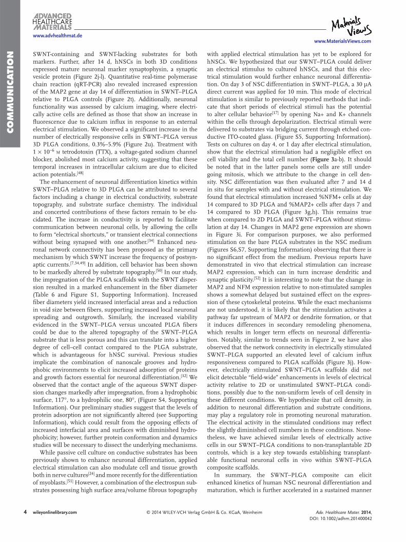
with applied electrical stimulation has yet to be explored for hNSCs. We hypothesized that our SWNT–PLGA could deliver an electrical stimulus to cultured hNSCs, and that this elec-trical stimulation would further enhance neuronal differentia-tion. On day 3 of NSC differentiation in SWNT–PLGA, a 30 µA direct current was applied for 10 min. This mode of electrical stimulation is similar to previously reported methods that indi-cate that short periods of electrical stimuli has the potential to alter cellular behavior [ 17 ] by opening Na+ and K+ channels within the cells through depolarization. Electrical stimuli were delivered to substrates via bridging current through etched con-ductive ITO-coated glass. (Figure S5, Supporting Information). Tests on cultures on day 4, or 1 day after electrical stimulation, show that the electrical stimulation had a negligible effect on cell viability and the total cell number ( Figure 3 a-b). It should be noted that in the latter panels some cells are still under-going mitosis, which we attribute to the change in cell den-sity. NSC differentiation was then evaluated after 7 and 14 d in situ for samples with and without electrical stimulation. We found that electrical stimulation increased %NFM+ cells at day 14 compared to 3D PLGA and %MAP2+ cells after days 7 and 14 compared to 3D PLGA (Figure 3 g,h). This remains true when compared to 2D PLGA and SWNT–PLGA without stimu-lation at day 14. Changes in MAP2 gene expression are shown in Figure 3 i. For comparison purposes, we also performed stimulation on the bare PLGA substrates in the NSC medium (Figures S6,S7, Supporting Information) observing that there is no signifi cant effect from the medium. Previous reports have demonstrated in vivo that electrical stimulation can increase MAP2 expression, which can in turn increase dendritic and synaptic plasticity. [ 52 ] It is interesting to note that the change in MAP2 and NFM expression relative to non-stimulated samples shows a somewhat delayed but sustained effect on the expres-sion of these cytoskeletal proteins. While the exact mechanisms are not understood, it is likely that the stimulation activates a pathway far upstream of MAP2 or dendrite formation, or that it induces differences in secondary remodeling phenomena, which results in longer term effects on neuronal differentia-tion. Notably, similar to trends seen in Figure 2 , we have also observed that the network connectivity in electrically stimulated SWNT–PLGA supported an elevated level of calcium infl ux responsiveness compared to PLGA scaffolds (Figure 3 j). How-ever, electrically stimulated SWNT–PLGA scaffolds did not elicit detectable “fi eld-wide” enhancements in levels of electrical activity relative to 2D or unstimulated SWNT–PLGA condi-tions, possibly due to the non-uniform levels of cell density in these different conditions. We hypothesize that cell density, in addition to neuronal differentiation and substrate conditions, may play a regulatory role in promoting neuronal maturation. The electrical activity in the stimulated conditions may refl ect the slightly diminished cell numbers in these conditions. None-theless, we have achieved similar levels of electrically active cells in our SWNT–PLGA conditions to non-transplantable 2D controls, which is a key step towards establishing transplant-able functional neuronal cells in vivo within SWNT–PLGA composite scaffolds.
In summary, the SWNT–PLGA composite can elicit enhanced kinetics of human NSC neuronal differentiation and maturation, which is further accelerated in a sustained manner
Adv. Healthcare Mater. 2014, DOI: 10.1002/adhm.201400042
www.MaterialsViews.com
© 2014 WILEY-VCH Verlag GmbH & Co. KGaA, Weinheim wileyonlinelibrary.com 5
CO
MM
UN
ICATIO
N
www.advhealthmat.de
by a simple electrical stimulation regimen. Such substrates could be vital to expanding neuronal cells derived from, say, patient-derived iPSC for in vitro applications such as drug screening or disease modeling. In addition, interfacing human neuronal cells with SWNT composites opens up a wide range of applications to testing the plasticity, functionality, and sub-type specifi city of neurons following programmed regimen of electrical stimulation. Such a system could also be used to transplant human neuronal cells within biomaterial constructs into models of spinal cord or neurodegeneration in the brain
and serve to accelerate neuronal differentiation, improve inte-gration of exogenous cells within host tissue, and provide the ability to deliver or monitor electrical signals to or from the transplanted constructs.
We have introduced an integrated platform for growth and electrical stimulation of hNSCs combining three salient features: i) First the SWNT–PLGA substrates can be easily fabricated based on vacuum-driven impregnation of an electrospun fi brous substrate with a SWNT dispersion. ii) The resulting substrates combine the salient features of highly conductive SWNTs with
Adv. Healthcare Mater. 2014, DOI: 10.1002/adhm.201400042
Figure 3. Electrical stimulation further enhances neuronal differentiation of hNSCs in SWNT–PLGA substrates . hNSCs were cultured on SWNT–PLGA, and at day 3, selected substrates were electrically stimulated for 10 min at 30 µA. On day 4, one day after stimulation, cell viability testing determined that there was no signifi cant decrease in cell viability or number due to electrical stimulation (a,b). After 7 and 14 d, the cells were fi xed and all condi-tions were labeled with antibodies for NFM (green) and MAP2 (red) (c–f). The percentage of NFM and MAP2 cells for each condition are shown (g,h). On day 7, there is a signifi cant increase in the percentage of MAP2-positive cells on the electrically stimulated SWNT–PLGA substrates versus 2D PLGA ( p < 5 × 10 −4 ). On day 14, electrical stimulation signifi cantly increased percentage MAP2-positive cells compared to 2D PLGA ( p = 0.008), 3D PLGA ( p < 1 × 10 −8 ), and SWNT–PLGA alone ( p = 0.02). PCR analysis of MAP2 gene fold expression (i) showed responsiveness following electrical stimula-tion. The percentage of cells responsive to electrical stimulation via calcium infl ux was quantifi ed (j); Cells on SWNT–PLGA with ( p < 1 × 10 −3 ) and without ( p < 2 × 10 −6 ) stimulation are signifi cantly more responsive compared to 2D PLGA. A comparison study for PLGA substrates containing no SWNT but electrically stimulated can be found in the Supporting Information. *Signifi cantly different from 3D PLGA + Signifi cantly different from 3D PLGA and SWNT-PLGA (ANOVA with Bonferroni–Holm Correction). Scale bars on NFM/MAP2 images are 50 µm (c–f).
www.MaterialsViews.com
© 2014 WILEY-VCH Verlag GmbH & Co. KGaA, Weinheimwileyonlinelibrary.com6
CO
MM
UN
ICATI
ON
www.advhealthmat.de
the topography of electrospun substrates, and were found to be highly biocompatible for the support of hNSCs derived from iPSC’s. iii) The application of electrical stimulation to the con-ductive substrates was found to elicit an enhanced effect on the hNSC differentiation process. Thus, the composite substrates are promising candidates for probing neurogenesis and neural activity, given the high level of biocompatibility, 3-D geom-etries, ECM-mimetic topography, and tunable differentiation/maturation cues. In addition, a conductive polymer scaffold may improve cell survival and functional integration after transplanta-tion in vivo by providing structural support for transplanted cells and facilitating synaptogenesis with host cells. It is also antici-pated that the novel SWNT composite fabrication technique presented here can be extended to similar electrospun scaffolds fabricated from different polymers, which would permit tuning of scaffold material properties for specifi c in vivo applications. Similarly, the amount of SWNT that can be deposited within the substrate can be controlled, allowing future studies into the effects of SWNT concentration and topography on differentia-tion. This will allow for future studies to determine the optimal substrate and stimulation conditions to maximally promote neu-ronal differentiation and maturation, which may lead to trans-plantable cell-scaffold constructs for CNS regeneration.
Experimental Section Preparation of SWNT Dispersion : All chemicals were of reagent grade
or higher. An aqueous dispersion of SWNT was prepared containing 0.4 (wt% by vol) of SWNT (Unidym), BSA (Sigma–Aldrich), and ascorbic acid (Sigma–Aldrich). The dispersion was mixed with a horn tip sonicator (Mixsonix S400) for 10 min at a pulsed rate of one second on and one second off at 40 amperes.
Fabrication of PLGA Scaffolds : Fibrous PLGA scaffolds were made by dissolving 15% weight/volume PLGA (Sigma–Aldrich, 85:15 PLA:PGA) in 1,1,1,3,3,3-Hexafl uoro-2-propanol (Sigma–Aldrich, HFIP) with gentle agitation. This solution was electrospun with a 23-gauge needle and a potential difference of +18kV at a distance of 18 cm onto a grounded fl at plate collector.
Impregnation of PLGA Scaffolds with SWNT Dispersions : A 5 cm by 5 cm PLGA scaffold was placed on a Nalgene 0.2 µm pore fi lter with a 150 mL capacity. The scaffold was sealed in place by the upper cup of the fi lter. A volume of 3 mL of the dispersion was then placed onto the scaffold and pulled through the scaffold by vacuum. The conductivity of the substrates was determined from resistance measurements shown in the Supporting Information and compared with the conductivity of the dried SWNT and the PLGA scaffolds without SWNT. A micro-BCA assay (Pierce Biotechnology) was performed to assess any differences in adsorbed level of protein (laminin), as elaborated in the Supporting Information.
Scanning Electron Microscopy : All images were imaged using a Hitachi S-4500 Field emission SEM. PLGA and PLGA–SWNT substrates were sputtercoated with platinum prior to imaging.
Fabrication of PLGA Thin Film Coverslips : A 1% (w/v) PLGA polymer solution was prepared by dissolving the polymer in HFIP overnight at room temperature. The polymer solution was then spin-coated onto 12 mm glass coverslips. The thin fi lms were dried for at least 24 h under vacuum.
iPSC to NSC Differentiation : Induced pluripotent stem cells (HFF1-iPSCs, gift from the Rutgers University Cell and DNA Repository, RUCDR) were differentiated to neural stem cells (hNSCs), using Neurobasal Media (Life Technologies) with 1× N2 (Life Technologies), 1x B27 without vitamin A (Life Technologies), 1× ITS (Life Technologies), and 2 × 10 −3 M L -glutamine (Invitrogen) supplemented with 500 ng mL −1
Noggin (Peprotech). hNSCs were further propagated in proliferation media consisting of 50% DMEM/F12 (Life Technologies) with 2 × 10 −3 M L -glutamine (Invitrogen) and 50% Neurobasal Media (Life Technologies) supplemented with 0.5x N2 supplement (Life Technologies), 0.5x B27 supplement without Vitamin A (Life Technologies), and 20 ng mL −1 bFGF (Peprotech).
hNSC Priming for Differentiation : In order to prime hNSCs for differentiation, they were cultured in proliferation media with bFGF withdrawn once the cells reached 70% confl uence, where the proliferation media consisted of Neurobasal Media (Life Technologies), DMEM/F12 (Life Technologies), 0.5x N2 (Life Technologies), and B27 without vitamin A (Life Technologies) without any additional growth factors. The hNSCs would subsequently be switched to differentiation media consisting of Neurobasal Media (Life Technologies), 1x B27 (Life Technologies), and 1× Glutamax (Invitrogen) with 10 ng mL −1 BDNF (Peprotech).
hNSC Neuronal Differentiation Analysis : Neuronal differentiation was evaluated using immunocytochemistry and qRT-PCR. The presence of MAP2 and NFM was evaluated using primary antibodies for those proteins, anti-MAP2 (Becton Dickinson), and anti-NFM (Invitrogen), linked to secondary antibodies labeled at 594 nm and 488 nm, respectively. Cell nuclei were labeled with DAPI. Images were taken with an Olympus IX81 Inverted Fluorescent Microscope. Each marker’s presence per cell was quantifi ed using the ImageJ software. For the qRT–PCR analysis MAP2 (TaqMan Hs 00258900_m1) was evaluated for undifferentiated NSCs and for each experimental condition after 14 d of differentiation. Undifferentiated NSCs were used as the reference sample. For NFM and MAP2 percentages, the average number of cells per data point was 6165 cells with a minimum of 1429 and a maximum of 10 777. For electrical activity, >1000 cells were used per data point.
Immunostaining : The NSCs in substrates were processed for immunocytochemistry by fi xing the live cells with 0.4% paraformaldehyde (paraformaldehyde) for 30 min at room temperature. The sample was then washed three times with phosphate buffered saline (PBS). To block and permeabolize the sample, it was immersed in a solution PBS based solution of 10% normal goat serum (Fisher), 1% BSA (Sigma–Aldrich), and 0.1% Triton X (Sigma–Aldrich) for 1 h at room temperature. All antibody and isotype control solutions were made in 10% normal goat serum and 1% BSA and were incubated on the samples for 1 h at room temperature. Three washes with PBS, minimum of 15 min each, cleared the sample for 1 h at room temperature with the secondary, fl uorescent antibody. Three washes with PBS followed secondary antibody incubation. DAPI was used to mark the nuclei of each cell and was added at a fi nal concentration of 1 µg mL −1 for 5 min to the samples before three PBS washes. Each sample was then mounted onto a glass coverslip using ProLong Gold Antifade Reagent (Invitrogen) for imaging. The samples were not sectioned. All imaging was done on either an Olympus IX81 Inverted Fluorescent Microscope. For the immunocytochemistry imaging four images were taken of each sample in random locations on the substrate or coverslip. Each image was quantifi ed until a minimum of three images and 150 cells were counted. Quantifi cation is shown as the mean ± standard error of three independent experiments, where at least four frames from each of two biological replicates were analyzed for each experiment. DAPI staining was used to quantify how many cells were present.
Electrical Stimulation Set-up : A circuit board was constructed allowing for the application of external electrical stimuli. Each electrode starts as an ITO-covered glass slide. The middle portion is etched with solution of 20% HCl and 5% HNO 3 to create a non-conductive gap, forming two electrodes. [ 38 ] This ensures that the applied current travels through the SWNT–PLGA and not around a path of least resistance. SWNT–PLGA are fi tted with cell culture rings to house the cells and growth media. The power source consists of a 2.5 volt watch battery. The ITO glass slides had a resistance of 200 Ω (±50 Ω) whereas the substrate contained a resistance of 4.4 kΩ (±6.5 kΩ) on the top and 10.7 kΩ (±9.3 kΩ) on the bottom. Electrical stimulation of the cells was applied for 10 min on the third day of differentiation under the experimental conditions.
Cell Viability Analysis : To determine cell viability, cells were incubated for 30 min at 37 °C with 4 × 10 −6 M calcein AM and 1 µg mL −1 propidium
Adv. Healthcare Mater. 2014, DOI: 10.1002/adhm.201400042
www.MaterialsViews.com
© 2014 WILEY-VCH Verlag GmbH & Co. KGaA, Weinheim wileyonlinelibrary.com 7
CO
MM
UN
ICATIO
N
www.advhealthmat.de
iodide (both from Life Technologies), which labeled live and dead cells, respectively. Cells were then imaged by acquiring z-stacks using a Leica TCS SP2 confocal microscope. This stacking, combined with a higher magnifi cation allows for a more accurate percentages of NFM+ and MAP2+ cells in each condition. For each condition, there was a minimum of two biological replicates in each of the three experiments. Images were taken with a 40× objective and quantifi ed for cell number and percentage of NFM or MAP2-positive cells using custom ImageJ macros. Maximum intensity projections of each stack were constructed and used for quantifi cation of cell viability. Each cell in one frame was counted as either live or dead using ImageJ software (NIH).
Quantitative Real-Time Polymerase Chain Reaction : Total RNA was extracted from hNSCs on day 14 in each condition using the RNEasy kit (Qiagen, Valencia, CA, USA) according to the manufacturer’s instructions, including removal of genomic DNA using RNase-free DNase. A high-capacity cDNA reverse transcription kit (Applied Biosystems, Foster City, CA, USA) containing random primers was used to reverse transcribe RNA from each sample to cDNA. TaqMan gene expression master mix and TaqMan gene expression assays (Applied Biosystems) were used for template amplifi cation of 10 ng cDNA/reaction. qRT–PCR was carried out on a 7500 Fast Real-Time PCR instrument (Applied Biosystems). Relative gene expression was calculated using the ΔΔCT method, normalizing to glyceraldehyde 3-phosphate dehydrogenase (GAPDH) as the endogenous control and undifferentiated hNSCs in standard 2-D culture. Graphs represent mean ± standard deviation. The TaqMan gene expression assays MAP2 (Hs00258900_m1) and GAPDH (4326317E) (Applied Biosystems) were used in these studies.
Calcium Imaging : Electrical activity was assessed using a calcium indicator dye, which refl ects changes intensity in response to changes in calcium levels. 3 × 10 −6 M Fluo-4 AM (Invitrogen) was loaded into the cells in an imaging solution, which consisted of 140 × 10 −3 M NaCl, 5 × 10 −3 M KCl, 2 × 10 −3 M CaCl 2 , 2 × 10 −3 M MgCl 2 , 10 × 10 −3 M HEPES, and 10 × 10 −3 M Glucose, supplemented with 0.2% pluronic F127 (Invitrogen). The chamber consisted of a two well LabTek (Thermo Scientifi c) modifi ed to include a platinum wire on either side of the well, acting as electrodes. To suspend the immersed substrate for image capture, a coverslip was altered to include four plastic pillars, each approximately 1 mm high. A second coverslip was placed on top of the pillars, forming a containment space for the sample. Each sample was excited using a 10 V pulse train with 10 ms pulse width and 100 ms pulse spacing. The dye’s response was captured at 1 Hz on a Leica SP2 Confocal Microscope. Stacks were made from the images in ImageJ and shifting in the sample was corrected using normalized correlation coeffi cient template matching. Each cell in the image was selected as a region of interest, and the cells’ average fl uorescence at each frame exported to MATLAB. A MATLAB program detected fl uorescence spikes in terms of the change in fl uorescence over initial fl uorescence (d F / F 0 ) due to the stimulation with a threshold of a 1d F / F 0 rise over a 10 s window and calculated the percentage of cells in the frame that showed this activity, which was manually checked for each series of images.
Statistics : One-way ANOVA was used to test the statistical signifi cance between conditions at each individual time-point with a Bonferroni–Holm’s post-hoc test. Samples with a p value below 0.05 were considered signifi cant.
Supporting Information Supporting Information is available from the Wiley Online Library or from the author.
Acknowledgements J.L., J.T.T., and G.H. contributed equally to this work. This work was funded in part by NIH EB 001046 (P.V.M.), NSF IGERT DGE 0801620
(P.V.M.), NJSCR Exploratory Research Grant (P.V.M.), a GAANN award from the Department of Education in pharmaceutical engineering and NIH R01 EB007467 (A.V.N.). The content is solely the responsibility of the authors and does not necessarily represent the offi cial views of the NIH or DoE. The authors would like to thank Valery Starovoytov for expert guidance and assistance with SEM. The authors would like to thank Jen Moore and the RUCDR facility at Rutgers University for the gift of the iPSC cell line. J.L. thanks Nedjma Bendiab for helpful discussion relating to Raman measurements. Finally, the authors thank Prof. Asefa and Yuying Meng for help with UV–vis measurements.
Received: January 19, 2014 Revised: March 17, 2014
Published online:
[1] J. W. Xie , M. R. MacEwan , S. M. Willerth , X. R. Li , D. W. Moran , S. E. Sakiyama-Elbert , Y. N. Xia , Adv. Funct. Mater. 2009 , 19 , 2312 .
[2] J. F. Cherry , A. L. Carlson , F. L. Benarba , S. D. Sommerfeld , D. Verma , G. Loers , J. Kohn , M. Schachner , P. V. Moghe , Biointer-phases 2012 , 7 , 16 .
[3] H. F. Lu , S. X. Lim , M. F. Leong , K. Narayanan , R. P. K. Toh , S. J. Gao , A. C. A. Wan , Biomaterials 2012 , 33 , 9179 .
[4] H. Kim , M. J. Cooke , M. S. Shoichet , Trends Biotechnol. 2012 , 30 , 55 . [5] G. T. Christopherson , H. Song , H. Q. Mao , Biomaterials 2009 , 30 ,
556 . [6] J. W. Xie , S. M. Willerth , X. R. Li , M. R. Macewan , A. Rader ,
S. E. Sakiyama-Elbert , Y. N. Xia , Biomaterials 2009 , 30 , 354 . [7] G. Cellot , F. M. Toma , Z. K. Varley , J. Laishram , A. Villari ,
M. Quintana , S. Cipollone , M. Prato , L. Ballerini , J. Neurosci. 2011 , 31 , 12945 .
[8] C. D. McCaig , A. M. Rajnicek , B. Song , M. Zhao , Physiol. Rev. 2005 , 85 , 943 .
[9] M. S. Graves , T. Hassell , B. L. Beier , G. O. Albors , P. P. Irazoqui , Ann. Biomed. Eng. 2011 , 39 , 1759 .
[10] N. M. Geremia , T. Gordon , T. M. Brushart , A. A. Al-Majed , V. M. K. Verge , Exp. Neurol. 2007 , 205 , 347 .
[11] Y. N. Cho , R. Ben Borgens , J. Biomed. Mater. Res. Part A 2010 , 95A , 510 .
[12] X. L. Luo , C. L. Weaver , D. D. Zhou , R. Greenberg , X. Y. T. Cui , Bio-materials 2011 , 32 , 5551 .
[13] K. Kimura , Y. Yanagida , T. Haruyama , E. Kobatake , M. Aizawa , J. Biotechnol. 1998 , 63 , 55 .
[14] L. Ghasemi-Mobarakeh , M. P. Prabhakaran , M. Morshed , M. H. Nasr-Esfahani , S. Ramakrishna , Tissue Eng. Part A 2009 , 15 , 3605 .
[15] L. H. Huang , X. L. Zhuang , J. Hu , L. Lang , P. B. Zhang , Y. S. Wang , X. S. Chen , Y. Wei , X. B. Jing , Biomacromolecules 2008 , 9 , 850 .
[16] J. Y. Lee , J. W. Lee , C. E. Schmidt , J. R. Soc. Interface 2009 , 6 , 801 . [17] C. E. Schmidt , V. R. Shastri , J. P. Vacanti , R. Langer , Proc. Natl.
Acad. Sci. U.S.A. 1997 , 94 , 8948 . [18] B. C. Thompson , R. T. Richardson , S. E. Moulton , A. J. Evans ,
S. O’Leary , G. M. Clark , G. G. Wallace , J. Controlled Release 2010 , 141 , 161 .
[19] Z. Zhang , M. Rouabhia , Z. X. Wang , C. Roberge , G. X. Shi , P. Roche , J. M. Li , L. H. Dao , Artif. Organs 2007 , 31 , 13 .
[20] A. K. Bakhshi , G. Bhalla , J. Scientifi c Ind. Res. 2004 , 63 , 715 . [21] L. Ghasemi-Mobarakeh , M. P. Prabhakaran , M. Morshed ,
M. H. Nasr-Esfahani , H. Baharvand , S. Kiani , S. S. Al-Deyab , S. Ramakrishna , J. Tissue Eng. Regen. Med. 2011, 5, E17.
[22] Y. J. Huang , H. C. Wu , N. H. Tai , T. W. Wang , Small 2012 , 8 , 2869 . [23] M. P. Mattson , R. C. Haddon , A. M. Rao , J. Mol. Neurosci. 2000 , 14 , 175 . [24] S. Y. Park , J. Park , S. H. Sim , M. G. Sung , K. S. Kim , B. H. Hong ,
S. Hong , Adv. Mater. 2011 , 23 , H263 - + .
Adv. Healthcare Mater. 2014, DOI: 10.1002/adhm.201400042

www.MaterialsViews.com
© 2014 WILEY-VCH Verlag GmbH & Co. KGaA, Weinheimwileyonlinelibrary.com8
CO
MM
UN
ICATI
ON
www.advhealthmat.de
Adv. Healthcare Mater. 2014, DOI: 10.1002/adhm.201400042
[25] A. Fabbro , S. Bosi , L. Ballerini , M. Prato , ACS Chem. Neurosci. 2012 , 3 , 611 .
[26] R. A. Dubin , G. C. Callegari , J. Kohn , A. V. Neimark , IEEE Trans. Nanobiosci. 2008 , 7 , 11 .
[27] D. Y. Lewitus , J. Landers , J. R. Branch , K. L. Smith , G. Callegari , J. Kohn , A. V. Neimark , Adv. Funct. Mater. 2011 , 21 , 2624 .
[28] J. K. Alexander , B. Fuss , R. J. Colello , Neuron Glia Biol. 2006 , 2 , 93 . [29] T. Gordon , E. Udina , V. M. K. Verge , E. I. P. de Chaves , Motor Con-
trol 2009 , 13 , 412 . [30] M. D. Wood , R. K. Willits , J. Neural Eng. 2009 , 6 , 8 . [31] E. Jan , N. A. Kotov , Nano Lett. 2007 , 7 , 1123 . [32] T. I. Chao , S. H. Xiang , C. S. Chen , W. C. Chin , A. J. Nelson ,
C. C. Wang , J. Lu , Biochem. Biophys. Res. Commun. 2009 , 384 , 426 . [33] V. Lovat , D. Pantarotto , L. Lagostena , B. Cacciari , M. Grandolfo ,
M. Righi , G. Spalluto , M. Prato , L. Ballerini , Nano Lett. 2005 , 5 , 1107 .
[34] G. Cellot , E. Cilia , S. Cipollone , V. Rancic , A. Sucapane , S. Giordani , L. Gambazzi , H. Markram , M. Grandolfo , D. Scaini , F. Gelain , L. Casalis , M. Prato , M. Giugliano , L. Ballerini , Nat. Nanotechnol. 2009 , 4 , 126 .
[35] H. H. Liao , R. L. Qi , M. W. Shen , X. Y. Cao , R. Guo , Y. Z. Zhang , X. Y. Shi , Colloid. Surf. B-Biointerfaces 2011 , 84 , 528 .
[36] L. Y. Yeo , J. R. Friend , J. Exp. Nanosci. 2006 , 1 , 177 . [37] K. Balani , R. Anderson , T. Laha , M. Andara , J. Tercero , E. Crumpler ,
A. Agarwal , Biomaterials 2007 , 28 , 618 . [38] M. K. Gheith , T. C. Pappas , A. V. Liopo , V. A. Sinani , B. S. Shim ,
M. Motamedi , J. R. Wicksted , N. A. Kotov , Adv. Mater. 2006 , 18 , 2975 . [39] T. Uchida , A. Yagi , Y. Oda , Y. Nakada , S. Goto , Chem. Pharm. Bull.
1996 , 44 , 235 . [40] S. Y. Park , D. S. Choi , H. J. Jin , J. Park , K. E. Byun , K. B. Lee ,
S. Hong , ACS Nano 2011 , 5 , 4704 .
[41] J. Kleis , P. Hyldgaard , E. Schroder , Comput. Mater. Sci. 2005 , 33 , 192 .
[42] A. V. Neimark , S. Ruetsch , K. G. Kornev , P. I. Ravikovitch , Nano Lett. 2003 , 3 , 419 .
[43] M. S. Dresselhaus , G. Dresselhaus , R. Saito , A. Jorio , Phys. Rep.-Rev. Section Phys. Lett. 2005 , 409 , 47 .
[44] M. HajjHassan , V. Chodavarapu , S. Musallam , Sensors 2008 , 8 , 6704 .
[45] E. J. Petersen , L. Zhang , N. T. Mattison , D. M. O’Carroll , A. J. Whelton , N. Uddin , N. Tinh , Q. Huang , T. B. Henry , R. D. Holbrook , K. L. Chen , Environ. Sci. Technol. 2011 , 45 , 9837 .
[46] Y. Xiong , Y. S. Zeng , C. G. Zeng , B. L. Du , L. M. He , D. P. Quan , W. Zhang , J. M. Wang , J. L. Wu , Y. Li , J. Li , Biomaterials 2009 , 30 , 3711 .
[47] N. Zhang , T. G. Kang , Y. Xia , Q. P. Wen , X. D. Zhang , H. Y. Li , Y. Hu , H. G. Hao , D. Zhao , D. Sun , Y. P. Yan , G. X. Zhang , J. X. Yang , Eur. J. Pharmacol. 2012 , 697 , 32 .
[48] H. Shimada , Y. Okada , K. Ibata , H. Ebise , S. Ota , I. Tomioka , T. Nomura , T. Maeda , K. Kohda , M. Yuzaki , E. Sasaki , M. Nakamura , H. Okano , PLoS One 2012 , 7 , e49469 .
[49] A. Mazzatenta , M. Giugliano , S. Campidelli , L. Gambazzi , L. Businaro , H. Markram , M. Prato , L. Ballerini , J. Neurosci. 2007 , 27 , 6931 .
[50] E. S. Fioretta , J. O. Fledderus , E. A. Burakowska-Meise , F. P. T. Baaijens , M. C. Verhaar , C. V. C. Bouten , Macromol. Biosci. 2012 , 12 , 577 .
[51] A. F. Quigley , J. M. Razal , M. Kita , R. Jalili , A. Gelmi , A. Penington , R. Ovalle-Robles , R. H. Baughman , G. M. Clark , G. G. Wallace , R. M. I. Kapsa , Adv. Healthcare Mater. 2012 , 1 , 801 .
[52] Q. Zhou , Q. Zhang , X. Q. Zhao , Y. Y. W. Duan , Y. Lu , C. Y. Li , T. Li , Brain Res. 2010 , 1311 , 148 .