Carbon monoxide, smoking and atherosclerosis POUL ASTRUP
10
Postgraduate Medical Journal (October 1973) 49, 697-706. Carbon monoxide, smoking and atherosclerosis POUL ASTRUP M.D. Professor of Clinical Chemistry, Department of Clinical Chemistry, Rigshospitalet, 9 Blegdamsvej, DK-2100 Copenhagen, Denmark THE toxic effect of carbon monoxide on the animal organism has probably been known by man since the discovery of fire, and we know it has been recog- nized as a dangerous poison in ancient times. It was Claude Bernard who first studied its mode of action, but it is J. S. Haldane who is considered as the real pioneering investigator of the physiology and toxicology of carbon monoxide. Together with some of his co-workers he performed the now classical studies on the effects on man of carbon monoxide exposure. His first paper appeared in 1895. Haldane considered, as a result of his investigations, that the only toxic effect of carbon monoxide was its ability to bind to haemoglobin at a much higher degree than oxygen, thus displacing oxygen in oxyhaemoglobin and depriving blood of its oxygen transport ability. As long as this transport was not seriously im- paired, carbon monoxide was regarded as relatively harmless. This was supported by the findings of many physiologists, showing that concentrations of up to 20% of carboxyhaemoglobin had little or no effect at rest on physiological parameters, such as heart rate, cardiac output, respiration, blood pressure, etc. This is just the opposite effect to that observed for moderate hypoxia, where the venous and tissue oxygen tensions are quite normal which is not the case, however, after moderate exposure to carbon monoxide, as was shown by Campbell in England in 1929. This lowering of the tissue oxygen tension is due to the lack of a cardio-respiratory compensating adjustment, and to the displacement of the oxyhaemoglobin dissociation curve to the left, first investigated by J. B. S. Haldane in 1912, and later in more detail by Roughton & Darling (1944) and by others. The displacement has never attained any physiological interest. An important breakthrough in carbon monoxide physiology was made in 1951 by the Swede T. Sjostrand, who discovered that carbon monoxide was formed continuously in the human body by the catabolism of haemoglobin. This explains the normal carboxyhaemoglobin concentration of about 0 5 % in man, increasing to about 3 % by increased haemo- lysis. This discovery added to the conception of carbon monoxide as a relatively harmless gas as long as it did not interfere seriously with the oxygen transport of the blood. During the last 10 years the interest in the physio- logical and pathological effects of moderate carbon monoxide exposure has increased considerably, mainly due to the concern about the risks of the growing air pollution, especially that due to car exhaust, which is the major source of the 250 million tons of global production of carbon monoxide per year. The gas is released to the air and since the background level does not increase, it is assumed that oxidation to carbon dioxide takes place in the upper atmosphere. It should be stressed that non-smokers do not run the risk of getting significantly elevated carboxy- haemoglobin levels from car exhaust in the streets, which has been shown in many studies, and clearly demonstrated recently by the findings of carboxy- haemoglobin levels of only 1 -4-30 % in non-smoking taxi drivers in London (Jones et al., 1972). This is in contrast to the much higher carboxyhaemoglobin levels, up to 20 %, found in inhaling tobacco smokers. Also people exposed to carbon monoxide for hours or days in more or less closed compartments (garages, tunnels, mines, submarines, etc.) with carbon monoxide release may also obtain similar high or even higher carboxyhaemoglobin levels. Now the question is: do carboxyhaemoglobin con- centrations up to 20% exert measurable physiolo- gical or pathological effects? A few years ago the answer would have been no, but today it would undoubtedly be yes. The central nervous system seems to be influenced. This was shown in the forties by McFarland and his associates (1970) by demon- strating impaired discrimination of small differences in light intensity at 2 and 4% carboxyhaemoglobin respectively. Also various performance in tests, for instance the estimation of time intervals without having a clock and the duration of auditory signals, are found decreased by some investigators at carb- oxyhaemoglobin levels about 5 % (Beard & Grand- staff, 1970). Also the myocardium may be affected by small
Transcript of Carbon monoxide, smoking and atherosclerosis POUL ASTRUP
Postgraduate Medical Journal (October 1973) 49, 697-706.
Carbon monoxide, smoking and atherosclerosis
POUL ASTRUPM.D.
Professor of Clinical Chemistry, Department of Clinical Chemistry,Rigshospitalet, 9 Blegdamsvej, DK-2100 Copenhagen, Denmark
THE toxic effect of carbon monoxide on the animalorganism has probably been known by man sincethe discovery of fire, and we know it has been recog-nized as a dangerous poison in ancient times. It wasClaude Bernard who first studied its mode of action,but it is J. S. Haldane who is considered as the realpioneering investigator of the physiology andtoxicology of carbon monoxide. Together with someof his co-workers he performed the now classicalstudies on the effects on man of carbon monoxideexposure. His first paper appeared in 1895. Haldaneconsidered, as a result of his investigations, that theonly toxic effect of carbon monoxide was its abilityto bind to haemoglobin at a much higher degree thanoxygen, thus displacing oxygen in oxyhaemoglobinand depriving blood of its oxygen transport ability.As long as this transport was not seriously im-
paired, carbon monoxide was regarded as relativelyharmless. This was supported by the findings ofmany physiologists, showing that concentrations ofup to 20% of carboxyhaemoglobin had little or noeffect at rest on physiological parameters, such asheart rate, cardiac output, respiration, bloodpressure, etc. This is just the opposite effect to thatobserved for moderate hypoxia, where the venousand tissue oxygen tensions are quite normal whichis not the case, however, after moderate exposure tocarbon monoxide, as was shown by Campbell inEngland in 1929. This lowering of the tissue oxygentension is due to the lack of a cardio-respiratorycompensating adjustment, and to the displacementof the oxyhaemoglobin dissociation curve to the left,first investigated by J. B. S. Haldane in 1912, andlater in more detail by Roughton & Darling (1944)and by others. The displacement has never attainedany physiological interest.An important breakthrough in carbon monoxide
physiology was made in 1951 by the Swede T.Sjostrand, who discovered that carbon monoxidewas formed continuously in the human body by thecatabolism ofhaemoglobin. This explains the normalcarboxyhaemoglobin concentration of about 0 5%in man, increasing to about 3% by increased haemo-lysis. This discovery added to the conception of
carbon monoxide as a relatively harmless gas as longas it did not interfere seriously with the oxygentransport of the blood.During the last 10 years the interest in the physio-
logical and pathological effects of moderate carbonmonoxide exposure has increased considerably,mainly due to the concern about the risks of thegrowing air pollution, especially that due to carexhaust, which is the major source of the 250 milliontons of global production of carbon monoxide peryear. The gas is released to the air and since thebackground level does not increase, it is assumed thatoxidation to carbon dioxide takes place in the upperatmosphere.
It should be stressed that non-smokers do not runthe risk of getting significantly elevated carboxy-haemoglobin levels from car exhaust in the streets,which has been shown in many studies, and clearlydemonstrated recently by the findings of carboxy-haemoglobin levels of only 1 -4-30% in non-smokingtaxi drivers in London (Jones et al., 1972). This is incontrast to the much higher carboxyhaemoglobinlevels, up to 20 %, found in inhaling tobacco smokers.Also people exposed to carbon monoxide for hoursor days in more or less closed compartments(garages, tunnels, mines, submarines, etc.) withcarbon monoxide release may also obtain similarhigh or even higher carboxyhaemoglobin levels.Now the question is: do carboxyhaemoglobin con-
centrations up to 20% exert measurable physiolo-gical or pathological effects? A few years ago theanswer would have been no, but today it wouldundoubtedly be yes. The central nervous systemseems to be influenced. This was shown in the fortiesby McFarland and his associates (1970) by demon-strating impaired discrimination of small differencesin light intensity at 2 and 4% carboxyhaemoglobinrespectively. Also various performance in tests, forinstance the estimation of time intervals withouthaving a clock and the duration of auditory signals,are found decreased by some investigators at carb-oxyhaemoglobin levels about 5% (Beard & Grand-staff, 1970).
Also the myocardium may be affected by small
Poul Astrup
carboxyhaemoglobin concentrations, since the utiliz-ation of available oxygen is very high at rest, andsince the binding of carbon monoxide to myoglobinis higher than its binding to haemoglobin. Thusabout 5-10% carboxyhaemoglobin concentrationsgive approximately 3 times higher carboxymyoglobinconcentrations (Coburn, 1970) which, of course, toa considerable extent interferes with the oxygentransport function of myoglobin. It has been demon-strated that 5-10% carboxyhaemoglobin in manleads to a decreased coronary arteriovenous oxygendifference (Ayres et al., 1970), and to an increasedcoronary blood flow. Similar carboxyhaemoglobinlevels have been shown to intensify myocardialischemia and to enhance development of arrhythmiaduring exercise in subjects older than 40 years(Knelson, 1972), indicating that limited capacity toincrease blood flow due to coronary obliterations,increases the susceptibility of carbon monoxideexposure.
Severe damage of the myocardium has also beenfound by ultrastructural studies in rabbits exposedfor 2 weeks to carbon monoxide, leading to a carb-oxyhaemoglobin concentration of approximately18% (Kjeldsen et al., 1972). The changes are verysimilar to changes following severe hypoxaemia, andinclude the formation of intercellular oedema, des-truction of mitochondria, etc.Our findings of high carboxyhaemoglobin levels
in the blood of many heavy smokers, especially insmokers with peripheral arteriosclerosis, have led usto the hypothesis that it might be the carbonmonoxide in the tobacco smoke, which is responsiblefor the much greater risk for smokers of developingarteriosclerosis in comparison to non-smokers.Today I am going to give the main results of our
experimental work which in our opinion definitelyprove that carbon monoxide has a damaging effecton the arterial walls, leading to an increased per-meability for various plasma components, to theformation of subendothelial oedema and to increasedatheromatosis. The results indicate that the muchhigher risk for smokers of developing arterial diseasein comparison to non-smokers is, at least mainly,due to the inhaled carbon monoxide in the tobaccosmoke and not to nicotine. There is no evidence fromanimal experiments that nicotine has an atherogeniceffect.For our animal experiments airtight chambers
were constructed in which rabbit cages could beplaced, and through which various gas mixturescould be passed. Each of the chambers we use nowcan hold eighteen rabbit cages. The gas mixtureswere made by mixing atmospheric air with carbonmonoxide, oxygen, and nitrogen respectively. Forthe various series we have used different techniques,which I am not going to describe in detail. In the
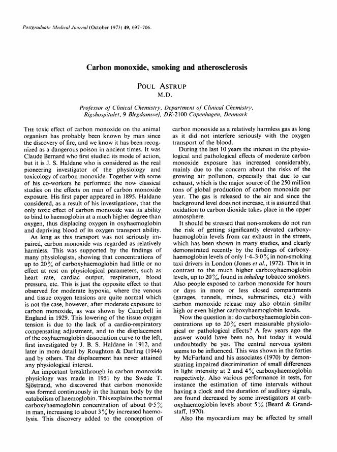
very first experiment cholesterol fed rabbits werecontinuously exposed to 170 ppm carbon monoxidefor 10 weeks giving carboxyhaemoglobin concentra-tions about 15%. This resulted in a cholesterolcontent of the aorta which was 2-5 times higher(P<0 001) than in the control rabbits, which hadnot been exposed to carbon monoxide, but were alsofed cholesterol (Astrup et al., 1967). We have re-peated the experiment several times and alwaysfound an enhancing effect of continuous carbonmonoxide exposure on cholesterol accumulation,and this has now been confirmed in other labora-tories, also by using primates (Birnstingl et al., 1970;Webster et al., 1970). By intermittent exposure ofgroups of eighteen rabbits each to carbon monoxide,12 or 4 hr a day, we obtained respectively 3 and 5times higher cholesterol accumulation than by con-tinuous exposure as demonstrated in Fig. 1, suggest-ing a certain adaptation of continuous exposure.
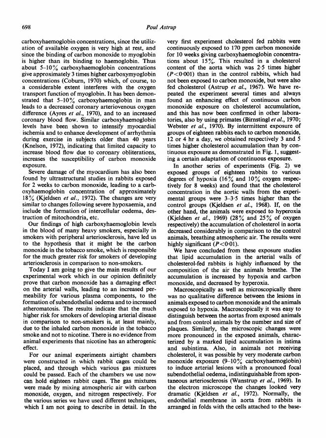
In another series of experiments (Fig. 2) weexposed groups of eighteen rabbits to variousdegrees of hypoxia (16% and 10% oxygen respec-tively for 8 weeks) and found that the cholesterolconcentration in the aortic walls from the experi-mental groups were 3-3 5 times higher than thecontrol groups (Kjeldsen et al., 1968). If, on theother hand, the animals were exposed to hyperoxia(Kjeldsen et al., 1969) (28% and 25% of oxygenrespectively) the accumulation of cholesterol in aortadecreased considerably in comparison to the controlanimals, breathing atmospheric air. The results werehighly significant (P<0-01).We have concluded from these exposure studies
that lipid accumulation in the arterial walls ofcholesterol-fed rabbits is highly influenced by thecomposition of the air the animals breathe. Theaccumulation is increased by hypoxia and carbonmonoxide, and decreased by hyperoxia.
Macroscopically as well as microscopically therewas no qualitative difference between the lesions inanimals exposed to carbon monoxide and the animalsexposed to hypoxia. Macroscopically it was easy todistinguish between the aortas from exposed animalsand from control animals by the number and size ofplaques. Similarly, the microscopic changes weremore pronounced in the exposed animals, charac-terized by a marked lipid accumulation in intimaand subintima. Also, in animals not receivingcholesterol, it was possible by very moderate carbonmonoxide exposure (9-10% carboxyhaemoglobin)to induce arterial lesions with a pronounced focalsubendothelial oedema, indistinguishable from spon-taneous arteriosclerosis (Wanstrup et al., 1969). Inthe electron microscope the changes looked verydramatic (Kjeldsen et al., 1972). Normally, theendothelial membrane in aorta from rabbits isarranged in folds with the cells attached to the base-
698
Carbon monoxide, smoking and atherosclerosis 699
5-
4
3
2-
2 - ---- -X -- -l----- ----
daily exp 24h 12h 4h atm air
ratio 00 3 24 5 08 0 40
FIG. 1. Relative values of aortic cholesterol in choles-terol-fed rabbits exposed to 0-018% carbon monoxidefor 24, 12, or 4 hr daily, or to atmospheric air for10 weeks. Each experiment comprised eighteen rabbits.
4
3
2
02% 28 26 21 16 l0ratio 0 50 0 66 1l00 265 3 40
FIG. 2. Relative values of aortic cholesterol in choles-terol-fed rabbits breathing air with varying oxygencontent for 10 weeks. Each experiment compriseseighteen rabbits.
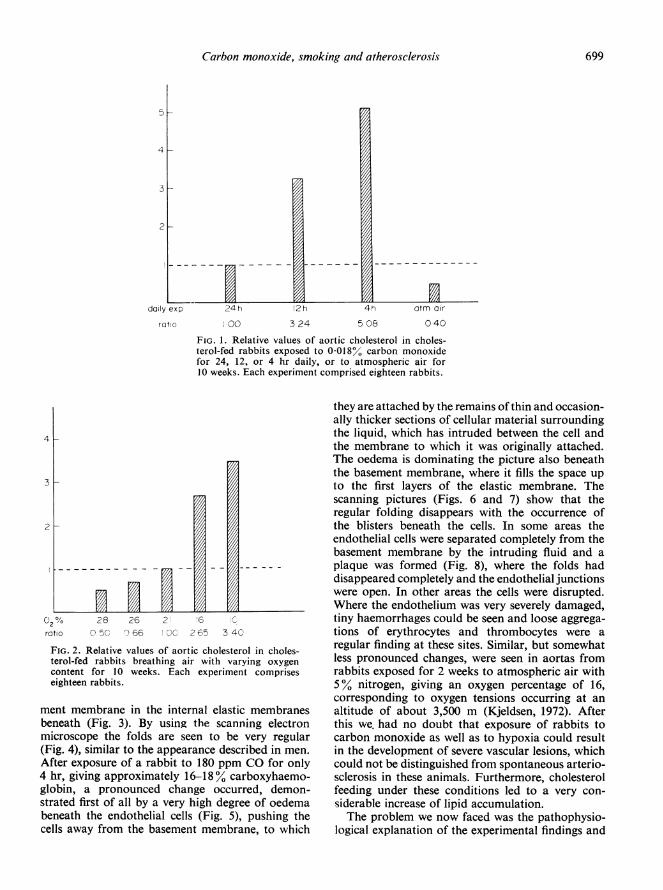
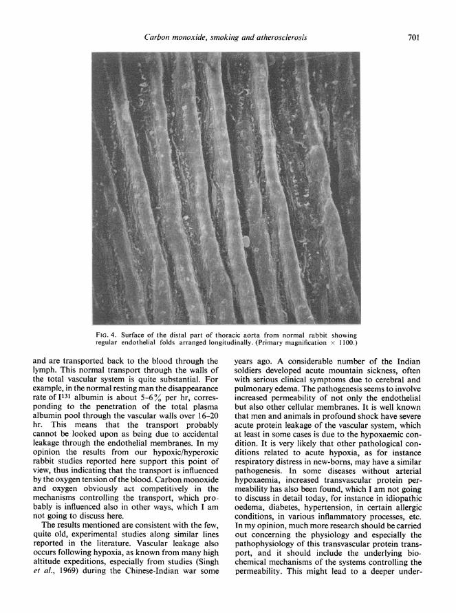
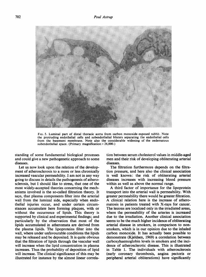
ment membrane in the internal elastic membranesbeneath (Fig. 3). By using the scanning electronmicroscope the folds are seen to be very regular(Fig. 4), similar to the appearance described in men.After exposure of a rabbit to 180 ppm CO for only4 hr, giving approximately 16-18% carboxyhaemo-globin, a pronounced change occurred, demon-strated first of all by a very high degree of oedemabeneath the endothelial cells (Fig. 5), pushing thecells away from the basement membrane, to which
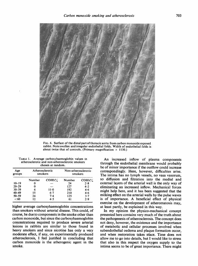
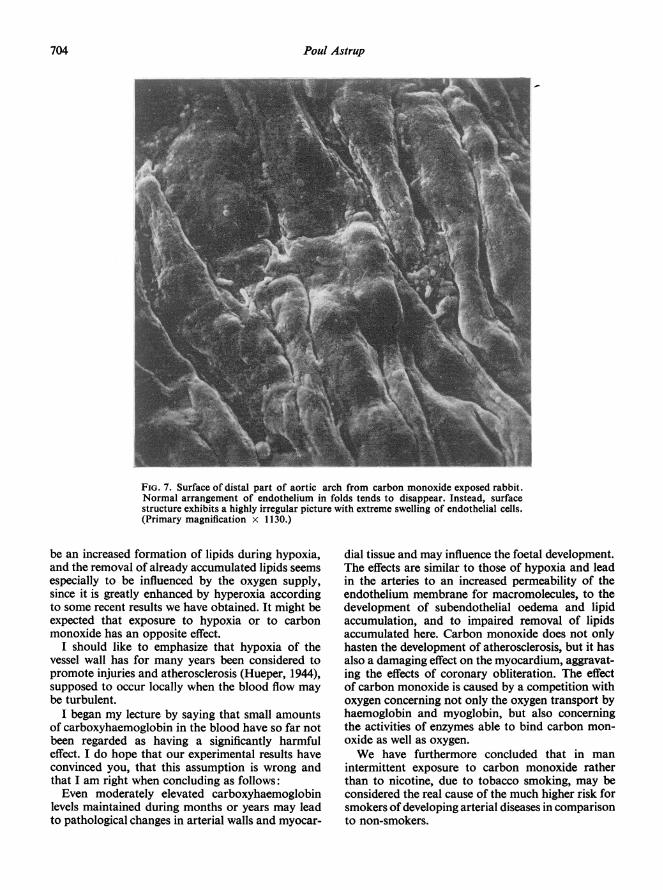
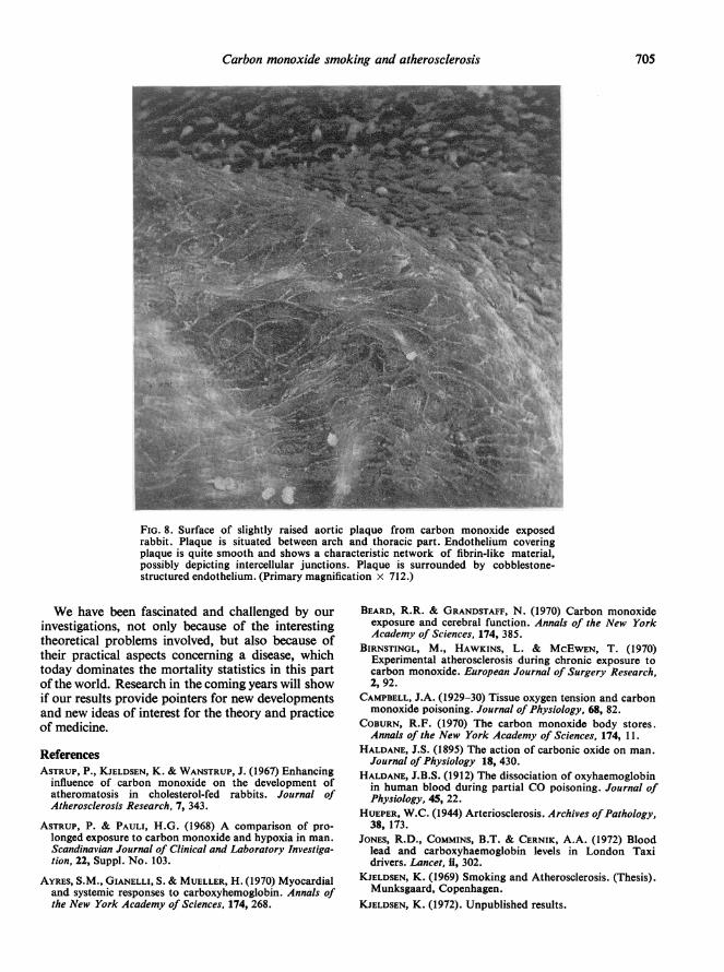
they are attached by the remains ofthin and occasion-ally thicker sections of cellular material surroundingthe liquid, which has intruded between the cell andthe membrane to which it was originally attached.The oedema is dominating the picture also beneaththe basement membrane, where it fills the space upto the first layers of the elastic membrane. Thescanning pictures (Figs. 6 and 7) show that theregular folding disappears with the occurrence ofthe blisters beneath the cells. In some areas theendothelial cells were separated completely from thebasement membrane by the intruding fluid and aplaque was formed (Fig. 8), where the folds haddisappeared completely and the endothelial junctionswere open. In other areas the cells were disrupted.Where the endothelium was very severely damaged,tiny haemorrhages could be seen and loose aggrega-tions of erythrocytes and thrombocytes were aregular finding at these sites. Similar, but somewhatless pronounced changes, were seen in aortas fromrabbits exposed for 2 weeks to atmospheric air with5% nitrogen, giving an oxygen percentage of 16,corresponding to oxygen tensions occurring at analtitude of about 3,500 m (Kjeldsen, 1972). Afterthis we, had no doubt that exposure of rabbits tocarbon monoxide as well as to hypoxia could resultin the development of severe vascular lesions, whichcould not be distinguished from spontaneous arterio-sclerosis in these animals. Furthermore, cholesterolfeeding under these conditions led to a very con-siderable increase of lipid accumulation.The problem we now faced was the pathophysio-
logical explanation of the experimental findings and
700 Poul Astrup~~~~~~~~~~~~~~~~~~~~~~~~~~~~~~~~.. 5.||.t......
...... RNP~~~~~~~. -.1
~~~~~~~~~~~~~~~~~~~~~~~~~~~~~~~~~~~~~~~~~~~~. ... .. .. .. ; ... ... ...... . .. ... ..~~~~~~~~~~~~~~~~~~~~~~~......
I ';:j~
:l'( DSt:~K~*~...... ..O'.....
4F.4E E E ;3._
,~~~~~~~~~~~~~~~~~~~~~~~~~~~~~~~~~~~~~~~~~~~~~~~~~~~~~~~~~~~~~~~~~~~~~~~~~~~~~~~~~~~~~~~......
FIG. 3. Luminal part of thoracic aorta from normal rabbit. (Primary magnification x 3300.)
the relation to the pathogenetic mechanisms in thedevelopment of arteriosclerosis in man. The findingsthat rabbits exposed to carbon monoxide quite oftenhad fluid with a high protein content, 3-4%, in theserous cavities, i.e. pleura, pericardium, and peri-toneum, led to the hypothesis that the arterialinjuries were caused by increased permeability ofthe endothelial membranes, leading to subendo-thelial oedema as demonstrated by the microscopicand electron microscopic findings. To evaluate thishypothesis in 1967 we made a comparison betweenchanges in some physiological and biochemical para-meters measured in human individuals duringexposure to carbon monoxide for 10 days and lateron during exposure to hypoxia over a period of 10days at the high altitude laboratory at Jungfraujochin Switzerland, 3,454 m above sea level (Astrup &Pauli, 1968). It was demonstrated, that carbon mon-oxide exposure (20-25% carboxyhaemoglobin) ledto a 50% increase of glomerular filtration rate duringthe first day of exposure (Pauli et al., 1968); to anincreased disappearance rate from the blood of in-jected radionated serum 1131 albumin (Siggaard-Andersen et al., 1968), and probably to an increase
in capillary filtration rate measured plethysmo-graphically on the calf (Siggaard-Andersen et al.,1967). This supported the hypothesis of an increasedvascular permeability due to carbon monoxide. Thetransvascular protein flux during carbon monoxideexposure has recently been studied in more detail inmy department (Parving, 1972; Parving et al., 1972)by measuring in human individuals the disappearancerate of 1131 albumin injected intravenously, and byfollowing the protein flux in lymph in dogs. It wasconfirmed that the disappearance rate of I131 albumin,after exposure to carbon monoxide (20-25% COHb)for 3 hr, is increased, on average 50%.The occurrence of increased permeability during
exposure to carbon monoxide could also be demon-strated in dogs, where the lymph flow and the proteinflux in the thoracic duct increased considerably. Itwas of interest that the increase in protein flux wasrelatively more marked for the high molecularproteins than for the low molecular ones (Parvinget al., 1972).When discussing these results, it should be em-
phasized that proteins and other macromoleculespenetrate the vascular wall under normal conditions
Carbon monoxide, smoking and atherosclerosis 701
5m: Mr.uS
FIG. 4. Surface of the distal part of thoracic aorta from normal rabbit showingregular endothelial folds arranged longitudinally. (Primary magnification x 1100.)
and are transported back to the blood through thelymph. This normal transport through the walls ofthe total vascular system is quite substantial. Forexample, in the normal resting man the disappearancerate ofI131 albumin is about 5-6% per hr, corres-ponding to the penetration of the total plasmaalbumin pool through the vascular walls over 16-20hr. This means that the transport probablycannot be looked upon as being due to accidentalleakage through the endothelial membranes. In myopinion the results from our hypoxic/hyperoxicrabbit studies reported here support this point ofview, thus indicating that the transport is influencedby the oxygen tension ofthe blood. Carbon monoxideand oxygen obviously act competitively in themechanisms controlling the transport, which pro-bably is influenced also in other ways, which I amnot going to discuss here.The results mentioned are consistent with the few,
quite old, experimental studies along similar linesreported in the literature. Vascular leakage alsooccurs following hypoxia, as known from many highaltitude expeditions, especially from studies (Singhet al., 1969) during the Chinese-Indian war some
years ago. A considerable number of the Indiansoldiers developed acute mountain sickness, oftenwith serious clinical symptoms due to cerebral andpulmonary edema. The pathogenesis seems to involveincreased permeability of not only the endothelialbut also other cellular membranes. It is well knownthat men and animals in profound shock have severeacute protein leakage of the vascular system, whichat least in some cases is due to the hypoxaemic con-dition. It is very likely that other pathological con-ditions related to acute hypoxia, as for instancerespiratory distress in new-borns, may have a similarpathogenesis. In some diseases without arterialhypoxaemia, increased transvascular protein per-meability has also been found, which I am not goingto discuss in detail today, for instance in idiopathicoedema, diabetes, hypertension, in certain allergicconditions, in various inflammatory processes, etc.In my opinion, much more research should be carriedout concerning the physiology and especially thepathophysiology of this transvascular protein trans-port, and it should include the underlying bio-chemical mechanisms of the systems controlling thepermeability. This might lead to a deeper under-
702 Poul Astrup
1...
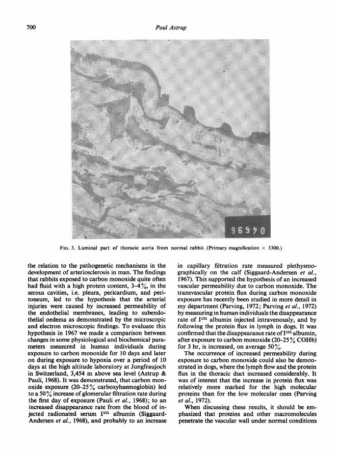
FIG. 5. Luminal part of distal thoracic aorta from carbon monoxide exposed rabbit. Notethe protruding endothelial cells and subendothelial blisters separating the endothelial cellsfrom the basement membrane. Note also the considerable widening of the oedematoussubendothelial space. (Primary magnification x 26,000.)
standing of some fundamental biological processesand could give a new pathogenetic approach to somediseases.
Let us now look upon the relation of the develop-ment of atherosclerosis to a more or less chronicallyincreased vascular permeability. I am not in any waygoing to discuss in details the pathogenesis of athero-sclerosis, but I should like to stress, that one of themost widely-accepted theories concerning the mech-anisms involved is the so-called filtration theory. Itsays, that plasma components filter into the arterialwall from the luminal side, especially when endo-thelial injuries occur, and under certain circum-stances accumulate here forming plaques, with orwithout the occurrence of lipids. This theory issupported by clinical and experimental findings; andparticularly by the observation that most of thelipids accumulated in atheromata are derived fromthe plasma lipids. The lipoproteins filter into thewall, where under unfavourable conditions the lipidsmay be released and be deposited. It is quite obviousthat the filtration of lipids through the vascular wallwill increase when the lipid concentration in plasmaincreases. Thus the probability of deposition of lipidwill increase. The clinical significance of this may beillustrated for instance by the almost linear correla-
tion between serum cholesterol values in middle-agedmen and their risk of developing obliterating arterialdiseases.The filtration furthermore depends on the filtra-
tion pressure, and here also the clinical associationis well known: the risk of obliterating arterialdiseases increases with increasing blood pressurewithin as well as above the normal range.A third factor of importance for the lipoprotein
transport into the arterial wall is permeability. Withgreater permeability there would be greater filtration.A clinical relation here is the increase of athero-matosis in patients treated with X-rays for cancer.The lesions are localized only in the irradiated areas,where the permeability of the arteries is increaseddue to the irradiation. Another clinical associationseems to be the much higher incidence of obliteratingarterial disease in smokers, in comparison to non-smokers, which- is in our opinion due to the inhaledcarbon monoxide. It has actually been possible todemonstrate (Kjeldsen, 1969) a correlation betweencarboxyhaemoglobin levels in smokers and the inci-dence of atherosclerotic disease. This is illustratedin Table 1. The individuals with atherosclerosis(early coronary thrombosis, angina pectoris orperipheral arterial obliterations) have significantly
Carbon monoxide smoking and atherosclerosis 703
lnn---rj_es_
S-F
-F9_-
9_s
'9
.
F .: :F
FIG . 6. Surface of the distal part ofthoracic aorta from carbon monoxide exposedrabbit. Note swollen and irregular endothelial folds. Width of endothelial folds isabout twice that of controls. (Primary magnification x 1130.)
TABLE 1. Average carboxyhaemoglobin values inatherosclerotic and non-atherosclerotic smokers
chosen at random.
Age Atherosclerotic Non-atheroscleroticgroups smokers smokers
Number COHb% Number COHb%.10-19 0 - 24 2-820-29 0 - 127 4-230-39 6 11-0 192 4-640-49 15 6-7 210 4-650-59 24 7-4 125 3-7<60 12 4 5 61 2-9
higher average carboxyhaemoglobin concentrationsthan smokers without arterial disease. This could, ofcourse, be due to components in the smoke other thancarbon monoxide, but since the carboxyhaemoglobinconcentrations required to produce severe arteriallesions in rabbits are similar to those found inheavy smokers and since nicotine has only a verymoderate effect, if any, on experimentally producedatherosclerosis, I feel justified in concluding thatcarbon monoxide is the atherogenic agent in thesmoke.
An increased inflow of plasma componentsthrough the endothelial membrane would probablybe of minor importance if the outflow could increasecorrespondingly. Here, however, difficulties arise.The intima has no lymph vessels, no vasa vasorum,so diffusion and filtration into the medial andexternal layers of the arterial wall is the only way ofeliminating an increased inflow. Mechanical forcesmight help here, and it has been suggested that themilking effect on the arterial walls by the pulse wavesis of importance. A beneficial effect of physicalexercise on the development of atheromatosis may,at least partly, be explained in this way.
In my opinion the physico-mechanical conceptpresented here contains very much of the truth aboutthe pathogenesis of atherosclerosis. The concept doesnot deny, however, the existence and the importanceof metabolic and cellular processes involved whensubendothelial oedema and plaque formation occur,and when restoration takes Dlace. Time does notallow me to go into details, but I would like to stressthat also in this respect the oxygen supply to theintima seems to be of great importance. There might
704 Poul Astrup
A!l
_t
FIG. 7. Surface of distal part of aortic arch from carbon monoxide exposed rabbit.Normal arrangement of endothelium in folds tends to disappear. Instead, surfacestructure exhibits a highly irregular picture with extreme swelling of endothelial cells.(Primary magnification x 1130.)
be an increased formation of lipids during hypoxia,and the removal of already accumulated lipids seemsespecially to be influenced by the oxygen supply,since it is greatly enhanced by hyperoxia accordingto some recent results we have obtained. It might beexpected that exposure to hypoxia or to carbonmonoxide has an opposite effect.
I should like to emphasize that hypoxia of thevessel wall has for many years been considered topromote injuries and atherosclerosis (Hueper, 1944),supposed to occur locally when the blood flow maybe turbulent.
I began my lecture by saying that small amountsof carboxyhaemoglobin in the blood have so far notbeen regarded as having a significantly harmfuleffect. I do hope that our experimental results haveconvinced you, that this assumption is wrong andthat I am right when concluding as follows:Even moderately elevated carboxyhaemoglobin
levels maintained during months or years may leadto pathological changes in arterial walls and myocar-
dial tissue and may influence the foetal development.The effects are similar to those of hypoxia and leadin the arteries to an increased permeability of theendothelium membrane for macromolecules, to thedevelopment of subendothelial oedema and lipidaccumulation, and to impaired removal of lipidsaccumulated here. Carbon monoxide does not onlyhasten the development of atherosclerosis, but it hasalso a damaging effect on the myocardium, aggravat-ing the effects of coronary obliteration. The effectof carbon monoxide is caused by a competition withoxygen concerning not only the oxygen transport byhaemoglobin and myoglobin, but also concerningthe activities of enzymes able to bind carbon mon-oxide as well as oxygen.We have furthermore concluded that in man
intermittent exposure to carbon monoxide ratherthan to nicotine, due to tobacco smoking, may beconsidered the real cause of the much higher risk forsmokers ofdeveloping arterial diseases in comparisonto non-smokers.
Carbon monoxide smoking and atherosclerosis 705
FIG. 8. Surface of slightly raised aortic plaque from carbon monoxide exposedrabbit. Plaque is situated between arch and thoracic part. Endothelium coveringplaque is quite smooth and shows a characteristic network of fibrin-like material,possibly depicting intercellular junctions. Plaque is surrounded by cobblestone-structured endothelium. (Primary magnification x 712.)
We have been fascinated and challenged by ourinvestigations, not only because of the interestingtheoretical problems involved, but also because oftheir practical aspects concerning a disease, whichtoday dominates the mortality statistics in this partof the world. Research in the coming years will showif our results provide pointers for new developmentsand new ideas of interest for the theory and practiceof medicine.
ReferencesASTRUP, P., KJELDSEN, K. & WANSTRUP, J. (1967) Enhancing
influence of carbon monoxide on the development ofatheromatosis in cholesterol-fed rabbits. Journal ofAtherosclerosis Research, 7, 343.
ASTRUP, P. & PAULI, H.G. (1968) A comparison of pro-longed exposure to carbon monoxide and hypoxia in man.Scandinavian Journal of Clinical and Laboratory Investiga-tion, 22, Suppl. No. 103.
AYRES, S.M., GIANELLI, S. & MUELLER, H. (1970) Myocardialand systemic responses to carboxyhemoglobin. Annals ofthe New York Academy of Sciences, 174, 268.
BEARD, R.R. & GRANDSTAFF, N. (1970) Carbon monoxideexposure and cerebral function. Annals of the New YorkAcademy of Sciences, 174, 385.
BIRNSTINGL, M., HAWKINS, L. & MCEWEN, T. (1970)Experimental atherosclerosis during chronic exposure tocarbon monoxide. European Journal of Surgery Research,2, 92.
CAMPBELL, J.A. (1929-30) Tissue oxygen tension and carbonmonoxide poisoning. Journal of Physiology, 68, 82.
COBURN, R.F. (1970) The carbon monoxide body stores.Annals of the New York Academy of Sciences, 174, 11.
HALDANE, J.S. (1895) The action of carbonic oxide on man.Journal of Physiology 18, 430.
HALDANE, J.B.S. (1912) The dissociation of oxyhaemoglobinin human blood during partial CO poisoning. Journal ofPhysiology, 45, 22.
HUEPER, W.C. (1944) Arteriosclerosis. Archives ofPathology,38, 173.
JONES, R.D., COMMINS, B.T. & CERNIK, A.A. (1972) Bloodlead and carboxyhaemoglobin levels in London Taxidrivers. Lancet, ii, 302.
KJELDSEN, K. (1969) Smoking and Atherosclerosis. (Thesis).Munksgaard, Copenhagen.
KJELDSEN, K. (1972). Unpublished results.
706 Poul Astrup
KJELDSEN, K., ASTRUP, P. & WANSTRUP, J. (1969) Reversalof rabbit atheromatosis by hyperoxia. Journal of Atheros-clerosis Research, 10, 173.
KJELDSEN, K., ASTRUP, P. & WANSTRUP, J. (1972) Ultra-structural intimal changes in the rabbit aorta after amoderate carbon monoxide exposure. Atherosclerosis,16, 67.
KJELDSEN, K., THOMSEN, H.K. & ASTRUP, P. (1972) Theeffects of carbon monoxide on myocardium. Submitted forpublication.
KJELDSEN, K., WANSTRUP, J. & ASTRUP, P. (1968) Enhancinginfluence of arterial hypoxia on the development ofatheromatosis in cholesterol-fed rabbits. Journal ofAtherosclerosis Research, 8, 835.
KNELSON, J.H. (1972) Verein Deutscher Ingenieure Berichte,180, 99.
McFARLAND, R.A. (1970) The effects of exposure to smallquantities of carbon monoxide on vision. Annals of theNew York Academy of Sciences, 174, 301.
PARVING, H.-H. (1972) The effect of hypoxia and carbonmonoxide exposure on plasma volume and capillarypermeability to albumin. Scandinavian Journal of Clinicaland Laboratory Investigation, 30, 49.
PARVING, H.-H., OHLSSON, K., BUCHARDT-HANSEN, H.J. &RORTH, M. (1972). Effect of carbon monoxide exposureon capillary permeability to albumin and a2-macroglobulin.Scandinavian Journal of Clinical and Laboratory Investiga-tion, 29, 381.
PAULI, H.G., TRUNIGER, B., LARSEN, J.K. & MULHAUSEN,
R.O. (1968) Renal function during prolonged exposure tohypoxia and carbon monoxide. Scandinavian Journal ofClinical and Laboratory Investigation, 22, Suppl. No. 103,55.
ROUGHTON, F.J.W. & DARLING, R.C. (1944) Effect of carbonmonoxide on oxyhemoglobin dissociation curve. AmericanJournal of Physiology, 141, 17.
SIGGAARD-ANDERSEN, J., BONDE PETERSEN, F., HANSEN, T.I.& MELLEMGAARD, K. (1968) Plasma volume and vascularpermeability during hypoxia and carbon monoxide ex-posure. Scandinavian Journal of Clinical and LaboratoryInvestigation, 22, Suppl. No. 103, 39.
SIGGAARD-ANDERSEN, J., KJELDSEN, K., BONDE PETERSEN, F.& ASTRUP, P. (1967) A possible connection between carbonmonoxide exposure, capillary filtration rate, and athero-sclerosis. Acta Medica Scandinavica, 182, 397.
SINGH, I., KHANNA, P.K., SRIVASTAVA, M.C., LAL, M.,Roy, S.B. & SUBRAMANYAM, C.S.V. (1969) Acute mountainsickness. New England Journal of Medicine, 280, 175.
SJOSTRAND, T. (1951) The in vitro formation of carbonmonoxide in blood. Acta Physiologica Scandinavica, 24,314.
WANSTRUP, J., KJELDSEN, K. & ASTRUP, P. (1969) Accelera-tion of spontaneous changes in rabbit aorta by a pro-longed moderate carbon monoxide exposure. ActaPathologica et Microbiologica Scandinavica, 75, 353.
WEBSTER, W.S., CLARKSON, T.B. & LOFLAND, H.B. (1970)Carbon monoxide-aggravated atherosclerosis in thesquirrel monkey. Experimental and Molecular Pathology,13, 36.