Carbon dioxide (CO2) angiography as an option for endovascular … · 2017-05-29 ·...
8
Vol.:(0123456789) 1 3 Int J Cardiovasc Imaging DOI 10.1007/s10554-017-1175-2 ORIGINAL PAPER Carbon dioxide (CO 2 ) angiography as an option for endovascular abdominal aortic aneurysm repair (EVAR) in patients with chronic kidney disease (CKD) Chiara De Angelis 1 · Francesco Sardanelli 2,3 · Matteo Perego 4 · Marco Alì 5 · Francesco Casilli 6 · Luigi Inglese 7 · Giovanni Mauri 2,8 Received: 13 February 2017 / Accepted: 18 May 2017 © European Union 2017 conservatively managed. In one patient, CO 2 angiograms were considered of too low quality for guiding the proce- dure and 200 ml of ICM were administered. Overall, 11 patients (84.6%) underwent a successful EVAR under the guidance of the sole CO 2 angiography. No patients suf- fered from major complications, including those typically CO 2 -related. Two patients suffered from abdominal pain during the procedure secondary to a transient splanchnic perfusion’s reduction due to CO 2 , and one patient had a worsening of renal function probably caused by a choles- terol embolization during the procedure. In patients with CKD, EVAR under CO 2 angiography guidance is feasible, effective, and safe. Abstract To assess feasibility, efficacy and safety of carbon dioxide (CO 2 ) digital subtraction angiography (DSA) to guide endovascular aneurysm repair (EVAR) in a cohort of patients with chronic kidney disease (CKD). After Ethical Committee approval, the records of 13 patients (all male, mean age 74.6 ± 8.0 years) with CKD, who underwent EVAR to exclude an abdominal aortic aneurysm (AAA) under CO 2 angiography guidance, were reviewed. The AAA to be excluded had a mean diameter of 52.0 ± 8.0 mm. CO 2 angiography was performed by auto- matic (n = 7) or hand (n = 6) injection. The endograft was correctly placed and the AAA was excluded in all cases, without any surgical conversions. Two patients (15.4%) had an endoleak: one type-Ia, detected by CO 2 -DSA and effectively treated with prosthesis dilatation; one type-III, detected by CO 2 -DSA, confirmed using 10 ml of ICM, and * Chiara De Angelis [email protected] Francesco Sardanelli [email protected] Matteo Perego [email protected] Marco Alì [email protected] Francesco Casilli [email protected] Luigi Inglese [email protected] Giovanni Mauri [email protected] 1 Scuola di Specializzazione in Radiologia, Postgraduation School in Radiology, Università degli Studi di Milano, Via Festa del Perdono 7, 20122 Milan, Italy 2 Radiology Unit, IRCCS Policlinico San Donato, Via Morandi 30, San Donato Milanese, 20097 Milan, Italy 3 Department of Biomedical Sciences for Health, Università degli Studi di Milano, via Morandi 30, San Donato Milanese, 20097 Milan, Italy 4 Corso di Laurea in Tecniche di Radiologia Medica per Immagini e Radioterapia, Università degli Studi di Milano, via Festa del Perdono 7, 20122 Milan, Italy 5 Integrated Biomedical Research, Università degli Studi di Milano, Via Mangiagalli 31, 20133 Milan, Italy 6 Interventional Cardiology Unit, Istituto Clinico Sant’Ambrogio, Milan, Italy 7 Interventistica Cardiovascolare, Gruppo Sanitario Policlinico di Monza, Milan, Italy 8 Division of Interventional Radiology, European Institute of Oncology, via Ripamonti 435, 20141 Milan, Italy
Transcript of Carbon dioxide (CO2) angiography as an option for endovascular … · 2017-05-29 ·...

Vol.:(0123456789)1 3
Int J Cardiovasc Imaging DOI 10.1007/s10554-017-1175-2
ORIGINAL PAPER
Carbon dioxide (CO2) angiography as an option for endovascular abdominal aortic aneurysm repair (EVAR) in patients with chronic kidney disease (CKD)
Chiara De Angelis1 · Francesco Sardanelli2,3 · Matteo Perego4 · Marco Alì5 · Francesco Casilli6 · Luigi Inglese7 · Giovanni Mauri2,8
Received: 13 February 2017 / Accepted: 18 May 2017 © European Union 2017
conservatively managed. In one patient, CO2 angiograms were considered of too low quality for guiding the proce-dure and 200 ml of ICM were administered. Overall, 11 patients (84.6%) underwent a successful EVAR under the guidance of the sole CO2 angiography. No patients suf-fered from major complications, including those typically CO2-related. Two patients suffered from abdominal pain during the procedure secondary to a transient splanchnic perfusion’s reduction due to CO2, and one patient had a worsening of renal function probably caused by a choles-terol embolization during the procedure. In patients with CKD, EVAR under CO2 angiography guidance is feasible, effective, and safe.
Abstract To assess feasibility, efficacy and safety of carbon dioxide (CO2) digital subtraction angiography (DSA) to guide endovascular aneurysm repair (EVAR) in a cohort of patients with chronic kidney disease (CKD). After Ethical Committee approval, the records of 13 patients (all male, mean age 74.6 ± 8.0 years) with CKD, who underwent EVAR to exclude an abdominal aortic aneurysm (AAA) under CO2 angiography guidance, were reviewed. The AAA to be excluded had a mean diameter of 52.0 ± 8.0 mm. CO2 angiography was performed by auto-matic (n = 7) or hand (n = 6) injection. The endograft was correctly placed and the AAA was excluded in all cases, without any surgical conversions. Two patients (15.4%) had an endoleak: one type-Ia, detected by CO2-DSA and effectively treated with prosthesis dilatation; one type-III, detected by CO2-DSA, confirmed using 10 ml of ICM, and
* Chiara De Angelis [email protected]
Francesco Sardanelli [email protected]
Matteo Perego [email protected]
Marco Alì [email protected]
Francesco Casilli [email protected]
Luigi Inglese [email protected]
Giovanni Mauri [email protected]
1 Scuola di Specializzazione in Radiologia, Postgraduation School in Radiology, Università degli Studi di Milano, Via Festa del Perdono 7, 20122 Milan, Italy
2 Radiology Unit, IRCCS Policlinico San Donato, Via Morandi 30, San Donato Milanese, 20097 Milan, Italy
3 Department of Biomedical Sciences for Health, Università degli Studi di Milano, via Morandi 30, San Donato Milanese, 20097 Milan, Italy
4 Corso di Laurea in Tecniche di Radiologia Medica per Immagini e Radioterapia, Università degli Studi di Milano, via Festa del Perdono 7, 20122 Milan, Italy
5 Integrated Biomedical Research, Università degli Studi di Milano, Via Mangiagalli 31, 20133 Milan, Italy
6 Interventional Cardiology Unit, Istituto Clinico Sant’Ambrogio, Milan, Italy
7 Interventistica Cardiovascolare, Gruppo Sanitario Policlinico di Monza, Milan, Italy
8 Division of Interventional Radiology, European Institute of Oncology, via Ripamonti 435, 20141 Milan, Italy

Int J Cardiovasc Imaging
1 3
Keywords Angiography · Abdominal aortic aneurysm (AAA) · Carbon dioxide (CO2) · Endovascular aneurysm repair (EVAR) · Iodinated contrast material (ICM) · Chronic kidney disease (CKD)
Introduction
Endovascular aneurysm repair (EVAR) has become the treatment of choice for abdominal aortic aneurysms (AAA) in patients with suitable anatomy, especially in elderly patients with comorbidities because it is less inva-sive than open surgery [1, 2]. The standard endovascular procedure implies the use of iodinated contrast material (ICM) to guide digital subtraction angiography (DSA), but risks of nephrotoxicity and allergic reaction may represent a contraindication to their administration [3]. Notably, the incidence of chronic kidney disease (CKD) in EVAR patients has been reported to be about 7–25% [4, 5] and acute kidney injury after EVAR 2–16%. Mor-tality in patients with acute kidney injury after EVAR can be as high as 30–50% [6]. Thus, finding an alternative method to guide an EVAR procedure, allowing for avoid-ing or reducing the amount of administered ICM, would be extremely helpful.
Carbon dioxide (CO2) is a gas with chemical and physical properties that makes it a theoretical ideal con-trast material for angiography in this setting. It has spe-cific advantages because it is not allergenic and not toxic, 20-fold more soluble than O2 in blood and 400-fold less viscous than ICMs. Moreover, it is very cheap and does not necessarily require dedicated hardware. Gaseous CO2 can be used as a negative intra-arterial contrast material below the diaphragm instead of ICM and CO2-DSA can be con-sidered a good alternative to ICM-DSA in patients at risk of contrast nephrotoxicity or with previous known allergic reactions to ICM [1, 7, 8]. In particular, CO2 is indicated in patients with reduced renal function (serum creati-nine >1.5 g/dl) that should undergo diagnostic or therapeu-tic DSA, because ICM may induce nephropathy [9]. It may also be used in combination with ICM, in order to reduce its amount during the procedures [8, 10, 11]. Moreover, CO2 can be safely used as a contrast material in the pediat-ric age, especially in case of contraindications to ICM such as renal transplant [12]. The CO2-DSA has been reported to be feasible and safe in small series of patients for diagnos-tic and interventional endovascular procedures under the diaphragm, in patients with contraindications to ICM [1–3, 7, 13–15].
The aim of this study was to assess feasibility, safety and efficacy of CO2-DSA to guide EVAR procedures for AAA in patients with CKD.
Materials and methods
Study population
The Institutional Review Board approved this study (Ethi-cal Committee of the BLINDED; authorization number BLINDED on DATE BLINDED). It was retrospectively evaluated clinical data and DSA images in a consecutive series of 13 patients, all male (mean age 74.6 ± 8.0 years), with CKD as a contraindication to ICM (none of the treated patients had allergy) who underwent EVAR procedures for the treatment of an AAA at our Institution between 2010 and 2015.
In order to avoid the administration of ICM, patients were studied before the procedure with ultrasound and unenhanced computed tomography (CT) or magnetic reso-nance imaging (MRI) for planning the EVAR. The type of endoprosthesis was chosen according to the patient’s char-acteristics and size of the AAA and of the landing zone.
Procedure
Before the procedure, the anesthesiologist performed firstly a spinal anesthesia. Sedation was avoided, as respiratory depression and hypotension caused by air contamination could be mistaken for adverse effects of sedatives and/or analgesics. For these reasons, patient’s vital signs (heart and respiratory rates, electrocardiogram) were monitored: relevant changes should be considered as a possible sign of excessive CO2 dose or air contamination.
Then, the patient was positioned on a standard angio-graphic table in Trendelenburg position. No specific prepa-ration was required. No hydration was needed in patients with renal impairment treated using only CO2; instead, patients had to be hydrated with intravenous isotonic saline solution before the procedure in case of use of ICM, to pre-serve renal function [8]. Sedation was avoided, as respira-tory depression and hypotension caused by air contamina-tion could be mistaken for adverse effects of sedatives and/or analgesics. For these reasons, patient’s vital signs (heart and respiratory rates, electrocardiogram) were monitored: relevant changes should be considered as a possible sign of excessive CO2 dose or air contamination.
All patients were punctured from bilateral femoral access. An angiographic catheter was positioned in the appropriate location and was connected to a manual or automatic injection system (see below), taking care of pre-venting an “explosive” delivery by initially filling it with a small amount of CO2 (90% of CO2 is injected in the last 0.5 s during a 4 s injection) [16]. Considering the risk of injecting high CO2 volumes, the catheter was not directly connected to the CO2 cylinder containing a large gas vol-ume at a very high pressure. CO2 was delivered into the

Int J Cardiovasc Imaging
1 3
vessel by using a handheld syringe or by a plastic bag sys-tem. In case of doubt at DSA images obtained with CO2, or if the operator judged the CO2 images inadequate for the correct performance of the EVAR procedure, ICM admin-istration was performed.
The manual injection system used was “home-made” and composed by three Luer-lock syringes linked together forming a “reservoir”, connected to another 20-ml syringe for diagnostic injection and to a disposable gas cylin-der filled with 99% laboratory-grade CO2 through a filter (Fig. 1). The CO2 canister (cylinder) is medical grade. The filter (0.2 µm) was necessary was specific to hold big par-ticles (such as air) and purify the injected CO2. Moreover, the filter prevented CO2 emission into the room, that could be toxic and undetectable by the operators.
The automatic apparatus for the injection of CO2 (Grupo Grifols, S.A., Parets del Valles, Spain) was composed by a Luer-lock syringe connected with a CO2 delivery system from a gas cylinder. The injection was guided by a moni-tor that displayed injection parameters (dose, pressure and slope). Before the procedure, in order to remove air from the circuit and the catheter, an injection test was performed into sterile saline solution, outside the patient (Fig. 2). An intravenous bolus of 0.5–1 mg of glucagon was adminis-tered to decrease the effects of bowel gas motion [10, 17].
Another available plastic bag system is produced by Merit Medical system, Inc (South Jordan, Utah, USA) [18]. Another recently introduced, FDA-approved porta-ble CO2 delivery system is the CO2mmander with Angio-Assist (PMDA, LLC, Ft.Myers, FL, USA). It allows for the delivery of CO2 at a low pressure to any reservoir
such as the plastic bag or a large syringe with the Luer-lock fitting. The AngioAssist is a unique stopcock system with valves that control direction of gas flow, allowing gas delivery in a non-explosive fashion using a 60 cc res-ervoir syringe and a 30 cc injection syringe.
During the procedure, angiographic images docu-mented the positioning of the prosthesis. Modern equip-ment for image quality and processing system, with 1024 × 1024 matrix and rapid data manipulation, greatly increased the ability to utilize CO2 as a contrast mate-rial. Acquisition programs had a high frame rate (>6 frames/s), as the gas propagated quickly into the vessels, and a high kilo-voltage to lower the skin dose but keep the contrast discrimination [1, 8].
The CO2 injection was performed using automatic delivery system in seven patients and manual delivery system in the remaining six patients (Figs. 1, 2).
Image and data analysis
Angiographic images and clinical records of the patients were evaluated to determine the EVAR technical success and rate of major and minor complications related to the CO2-DSA procedure. Also, clinical results of EVAR pro-cedure were assessed.
The angiographic procedure was considered com-pletely successful if no use of ICM was required, and par-tially successful if less than 20 ml of ICM were necessary to complete the EVAR placement.
Fig. 1 Manual CO2 injection system. This figure illustrates the man-ual system for CO2 injection, composed by: three luer lock syringes connected in series (asterisk, a) that represent a “reservoir”, con-
nected to another syringe (arrow, a) used for diagnostic injection and connected, through a filter (F, a), to a disposable gas cylinder (B, b)

Int J Cardiovasc Imaging
1 3
The EVAR procedure was considered completely suc-cessful if the aneurysm was correctly excluded, the graft correctly placed and no endoleak was caused.
Complications were evaluated and categorized as major or minor according to the SIR guidelines [19].
Statistical analysis
Descriptive statistics tests were used. The distribution was reported as percent and 95% confidential interval (95% CI).
Results
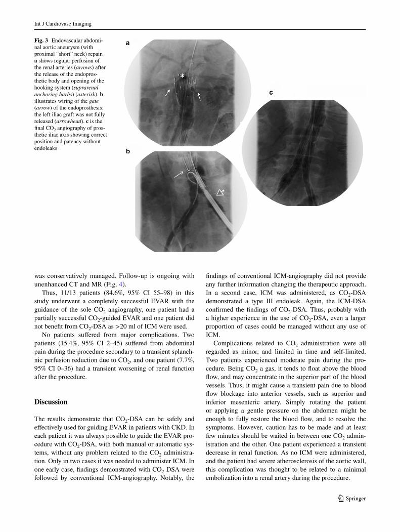
In all thirteen patients it was possible to correctly identify the anatomical landmarks (aortic and iliac landing zones) for a successful graft deployment with CO2. All grafts were finally correctly placed and the AAA excluded with no need of surgical conversions. The mean diameter of AAA in the 13 patients was 52.0 ± 8.0 mm (mean ± standard deviation).
A successful case is shown in Fig. 3.CO2-DSA was performed by automatic (n = 7) or hand
(n = 6) injection. In one of the first patients treated, the quality of CO2 images was considered insufficient to cor-rectly visualize the procedure, and 200 ml of ICM was administered. However, in this case no further informa-tion changing the strategy were finally derived from the ICM-DSA images.
One patient developed a type-Ia endoleak, caused by incomplete attachment of the proximal portion of the prosthesis immediately after the EVAR deployment; this endoleak was detected by CO2-DSA and effectively treated with prosthesis dilatation. A second patient devel-oped a type-III endoleak immediately after the EVAR deployment, caused by a graft defect or a graft module disconnection; this endoleak was detected by CO2-DSA. However, as this might be a critical clinical decision whether to treat or not to treat such endoleak, in this case a conventional DSA with 10 ml of ICM was per-formed. The findings of conventional DSA confirmed the diagnosis obtained with CO2-DSA and the endoleak
Fig. 2 Automatic CO2 injection system. Disposable kit, already assembled, composed by a luer lock 60 ml syringe (S) connected to a valvulated system (V) provided with a filter (arrow) for the connec-tion to the gas cylinder (a). The syringe is also connected to a flow reader system (asterisk, a) for the synchronization with the CO2 auto-matic injector. This assembled kit is then inserted in the piston-car-riage of the machinery (b); S indicated the syringe, arrow indicates the filter, V indicates the valvulated system and asterisk indicates the flow reader (b). The piston-carriage is then closed (c). S indicated
the syringe (c). d and e show the control panel with its parameters dose, pressure and slope. Arrow in e indicates the push-button to start the injection. f shows the “injection test” of CO2 outside the patient in sterile saline solution, mandatory before the procedure because it removes air from the circuit and the catheter (arrow). Finally, g shows the system assembled in each part, ready to be used. The disposable gas cylinder (B) is provided with a manometer (M) to control its replenishment status. S indicates the syringe (g)
Int J Cardiovasc Imaging
1 3
was conservatively managed. Follow-up is ongoing with unenhanced CT and MR (Fig. 4).
Thus, 11/13 patients (84.6%, 95% CI 55–98) in this study underwent a completely successful EVAR with the guidance of the sole CO2 angiography, one patient had a partially successful CO2-guided EVAR and one patient did not benefit from CO2-DSA as >20 ml of ICM were used.
No patients suffered from major complications. Two patients (15.4%, 95% CI 2–45) suffered from abdominal pain during the procedure secondary to a transient splanch-nic perfusion reduction due to CO2, and one patient (7.7%, 95% CI 0–36) had a transient worsening of renal function after the procedure.
Discussion
The results demonstrate that CO2-DSA can be safely and effectively used for guiding EVAR in patients with CKD. In each patient it was always possible to guide the EVAR pro-cedure with CO2-DSA, with both manual or automatic sys-tems, without any problem related to the CO2 administra-tion. Only in two cases it was needed to administer ICM. In one early case, findings demonstrated with CO2-DSA were followed by conventional ICM-angiography. Notably, the
findings of conventional ICM-angiography did not provide any further information changing the therapeutic approach. In a second case, ICM was administered, as CO2-DSA demonstrated a type III endoleak. Again, the ICM-DSA confirmed the findings of CO2-DSA. Thus, probably with a higher experience in the use of CO2-DSA, even a larger proportion of cases could be managed without any use of ICM.
Complications related to CO2 administration were all regarded as minor, and limited in time and self-limited. Two patients experienced moderate pain during the pro-cedure. Being CO2 a gas, it tends to float above the blood flow, and may concentrate in the superior part of the blood vessels. Thus, it might cause a transient pain due to blood flow blockage into anterior vessels, such as superior and inferior mesenteric artery. Simply rotating the patient or applying a gentle pressure on the abdomen might be enough to fully restore the blood flow, and to resolve the symptoms. However, caution has to be made and at least few minutes should be waited in between one CO2 admin-istration and the other. One patient experienced a transient decrease in renal function. As no ICM were administered, and the patient had severe atherosclerosis of the aortic wall, this complication was thought to be related to a minimal embolization into a renal artery during the procedure.
Fig. 3 Endovascular abdomi-nal aortic aneurysm (with proximal “short” neck) repair. a shows regular perfusion of the renal arteries (arrows) after the release of the endopros-thetic body and opening of the hooking system (suprarenal anchoring barbs) (asterisk). b illustrates wiring of the gate (arrow) of the endoprosthesis; the left iliac graft was not fully released (arrowhead). c is the final CO2 angiography of pros-thetic iliac axis showing correct position and patency without endoleaks

Int J Cardiovasc Imaging
1 3
Although the use of CO2-DSA was first reported by Hawkins in 1982 [20], since then CO2 has been described as a possible alternative to ICM for angiography in few reports [1, 2, 7, 20].
Different delivery systems can be used. Automatic injec-tion implies a closed system with a fast learning curve. It is self-priming, easily adjustable and does not cause explo-sive delivery nor air contamination. Hand injection implies an open or semi-open system implying a moderate risk of explosive delivery and possible air contamination; moreo-ver, gas delivery is less precise and the learning curve might be longer. Thus, if available, automatic injection sys-tems should be preferred. On the other hand, the manual injection systems are extremely simple and cheap, and in our experience not burdened by any complication. This sys-tem could be thus considered in case an automatic system is not available. Digital subtraction is particularly helpful, due to the low CO2 density [20].
Theoretically, unlimited amounts of CO2 may be admin-istered for CO2-DSA because the gas is effectively elimi-nated by means of respiration, keeping in mind to allow
sufficient time for its clearance (i.e., 2–3 min between subsequent injections) [10]. Using CO2 in combination with ICM reduce the amount of ICM administered [8, 10], thus being helpful to reduce the renal damage related to ICM administration. Other advantages of CO2 arteriog-raphy include avoidance of fluid overload, use of thinner catheters, visualization of stents and costs lower than ICM procedures.
The use of CO2 has no absolute contraindications. A relative contraindication does exist in patients with an atrial or ventricular septal defect (CO2 bubbles may enter the systemic circulation via the patent foramen ovale or ventricular septal defects) or a pulmonary arteriovenous malformation because of the possibility of paradoxi-cal gas embolism. The CO2 should be used with caution in patients with pulmonary insufficiency or pulmonary hypertension because a diagnostic dose of CO2 may cause an increase in pulmonary arterial pressure [14]. Seizures, loss of consciousness, brief respiratory arrest, or some combination of these complications have been reported in some patients when CO2 reached the cerebral arteries
Fig. 4 Endovascular abdominal aortic aneurysm repair. The patient was a 71 year-old man suffering from arterial hypertension, dys-lipidemia, and stage III chronic kidney disease (plasma creatinine 2.97 mg/dl). Infrarenal abdominal aortic aneurysm was detected by ultrasound and CT scans (not shown), with evidence of an increasing size. Baseline aortography is shown in image (a), iliac arteries angio-graphic study in image (b), both performed with repeated boluses of CO2. Image (c) shows the positioning of the endovascular prosthesis to exclude the aortic aneurysm. Image d shows the proximal sealing
optimization by elastomeric balloon dilatation. Final CO2 angiogra-phy (e, f) showed successful angiographic patency and correct posi-tion of prosthetic elements, the absence of proximal endoleak, and the presence of type III endoleak at the level of left prosthetic elements overlap (arrow). No treatment was performed; further controls dem-onstrated the endoleak resolution. The presence of type III endoleak at the level of left prosthetic elements overlap was demonstrated with CO2-DSA (arrow, g) and confirmed with C-DSA after 10 ml of ICM (arrow, h)

Int J Cardiovasc Imaging
1 3
[13, 21]. Therefore, arterial CO2 injections should be not performed above the diaphragm [8, 10]. Other complica-tions resulting from intravascular CO2 injection may be severe, but are generally rare [10]. They often relate to an incorrect application of technique that may result in air contamination and cause serious complications (vapor lock in the pulmonary artery with consequent hypoten-sion), or to an inadvertent injection of excessive volumes of CO2.
Overall, CO2-DSA is a good alternative choice to per-form diagnostic or therapeutic interventional procedures in patients with contraindications to ICM, i.e. allergy or CKD. Of note, patients with AAA requiring EVAR are fre-quently in the elderly and have comorbidities which imply contraindications not only to surgery but also to ICM injec-tion (CKD and allergy to ICM). The CO2 seems to be the favorite alternative option for these patients, and EVAR procedures guided by CO2-DSA allow to treat a larger number of patients.
This study agrees with some other previously published studies. In particular, in 2012 Criado et al. [7] published their experience of CO2 angiography as the preferred angiographic contrast agent for EVAR in 114 consecutive patients. They concluded that CO2-guided EVAR is techni-cally feasible and safe, eliminating or reducing the need for ICM [7]. In 2015, Sueyoshi et al. [2] found that CO2-DSA is reliable for the detection of endoleaks in the EVAR procedures.
In a wider framework, this work should be thought as one of the ways to be run for reducing contrast adminis-tration (and, whenever possible also X-ray radiation) to EVAR patients, a need that is absolutely important for their follow-up. One possible way in that direction is to use non-contrast MRI [22, 23].
This study has some limitations. First, the small num-ber of patients. However, these 13 patients are the entire consecutive series treated with CO2-DSA-guided EVAR at our center from 2010 to 2015. Second, it is a retrospective study, as it frequently happens for procedures alternative to conventional methods to be used in a small proportions of patients.
In conclusion, this study confirm that CO2 angiography is feasible, effective and safe to guide EVAR procedure in patients with CKD, using both manual or automatic deliv-ery systems. This approach can allow for treating patients preventing ICM-nephropathy in case of CKD.
An open issue to be investigated is the possibility to use CO2-DSAs not only in special subgroups of patients but as a standard approach for infra-diaphragmatic interventional procedures. Thus, the majority of patients could benefit from avoidance or minimal use of ICM. This way should be explored with large randomized controlled trials. First reported results from two trials seem highly promising [3,
24]. In the current era of patient-centered medicine, more efforts should be dedicated to this approach.
Funding This research received no specific grant from any funding agency in the public, commercial, or not-for-profit sectors.
Compliance with ethical standards
Conflict of interest The authors declare no conflict of interest.
References
1. Koutouzi G, Henrikson O, Roos H et al (2015) EVAR guided by 3D image fusion and CO2 DSA: a new imaging combination for patients with renal insufficiency. J Endovasc Ther 22:912–917. doi:10.1177/1526602815605468
2. Sueyoshi E, Nagayama H, Sakamoto I, Uetani M (2015) Car-bon dioxide digital subtraction angiography as an option for detection of endoleaks in endovascular abdominal aortic aneu-rysm repair procedure. J Vasc Surg 61:298–303. doi:10.1016/j.jvs.2014.07.088
3. De Almeida MC, De Arruda MA, Passos Teivelis M et al (2014) Carbon dioxide is a cost-effective contrast medium to guide revascularization of TASC A and TASC B femoropopliteal occlusive disease. Ann Vasc Surg 28:1473–1478. doi:10.1016/j.avsg.2014.03.021
4. Chuter TA, Reilly LM, Faruqi RM et al (2000) Endovascu-lar aneurysm repair in high-risk patients. J Endovasc Ther 31:122–133
5. Wald R, Waikar SS, Liangos O et al (2006) Acute renal failure after endovascular vs open repair of abdominal aortic aneurysm. J Vasc Surg. doi:10.1016/j.jvs.2005.11.053
6. Mehta M, Veith FJ, Lipsitz EC et al (2004) Is elevated creatinine level a contraindication to endovascular aneurysm repair? J Vasc Surg 39:118–123. doi:10.1016/S0741-5214(03)01041-3
7. Criado E, Upchurch GR, Young K et al (2012) Endovascular aortic aneurysm repair with carbon dioxide-guided angiography in patients with renal insufficiency. J Vasc Surg 55:1570–1575. doi:10.1016/j.jvs.2011.11.142
8. Kessel DO, Robertson I, Patel J et al (2002) Carbon-dioxide-guided vascular interventions: technique and pitfalls. Cardiovasc Interv Radiol 25:476–483. doi:10.1007/s00270-002-1925-x
9. Kroneberger C, Enzweiler CN, Schmidt-Lucke A et al (2015) Contrast-induced nephropathy in patients with chronic kid-ney disease and peripheral arterial disease. Acta Radiol Open 4:2058460115583034. doi:10.1177/2058460115583034
10. Eschelman DJ, Sullivan KL, Bonn J, Gardiner GA (1998) Car-bon dioxide as a contrast agent to guide vascular interventional procedures. AJR Am J Roentgenol 171:1265–1270. doi:10.2214/ajr.171.5.9798858
11. Nadolski GJ, Stavropoulos SW (2013) Contrast alternatives for iodinated contrast allergy and renal dysfunction: options and lim-itations. J Vasc Surg 57:593–598. doi:10.1016/j.jvs.2012.10.009
12. Kriss VM, Cottrill CM, Gurley JC (1997) Carbon dioxide (CO2) angiography in children. Pediatr Radiol 27:807–810. doi:10.1007/s002470050239
13. Ehrman KO, Taber TE, Gaylord GM et al (1994) Comparison of diagnostic accuracy with carbon dioxide versus iodinated con-trast material in the imaging of hemodialysis access fistulas. J Vasc Interv Radiol 5:771–775
14. Hawkins IF, Caridi JG (1998) Carbon dioxide (CO2) digital sub-traction angiography: 26-year experience at the University of Florida. Eur Radiol 8:391–402. doi:10.1007/s003300050400

Int J Cardiovasc Imaging
1 3
15. Kerns SR, Hawkins IF (1995) Carbon dioxide digital subtrac-tion angiography: expanding applications and technical evo-lution. AJR Am J Roentgenol 164:735–741. doi:10.2214/ajr.164.3.7863904
16. Seeger JM, Self S, Harward TR et al (1993) Carbon dioxide gas as an arterial contrast agent. Ann Surg 217:688
17. Back MR, Caridi JG, Hawkins IF, Seeger JM (1998) Angiogra-phy with carbon dioxide (CO2). Surg Clin North Am 78:575–591. doi:10.1016/S0039-6109(05)70335-2
18. Ho JP, Cho KJ, Ko PJ, Chu SY, Gopinathan A (2016) Practical guide to surgical and endovascular hemodialysis access manage-ment. World Scientific, Beaverton
19. Sacks D, McClenny TE, Cardella JF, Lewis CA (2003) Society of Interventional Radiology clinical practice guidelines. J Vasc Interv Radiol 14:S199–S202
20. Hawkins IF (1982) Carbon dioxide digital subtraction arteriogra-phy. AJR Am J Roentgenol 139:19–24. doi:10.2214/ajr.139.1.19
21. Coffey R, Quisling RG, Mickle JP et al (1984) The cerebro-vascular effects of intraarterial CO2 in quantities required for diagnostic imaging. Radiology 151:405–410. doi:10.1148/radiology.151.2.6424174
22. Resta EC, Secchi F, Giardino A et al (2013) Non-contrast MR imaging for detecting endoleak after abdominal endovascular aortic repair. Int J Cardiovasc Imaging 29:229–235. doi:10.1007/s10554-012-0060-2
23. Iozzelli A, D’Orta G, Aliprandi A et al (2009) The value of true-FISP sequence added to conventional gadolinium-enhanced MRA of abdominal aorta and its major branches. Eur J Radiol 72:489–493. doi:10.1016/j.ejrad.2008.09.004
24. Liss P, Eklöf H, Hellberg O et al (2005) Renal effects of CO2 and iodinated contrast media in patients undergoing renovascu-lar intervention: a prospective, randomized study. J Vasc Interv Radiol 16:57–65. doi:10.1097/01.RVI.0000144807.81633.79