Carbohydrate heterogeneity of vesicular stomatitis virus G glycoprotein allows localization of the...
12
ARCHIVES OF BIOCHEMISTRY AND BIOPHYSKS Vol. 219, No. 1, November, pp. 128-139, 1982 Carbohydrate Heterogeneity of Vesicular Stomatitis Virus G Glycoprotein Allows Localization of the Defect in a Glycosylation Mutant of CHO Cells PAMELA STANLEY Department of Cell Biology, Albert Einstein College of Medicim, 1300 Morris Park Auenue, Bronx, New York 10461 Received June 23, 1982 The carbohydrate portion of the G glycoprotein of vesicular stomatitis virus (VSV) grown in CHO cells (CHO/VSV) has been fractionated on Bio-Gel-P6, concanavalin A-Sepharose, and pea lectin-agarose. The results suggest that, in addition to sialic acid and fucose heterogeneity, the asparagine-linked complex carbohydrate moieties of CHO/VSV also display branching heterogeneity. Although the majority of the glyco- peptides bind to concanavalin A-Sepharose in a manner typical of certain biantennary carbohydrate structures, a significant proportion do not bind to the lectin. The latter behavior is typical of tri- or tetraantennary (branched) carbohydrate structures. The CHO/VSV glycopeptides which do not bind to concanavalin A-Sepharose separate into bound and unbound fractions on pea lectin-agarose suggesting that they include at least two different types of (branched) carbohydrate structures. Glycopeptides from the G glycoprotein of VSV grown in two, independently derived CHO glycosylation mutants which belong to complementation group 4 (Lec4 mutants) were examined in the same manner. In contrast to glycopeptides from CHO/VSV, glycopeptides from Lec4/VSV which passed through concanavalin A-Sepharose did not contain a component which subsequently bound to pea lectin-agarose. A glycopeptide fraction with these lectin- binding properties was also missing from cell surface glycopeptides derived from Lec4 cells. The combined results are consistent with the hypothesis that Lec4 CHO glyco- sylation mutants lack a glycosyltransferase activity responsible for the addition of a (branch) N-acetylglucosamine residue linked 61,s to mannose. This laboratory has previously described a variety of genetically distinct, lectin-re- sistant mutants of Chinese hamster ovary (CHO)l cells which express altered car- bohydrate at the cell surface (1,2). A major objective in characterizing these mutants is the molecular localization of their struc- tural carbohydrate defects so that the en- ’ Abbreviations used: CHO, Chinese hamster ovary; VSV, vesicular stomatitis virus; GlcNAc, N-acetyl- glucosamine; SA, sialic acid; FCS, fetal calf serum. SDS, sodium dodecyl sulfate; PBS, phosphate-buff- ered saline; TCA, trichloroacetic acid, Con A, con- canavalin A; PSA, pea lectin; IgG, immunoglobulin G; GS, biantennary IgG glycopeptide. zymatic basis of each mutation may ulti- mately be defined. One approach which has proved successful in uncovering cer- tain types of altered carbohydrate struc- tures, is the study of glycopeptides derived from vesicular stomatitis virus (VSV) grown in mutant cells (3,4). Purified VSV contains only one glycoprotein G, which is glycosylated at asparagine residues 163 and 320 (5) by host cell enzymes (3, 6, 7). The carbohydrate moieties at both sites contain typical core structures of N-ace- tylglucosamine (GlcNAc) and mannose (Man) and antennae of GlcNAc, galac- tose (Gal), and sialic acid (SA) sequences (7-10). 0003-9ss1/xz/1:~012x-12$02.00/0 Copyright 0 1982 by Academic Press, Inc. All rights of reproduction in any form reserved. 128
-
Upload
pamela-stanley -
Category
Documents
-
view
212 -
download
0
Transcript of Carbohydrate heterogeneity of vesicular stomatitis virus G glycoprotein allows localization of the...

ARCHIVES OF BIOCHEMISTRY AND BIOPHYSKS Vol. 219, No. 1, November, pp. 128-139, 1982
Carbohydrate Heterogeneity of Vesicular Stomatitis Virus G Glycoprotein
Allows Localization of the Defect in a Glycosylation Mutant of CHO Cells
PAMELA STANLEY
Department of Cell Biology, Albert Einstein College of Medicim,
1300 Morris Park Auenue, Bronx, New York 10461
Received June 23, 1982
The carbohydrate portion of the G glycoprotein of vesicular stomatitis virus (VSV) grown in CHO cells (CHO/VSV) has been fractionated on Bio-Gel-P6, concanavalin A-Sepharose, and pea lectin-agarose. The results suggest that, in addition to sialic acid and fucose heterogeneity, the asparagine-linked complex carbohydrate moieties of CHO/VSV also display branching heterogeneity. Although the majority of the glyco- peptides bind to concanavalin A-Sepharose in a manner typical of certain biantennary carbohydrate structures, a significant proportion do not bind to the lectin. The latter behavior is typical of tri- or tetraantennary (branched) carbohydrate structures. The CHO/VSV glycopeptides which do not bind to concanavalin A-Sepharose separate into bound and unbound fractions on pea lectin-agarose suggesting that they include at least two different types of (branched) carbohydrate structures. Glycopeptides from the G glycoprotein of VSV grown in two, independently derived CHO glycosylation mutants which belong to complementation group 4 (Lec4 mutants) were examined in the same manner. In contrast to glycopeptides from CHO/VSV, glycopeptides from Lec4/VSV which passed through concanavalin A-Sepharose did not contain a component which subsequently bound to pea lectin-agarose. A glycopeptide fraction with these lectin- binding properties was also missing from cell surface glycopeptides derived from Lec4 cells. The combined results are consistent with the hypothesis that Lec4 CHO glyco- sylation mutants lack a glycosyltransferase activity responsible for the addition of a (branch) N-acetylglucosamine residue linked 61,s to mannose.
This laboratory has previously described a variety of genetically distinct, lectin-re- sistant mutants of Chinese hamster ovary (CHO)l cells which express altered car- bohydrate at the cell surface (1,2). A major objective in characterizing these mutants is the molecular localization of their struc- tural carbohydrate defects so that the en-
’ Abbreviations used: CHO, Chinese hamster ovary; VSV, vesicular stomatitis virus; GlcNAc, N-acetyl- glucosamine; SA, sialic acid; FCS, fetal calf serum. SDS, sodium dodecyl sulfate; PBS, phosphate-buff- ered saline; TCA, trichloroacetic acid, Con A, con- canavalin A; PSA, pea lectin; IgG, immunoglobulin G; GS, biantennary IgG glycopeptide.
zymatic basis of each mutation may ulti- mately be defined. One approach which has proved successful in uncovering cer- tain types of altered carbohydrate struc- tures, is the study of glycopeptides derived from vesicular stomatitis virus (VSV) grown in mutant cells (3,4). Purified VSV contains only one glycoprotein G, which is glycosylated at asparagine residues 163 and 320 (5) by host cell enzymes (3, 6, 7). The carbohydrate moieties at both sites contain typical core structures of N-ace- tylglucosamine (GlcNAc) and mannose (Man) and antennae of GlcNAc, galac- tose (Gal), and sialic acid (SA) sequences (7-10).
0003-9ss1/xz/1:~012x-12$02.00/0 Copyright 0 1982 by Academic Press, Inc. All rights of reproduction in any form reserved.
128

LOCALIZATION OF GLYCOSYLATION LESION IN A CHO MUTANT 129
Glycopeptides derived by Pronase diges- tion of VSV G glycoprotein elute as four species termed So, Si, SZ, and S3 on Bio- Gel-P6 (9, 11). Following removal of sialic acid, only the species corresponding to S, is observed suggesting that So, Si, and S, represent structures containing decreasing numbers of sialic acid residues (9, 11). Structural studies of the carbohydrate as- sociated with G from VSV grown in BHK cells (9, 11) suggest that the saccharide units are triantennary and appear similar to the asparagine-linked carbohydrate moiety of fetuin (12, 13). Although not evident in the structure reported by Read- ing et al. (lo), heterogeneity was observed in the number of sialic acid and fucose res- idues per molecule by other authors (8, 9, 11, 14).
The carbohydrate units of G from VSV grown in CHO cells appear similar to those of BHK-grown VSV when profiles of Pro- nase glycopeptides on Bio-Gel-P6 before and after removal of sialic acid residues are compared (3, 9, 14). However, when sub- jected to lectin affinity chromatography, the glycopeptides from CHO-grown VSV exhibit a heterogeneity which cannot be accounted for by variation in the numbers of sialic acid and/or fucose residues. In this paper, we present evidence which suggests that the majority of the carbohydrate of G from CHO-grown VSV consists of bian- tennary structures which bind to conca- navalin A-Sepharose. The remainder in- clude at least two types of (branched) car- bohydrate structures-one type binds to pea lectin-agarose while the other does not. CHO glycosylation mutants from complementation group 4 (now designated Lec4 mutants; see Ref. (2)) do not syn- thesize the latter type of (branched) car- bohydrate species.
MATERIALS AND METHODS
Cells and Virus Culture
Parental CHO cells (Pro-5 and Gatt2) and two in- dependent mutants from complementation group 4 (Pro-Lec4.12.2 and GattLec4.2D) were cultured at 37°C in complete alpha medium containing 10% horse serum and 2% fetal calf serum (FCS) as previously
described (2, 4). Alpha medium and sera were ob-
tained from GIBCO Laboratories, Grand Island, New York. Stocks of VSV (Indiana strain) were grown
from a clone derived in this laboratory by three plaque
purifications of a virus stock originally supplied by
Don Summers (University of Utah). Pro-5 CHO cells growing in suspension were concentrated by centrif-
ugation to approximately 2 X lo7 cells/ml and mixed with VSV at 0.1 pfu/cell. After 1 h at 37”C, the cells
were diluted to approximately lOa cells/ml in alpha medium containing 2% FCS and incubated with stir-
ring at 37°C for 8 h. The culture medium was clarified by centrifugation at 3000 rpm for 20 min in a PR-2
International centrifuge and typically contained in-
fectious virus at approximately 5 X 10⁷-5 X 10s pfu/ ml. To obtain more concentrated stocks, the medium
was centrifuged at 25,000 rpm for 2 h at 4°C in the SW28 rotor of a Beckman ultracentrifuge. The su-
pernatant was discarded, and the crude viral pellet resuspended in a small volume of ET buffer (10 mM
Tris-HCl, 1 mM ethylenediaminetetraacetic acid, pH 7.0) containing 10% dimethylsulfoxide and stored fro-
zen in 0.1.ml aliquots at -120°C. The infectivity of
these preparations was usually 5 X log-5 X 10” pfu/ml.
Radioisotopes
All radioisotopes and scintillation fluid were pur- chased from Amersham Radiochemical Corporation,
Arlington Heights, Illinois. These include: D-
[6-3H]glucosamine hydrochloride (20-40 Ci/mmol); D- [ l-‘*Cl -glucosamine hydrochloride (50-60 mCi/
mmol); L-[6-sH]fucose (16.6 Ci/mmol); [“HIacetic an- hydride (5 mCi/lO mg); and N-acetyl[4, 5, 6, 7, 8, 9-
“Clneuraminic acid. Aqueous samples were counted in ACS II in a Beckman scintillation counter LS9000
which had a counting efficiency of approximately 60% for ‘H and 80% for i4C. The background for open- channel counting was approximately 30 cpm for ‘H
and 12 cpm for 14C.
Preparation of Radiolabeled VS V and Cells
VSV was labeled by incorporation of [aH] or
[i4C]glucosamine as follows: Cells growing exponen- tially in suspension culture were pelleted by centrif-
ugation, washed once with alpha medium, and resus-
pended at 2.5 X lo7 cells/ml in alpha medium con- taining VSV at approximately lo-40 pfu/cell. The cell
suspension was incubated 1 h at 37°C with occasional mixing and subsequently diluted to 1 X lOa-1.5 X lOa cells/ml in alpha medium containing 2% heat-inac- tivated FCS. After 3 h in suspension culture at 37”C,
the cells were pelleted by centrifugation, washed once with alpha medium, and resuspended in alpha me- dium containing glucose at 0.5 mg/ml and supple-
mented with glucosamine (8 pCi/ml [3H]glucosamine)

130 PAMELA STANLEY
and (4 @i/ml [i4C]glucosamine) and 2% dialyzed FCS. Following overnight incubation in suspension
at 37”C, the culture medium was clarified by centrif- ugation at 3000 rpm for 15 min and the supernatant
layered directly over 28.6-ml linear gradients of po- tassium tartrate (15 to 33% (w/w) in ET buffer). The
gradients were centrifuged to equilibrium (2 h at 22,500 rpm at 4°C in the SW28 rotor of a Beckman
ultracentrifuge). The virus band was harvested by
needle, diluted into ET buffer, and pelleted by cen- trifugation at 25,000 rpm for 2 h at 4°C. The virus
pellet was resuspended in a small volume of 5 mM
Tris-HCl buffer, pH 8.5, and stored at -12O’C. SDS-
gel analysis of purified glucosamine-labeled CHO/ VSV revealed one major labeled band which comi-
grated with authentic G glycoprotein. To obtain labeled glycopeptides enriched for cell
surface carbohydrates, exponentially growing, lo-ml suspension cultures of parental and Lec4 cells were suspended at 2 X lo5 cells/ml in the presence of alpha
medium 10% FCS containing either [sH]glucosamine hydrochloride (60 &i/ml) or [3H]fucose (11 Ci/ml).
After 50 h, the cells had proliferated to 1 X lOs-1.6 X 10s cells/ml. At this time they were pelleted, the
supernatant removed and the cells were mixed with 1.8 X lo7 unlabeled cells. The combined cells were
centrifuged and washed three times with phosphate-
buffered saline containing 1 mM CaClx and 1 mM
MgClx (PBS), pH 7.4. They were subsequently sus- pended in 3.6 ml of PBS prior to digestion with Pro-
nase.
Enzyme Digestions
To obtain Pronase glycopeptides, purified radiola-
beled VSV was suspended in 5 mM Tris-HCl, pH 8.5, and 3 mM CaCl, containing 1 mg/ml Pronase (B
Grade; Calbiochem, La Jolla, Calif.; predigested at 50°C for 1 h). A drop of toluene was added to the
incubation mixture, which was tightly capped and in- cubated at 37°C. After 24 h, Pronase was added again
to a final concentration of 2 mg/ml. The incubation was continued for 24 h when 295% of the radiolabel was TCA-soluble. The digestion was stopped by boil-
ing the incubation mixture for 1 min and insoluble material was removed by a 5-min centrifugation in
a Brinkman microfuge. Tryptic glycopeptides were generated by incubating
purified VSV in the presence of 1 mM Tris-HCl, pH 8.5, containing 1 mg/ml Trypsin-TPCK (Millipore Corporation, Freehold, N. J.) at 37’C for 24 h. The digestion was stopped by boiling the sample for 1 min.
Insoluble material was removed by high-speed cen- trifugation in a Brinkman microfuge and the super- natant was collected. Neuraminidase treatment of tryptic glycopeptides was performed by incubating the latter with 20 munit of neuraminidase for 16 hat 37’C following adjustment of the buffer to 0.05 M citrate
phosphate of pH 6.5. Purified neuraminidase was a gift from J. R. Etchison (University of California,
Davis). Under these conditions of digestion, the en- zyme preparation exhibited negligible glycosidase ac-
tivities as measured against a variety ofp-nitrophenyl sugar substrates (Etchison, personal communication).
Pronase glycopeptides enriched for cell surface car-
bohydrates were obtained by incubating washed, la- beled cells in 4 ml PBS, pH 7.4, containing 1 mg/ml
Pronase. After 100 min at 37”C, the cell suspension was centrifuged to remove cells and the supernatant
was incubated overnight at 50°C in the presence of
sodium axide (0.02 mg/ml). The next day fresh Pro- nase was added at 1 mg/ml and the incubation con-
tinued at 50°C overnight. The reaction mixtures were clarified by centrifugation at 3000 rpm for 20 min and
desalted on Bio-Gel-P2. Radiolabeled material which eluted with the void volume was pooled for further
characterization.
Column Chromatography
(0) Gel filtration. Bio-Gel-P6 (minus 400) and Bio-
Gel-P2 (minus 400) were obtained from Bio-Rad Laboratories, Richmond, California. Each gel was
suspended in deionized, distilled water or buffer and extensively defined before column preparation. Bio-
Gel-P2 columns (1.5 X 46 cm) were used under pres- sure for desalting glycopeptides at a flow rate of ap-
proximately 50 ml/h of deionized water. Bio-Gel-P6 columns (0.9 X 190 cm) equilibrated in 0.1 M
NHIHCO,, 0.02 mg/ml sodium azide and pumped at a flow rate of approximately 16 ml/h were used to separate Pronase glycopeptides.
(b) Zon erchange. DEAE-Sephacel (Pharmacia Fine Chemicals, Uppsala, Sweden) columns (0.9 X 20 cm) were equilibrated with 1 mM Tris-HCl, pH 8.5, at a
flow rate of approximately 25 ml/h. Samples were applied in 1 mM Tris-HCl, pH 8.5, and 20 fractions
(35 ml) were collected in this buffer. This was followed by a 200.ml, two-chamber salt gradient of 0 to 0.1 M
NaCl in 1 mM Tris-HCI, pH 8.5, and subsequently by elution with 1 M NaCl. The linearity of the gradient
was determined using a conductivity meter. (c) Z&tin affinity. Concanavalin A (Con A)-Se-
pharose (Pharmacia Fine Chemicals) was equilibrated
in Con A buffer, pH 7.3, containing 1.0 M sodium chlo- ride, 0.1 M sodium acetate, 0.01 M MgCl*, 0.01 M CaC12, 0.01 M MnCle, 0.001 mg/ml polyethylene gly- col, and 0.02 mg/ml sodium aside, packed under grav- ity into a column 0.6 X 14 or 0.6 X 22 cm and.washed with the sample buffer at a flow rate of approximately
25 ml/h. Glycopeptides which bound to the column were eluted with Con A buffer containing 200 mM
ol-methylmannoside (Sigma Chemical Co., St. Louis, MO.) or a linear, loo-ml gradient of 0 to 200 mM 01- methylmannoside in Con A buffer. Pea lectin (PSA)- agarose (Vector Laboratories, Burlingame, Calif.) was
LOCALIZATION OF GLYCOSYLATION LESION IN A CHO MUTANT 131
packed by gravity in a column of 0.6 X 22 cm and equilibrated in Con A buffer at a flow rate of ap-
proximately 25 ml/h. Glycopeptides which bound to
the lectin were eluted with 200 mM a-methylman- noside in Con A buffer. Lectin affinity columns were
stored in Con A buffer containing 200 mM cu-meth- ylmannoside and washed with at least 10 column vol
of Con A buffer prior to use. They retained their bind- ing properties for more than 1 year.
All column chromatographies were performed at room temperature. Recoveries were routinely 80-
100% of applied radiolabel.
Glycopeptide Markers
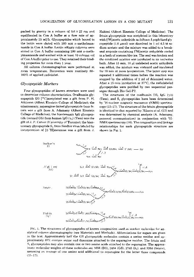
Four glycopeptides of known structure were used
to determine column characteristics. Ovalbumin gly- copeptide D3 [‘“Clacetylated was a gift from P. H.
Atkinson (Albert Einstein College of Medicine); the triantennary, asparagine-linked glycopeptide from fe- tuin was a gift from A. Adamany (Albert Einstein
College of Medicine); the biantennary IgG glycopep-
tide (termed GS) from human IgG (rl) (Tern) was the gift of J. P. Carver (University of Toronto); the bian-
tennary glycopeptide S, from Sindbis virus labeled by incorporation of [2-3H]mannose was a gift from J.
Ovalbumin 03
rlIgG (Tern) (GS)
Fetuin.
Hakimi (Albert Einstein College of Medicine). The
fetuin glycopeptide was acetylated in this laboratory
with [3H]acetic anhydride as follows: Lyophilized gly- copeptide (1.0 pmol) was dissolved in 1 ml 4.5 M so-
dium acetate and the mixture was added to a break- seal ampoule containing [3H]acetic anhydride cooled
in a bath of acetone/dry ice. The seal was broken and the combined mixture was incubated in an ice/water
bath. After 15 min, 10 ~1 unlabeled acetic anhydride was added, the mixture was vortexed and incubated
for 10 min at room temperature. The latter step was repeated 3 additional times before the reaction was
stopped by the addition of 2 ml of deionized water.
After a 15-min incubation at 37’C, the radiolabeled glycopeptides were purified by two sequential pas-
sages through Bio-Gel-PP. The structures of the ovalbumin D3, IgG (71)
(Tern), and S, glycopeptides have been determined by ‘H-nuclear magnetic resonance (NMR) spectros-
copy (15-17). The structure of the fetuin glycopeptide is identical to that reported by Nilsson et al. (13) and
was determined by chemical analysis (A. Adamany, personal communication) in conjunction with ‘H-
NMR spectroscopy (16). The composition and linkage relationships for each glycopeptide structure are
shown in Fig. 1.
FIG. 1. The structures of glycopeptides of known composition used as marker molecules for an- alytical column chromatography (see Materials and Methods). Abbreviations for sugars are given in the text. Approximately half the GS glycopeptide molecules contain a serine residue and ap- proximately 40% contain serine and threonine attached to the asparagine residue. The fetuin and
S, glycopeptides may also contain one or two amino acids attached to the asparagine. The approx- imate molecular weights of these molecules are 1656 (D3), 2464 (GS), 2745 (S,), and 3264 (fetuin), assuming an average of one amino acid additional to asparagine for the latter three compounds (15-17).

132 PAMELA STANLEY
Analytical Methods
All chemicals were reagent grade. Sugars used to calibrate the Bio-Gel columns (dextran, stachyose,
raffinose, cellobiose, and galactose) were detected by
the phenol-sulfuric acid assay for neutral sugars (18). Sialic acid was measured by the thiobarbituric acid
assay (19). Radiolabeled sialic acid was identified by
paper chromatography in parallel with authentic [‘?]sialic acid. Samples were spotted onto What-
man No. 1 paper in 200 ~1 50% ethanol. The chro- matogram was developed with n-butyl acetate:glacial
acetic acidwater (3:2:1) for 15.5 h at room tempera- ture. Radioactive substances were located by cutting
the dried paper into l-cm’ strips, incubating them in 1 ml water for 30 min at room temperature, and count- ing the mixture in 10 ml ACS II scintillation fluid.
RESULTS AND DISCUSSION
Gel Filtration of Pronase Glycopeptides
This laboratory has previously shown that Pronase glycopeptides derived from Lec4/VSV G glycoprotein exhibit a re- duced amount of the species termed So when chromatographed on Bio-Gel-P6 (4). These results were obtained with VSV grown in monolayer cultures of parental CHO cells and two independent Lec4 mu- tants. However, Lec4 cells tend to form loose monolayers and to detach from the dish before reaching confluency (4), mak- ing it difficult to obtain monolayers ap- propriate for sustaining a VSV infection. Since in suspension culture Lec4 cells grow as well as parental CHO cells, it was im- portant to determine whether VSV from suspension-grown cells would exhibit a glycosylation defect similar to that previ- ously observed in monolayer-grown virus.
Figure 2 shows the elution profiles on Bio-Gel-P6 of Pronase glycopeptides ob- tained from glucosamine-labeled, suspen- sion-grown parental/VSV ([3H]glucos- amine) and Pro-Lec4/VSV ( [‘4C]glucos- amine). The virus preparations were mixed prior to digestion with Pronase. Parental/ VSV exhibits the four expected species (So, S1, Sa, and SJ, while Lec4/VSV exhibits markedly reduced amounts of So. The gly- copeptides S1, Sp, and S, are present in Lec4/VSV Pronase glycopeptides and, al- though their elution profile does not differ significantly from their counterparts in
FIG. 2. Pronase glycopeptides of parental/VSV (3H) and Lec4/VSV (i4C) grown in suspension culture.
Purified glucosamine-labeled VSV was prepared from Pro-5 and Pro-Lec4.12-2 cells, mixed, digested with
Pronase, and chromatographed on Bio-Gel-P6 as de- scribed under Materials and Methods. The sample
was loaded in 200 ~1 buffer containing 500 pg dextran ( V,,) and 500 pg galactose (Vi, which occurred at frac-
tion 217). Fraction size was 0.47 ml. Arrows indicate elution positions of GS and D3 which were chro-
matographed in a parallel column with dextran and galactose markers.
parental/VSV, their relative proportions do appear to differ somewhat. Following neuraminidase treatment, So, S1, and SZ from parental/VSV and Lec,/VSV are converted to a broad peak which elutes at the position of S3 ((3), Stanley, unpub- lished observations). Based on the re- ported structure of the carbohydrate of G from BHK/VSV (9, lo), S3 would be ex- pected to be a triantennary carbohydrate moiety terminating in Gal residues. How- ever, the biantennary glycopeptide GS eluted before S3 on Bio-Gel-P6 (Fig. 2). Although it is known that the behaviour of charged glycopeptides on Bio-Gel is not strictly a function of molecular size, (20), these results suggested that S3 at least may consist predominantly of biantennary car- bohydrate structures.
Profiles essentially identical to Fig. 2 were obtained when suspension- grown Gatt/VSV ( [‘4C]glucosamine) and Gat-Lecli/VSV ( [3H]glucosamine) were compared in a similar experiment (data not shown). The profiles were not signif- icantly affected by the particular isotope of radioactive glucosamine incorporated. The combined results show that Pronase glycopeptides from monolayer (4) and sus- pension-grown VSV behave similarly on

LOCALIZATION OF GLYCOSYLATION LESION IN A CHO MUTANT 133
10 40 50 FRACTI& NUMBER
FIG. 3. Pronase glycopeptides of parental/VSV
(‘“C) and Lec4/VSV (sH) on Con A- Sepharose. Pro- nase digests of Pro-5/VSV (24,600 cpm), Pro-Lec-
4.12-2/VSV (302,000 cpm), and Gatt2Lec4.2D/VSV (56,600 cpm) were suspended in 460 &l Con A buffer
and chromatographed separately on Con A-Sephar- ose (0.6 X 14 cm) as described under Materials and Methods. Fractions of 600 ~1 were collected and 25
or 50 al/fraction was assayed for 3H and W. Con A buffer containing 0.1 M a-methylmannoside was ap-
plied at fraction 31 (arrow).
Bio-Gel-P6 and that Lec4/VSV glycopep- tides produced by either method, exhibit a deficiency of S,,.
The glycopeptides shown in Fig. 2 were generated by Pronase digestion of intact, purified VSV labeled with glucosamine during a single cycle of infection. The ab- sence of significant amounts of radiola- beled material eluting after S3 or at the Vi suggests that interconversion of glucos- amine to amino acids did not occur to an appreciable extent. These data are consis- tent with the fact that gel electrophoretic analysis of purified CHO/VSV labeled with glucosamine reveals only one major band which comigrates with the G glyco- protein ((3), Stanley, unpublished results). The latter also shows that significant con-
tamination of VSV preparations with la- beled cellular glycoproteins of different molecular size does not occur. In fact, the profiles in Fig. 2 are indistinguishable from those previously obtained following partial purification of G glycoprotein from CHO/ VSV (3). Based on these findings, the pu- rification of G is not a necessary prereq- uisite to studying the structure of its car- bohydrate moieties. Therefore, all proteo- lytic enzyme digestions were performed on purified intact virions.
Con A-Sepharose Chromatography of Pronase Glycopeptides
When glucosamine-labeled CHO/VSV Pronase glycopeptides were subjected to affinity chromatography on Con A-Se- pharose, approximately 20% of the label passed through while 80% bound to the column (Fig. 3). The majority of the Lec4/ VSV Pronase glycopeptides prepared from both independent Lec4 mutants also bound to the Con A column. However, a small proportion of the latter (approximately 10%) was recovered in the void volume of the initial effluent. Previous studies have shown that glycopeptides which contain branch GlcNAc residues do not bind to Con A-Sepharose, whereas many bianten- nary carbohydrate structures do bind to the lectin and require a-methylmannoside for elution (21-24). These observations were confirmed in our laboratory by ex- amining the behavior of glycopeptides of known structure (see Materials and Meth- ods). The triantennary glycopeptide from fetuin was shown to exhibit no binding to Con A-Sepharose, whereas the bianten- nary glycopeptide from Sindbis virus (S,) was shown to bind to the column and to be specifically eluted with a-methylman- noside (data not shown). The results in Fig. 3 suggest that, while the majority of the glycopeptides of suspension-grown pa- rental/VSV and Lec4/VSV appear to be biantennary (Con A bound), a small pro- portion behave like tri- or tetraantennary structures. It is the latter species which is reduced in Lec4/VSV.
To determine how the glycopeptide frac- tions separated on Con A-Sepharose (Fig.

134 PAMELA STANLEY
3) correspond to the species observed on Bio-Gel-P6 (Fig. 2), the glycopeptides which came through Con A (Con A-T) and those which bound to Con A (Con A-B) were pooled separately and desalted. Anal- ogous glycopeptides from parental/VSV and Lec4/VSV were mixed and chromato- graphed on Bio-Gel-PG. The Con A-T ma- terial eluted mainly in the region corre- sponding to S, with some radiolabel elut- ing at the void volume. The Con A-B material contained species which eluted at the positions expected for S1, SB, and S,. It did not contain appreciable radiolabel corresponding to S, or eluting at the void volume. These results indicated that the proportion of carbohydrate occurring as branched structures in suspension-grown CHO/VSV is small and that it is largely accounted for by S,. The species termed Si, Sz, and S3 appear to comprise mainly biantennary structures. Further evidence in support of these conclusions is pre- sented in Figs. 6 and 7.
Properties of Tryptic Glycopeptides from ParentallVS V and Lec4/VS V
Since the alteration expressed by Lec4/ VSV apparently affects a small proportion of the total glycopeptides, it seemed im- portant to determine whether the lesion occurred at both glycosylation sites in the G glycoprotein. Previous studies have shown that the two glycosylation sites in G from CHO/VSV may be separated as desialylated, tryptic glycopeptides by ion- exchange chromatography (3, 14, 25). In order to compare the carbohydrate units at each glycosylation site in G from pa- rental/VSV and Lec4/VSV, glucosamine- labeled tryptic peptides were generated from purified virus, treated with neur- aminidase, and subjected to chromatog- raphy on DEAE-Sephacel (Fig. 4). In each case, two major peaks were observed (I and II). The peak marked SA was identified as radiolabeled sialic acid by paper chroma- tography. The origin of the other small peaks is unknown but their presence is reproducible under apparently complete digestion conditions.
The elution positions of peaks I and II
FRACTION NUMBER
FIG. 4. Neuraminidase-treated tryptic glycopep-
tides of parental/VSV (I%) and Lec4/VSV f3H) on DEAE-Sephacel. Desalted glycopeptides from Pro-5/
VSV (7500 cpm), Pro-Lec4.12-2/VSV (50,000 cpm), and GatmLec4.2D/VSV (30,000 cpm) were dissolved
in 0.5 ml of 1 mM Tris-HCl, pH 8.5, and chromato- graphed on DEAE-Sephacel as described under Ma-
terials and Methods. Fractions of 1.75 ml were col- lected and 500 or 800.al samples of each fraction were counted. A linear gradient of 0 to 0.1 M NaCl was
applied at fraction 21 (arrow) and the theoretical gra- dient of Cl- is indicated (- - -). NaCl (1 M) was ap-
plied at fraction 131 (arrow).
were similar for parental/VSV and Lec4/ VSV glycopeptides prepared from both in- dependent Lec4 mutants (Fig. 4). How- ever, the proportion of label eluting with peak II was higher for glycopeptides de- rived from Lec4/VSV (approximately 77%) compared with parental/VSV (approxi- mately 55%). This suggested that perhaps the reduced amount of Con A-T glycopep- tides observed in Lec4/VSV might be oc- curring only at glycosylation site I.
To investigate this question, peaks I and II were pooled separately, desalted, di- gested with Pronase and their behavior examined on Con A-Sepharose. From the results in Fig. 5, it appears that both gly- cosylation sites in parental/VSV contain carbohydrate moieties which do not bind to Con A, although proportionately more Con A-T glycopeptides occur at glycosy-

LOCALIZATION OF GLYCOSYLATION LESION IN A CHO MUTANT 135
PEAK1 PEAKII
4 \
1°C) Parent 4
L” FRA??ION NUMB-i-R
FIG. 5. Pronase glycopeptides from glycosylation
sites I and II of parental/VSV (I%) and Lec4/VSV (3H) on Con A-Sepharose. Peaks I and II from Fig.
4 were pooled separately, Peak II glycopeptides were desalted, and Peaks I and II glycopeptides were di-
gested with Pronase. The digests were dissolved in 400 ~1 Con A buffer and chromatographed on Con A-
Sepharose (0.6 X 14 cm) as described in Fig. 3.
lation site I. By contrast Lec4/VSV exhib- its proportionately less Con A-T glycopep- tides at glycosylation site I and no de- tectable Con A-T glycopeptides at glycosylation site II. Therefore, Lec4/VSV appears to be missing Con A-T glycopep- tides from both glycosylation sites. The results in Fig. 5 also suggest that removal of sialic acid residues does not alter the elution pattern of the VSV glycopeptides on Con A-Sepharose. This point was con- firmed by examining neuraminidase- treated Pronase glycopeptides directly on Con A-Sepharose (data not shown) and has previously been reported, for other de- sialylated glycopeptides (23, 24).
Further Characterization of Pronase Glycopeptides
Since the carbohydrate defect expressed by Lec4/VSV was observed at both gly- cosylation sites of G, further characteriza- tion was performed with total Pronase gly-
copeptides. To avoid contamination with even a small amount of undigested mate- rial, Pronase glycopeptides from parental/ VSV and Lec4/VSV were chromato- graphed separately on Bio-Gel-P6 and SO, Si, Sz, and S3 (A and B) species were pooled (Fig. 6). Each of these preparations was subjected to affinity chromatography on Con A-Sepharose. Representative results obtained with S,, and S, are shown in Fig. 7. The only species which contained pre- dominantly Con A-T material was So from both parental/VSV and Lec4/VSV glyco- peptides. The proportion of the total ra- dioactivity which bound to Con A-Se-
3- so Sl 52 53
VO (“C) Parent
- 1
2-
, ! 40 50 60 70 80 90
FRACTION NUMBER
FIG. 6. Preparation of Pronase glycopeptides So, Si,
S,,and Ss. Purified Pro-5/VSV (-340,000 cpm i4C) and Pro-Lec4.12.2/VSV (-300,000 cpm 3H) were digested with Pronase and chromatographed on two
different columns of Bio-Gel-PG. The sample was loaded in 150 ~1 buffer containing dextran, stachyose, raffinose, cellobiose, and galactose and 0.4-ml frac-
tions were collected. Samples of 10 or 20 ~1 were as- sayed per fraction. The parent/VSV column flowed at approximately 8 ml/h, whereas the Lecl/VSV col-
umn flowed much slower (-2 ml/h). Fractions des- ignated So, Si, S,, Ss.+, and Sss were pooled as shown
c-j.

136 PAMELA STANLEY
pharose for parental/VSV was 10 (S,,), 86 (S,), 92 (S,), 87 (S,,), and 92% (S,s). For Lec4/VSV, the corresponding proportions of Con A-B material were 31 (So), 99 (S,), 97 (Sz), 97 (S& and 99% @,a). Clearly, the majority of the Con A-T (branched) carbohydrate structures are found in the S,, fraction whereas S1, Sg, and S, contain predominantly Con A-B (biantennary) structures. Although contamination of ad- jacent pooled species might be predicted since separation is not complete on the Bio-Gel-P6 column, it seems likely that at least some branched species coelute with Sz and S3 from parental/VSV, probably as a result of heterogeneity in their sialic acid content. Each of the Pronase glycopeptide preparations which bound to Con A-Se- pharose was eluted with a gradient of O- 200 mM cz-methylmannoside. However, no significant differences in elution positions were observed between parental- and mu- tant-derived species. In all cases, elution of the peak of labeled glycopeptides was achieved at about 3 mM a-methylmanno- side and all radiolabel was eluted by 20 mM
cY-methylmannoside.
PSA-Agarose Chromatography of Pronase Glycopeptides
The binding characteristics of certain carbohydrate structures on a pea lectin (PSA) affinity column have been described by Kornfeld et al. (26). Biantennary car- bohydrate moieties, which contain fucose linked c~1,6 to the GlcNAc residue adjacent to asparagine, bind to PSA-agarose, as does a triantennary species which contains (in addition to the crl,6-linked fucose res- idue) a branch GlcNAc residue linked 01,s to mannose. By contrast, tri- or tetraan- tennary moieties containing branch GlcNAc residues linked /31,4 to mannose do not bind to PSA-agarose, even if they also contain a branch GlcNAc linked pl,S to mannose and an al,6-linked fucose res- idue. Examination of the glycopeptides of known structure from fetuin and Sindbis virus yielded results consistent with these findings. The triantennary fetuin glyco- peptide, which contains a branch GlcNAc linked @1,4 to mannose, was not bound by
FIG. 7. Pronase glycopeptides So and S, on Con A-
Sepharose. Samples of S, and S, containing 3000- 5000 cpm from Fig. 6 were lyophilized, resuspended
in 400 pl Con A buffer and chromatographed on Con A-Sepharose (0.6 X 22 cm). Fractions of approxi- mately 960 ~1 were collected and 500 pi/fraction was
counted for 3H or r4C. A loo-ml gradient of 0 to 200 mM cu-methylmannoside was applied at fraction 16
(arrow). Progression of these gradients was deter- mined by phenol-sulfuric acid assay of 5.~1 aliquots
of each fraction.
PSA-agarose, whereas the biantennary glycopeptide S1, which possesses the ap- propriate al,6 fucose residue, bound spe- cifically to the column (data not shown).
To determine whether the Con A-T (branched) species of CHO/VSV glycopep- tides displayed heterogeneity, So material which had been passed through Con A- Sepharose (Fig. 7) was chromatographed on PSA-Sepharose. Two labeled species were separated (Fig. 8). While approxi- mately 70% of the Con A-T glycopeptides passed through PSA-Sepharose (PSA-T glycopeptides), about 30% bound to the column and was specifically eluted with c-Y-methylmannosde. By contrast, all the S,, glycopeptides from Lec4/VSV which passed through Con A-Sepharose (Fig. 7), also passed through PSA-Sepharose (Fig. 8). That is, no detectable Con A-T, PSA- B material was observed from VSV grown in either of the independent Lec4 mutants. This suggested that the defect in Lec4 cells is the lack of synthesis of a Con A-T (branched) carbohydrate species which is bound by PSA-agarose.
To determine whether the results with Lec4/VSV reflect a mutant defect which

LOCALIZATION OF GLYCOSYLATION LESION IN A CHO MUTANT 137
- 10 20 30 40 50 60
FRACTION NUMBER
FIG. 8. Pronase glycopeptides So and S, which pass
through Con A-Sepharose chromatographed on PSA- agarose. The Con A-T glycopeptides from Fig. 7 were
pooled separately.The parental/VSV glycopeptides
(-2000 cpm) were desalted and loaded in 400 al Con A buffer onto PSA-agarose. The Pro-Lec4.122/VSV
glycopeptides (- 1000 cpm) were loaded directly in 2.9 ml Con A buffer onto PSA-agarose. The
GattLec4.2D glycopeptides (-1500 cpm) were Pro- nase glycopeptides (not Ss material) which had passed
through Con A- Sepharose and been desalted prior to being loaded in 400 ~1 Con A buffer onto PSA- agarose. Each fraction (500 al) was counted in total.
At fraction 31, elution of bound glycopeptides was achieved with Con A buffer containing 200 mM a-
methylmannoside (arrow).
is also expressed at the cell surface (as was previously indicated by the specific lectin- binding properties of Lec4/VSV (4)), Pro- nase glycopeptides were prepared by Pro- nase digestion of intact parental and Lec4 CHO cells which had been grown for 3 days in the presence of [“Hlfucose. The digest was desalted and applied to Con A-Se- pharose (Fig. 9). The Con A-T material was pooled and applied directly to PSA- agarose (Fig. 9). Consistent with the be- havior of the viral glycopeptides, the Con A-T glycopeptides from parental CHO cells fractionated into PSA-T and PSA-B glycopeptides on PSA-agarose. However, the corresponding Con A-T glycopeptides from Lec4 cells contained only PSA-T moieties. A similar result was obtained
with glucosamine-labeled cell surface gly- copeptides (data not shown). Therefore, cellular-derived, as well as viral Pronase glycopeptides from Lec4 cells, are missing a carbohydrate species synthesized by parental CHO cells. This glycopeptide be- haves like a branched carbohydrate struc- ture which does not bind to Con A-Se- pharose but which does bind to PSA- agarose.
Based on the lectin-binding properties of structurally characterized glycopeptides ((21-24, 26) and this manuscript) and on its elution position on Bio-Gel-P6, the spe- cies missing from Lec4/VSV exhibits the properties of a triantennary carbohydrate moiety in which the branch GlcNac is linked fi1,6 to mannose. A likely hypoth- esis to explain the mutation in Lec4 cells is that these cells are missing a specific /31,6-N-acetylglycosaminyltransferase ac- tivity. Consistent with this hypothesis is the fact that a pentasaccharide which con- tains a fil,G-GlcNAc attached to a disub- stituted mannose residue appears to be the most complementary structure which rec- ognizes the leukoagglutinin from PHA (PHA-L, (27)). Lec4 cells as well as Lec4/ VSV exhibit a markedly reduced ability to bind lz51-PHA-L (4). The mutant cells are also highly resistant to the toxicity of PHA-L (>lOOO-fold compared with paren- tal CHO cells (28)). Finally, the Lec4 mu- tation behaves recessively in somatic cell hybrids (29), which is consistent with the loss of an enzymatic activity. Another mu- tant (PhaR2.1) selected for resistance to PHA-L from a population of mouse lym- phoma cells (30) also appears to be defi- cient in a glycopeptide species containing fll,6-linked GlcNAc residues (R. Cum- mings and S. Kornfeld, personal commu- nication).
Clearly, conclusions regarding the struc- ture of the carbohydrate unit missing from Lec4/VSV must remain tentative until direct structural analyses are performed. We are currently pursuing this objective by examining lectin-affinity purified Pro- nase glycopeptides from CHO/VSV by ‘H- NMR spectroscopy. Complete structural information may be obtained by this ap- proach because of the large library of

138 PAMELA STANLEY
Parent Pro- kc 4
II’,’ CON A
3 2
2 3 “0 I 5 1
u_
;: OI “; 2 4
0 ; 3
5, 2
I
20 ‘40 60 20 40 60
FRACTION NUMBER
FIG. 9. Cell surface Pronase glycopeptides chro- matographed on Con A-Sepharose and PSA-agarose. Pronase glycopeptides of fucose-labeled Pro-Lec4 and
Pro-5 CHO cells were desalted on Bio-Gel-P2. The glycopeptides which eluted with the V,( -37,000 cpm
Pro-5 and 24,000 cpm Lec4 glycopeptides) were dis- solved in 400 ~1 Con A buffer and loaded onto Con
A-Sepharose (0.6 X 22 cm). Thirty fractions (400 ~1) were collected before a loo-ml gradient of 0 to 200
mM n-methylmannoside was applied (arrow). An al- iquot (50 ~1) of every second fraction was counted.
The Con A-T glycopeptides were subsequently pooled and approximately 1000 cpm of each preparation was
loaded in 500 or 600 ~1 of Con A buffer onto PSA- agarose. This column was run as described for the Con A-Sepharose column except that the fraction
volume was 480 ~1 and 200 mM oc-methylmannoside
was used directly to elute bound glycopeptides (ar- row).
known carbohydrate compounds for which chemical shifts have already been assigned (16, 31). In preliminary experiments, we have studied the Con A-B glycopeptides of CHO/VSV (32). They appear identical to the biantennary carbohydrate moieties associated with Sindbis virus glycopep- tides (17). The spectra reveal heteroge- neity in the number of sialic acid and fu- case residues per molecule in agreement with the heterogeneity observed on Bio- Gel-P6 for Con A-B glycopeptides and the fact that only about 30% of the labeled Con A-B glycopeptides bind to PSA-aga- rose (Stanley, unpublished observations). We have also examined the glycopeptide fraction from parental/VSV which passes through Con A-Sepharose and subse- quently binds to PSA-agarose. As pre-
dicted, this fraction exhibits anomeric and N-acetyl proton resonances consistent with the presence of a /31,6-linked GlcNAc res- idue (32). Further studies should enable a complete structure to be deduced for this carbohydrate moiety.
The carbohydrate units of the G glyco- protein from CHO/VSV clearly require further structural characterization. The heterogeneity observed upon fractionation of Pronase glycopeptides from G indicates that a range of asparagine-linked complex carbohydrate moieties may occur at each glycosylation site. The molecular basis for this variability is intriguing. Comparisons of the behavior on Con A-Sepharose of glycopeptides derived from monolayer- grown and suspension-grown CHO/VSV indicate that culture conditions may affect the proportion of branched versus bian- tennary carbohydrate moieties (Stanley, unpublished observations). Whatever the basis of the observed heterogeneity, the variety of carbohydrate structures associ- ated with G from CHO/VSV have proved extremely useful in localizing the glyco- sylation defects expressed by many of our CHO glycosylation mutants ((3,4), Camp- bell and Stanley, unpublished observa- tions).
ACKNOWLEDGMENTS
The author is indebted to Grace Vivona for excel-
lent technical assistance and thanks Anthony Ada- many and Paul Atkinson for critical comments on the
manuscript. This work was supported by a grant from the American Cancer Society (BC332A). P.S. is also the recipient of a senior faculty award from the
American Cancer Society. Partial salary support was provided for a period by the Core Support for Cancer
Research Center Grant (NIH NC1 P30 CA 13330-10).
REFERENCES
1. STANLEY, P. (1980) in Biochemistry of Proteo- glycans and Glycoproteins (Lennarz, W. J., ed.), pp. 161-189, Plenum, New York.
2. STANLEY, P. (1982) in Methods in Enzymology (Colowick, S. P., and Kaplan, N. O., eds.), Ac- ademic Press, New York, in press.
3. ROBERTSON, M. A., ETCHISON, J. R., ROBERT-
SON, J. S., SUMMERS, D. F., AND STANLEY, P. (1978) Cell 13, 515-526.
4. STANLEY, P., AND SUDO, T. (1981) Cell 23, 763- 769.

LOCALIZATION OF GLYCOSYLATION LESION IN A CHO MUTANT 139
5. ROSE, J. K., AND GALLIONE, C. J. (1981) J. Viral.
39,519-528. 6. BURGE, B. W., AND HUANG, A. S. (1970) J. Viral.
6.176-182.
7. ETCHISON, J. R., AND HOLLAND, J. J. (1974) Vi- rology 60, 217-229.
8. ROBERTSON, J. S., ETCHISON, J. R., AND SUM- MERS, D. F. (1976) J. Virol. 19, 871-878.
9. ETCHISON, J. R., ROBERTSON, J. S., AND SUM-
MERS, D. F. (1977) Virology 78, 375-392. 10. READING, C. L., PENHOET, E. D., AND BALLOU,
C. E. (1978) J. Biol. Chem. 253, 5600-5612.
11. SEFTON, B. (1976) J. Virol. 17, 85-93.
12. BAENZIGER, J. U., AND FIETE, D. (1979) J. Biol. Chem. 254,2400-2407.
13. NILSSON, B., NORDEN, N. E., AND SVENSSON, S. (1979) J. Biol. Chem. 254, 4545-4553.
14. ETCHISON, J. R., ROBERTSON, J. S., AND SUM- MERS, D. F. (1981) J. Gen. Virol. 57, 43-52.
15. CARVER, J. P., GREY, A. A., WINNIK, F. M., HAK-
IMI, J., CECCARINI, C., AND ATKINSON, P. H. (1981) Biochemistry 20, 6600-6606.
16. CARVER, J. P., AND GREY, A. A. (1981) Biochem- istry 20,6607-6616.
17. HAKIMI, J., CARVER, J., AND ATKINSON, P. H. (1981) Biodwmistry 20, 7314-7319.
18. DUBOIS, M., GILLES, K. A., HAMILTON, J. K., REBERS, P. A., AND SMITH, F. (1956) Ad. Chem. 28, 350-356.
19. AMINOFF, D. (1961) Biochem. J. 81, 384-392.
20. JOHN,M.,TRENEL,G.,AND DELLWEG,H.(~~~~)
J. Chromatogr. 42, 476-484. 21. OGATA, S., MURAMATSU, T., AND KOBATA, A.
(1975) J. Biochem. 78, 687-696.
22. KRUSIUS, T. (1976) FEBS Lat. 66(l), 86-89. 23. NARASIMHAN, S., WILSON, J. R., MARTIN, E.,
AND SCHACHTER, H. (1979) Cancd J. Biochem. 57, 83-96.
24. BAENZIGER, J. U., AND FIETE, D. (1979) J. Biol. Chem. 254,789-795.
25. ROBERTSON, J. S., ETCHISON, J. R., AND SUM-
MERS, D. F. (1982) J. Gen. Virol. 58, 13-23. 26. KORNFELD, K., REITMAN, M. L., AND KORNFELD,
R. (1981) J. Biol. Chem. 256, 6633-6640.
27. HAMMARSTROM, S., HAMMARSTROM, M.-L., SUNDBLAND, G., ARNARP, J., AND LONNGREN,
J. (1982) Proc. Nat. Acad. Sci. USA 79, 1611- 1615.
28. STANLEY, P., CAILLIBOT, V., AND SIMINOVITCH, L. (1975) Cell 6, 121-128.
29. STANLEY, P., AND SIMINOVITCH, L. (1977) SO- matic Cell Genet. 3, 391-405.
30. TROWBRIDGE, I. S., HYMAN, R., FERSON, T., AND MAZAUSKAS, C. (1978) Eur. J. Immunol. 8, 716-723.
31. VLIEGENTHART, J. F. G., VAN HALBEEK, H., AND
DORLAND, L. (1981) Pure Appl. Chem. 53,45- 77.
32. STANLEY, P., AND ATKINSON, P. H. (1982) 12th Znt. Congr. Biochem. in press.