CAR T C - telmarc.com Papers/140CART.pdf · CAR T cells are chimeric antigen receptors on T cells....
35
CAR T CELLS AND CANCER Cancer treatment using immune cells has been examined for almost fifty years. The recent effort using T killer cells, CTL, and modifying their receptors to match tumors is called chimeric antigen receptors, CAR T cells, are specifically targeted at the tumor cell. We examine these cells from a high level and integrate that into several adjunct areas as well. Copyright 2016 Terrence P. McGarty, all rights reserved. Terrence P McGarty White Paper No 140 November, 2016
Transcript of CAR T C - telmarc.com Papers/140CART.pdf · CAR T cells are chimeric antigen receptors on T cells....

CARTCELLSANDCANCER
Cancer treatment using immune cells has been examined for almost fifty years. The recent effort using T killer cells, CTL, and modifying their
receptors to match tumors is called chimeric antigen receptors, CAR T cells, are specifically targeted at the tumor cell. We examine these cells from a high level and integrate that into several adjunct areas as well. Copyright
2016 Terrence P. McGarty, all rights reserved.
TerrencePMcGartyWhitePaperNo140November,2016
DRAFT WHITE PAPER CAR T CELLS AND CANCER
1 | P a g e
Notice
This document represents the personal opinion of the author and is not meant to be in any way
the offering of medical advice or otherwise. It represents solely an analysis by the author of
certain data which is generally available. The author furthermore makes no representations
that the data available in the referenced papers is free from error. The Author also does not
represent in any manner or fashion that the documents and information contained herein can
be used other than for expressing the opinions of the Author. Any use made and actions
resulting directly or otherwise from any of the documents, information, analyses, or data or
otherwise is the sole responsibility of the user and The Author expressly takes no liability for
any direct or indirect losses, harm, damage or otherwise resulting from the use or reliance upon
any of the Author's opinions as herein expressed. There is no representation by The Author,
express or otherwise, that the materials contained herein are investment advice, business
advice, legal advice, medical advice or in any way should be relied upon by anyone for any
purpose. The Author does not provide any financial, investment, medical, legal or similar advice
in this document or in its publications on any related Internet sites.

DRAFT WHITE PAPER CAR T CELLS AND CANCER
2 | P a g e
Contents
1 Introduction ............................................................................................................................. 3
1.1 History and Principles ...................................................................................................... 3
1.2 CAR T Cells ..................................................................................................................... 4
2 T Cells ..................................................................................................................................... 7
2.1 T Cell Dynamics............................................................................................................... 7
2.2 PD-1 Pathways ................................................................................................................. 9
3 CAR Cells ............................................................................................................................. 13
3.1 Generational Architecture .............................................................................................. 15
3.1.1 First Generation ...................................................................................................... 16
3.1.2 Second Generation .................................................................................................. 17
3.1.3 Third Generation ..................................................................................................... 18
4 Reverse Transcription and Gene Insertion ............................................................................ 19
5 The Entire Process ................................................................................................................ 22
5.1 Detailed Steps ................................................................................................................. 22
5.2 Switch Control ................................................................................................................ 23
6 Observations ......................................................................................................................... 25
6.1 Damage ........................................................................................................................... 25
6.2 Specific Damage ............................................................................................................ 26
7 Appendix: Production (Methods and Protocols)................................................................... 27
7.1 Maude et al ..................................................................................................................... 27
7.2 Wu et al .......................................................................................................................... 28
7.2.1 Construction of ON-switch CARs .......................................................................... 28
7.2.2 Culturing conditions for T cells and target cells ..................................................... 28
7.2.3 Lentiviral engineering of T cells and K562 target cells .......................................... 29
7.2.4 Verifying CAR expression on T cells ..................................................................... 29
7.2.5 Quantitation of IL-2 and/or IFN-y production ........................................................ 30
7.2.6 Quantitation of CD69 surface expression ............................................................... 30
7.2.7 Quantitation of T cell proliferation ......................................................................... 30
7.3 Ellebrecht ....................................................................................................................... 32
8 References ............................................................................................................................. 33

DRAFT WHITE PAPER CAR T CELLS AND CANCER
3 | P a g e
1 INTRODUCTION The immune system is a powerful tool that can be used in a variety of ways. One problem is that it seems that every day we discover another subtlety regarding how this functions. It is a tool, and a very powerful tool, that can be used as a scalpel or a butchering ax. With the advent of CARs, specifically designed killer T cells, CTLs or cytotoxic T cells, one can attack cancer cells, however at times this tool can explode in our hands. This paper is an attempt to examine the CAR T Cells as a tool which can be engineered. The problem we face however is that in engineering the tool we oftentimes do not have a full grasp of its effects. Our intent herein is not to provide a detailed up to date review of CARs but to provide a summary introduction to the potential they provide. This area is still very much a work in progress and as such is subject to ongoing change. 1.1 HISTORYANDPRINCIPLES Steven Rosenberg has been studying how best to use the immune system to fight cancer. His 1992 was a prescient piece that laid out the future opportunities. From then until now, some 25 years, we know a great deal about the immune system which was lacking then and furthermore we have a wealth of tools to manipulate the cells involved. From Kahilil et al we have an introduction to CARs which provide continuity from the work on monoclonal antibodies, MABs: In the past decade, advances in the use of monoclonal antibodies (mAbs) and adoptive cellular therapy to treat cancer by modulating the immune response have led to unprecedented responses in patients with advanced-stage tumors that would otherwise have been fatal. To date, three immune-checkpoint-blocking mAbs have been approved in the USA for the treatment of patients with several types of cancer, and more patients will benefit from immunomodulatory mAb therapy in the months and years ahead. Concurrently, the adoptive transfer of genetically modified lymphocytes to treat patients with hematological malignancies has yielded dramatic results, and we anticipate that this approach will rapidly become the standard of care for an increasing number of patients. In this Review, we highlight the latest advances in immunotherapy and discuss the role that it will have in the future of cancer treatment, including settings for which testing combination strategies and 'armored' CAR T cells are recommended. From Batlevi et al we have a discussion on the flow from MABs to CARs with a nexus to checkpoint inhibitors, namely PD-1 inhibitors: The success of the anti-CD20 monoclonal antibody rituximab in the treatment of lymphoid malignancies provided proof-of-principle for exploiting the immune system therapeutically. Since the FDA approval of rituximab in 1997, several novel strategies that harness the ability of T cells to target cancer cells have emerged.

DRAFT WHITE PAPER CAR T CELLS AND CANCER
4 | P a g e
Reflecting on the promising clinical efficacy of these novel immunotherapy approaches, the FDA has recently granted 'breakthrough' designation to three novel treatments with distinct mechanisms. First, chimeric antigen receptor (CAR)-T-cell therapy is promising for the treatment of adult and pediatric relapsed and/or refractory acute lymphoblastic leukemia (ALL). Second, blinatumomab, a bispecific T-cell engager (BiTE®) antibody, is now approved for the treatment of adults with Philadelphia-chromosome-negative relapsed and/or refractory B-precursor ALL. Finally, the monoclonal antibody nivolumab, which targets the PD-1 immune-checkpoint receptor with high affinity, is used for the treatment of Hodgkin lymphoma following treatment failure with autologous-stem-cell transplantation and brentuximab vedotin. Herein, we review the background and development of these three distinct immunotherapy platforms, address the scientific advances in understanding the mechanism of action of each therapy, and assess the current clinical knowledge of their efficacy and safety. We also discuss future strategies to improve these immunotherapies through enhanced engineering, biomarker selection, and mechanism-based combination regimens. One of the observations when dealing with cancer and the immune system is that once when on tries a specific approach one often finds new mechanisms which can either be used or must be thwarted. From Jackson et al there is a discussion of the work of CARs using CD-19 targets: The engineered expression of chimeric antigen receptors (CARs) on the surface of T cells enables the redirection of T-cell specificity. Early clinical trials using CAR T cells for the treatment of patients with cancer showed modest results, but the impressive outcomes of several trials of CD19-targeted CAR T cells in the treatment of patients with B-cell malignancies have generated an increased enthusiasm for this approach. Important lessons have been derived from clinical trials of CD19-specific CAR T cells, and ongoing clinical trials are testing CAR designs directed at novel targets involved in hematological and solid malignancies. In this Review, we discuss these trials and present strategies that can increase the antitumor efficacy and safety of CAR T-cell therapy. Given the fast-moving nature of this field, we only discuss studies with direct translational application currently or soon-to-be tested in the clinical setting. 1.2 CARTCELLS CAR T cells are chimeric antigen receptors on T cells. Chimeric because one designs them specifically for the target cells and essentially crated a multiheaded receptor that matches the antigen presented by the tumor cell.

DRAFT WHITE PAPER CAR T CELLS AND CANCER
5 | P a g e
We provide a simple example below for a third-generation CAR:
TCR zeta chain signaling domain
Hinge Region
Target Cell
T Cell
Membrane Bound Tumor Antigen ie CD 19
PPP
PPP
Costimulatory Domain 1
Costimulatory Domain 2
The function of this designed T cell it to allow a normal CTL, killer T cell, attach to a cancer cell with a recognizable antigen, and then to do what CTLs do well, allow the attacked cell to go into apoptosis, and just disappear, its constituents being used elsewhere.

DRAFT WHITE PAPER CAR T CELLS AND CANCER
6 | P a g e
We demonstrate this process graphically above.

DRAFT WHITE PAPER CAR T CELLS AND CANCER
7 | P a g e
2 T CELLS We review some of the key functions of T cells. The two types of T cells of interest are T helper and T killer or cytotoxic T cells, CTL. The CTL is the prime target of interest for it is the cell which can attach to a tumor cell and effect apoptosis of the tumor cell by its normal operations. The T helper supports the CTL by expressing IL-2 which allows for proliferation of that specific CTL type. 2.1 TCELLDYNAMICS The CTL has surface receptors as shown below. Two are extending well beyond the cell wall and the remaining four are below the cell wall and provide for intra cellular activation. The complex acts in unison attaching to targeted cells. Now the essence of CARs is to modify this receptor so as to effect targeting of tumor cells and their exposed antigens.
This CTL binding process is shown below. Simply the process is as follows: 1. An antigen presenting cell, APC, in this case a tumor cell, presents an antigen using the MHC I molecule. Also, the tumor cell may have another surface protein that results in the presentation of a tumor specific surface molecule like CD-19 in the case of hematological malignancies. The process starts with the ability to identify this molecule. 2. Then the CTL has a matching or cognate receptor which aligns with the MHC I and Ag combination and it attaches itself, and via CD-8 strongly binds to the cell, also using CD-3. 3. Upon binding the CTL can release cytokines or equivalents that result in the apoptosis of the cell.

DRAFT WHITE PAPER CAR T CELLS AND CANCER
8 | P a g e
APC or Cancer Cell
CTL/Tk
MHC I
Cancer Cell Ag
CD 3
T Cell Rcptr
CD 8
We show the apoptosis below. Here the bound CTL recognizes the cancer cell and then releases apoptosis inciting proteins.
DRAFT WHITE PAPER CAR T CELLS AND CANCER
9 | P a g e
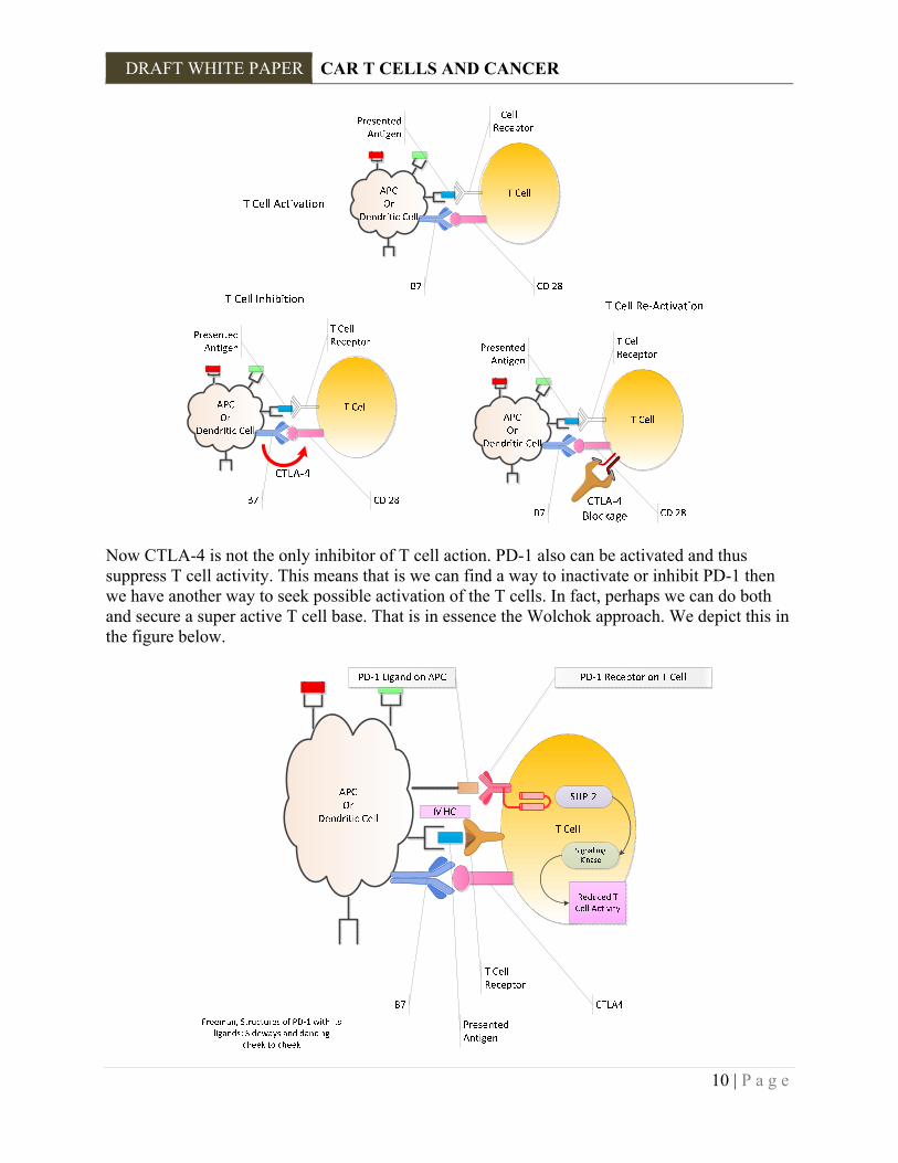
Thus, for any cancer cell we should be able to use this process, if we first know the Ag that is presented and second if we can create a receptor on a T killer cell, CTL, that recognizes that ligand and in turn can activate the apoptotic process. In the simplest terms this is how we might proceed. 1. Extract a tumor cell. 2. Ascertain the surface molecules and determine which one is unique to that type of cell and NOT common in other cells. You don't want the CTLs attacking everything. 3. Create a binding receptor for that ligand. 4. Extract the patient's CTL and insert by some reverse transcription manner, or CRISPR type approach the genes for that designed receptor. 5. Grow these modified cells in vitro using IL-2 or the like. 6. Insert these back in the patient. This is the "back of the envelope" approach to CAR therapy. Of course, there are many obstacles and the approach uses tools which may have to be gathered from afar. But as those who have developed CARs have shown it is doable. 2.2 PD‐1PATHWAYS Now what we have described above is not that simple. There are what are called a variety of "Checkpoint Inhibitors" that are an integral part of the control mechanisms of the immune system so that it does not go wild and destroy itself. Let us begin with a brief review of PD-1 pathways. We have previously discussed the CTLA-4 blockage and the current approaches used to inactivate that element of T cell suppression. We summarize that again in the figure below.
DRAFT WHITE PAPER CAR T CELLS AND CANCER
10 | P a g e
Now CTLA-4 is not the only inhibitor of T cell action. PD-1 also can be activated and thus suppress T cell activity. This means that is we can find a way to inactivate or inhibit PD-1 then we have another way to seek possible activation of the T cells. In fact, perhaps we can do both and secure a super active T cell base. That is in essence the Wolchok approach. We depict this in the figure below.
DRAFT WHITE PAPER CAR T CELLS AND CANCER
11 | P a g e
The paper by Okazaki and Honjo in 2007 also details many of the critical elements regarding the PD-1 and its ligands. It details many of the recognized disease states as well. As they state: Since the discovery of PD-1 in 1992, the biological function of PD-1 remained mystery for many years. Generation of Pdcd1mice and the discovery of its ligands turned around the situation and the function of PD-1 was unveiled thick and fast in these 5 years. Consequently, it became clear that PD-1 plays critical roles in the regulation of autoimmunity, tumor immunity, infectious immunity, transplantation immunity, allergy and immune privilege. The development of autoimmune diseases by Pdcd1 mice especially enchanted clinicians and promoted clinical research as well. Currently, many groups are trying to generate not only PD-1 antagonists for the treatment of cancer and infectious diseases but also PD-1 agonists for the treatment of autoimmune diseases, allergy and transplant rejection. Among these, humanized antibody against human PD-1 was approved by Food and Drug Administration of the United States as an investigational new drug in August 1, 2006. Clinical trials will test its clinical efficacy on cancer and infectious diseases. Now we can examine the features of PD-1. As Freeman states: T cell activation requires a TCR mediated signal, but the strength, course, and duration are directed by costimulatory molecules and cytokines from the antigen-presenting cell (APC). An unexpected finding was that some molecular pairs attenuate the strength of the TCR signal, a process termed co-inhibition. The threshold for the initiation of an immune response is set very high, with a requirement for both antigen recognition and costimulatory signals from innate immune recognition of ‘‘danger’’ signals. Paradoxically, T cell activation also induces expression of co-inhibitory receptors such as programmed death-1 (PD-1). Cytokines produced after T cell activation such as INF- and IL-4 up-regulate PD-1 ligands, establishing a feedback loop that attenuates immune responses and limits the extent of immune-mediated tissue damage unless overridden by strong costimulatory signals. PD-1 is a CD28 family member expressed on activated T cells, B cells, and myeloid cells. In proximity to the TCR signaling complex, PD-1 delivers a co-inhibitory signal upon binding to either of its two ligands, PD-L1 or PD-L2. Engagement of ligand results in tyrosine phosphorylation of the PD-1 cytoplasmic domain and recruitment of phosphatases, particularly SHP2 Additional insight can also be provided by examining the regulatory T cells as well. As Francisco et al state: Regulatory T cells (Tregs) and the PD-1: PD-ligand (PD-L) pathway is both critical to terminating immune responses. Elimination of either can result in the breakdown of tolerance and the development of autoimmunity. The PD-1: PD-L pathway can thwart self-reactive T cells

DRAFT WHITE PAPER CAR T CELLS AND CANCER
12 | P a g e
and protect against autoimmunity in many ways. In this review, we highlight how PD-1 and its ligands defend against potentially pathogenic self-reactive effector T cells by simultaneously harnessing two mechanisms of peripheral tolerance: (i) the promotion of Treg development and function and (ii) the direct inhibition of potentially pathogenic self-reactive T cells that have escaped into the periphery. Treg cells induced by the PD-1 pathway may also assist in maintaining immune homeostasis, keeping the threshold for T-cell activation high enough to safeguard against autoimmunity. PD-L1 expression on non-hematopoietic cells as well as hematopoietic cells endows PD-L1 with the capacity to promote Treg development and enhance Treg function in lymphoid organs and tissues that are targets of autoimmune attack. At sites where transforming growth factor-β is present (e.g. sites of immune privilege or inflammation), PD-L1 may promote the de novo generation of Tregs.
DRAFT WHITE PAPER CAR T CELLS AND CANCER
13 | P a g e
3 CAR CELLS CAR cells are essentially engineered T cells, specifically cytotoxic T lymphocytes, CTL, engineered to target specific cells such as those in various hematopoietic cell lines. such as leukemias and lymphomas. There is no fundamental reason that they cannot be used for solid tumors but there are certain operational barriers which must be overcome. As Kershaw et al note: There are two main types of antigen receptors used in genetic redirection. The first utilizes the native alpha and beta chains of a TCR specific for tumor antigen. The second is termed a chimeric antigen receptor (CAR), which is composed of an extracellular domain derived from tumor-specific antibody, linked to an intracellular signaling domain. Genes encoding these receptors are inserted into patient's T cells using viral vectors to generate tumor reactive T cells…. The specificity of CARs is derived from tumor-specific antibodies, which are relatively simple to generate through immunization of mice. Recombinant techniques can be used to humanize antibodies, or mice expressing human immunoglobulin genes can be used to generate fully human antibodies. Single-chain variable fragments of antibodies are used in the extracellular domain of CARs, which are joined through hinge and transmembrane regions to intracellular signaling domains. As Miller and Sadelain note: The advent of gene transfer technologies, in particular those enabling the transduction of human T lymphocytes using gibbon ape leukemia virus envelope-pseudotyped g-retroviral vectors, created new opportunities for immune intervention based on T cell engineering. Patients’ T cells, easily accessible in peripheral blood, can be genetically instructed to target tumors by transduction of receptors for antigen, utilizing either the physiological TCR or synthetic receptors now known as CARs. Both approaches have shown clinical successes, particularly in melanoma, targeting NYESO1, and in acute lymphoblastic leukemia, CARs are artificial, composite receptors for antigen that integrate principles of B cell and T cell antigen recognition. They are particularly attractive in that they elude human leucocyte antigen (HLA) restriction and are thus applicable to all patients irrespective of their HLA haplotypes, unlike TCRs. CARs may also overcome HLA downregulation by tumors, which deprives T cells of a ligand for their endogenous TCR. The critical function of CARs is, however, not to merely target the T cells to a tumor antigen, but to enhance T cell function. Thus, effective CARs further integrate principles of T cell costimulation and provide a broad spectrum of functional enhancements acquired by directly soliciting selected costimulatory pathways

DRAFT WHITE PAPER CAR T CELLS AND CANCER
14 | P a g e
Juillerat et al note: Adoptive immunotherapy using engineered T-cells has emerged as a powerful approach to treat cancer. The potential of this approach relies on the ability to redirect the specificity of T cells through genetic engineering and transfer of chimeric antigen receptors (CARs) or engineered TCRs1. Numerous clinical studies have demonstrated the potential of adoptive transfer of CAR T cells for cancer therapy but also raised the risks associated with the cytokine-release syndrome (CRS) and the “on-target off-tumor” effect. To date, few strategies have been developed to pharmacologically control CAR engineered T-cells and may rely on suicide mechanisms. Such suicide strategies leading to a complete eradication of the engineered T-cells will result in the premature end of the treatment. Consequently, implementing non-lethal control of engineered CAR T-cells represents an important advancement to improve the CAR T-cell technology and its safety. Small molecule based approaches that rely on dimerizing partner proteins have already been used to study, inter alia, the mechanism of T-cell receptor triggering15. Very recently, Lim and colleagues have adapted this approach to control engineered T-cells through the use of a multichain receptor. Here, we describe a strategy to create a switchable engineered CAR T-cells. Our approach is based on engineering a system that is directly integrated in the hinge domain that separate the scFv from the cell membrane. In addition, we chose to implement this strategy in a novel CAR architecture that relies on the FceRI receptor scaffold. The particularity of this design resides in the possibility to split or combine different key functions of a CAR such as activation and costimulation within different chains of a receptor complex, mimicking the complexity of the TCR native architecture. In this report, we showed that the hinge engineering approaches allowed to turn a T-cell endowed with an engineered CAR from an off-state to an on-state. By controlling the scFv presentation at the cell surface upon addition of the small molecule, our system allowed to further induce the cytolytic properties of the engineered T-cell. Overall, this non-lethal system offers the advantage of a “transient CAR T-cell” for safety while letting open the possibility of multiple specific cytotoxicity cycles using a small molecule drug. Principles of T Cell Engineering and CAR Design (A) Integration of B cell and T cell antigen recognition principles in the design of CARs. The heavy and light chain chains, which are components of the B cell receptor and Igs, are fused to the T-cell-activating z chain of the TCR-associated CD3 complex to generate non-MHC restricted, activating receptors capable of redirecting T cell antigen recognition and cytotoxicity.

DRAFT WHITE PAPER CAR T CELLS AND CANCER
15 | P a g e
(B and C) Integration of T cell activation and costimulation principles in dual signaling CARs designed to enhance T cell function and persistence in addition to retargeting T cell specificity. In (B), the physiological abTCR associated with the CD3 signaling complex is flanked by the CD28 costimulatory receptor. (C) shows a prototypic second-generation CAR, which comprises three canonical components: an scFv for antigen recognition, the cytoplasmic domain of the CD3z chain for T cell activation, and a costimulatory domain to enhance T cell function and persistence. Unlike the abTCR/CD3 complex, which comprises g, d, ε, and z signaling chains and is modulated by a multitude of costimulatory receptors, CARs possess in a single molecule the ability to trigger and modulate antigen-specific T cell functions. 3.1 GENERATIONALARCHITECTURE There are currently three generations of CAR T cell design. We examine each here. As Cartellieri et al note: In an attempt to extend the recognition specificity of T lymphocytes beyond their classical MHC-peptide complexes, a gene-therapeutic strategy has been developed that allows redirecting T cells to defined tumor cell surface antigens. This strategy uses both the cellular and humoral arm of the immune response by assembling an antigen-binding moiety, most commonly a single chain variable fragment (scFv) derived from a monoclonal antibody, together with an activating immune receptor. Once this artificial immune receptor is expressed at the surface of a modified T lymphocyte, upon binding of the scFv to its antigen an activating signal is transmitted into the lymphocyte, which in turn triggers its effector functions against the target cell (Figure 2). In the first attempts to reconfigure T cells with antibody specificity the variable parts of the TCR α and β chains were replaced with scFv fragments derived from monoclonal antibodies. These hybrid T-cell receptors were functionally expressed and recognized the corresponding antigens in a non-MHC-restricted manner. As a consequence of the finding, that CD3ζ chain signaling on its own is sufficient for T-cell activation, the first “true” chimeric single-chain receptors were created by fusing a scFv directly to the CD3ζ chain. At that time this concept was called the “T body approach”. Nowadays these types of artificial lymphocyte signaling receptors are commonly referred to as chimeric immune receptors (CIRs) or chimeric antigen receptors (CARs). The use of CARs to redirect T cells specifically against TAA-expressing tumor cells has a number of theoretical advantages over classical T-cell-based immunotherapies. In contrast to the long-lasting procedure of in vitro selection, characterization, and expansion of T-cell clones with native specificity for MHC tumor peptide complexes, genetic modification of polyclonal T-cell populations allows to generate TAA-specific T cells in one to two weeks. Engraftment with CARs enables T cells to MHC-independent antigen recognition; thus, major immune escape mechanisms of tumors such as downregulation of MHC molecules are efficiently bypassed.

DRAFT WHITE PAPER CAR T CELLS AND CANCER
16 | P a g e
Furthermore, proliferation and survival of modified T cells can be improved by the implementation of a multitude of signaling domains from different immune receptors in a single CAR 3.1.1 First Generation Following Cartellieri et al we note regarding all three generations that: Evolution of CAR signaling capacities. First generation CARs transmitted activating signals only via ITAM-bearing signaling chains like CD3ζ or FcεRIγ, licensing the engrafted T cells to eliminate tumor cells. Second generation CARs contain an additional costimulatory domain (CM I), predominantly the CD28 domain. Signaling through these costimulatory domain leads to enhanced proliferation, cytokine secretion, and renders engrafted T cells resistant to immunosuppression and induction of AICD. (Third Generation) Recent developments fused the intracellular part of a second costimulatory molecule (CM II) in addition to CD28 and ITAM-bearing signaling chains, thus generating tripartite signaling CARs. T cells engrafted with third generation CARs seem to have superior qualities regarding effector functions and in vivo persistence. The first generation shown below is the simplest.

DRAFT WHITE PAPER CAR T CELLS AND CANCER
17 | P a g e
VL
VH
P
TCR zeta chain signaling domain
Hinge Region
Target Cell
T Cell
Membrane Bound Tumor Antigen ie CD 19
P
P
3.1.2 Second Generation The second generation is as per below with the added element.

DRAFT WHITE PAPER CAR T CELLS AND CANCER
18 | P a g e
VL
VH
P
TCR zeta chain signaling domain
Hinge Region
Target Cell
T Cell
Membrane Bound Tumor Antigen ie CD 19
P
P
Costimulatory signaling domain: CD 28
3.1.3 Third Generation The third generation has added flexibility as shown below and described above.

DRAFT WHITE PAPER CAR T CELLS AND CANCER
19 | P a g e
VL
VH
TCR zeta chain signaling domain
Hinge Region
Target Cell
T Cell
Membrane Bound Tumor Antigen ie CD 19
PPP
PPP
Costimulatory Domain 1
Costimulatory Domain 2
4 REVERSE TRANSCRIPTION AND GENE INSERTION Now the insertion of the genes to create the previously described receptors uses a reverse transcription process. It is akin to what we see in HIV reverse transcription and specifically uses lentiviruses as the delivery mechanism.

DRAFT WHITE PAPER CAR T CELLS AND CANCER
20 | P a g e
As Naldini notes regarding lentiviruses: Major hurdles for hematopoietic-stem-cell (HSC) gene therapy include achieving efficient ex vivo gene transfer into long-term repopulating HSCs, preventing activation of oncogenes by the nearby integration of a vector and controlling transgene expression to avoid ectopic or constitutive expression that leads to toxicity. As compared to early generation gammaretroviral vectors (γ-RVs), HIV-derived lentiviral vectors result in more efficient gene transfer and stable, robust transgene expression in HSCs and their multilineage progeny. Extensive preclinical work indicated important features in vector biology and design that affect genotoxicity and highlighted strategies to alleviate it. The self-inactivating long terminal repeats (LTRs) and integration-site preferences of lentiviral vectors were shown to substantially alleviate insertional genotoxicity. When tested in γ-RVs, the self-inactivating LTR design was shown to improve the safety of this platform as well. Retrospective analysis of several earlier trials suggests that disease background, transgene function, ex vivo culture and the efficiency of host repopulation can all influence the likelihood that insertional genotoxicity will manifest in a trial. These data helped to shape the ideas that not all integrating vectors have the same effects and that genome-wide integration of improved vector designs, although still a mutagenic event, can be tolerated in the absence of aggravating circumstances. Self-inactivating lentiviral vectors are also being used to engineer T cells with chimeric antigen receptors (CARs) or T-cell antigen

DRAFT WHITE PAPER CAR T CELLS AND CANCER
21 | P a g e
receptors for use in adoptive immunotherapy for the treatment of cancer. The advantages of this new platform in comparison to earlier-generation γ-RVs, which perform satisfactorily in this cell target, are yet to be fully established. Lentiviral vectors are thought to give rise to more robust and stable transgene expression in T cells in vivo, and could facilitate more efficient and versatile ex vivo gene transfer while supporting coordinated expression of multiple transgenes. These advantages will become more relevant as the gene-therapy field implements refined strategies, such as improved T-cell manipulation to preserve T memory stem cells, or more demanding cell-engineering tasks, such as the co-expression of multiple CARs (to improve specificity) or a conditional safety switch/suicide gene (to improve safety).

DRAFT WHITE PAPER CAR T CELLS AND CANCER
22 | P a g e
5 THE ENTIRE PROCESS We now review the process below. We have initially presented a logical approach, then we explained how it could be accomplished and now we return and demonstrate how this could be accomplished. We explain in detail in the Appendix a multiplicity of such protocols in use today. 5.1 DETAILEDSTEPS
We demonstrate below another view of this process. It may have to be an iterative one since certain initial CARs may handle the initial targets but as the tumor is subject to mutation we may have to adjust on an ongoing basis for new targets.

DRAFT WHITE PAPER CAR T CELLS AND CANCER
23 | P a g e
5.2 SWITCHCONTROL Now the mechanism above may lose some elements of control and switch mechanisms to turn it on or off have been considered. From Wu et al a specific mechanism is presented with its advantages and possible concerns. They state: Cell-based therapies have emerged as a promising treatment modality for diseases such as cancer and autoimmunity. T cells engineered with synthetic receptors known as chimeric antigen receptors (CARs) have proven effective in eliminating chemotherapy resistant forms of B cell cancers. Such CAR T cells recognize antigens on the surface of tumor cells and eliminate them. However, CAR T cells also have adverse effects, including life threatening inflammatory side effects associated with their potent immune activity. Risks for severe toxicity present a key challenge to the effective administration of such cell-based therapies on a routine basis. The ON-switch CAR exemplifies a simple and effective strategy to integrate cell-autonomous decision-making (e.g., detection of disease signals) with exogenous, reversible user control. The rearrangement and splitting of key modular components provides a simple strategy for achieving
Patient Selection
Aphoresis
T Cell Activation
CAR Transduction
Expansion and Formulation
T cell Dosing
Conditioning

DRAFT WHITE PAPER CAR T CELLS AND CANCER
24 | P a g e
integrated multi-input regulation. This work also highlights the importance of developing optimized bio-inert, orthogonal control agents such as small molecules and light, together with their cellular cognate response components, in order to advance precision-controlled cellular therapeutics. We graphically demonstrate this mechanism below.
TCR zeta chain signaling domain
Hinge Region
Target Cell
T Cell
Membrane Bound Tumor Antigen ie CD 19
PPP
PPP
Costimulatory Domain 1
Costimulatory Domain 2
PPP
PPP
PPP
PPP
PPP
PPP
PPP
PPP
OFF ON
BindingSitefor
Activation
The authors continue: Titratable control of engineered therapeutic T cells through an ON-switch chimeric antigen receptor. A conventional CAR design activates T cells upon target cell engagement but can yield severe toxicity due to excessive immune response. The ON-switch CAR design, which has a split architecture, requires a priming small molecule, in addition to the cognate antigen, to trigger therapeutic functions. The magnitude of responses such as target cell killing can be titrated by varying the dosage of small molecule to mitigate toxicity. scFv, single-chain variable fragment; ITAM, immune receptor tyrosine-based activation motif.

DRAFT WHITE PAPER CAR T CELLS AND CANCER
25 | P a g e
6 OBSERVATIONS CAR T cell therapy has had successes and failures. It seems to be appropriate for hematological cancers and some related ones where immunodeficiency is an element. However, it often has some several unintended consequences. The immune system is a very powerful system in the body. Setting CTLs loose to do what they do best can be at times very overpowering. In addition, the use of these systems without a means to throttle them back can present a danger to a wide selection of patients. We examine some of these issues as follows. 6.1 DAMAGE As Brudno1 and Kochenderfer have noted: CAR T cells could damage tissues that express the antigen recognized by the CAR. This mechanism of toxicity can be minimized but not eliminated by an exhaustive search for expression of a targeted antigen on normal tissues during preclinical development of a CAR. Examples of this mechanism of toxicity have been reported in the literature. In one study, 3 patients with metastatic renal cell carcinoma who received infusions of autologous T cells transduced with aCAR targeting carboxyanhydrase- IX experienced grade increases in alanine aminotransferase, aspartate aminotransferase, or total bilirubin. Liver biopsies of affected patients revealed a cholangitis with a T-cell infiltration surrounding the bile ducts, and bile duct epithelial cells were unexpectedly found to express carboxy-anhydrase-IX. A patient with metastatic colorectal cancer who received an infusion of autologous CAR T cells directed against the antigenERBB2 (Her-2/neu) experienced acute respiratory distress and pulmonary edema requiring mechanical ventilation. The patient subsequently died. As Pegram et al note: CD19-targeted chimeric antigen receptor (CAR) T cells are currently being tested in the clinic with very promising outcomes. However, limitations to CAR T cell therapy exist. These include lack of efficacy against some tumors, specific targeting of tumor cells without affecting normal tissue and retaining activity within the suppressive tumor microenvironment. Whilst promising clinical trials are in progress, preclinical development is focused on optimizing CAR design, to generate “armored CAR T cells” which are protected from the inhibitory tumor microenvironment. Studies investigating the expression of cytokine transgenes, combination therapy with small molecule inhibitors or monoclonal antibodies are aimed at improving the anti-tumor efficacy of CAR T cell therapy. Other strategies aimed at improving CAR T cell therapy include utilizing dual CARs and chemokine receptors to more specifically target tumor cells. This review will describe the current clinical data and some novel “armored CAR T cell” approaches for improving anti-tumor efficacy therapy.

DRAFT WHITE PAPER CAR T CELLS AND CANCER
26 | P a g e
6.2 SPECIFICDAMAGE From FDA presentations, the following is a summary of such damages: Tumor Lysis Syndrome Cytokine Release Syndrome (CRS) Organ specific toxicities where: Tumor Lysis Syndrome:
1. Urinary symptoms 2. Renal failure from elevated uric acid levels 3. Abdominal pain 4. Electrolyte abnormalities
a. Hyperkalemia – weakness, cardiac rhythm abnormalities b. Hypocalcemia – cramps, tetany, cardiac rhythm abnormalities
CRS Clinical Manifestations:
1. Life-threatening 2. Hypotension 3. Fever 4. Hypoxia 5. Multi-organ failure 6. Coagulation disorders

DRAFT WHITE PAPER CAR T CELLS AND CANCER
27 | P a g e
7 APPENDIX: PRODUCTION (METHODS AND PROTOCOLS) We present summaries of two protocol descriptions. 7.1 MAUDEETAL From Maude et al in NEJM the following is an overview of their protocol:
1. Peripheral blood mononuclear cells (PBMCs) were collected by leukapheresis,
2. T cells were enriched by mononuclear cell elutriation, washed,
3. and expanded by addition of anti-CD3/CD28–coated paramagnetic beads for activation of T cells.2
4. The lentiviral vector containing the previously described CD19-BB-ζ transgene2
(produced by the Vector Core at the Children’s Hospital of Philadelphia) was added at the time of cell activation1
5. and was washed out on day 3 after culture initiation.
6. Cells were expanded on a rocking platform device (WAVE Bioreactor System) for 8 to 12
days.
7. On the final day of culture, the beads were removed by passage over a magnetic field and the CTL019 cells were harvested and cryopreserved in infusible medium.
8. Final product release criteria in the IND included the following:
a. cell viability ≥ 70%, b. CD3+ cells ≥ 80%, c. residual paramagnetic anti-CD3/CD28-coated paramagnetic beads ≤ 100 per 3x10⁶
cells, d. Endotoxin ≤ 3.5 EU/mL, e. Mycoplasma negative, f. Bacterial and fungal cultures negative, g. residual bovine serum albumin ≤ 1 μg/mL,
1 Note there are many Vector Core programs where specific molecules can be obtained. For example https://medicine.umich.edu/medschool/research/office-research/biomedical-research-core-facilities/vector https://www.med.unc.edu/genetherapy/vectorcore https://www.med.upenn.edu/gtp/vectorcore/ http://med.stanford.edu/gvvc/ https://sites.duke.edu/dvvc/ http://www.umassmed.edu/research/cores/viralvectorcore/

DRAFT WHITE PAPER CAR T CELLS AND CANCER
28 | P a g e
h. VSV-G DNA as a surrogate marker for replication competent lentivirus ≤ 50 copies per μg DNA,
i. transduction efficiency by flow cytometry ≥ 2%, transduction efficiency by vector DNA sequence 0.02 to 4 copies per cell.
7.2 WUETAL Now from Wu et al in Science we have further details: 7.2.1 Construction of ON-switch CARs The nucleotide sequence encoding a signal sequence, (was obtained as follows):
1. the anti-human CD19 antigen ligand binding scFv, and the human CD8a hinge and transmembrane domain …
2. Insertion of a Myc epitope tag immediately downstream of the signal sequence was
performed by PCR.
3. The human 4-1BB co-stimulation and CD3 ITAM signaling chains were cloned from cDNAs …
4. Sequences encoding the FKBP and the T2089L mutant of FRB domains were obtained
…
5. Sequences encoding the gibberellin-binding GID1 and GAI were also obtained …
6. The sequence encoding the anti-mesothelin scFv HN1 was synthesized by assembly PCR .
7. cloning techniques (PCR, restriction digestion, ligation, etc.) were applied to construct
CAR expression plasmids using a second-generation self-inactivating lentiviral vector. 7.2.2 Culturing conditions for T cells and target cells
1. A Jurkat T cell line engineered with a NFAT-dependent EGFP reporter gene …
2. Jurkat T cells, Raji B cells and Daudi B cells were maintained in RPMI- 1640 medium supplemented with 10% FBS, penicillin and streptomycin.
3. K562 target cell lines were provided … cultured in IMDM medium supplemented with
10% FBS.
4. Cell density and average cell size (to help with assessing activated or resting

DRAFT WHITE PAPER CAR T CELLS AND CANCER
29 | P a g e
5. Primary human CD4+ or CD8+ T cells were isolated from anonymous healthy donor’s blood after apheresis
6. Cells were enriched by negative selection
7. Isolated T cells were cryopreserved in RPMI-1640 medium supplemented with 20%
human AB serum and 10% DMSO until use.
8. Two days prior to lentiviral transduction, cells were thawed and cultured in human T cell medium
9. Recombinant human IL-2 was added to a final concentration of 30 lU/mL for CD4+ cells
and to 100 lU/mL for CD8+ T cells. 7.2.3 Lentiviral engineering of T cells and K562 target cells
1. Pantropic VSV-G pseudotyped lentivirus was produced from Lenti-X 293T cellsco-transfected with a pHR’SIN:CSW transgene expression vector and the viral packaging plasmids …using Lipofectamine
2. Infection medium/supernatant was collected 48 hours after transfection to transduce cells.
3. Twenty-four hours prior to viral transduction, primary human T cells were activated
4. Transduced primary T cells were maintained at ~106/mL in human T cell medium as
previously described
5. Transduced Jurkat and K562 cells were cultured for at least 9 days before experiments were conducted. Expression of transgenes was confirmed by either staining with fluorophore-conjugated antibodies or by detecting fluorescent reporter proteins
7.2.4 Verifying CAR expression on T cells
1. Jurkat or primary human T cells were resuspended in FACS wash buffer (PBS + 0.5% BSA + 0.1% sodium azide) and stained with an Alexa 488- or Alexa 647- conjugated anti-myc antibody
2. Stained cells were washed BD Cytofix (BD #554655), and processed
3. FlowJo software (TreeStar) was used to quantify Alexa dye and/or mCherry fluorescence
intensities.
4. Quantifying CD19 antigen ligand expression on target cells Target cells were pre-treated with human IgG in FACS wash buffer and then stained with an anti-human CD19-FITC antibody or isotype control
DRAFT WHITE PAPER CAR T CELLS AND CANCER
30 | P a g e
5. Stained cells were washed three times in wash buffer, fixed in a 1:1 mixture of the wash buffer.
6. FlowJo software was used to quantify FITC fluorescence intensity.
7.2.5 Quantitation of IL-2 and/or IFN-y production
1. Jurkat CD4+ T cells or primary CD4+ T cells expressing CARs were mixed with cognate (CD19+) or non-cognate (mesothelin+) K562 target cells at a 1:2 T cell target cell ratio.
2. The rapalog was serially diluted in medium and added to reaction mixtures.
3. Gibberellic acid acetoxymethyl ester was dissolved in ethanol and added to reaction
mixtures.
4. After overnight incubation, supernatants were collected and analyzed with IL-2 or IFN-y ELISA
5. Flow cytometry was performed to quantify NFAT-dependent GFP reporter expression in
Jurkat cells as a separate indicator for CAR activity. 7.2.6 Quantitation of CD69 surface expression
1. Primary CD4+ T cells expressing CARs were mixed with cognate (CD19+) or non-cognate (mesothelin+) K562 target cells at a 1:2 T celLtarget cell ratio in a U- bottom 96-well plate.
2. The rapalog A/C Heterodimerizer (Clontech Laboratories #635055) was serially diluted
in medium and added to reaction mixtures.
3. After overnight incubation, cells were pelleted by centrifugation at 400g for 5min.
4. Cells were resuspended in FACS wash buffer (PBS + 0.5% BSA + 0.1% sodium azide) and stained with an Alexa 488 conjugated anti-human CD69 antibody (BioLegend #310916).
5. Stained cells were washed three times in FACS wash buffer, fixed in a 1:1 mixture of the
wash buffer and BD Cytofix (BD #554655), and processed with a BD LSRII cytometer.
6. FlowJo software (TreeStar) was used to compare Alexa 488 fluorescence intensities of gated T cells (unique forward/side scatters) in samples.
7.2.7 Quantitation of T cell proliferation
1. Primary CD4+ T cells expressing the ON-switch CAR were labeled with CellTrace Violet (Life Technologies #C34557) following manufacturer’s instructions. K562 target cells expressing either the cognate ligand (CD19) or non-cognate ligand (mesothelin)
DRAFT WHITE PAPER CAR T CELLS AND CANCER
31 | P a g e
were treated with 25ug/mL mitomycin C (Molecular Toxicology, Inc. #60-100.20) for 30 minutes at 37°C to render target cells replication-incompetent.
2. T cells and target cells were mixed at 1:2 ratio, and the rapalog A/C Heterodimerizer
(Clontech Laboratories #635055) was added to desired final concentrations.
3. Cells were collected daily for flow cytometry analysis (BD LSRII) after incubation for 3, 4, 5 and 6 days.
4. Histograms of CellTrace Violet stained cells were generated using FlowJo software
(TreeStar).
5. Flow cytometry-based re-directed cytotoxicity/cell killing assay For Figure 4: The cognate (CD19+) and non-cognate (mesothelin+) K562 target cells were engineered with lentivirus to express the mCherry and GFP fluorescent proteins respectively, so that both cell types in a mixture could be simultaneously quantified by flow cytometry.
6. The two target cell lines were mixed at a 1:1 ratio and then co-incubated with resting
primary human CD8+ T cells at 5:2 T celLtarget cell ratio in a U-bottom 96 well plate.
7. Human IL-2 was added to a final concentration of 100 lU/mL in each reaction well.
8. The hetero-dimerizing rapalog (Clontech Laboratories #635055) was added to concentrations noted in figures.
9. After intended periods of incubation, samples were centrifuged at 400g for 5 minutes.
10. Pelleted cells were resuspended in FACS wash buffer (PBS +0.5% BSA + 0.1% sodium
azide) and fixed with an equal volume of BD Cytofix (BD #554655) prior to flow cytometry.
11. Control samples containing only the target cells were used to set a flow cytometry gate
for intact target cells based on forward and side scatter patterns that had been previously confirmed to exclude apoptosed cells.
12. The gate was applied to all reaction samples, and abundance of the two target cell types
was quantified.
13. A ratio of the surviving cognate target cells (mCherry+ GFP') to non-cognate target cells (mCherry' GFP+) was calculated for each sample to enumerate re-directed cytotoxic activities of T cells.
14. Flow cytometry data analysis was performed using FlowJo software (TreeStar).
15. Summary data plots were generated using Prism software (GraphPad).

DRAFT WHITE PAPER CAR T CELLS AND CANCER
32 | P a g e
7.3 ELLEBRECHT2 This is for a different vector.
2 http://www.sciencemag.org/cgi/content/full/science.aaf6756/DC1

DRAFT WHITE PAPER CAR T CELLS AND CANCER
33 | P a g e
8 REFERENCES
1. Andorsky, D., et al, Programmed death ligand 1 (PD-L1) is expressed by non-Hodgkin lymphomas and inhibits the activity of tumor-associated T cells, Clin Cancer Res Published OnlineFirst May 3, 2011.
2. Ascerito, P., et al, Melanoma: A model for testing new agents in combination therapies, Journal of Translational Medicine 2010, 8:38.
3. Batlevi et al, Novel immunotherapies in lymphoid malignancies, Nature Reviews Clinical Oncology, 13, 25–40, 2016
4. Bindu, G., Safety Issues with CAR T cells – Lessons Learnt, FDA, March 18, 2015, CIRM Webinar: CAR-T Cell Immunotherapy: Challenges and Opportunities Using Mature or Stem Memory T Cells
5. Brundo, J., J. Kochenderfer, Toxicities of chimeric antigen receptor T cells: recognition and management, Blood, 30 June 2016, Vol 127, No 26
6. Cartellieri, M., et al, Chimeric Antigen Receptor-Engineered T Cells for Immunotherapy of Cancer, Journal of Biomedicine and Biotechnology, Volume 2010, Article ID 956304
7. Cherkassky, L., et al, Human CAR T cells with cell-intrinsic PD-1 checkpoint blockade resist tumor-mediated inhibition, The Journal of Clinical Investigation, Volume 126 Number 8 August 2016
8. Dai et al, Chimeric Antigen Receptors Modified T-Cells for Cancer Therapy, JNCI J Natl Cancer Inst (2016) 108(7)
9. Davilla et al., How do CARs work? Early insights from recent clinical studies targeting CD19, OncoImmunology 1:9, 1577–1583; December 2012
10. Ellebrecht, et al, Reengineering chimeric antigen receptor T cells for targeted therapy of autoimmune disease, Science, 30 June 2016
11. Francisco, L. et al, The PD-1 Pathway in Tolerance and Autoimmunity, Immunol Rev. 2010 July; 236: 219–242.
12. Freeman, G., Structures of PD-1 with its ligands: Sideways and dancing cheek to cheek, PNAS July 29, 2008, vol. 105, no. 30, 10275–10276.
13. Grzywnowicz, M. et al, Programmed Death-1 and Its Ligand Are Novel Immunotolerant Molecules Expressed on Leukemic B Cells in Chronic Lymphocytic Leukemia, PLoS ONE, | www.plosone.org,1 April 2012, Volume 7, Issue 4, e35178.
14. Hiltbrunner, S., Exosomes - Immunomodulators In Cancer and Therapy, Karolinska PhD Thesis 2016.
15. Jackson, H., et al, Driving CAR T-cells forward, Nature Reviews Clinical Oncology, 13, pp 370–383, (2016)
16. Juillerat, et al, Design of chimeric antigen receptors with integrated controllable transient functions, Nature Scientific Reports, 11 January 2016.
DRAFT WHITE PAPER CAR T CELLS AND CANCER
34 | P a g e
17. Kahilil, et al, The future of cancer treatment: immunomodulation, CARs and combination immunotherapy, Nature Reviews Clinical Oncology, 13, 273–290, (2016)
18. Kershaw, M., Clinical application of genetically modified T cells in cancer therapy, Clinical & Translational Immunology (2014) 3
19. Maude et al, Chimeric Antigen Receptor T Cells for Sustained Remissions in Leukemia, NEJM, 371;16, October 16, 2014
20. Miller, J., M. Sadelain, The Journey from Discoveries in Fundamental Immunology to Cancer Immunotherapy, Cancer Cell 27, April 13, 2015
21. Miller, J., M. Sadelain, The Journey from Discoveries in Fundamental Immunology to Cancer Immunotherapy
22. Naldini, Gene therapy returns to centre stage, Nature, 15 October 2015, Vol 526
23. Okazaki, T., T., Honjo, PD-1 and PD-1 ligands: from discovery to clinical application, International Immunology, Vol. 19, No. 7, pp. 813–824, The Japanese Society for Immunology. 2007.
24. Pegram, H, et al, CD28z CARs and Armored CARs, Cancer J., 2014; 20(2): 127–133.
25. Protocol for: Hamid O, Robert C, Daud A, et al. Safety and tumor responses with lambrolizumab (anti–PD-1) in melanoma. N Engl J Med 2013.
26. Rosenberg, S., The Transformed Cell, Putnam (New York) 1992.
27. Roybal, et al, Precision Tumor Recognition by T Cells with Combinatorial Antigen-Sensing Circuits, Cell 164, 1–10, February 11, 2016
28. Urba, W., D. Longo, Redirecting T Cells, NEJM, 2011; 365:754-757
29. Walczak, H., Death Receptor–Ligand Systems in Cancer, Cell Death, and Inflammation, Cold Spring Harb Perspect Biol 2013;5: a008698
30. Wolchok, J., et al; Nivolumab plus Ipilimumab in Advanced Melanoma, this article was published on June 2, 2013, at NEJM.org. N Engl J Med 2013.
31. Wu et al, Remote control of therapeutic T cells through a small molecule–gated chimeric receptor, Science, 16 OCTOBER 2015, VOL 350 ISSUE 6258 293
32. Wu et al, Supplementary Materials, Remote control of therapeutic T cells through a small molecule–gated chimeric receptor, http://science.sciencemag.org/content/suppl/2015/09/23/science.aab4077.DC1