Capping of Tobacco Mosaic Virus RNA: Analysis of Viral-Coded Guanylyltransferase-Like Activity
THE .JOURNAL OF BIOLOCKXL CHEMISTRY G 1990 by The American Society for Biochemistry and Molecular Biology, Inc.
Capping of Tobacco Mosaic Virus RNA
Vol. 265, No. 14, Issue of May 15, pp. 7779%7786,199O Printed in U.S. A.
ANALYSIS OF VIRAL-CODED GUANYLYLTRANSFERASE-LIKE ACTIVITY*
(Received for publication, January 5, 1990)
David D. DuniganS and Milton Zaitlin From the Department of Plant Pathology, Cornell Uniuersity, Ithaca, New York 14853
The 5’ end of tobacco mosaic virus (TMV) genomic RNA is capped with 7-methylguanosine. A virus-coded polypeptide with guanylyltransferase activity has been investigated. This enzyme is responsible for forming the 5’ + 5’ linkage of guanosine 5’-monophosphate to the 5’-diphosphate of an acceptor RNA, thereby form- ing the cap. A critical step in the mechanism for cap formation in the eukaryotic nucleus is for guanylyl- transferase to bind covalently to guanosine B’-mono- phosphate with the hydrolysis of pyrophosphate when guanosine 5’-triphosphate is the substrate. The TMV 126-kilodalton protein, which is most probably a com- ponent of the TMV replicase, was found to have this activity. The mechanism of this reaction has been char- acterized biochemically.
Tobacco mosaic virus (TMV)’ is a plus-strand RNA plant virus. The 5’ end of the genomic RNA is terminated by 7- methylguanosine in a 5’ + 5’ linkage giving a type 0 cap structure (1). Although the general details of TMV replication are known (Z), the mechanism whereby the cap structure is attained by the viral RNA transcript has not been determined. TMV replication is limited to the cytoplasm of the infected cell and has no nuclear phase in its replication (3, 4). The virus-coded 126-kDa protein (TMV126kD protein) has been implicated in virus replication as a component of the replicase (3-5) and is limited to the cytoplasm (6). Capping of mRNA is normally a nuclear function, but as there is no nuclear associated viral phase, we postulate that TMV126kD protein would have the capping activity.
5’ RNA cap structures are known to arise through at least three different mechanisms. Influenza virus RNAs are capped by the transfer of oligonucleotides from the 5’ ends from cellular mRNAs to the virus mRNAs (7). Vesicular stomatitis virus removes the fl,r position phosphates from the 5’ end of the viral transcripts and then transfers guanosine 5’-diphos-
* This work was supported in part by Grant 87-CRCR-1-2549 from the Competitive Grants Program of the United States Department of Agriculture. The costs of publication of this article were defrayed in part by the payment of page charges. This article must therefore be hereby marked “aduertisement” in accordance with 18 U.S.C. Section 1734 solely to indicate this fact.
$ Supported in part by a fellowship from the Cornell Biotechnology Program, which is sponsored by the New York State Science and Technology Foundation, a consortium of industries, the United States Army Research Office, and the National Science Foundation. Present address: Dept. of Biology and Institute of Biomolecular Science, University of South Florida, Tampa, FL 33620-5150. Tel.: 813-974- 5259; Fax: 813-974-5273. To whom correspondence should be ad- dressed.
1 The abbreviations used are: TMV, tobacco mosaic virus; SDS, sodium dodecyl sulfate; PAGE, polyacrylamide gel electrophoresis; dd, dideoxy.
phate to the virus RNA (8). Eukaryotic mRNAs and other eukaryotic virus mRNAs are capped by yet another mecha- nism.
The details of 5’ RNA capping of eukaryotic mRNA have been studied for animal (9-17) and plant cells (18) as well as for reo (19,20) and vaccinia viruses (21-26); the reactions are essentially equivalent mechanistically. Four steps are required to achieve the type 0 structure; for cellular mRNAs these processes are thought to be limited to the nucleus (27). Fig. 1 outlines the mechanism for the type 0 structure formation. The first step (i) is to remove the y position phosphate from the 5’ end of the polymerase II transcript yielding a 5’- diphosphate “acceptor RNA,” which is the appropriate sub- strate for guanylyltransferase. Next, the guanylyltransferase is charged with guanosine 5’-monophosphate (GMP) by form- ing a covalent bond, where GTP is the substrate (step ii). The charged GMP-guanylyltransferase then transfers the GMP to the acceptor RNA linking the guanosine to the RNA tran- script through a 5’ + 5’ triphosphate (step iii). This base cap structure is then methylated at the 7 position of the terminal guanine by the transfer of a methyl group from S-adenosyl- methionine (AdoMet) catalyzed by guanine ‘l-methyltransfer- ase (step iv). The products of this step are the type 0 capped mRNA and S-adenosylhomocysteine.
In order to evaluate the subcellular origination of capping activity for the virus, we have taken advantage of the above mechanism to predict a reaction product specific to virus infection. In reaction step ii (Fig. l), the guanylyltransferase binds GMP covalently, forming a stable intermediate (en- zyme-GMP), where GTP is the substrate. By incubating either mock- or TMV-infected leaf tissue homogenates with [w~‘P]GTP and analyzing the products for radiolabeled co- valent intermediates (enzyme-GMP), we have investigated the possibility of a viral specific polypeptide involved in capping. These intermediates would represent candidates for a viral coded guanylyltransferase involved in the capping of the viral genomic RNA.
EXPERIMENTAL PROCEDURES
Plant and Virus Growth Conditions-The lower three fully ex- panded leaves of g-week-old A&cot&a tabacum L. cv. Turkish Sam- sun plants (approximately four or five fully expanded leaves) were inoculated with 0.2 mg/ml TMV Ul (common) strain in 0.05 M phosphate buffer at pH 6.9 with 0.05 mg/ml Celite as an abrasive. Mock-inoculated plants were rubbed with buffer and Celite. The inoculated plants were subsequently incubated at 28 “C for 7 days with the light cycle set at 16 h on/S h off in a growth chamber. At least three plants/condition were used.
Preparation of Leaf Tissue Homogenates-At the end of I days, the TMV-inoculated plants had signs of svstemic infection, showing the classic mosaic pattern. The upper lea& (not the primary inoculated leaves) were removed from the plant, washed thoroughly with deion- ized water, dried, and the midribs were removed and discarded. 100 grams of leaf tissue was homogenized with 200 ml of Bradley buffer (0.4 M sucrose, 20% glycerol, 10 mM KCI, 5 mM MgCl,, 50 mM Tris
7779
by guest on April 25, 2020
http://ww
w.jbc.org/
Dow
nloaded from

7780 TMV-encoded Guanylyltransferase-like Activity
ODO .Bb
I4 G(S)pppNpN-RNA + AdQMB, m m’GlS)pppNpN-RNA + AdoHcy
FIG. 1. Mechanistic scheme for 5’ + 5’ RNA capping in eukaryotic cells. Enz, enzyme; AdoHcy, adenosylhomocysteine.
(pH 7.5), 10 mM 2-mercaptoethanol) using an electric knife/razor blade homogenizer (28). The homogenate was filtered through one layer of Miracloth (Calbiochem) and collected in liquid nitrogen. The frozen homogenate was subsequently stored at -70 “C; approximately 40-70% loss of activity was noted at the end of 1 year. In some experiments, the leaf tissue homogenates were fractionated by cen- trifugation. Low speed centrifugation in a microcentrifuge at 13,000 X g for 10 min at 4 “C yielded supernatant (S-13) and pellet (P-13) fractions.
Standard Nucleotide-binding Assay-The basic reaction conditions were to mix 1 part of homogenate (usually 10 ~1) with 3 parts (30 ~1) of Bradley buffer containing radioactive nucleotides. The nucleotides were lyophilized in an evaporating centrifuge and then solubilized with the Bradley buffer. Typically there was 5-10 PCi of nucleotide (specific activity, 3,000 Ci/mmol)/incubation. Once the homogenate was added to the nucleotide-containing buffer, the samples were vortexed and then incubated at 25 “C. Samples of the incubation were quenched with electrophoresis sample buffer (final concentration, 0.0625 M Tris-HCl (pH 6.8), 2% SDS, 10% glycerol, 0.715 M 2- mercaptoethanol, 0.1 mg/ml bromphenol blue) by mixing 20 ~1 of sample with 5 ~1 of 5 x concentrated sample buffer, vortexed, and the quenched samples were then heated for 3 min in a boiling water bath. Just prior to electrophoresis, the samples were clarified by centrifugation at 13,000 x g for 2 min at room temperature. The supernatant fraction was then loaded onto a 12.5% acrylamide, so- dium dodecyl sulfate-polyacrylamide gel for electrophoresis (SDS- PAGE) (29). 5-20 ~1 of sample was electrophoresed at 10 mA constant (50-100-V variable)/gel until the dye front (bromphenol blue) was completely out of the gel into the lower tank buffer. This step separated the unincorporated radionucleotides from the gel, thus greatly reducing the background radioactivity. After electrophoresis, the gel was fixed in acetic acid/methanol/water (1O:lO:SO) for 30 min and then stained in the same solution containing 0.05% Coomassie Brilliant Blue G-250 and 0.05% Coomassie Brilliant Blue R-250 (Sigma) for 30 min at room temperature. The stained gel was then destained in the fixing solution for 15-24 h at room temperature with multiple changes of the solution. Under these conditions, noncova- lently bound nucleotides would be released from the polypeptide and washed out of the gel; therefore, radioactivity associated with these reaction products was considered to be covalently bound. The de- stained gel was dried onto Whatman 3MM paper and autoradi- orrraphed with preflashed (30) Kodak XAR film and intensifvine - _ screens and maintained ai -70 “C until the film was developed: Fluorography was performed essentially as described (31) by soaking the fixed gel in 1.0 M sodium salicylate for 1 h at room temperature and then immediately drying the gel; the preflashed film was mounted on the gel and maintained at -70 “C. Multiple exposures were taken for the purpose of deriving quantitative data from the film density. Quantitative analysis of the autoradiograms was made using an LKB GelScan XL scanning densitometer.
Zmmunoprecipitation Analysis of TMV-specific Nucleotide-binding Protein-The antibody preparation to the 126-kilodalton protein of TMV was described previously (6). The preimmune serum was taken from the same rabbit prior to injection with the antigen preparation. The nucleotide-binding reaction using both mock- and TMV-infected leaf tissue total homogenate was performed as described above, where the substrate was [a-32P]GTP; the reaction was allowed to go 2.5 h at 25 “C. One-third of the volume of this reaction was quenched with electrophoresis sample buffer and stored at -20 ‘C; the remaining two-thirds of the sample were divided and reacted with 2 ~1 of either the preimmune serum or the anti-TMV126kD protein antiserum. The immunoprecipitation technique was essentiall; as described (32) with the following modifications. (i) RIPA buffer had no Trasylol; (ii)
protein A-agarose (Sigma) was substituted for protein A-Sepharose; (iii) the final precipitate was boiled in gel electrophoresis sample buffer and then filtered through siliconized glass wool to remove the protein A-agarose. SDS-PAGE and autoradiography were performed as described above.
Inhibitor Assays of Nucleotide Binding to TMV126kD Protein- Possible inhibitors of [(u-32P]GTP binding to TMV126kD protein were included in the reaction tube with the [a-32P]GTP prior to lyophilization in the centrifuging evaporator and then resolubilized with Bradley buffer. After mixing with S-13 fraction and incubating at 25 “C, the reaction mixes we&quenched with SDS-PAGE sample buffer and analyzed bv SDS-PAGE/autoradiopanhv. In most studies. the values rep&ted were derived’ from dilution &ves where the possible inhibitor was diluted 1:lO. In other studies, the curves’ values were derived from dilution curves where the possible inhibitor was diluted 1:2 through a concentration at which the compound had been determined previously to be an effective inhibitor.
Nucleotide to Protein Bond Susceptibility Analyses-TMV-infected leaf tissue homogenate was fractionated by centrifugation, and the S-13 fraction was incubated with IL~-~‘P~GTP using the standard nucleotide-binding assay conditions.-To test the susceptibility of the radioactive phosphate bound to the protein, both nuclease and phos- phatase digestions were attempted. The effectiveness of the treat- ments was-determined by a qualitative evaluation of presence (+) or absence (-) of the 32P-TMV126kD protein. For PI nuclease digestion, an aliquot of the reaction mix was adjusted to 0.125 M sodium acetate, pH 5.0, and 25 fig/ml of Pl nuckase (Sigma); the mixture was incubated at 37 “C for 90 min and then auenched with SDS-PAGE sample buffer. For RNase T, digestion, an aliquot of the reaction mix was adjusted to 0.125 M sodium acetate, pH 5.0, and 0.5 units of RNase T, (Sigma); the mixture was incubated at 37 “C for 90 min and then quenched with SDS-PAGE sample buffer. For RNase A digestion, an aliquot of the reaction mix was adjusted to 0.1 M NaCl, 0.01 M Tris-Cl (pH 7.5), 0.005 M EDTA and 2.5 Kunitz units of RNase (Sigma); the mixture was incubated at 37 “C for 90 min and then quenched with SDS-PAGE sample buffer. For alkaline phos- phatase digestion, an aliquot of the reaction mix was adjusted to 0.2 M Tris-HCl at pH 9.0 and 0.1 units/p1 alkaline phosphatase (Boeh- ringer Mannheim); the mixture was then incubated at 37 “C for 2 h and then quenched with SDS-PAGE sample buffer. These samples were analyzed by SDS-PAGE and autoradiography along with an untreated aliquot of the reaction mix.
Subcellular- Fractionation of TMV-specific Nucleotide-binding Ac- tiuitv-Crude homoeenates of both mock- and TMV-infected leaf tissie in Bradley buffer were fractionated by differential centrifuga- tion. Pelleted fractions were resuspended in equivalent cell volumes of Bradley buffer. Three fractions were obtained. A low speed pellet fraction (P-10) resulted from centrifuging at 10,000 x g for 10 min at 4 “C. The resulting supernatant fraction was centrifuged at 100,000 X g for 1 h at 4 “C, which yielded a high speed pellet fraction (P-100) and a high speed supernatant fraction (S-100). The S-100 was further fractionated by chromatographic methods. The buffer was exchanged by passing the S-100 through Sephadex G-25 (coarse) equilibrated with KTl$IDP-10 buffer (16rn~ KCI, 50 mM Tris (pH ?.5), 5 mM MgC!12, 1 mM dithiothreitol. 0.1 mM phenvlmethvlsulfonvl fluoride), and the void volume was collected. The collected void volume fraction was subsequently fractionated with DEAE-cellulose (Whatman DE52) using a linear salt gradient from 10 to 500 mM KC1 in the same buffer. Absorbance at 280 nm was monitored continuously. Column fractions were collected, and samples were tested for salt concentration by measuring the conductivity. The remaining sample of all the fractions was dialyzed against KTMDP-10 buffer, and samples of the dialysate were used in the standard nucleotide-binding assay with [o~-~*P]GTP as the substrate to determine which fraction contained the virus-specific activity.
RESULTS
Infection-specific Radiolabeling with [m32PJGTP-When crude extracts of mock- or TMV-infected tobacco leaf tissue were incubated with [LX-~‘P]GTP, a virus infection-specific radiolabeled product of 120,000 approximate molecular weight was observed on autoradiograms (Fig. 2). With longer expo- sures or reaction times, polypeptides of molecular weight less than 120,000 were observed in both mock and infected tissue extracts. These lower molecular weight polypeptides were observed variably, but a guanylylated polypeptide of M, 60,000
by guest on April 25, 2020
http://ww
w.jbc.org/
Dow
nloaded from

T&W-encoded Guanylyltransferase-like Activity 7781
160 -
116 -
64 -
56 -
46 -
36 -
26 -
FIG. 2. TMV Mock- (M) and TMV-infected (I) leaf tissue homogenates were incubated with either [&‘P]GTP or [W”‘P]UTP, 500 pCi/ml of each, at 3,000 Ci/mmol specific activity, or [r-“‘P]GTP at 30 Ci/mmol specific activity. The reaction mixtures were incubated at 25 “C for 40 min and then quenched with SDS-PAGE sample buffer. The incubations were analyzed by SDS-PAGE and autoradiography, as described under “Experimental Procedures.” The numbers at the left of the figure represent the position of migration of the molecular weight standards X lo-“.
was often seen; this polypeptide may correspond to the cellular guanylyltransferase. Molecular weight analysis of guanylyl- transferase from plants has not been reported, but yeast (27) and mammalian (10) guanylyltransferases range from M, 52,000 to 68,000, respectively. We have not investigated this further. The radiolabeling of the virus infection-specific pro- tein required the radiolabeled phosphorus be in the (Y position. When the radiolabel was in the y position (Fig. 2, [r-“‘PI GTP), no virus-specific labeling could be observed. When [a- ,‘“P]UTP was used as a substrate (Fig. 2), no virus-specific radiolabeling was observed, in agreement with data shown in Fig. 4.
Zmmunoprecipitation Analysis of TMVl26kD Protein-The finding of the radiolabeled 120,000-dalton virus infection- specific protein suggested that this product may be the TMV126kD protein implicated in virus replication (3-5). Thus, the products of the nucleotide-binding reaction were immunoprecipitated with antisera made to the TMV126kD protein (6). Fig. 3 is an autoradiogram depicting the reaction products when the incubation was allowed to continue for 150 min as well as the immunoprecipitation products. During this long incubation, several secondary reaction products were observed in both the mock- and TMV-infected samples; how- ever, the primary product was the 120,000-dalton virus infec- tion-specific protein. When anti-TMV126kD protein anti- serum was incubated with these reaction products and then precipitated with protein A-agarose, there was specific precip- itation of the radiolabeled protein which corresponded with the observed virus infection-specific 120,000-dalton polypep- tide. No radiolabeled polypeptides were observed when preim- mune serum was reacted with the reaction products. Thus, the TMV126kD protein was shown to be a nucleotide-binding protein. (The diffuse band observed in both the mock and infected samples with anti-TMV126kD protein antiserum comigrates with the immunoglobulin G heavy chain and prob- ably represents nonspecific binding of the unincorporated [a-
Antisera o-,M”,26kD Pre.lmm”ne
Infection M I M IMI
e + TMV126kD
FIG. 3. Immunoprecipitation of the reaction products of the ~cx-~“PIGTP binding with the TMV126kD protein antisera. hock-‘(M) and TiX?V-infected (I) leaf tissue homogenates were incubated with [W”P]GTP for 2.5 h at 25 “C using the standard nucleotide-binding assay conditions, as described under “Experimen- tal Procedures.” One-third of the reaction mixture was quenched with SDS-PAGE sample buffer and stored at -20 “C. The remaining two- thirds were solit: one oortion was reacted with anti-TMV126kD protein antiserum (n-Tl\jIV126kD), and the other portion was reacted with preimmune rabbit serum. The immunocomplexes were precipi- tated with orotein A-aaarose and analvzed bv SDS-PAGE and auto-
” ”
radiography, along with the samples that were not immunoprecipi- tated. The samples that were not immunoprecipitated are shown here after 40 h of exposure to the film, whereas those samples that were reacted with serum (o-TMV126kD and preimmune) are shown here after 7 days of exposure. The arrow to the right of the figure indicates the position of migration of the TMV126kD protein.
“*P]GTP from the nucleotide-binding reaction mixture.) Nucleotide Specificity-If the TMV126kD protein was to
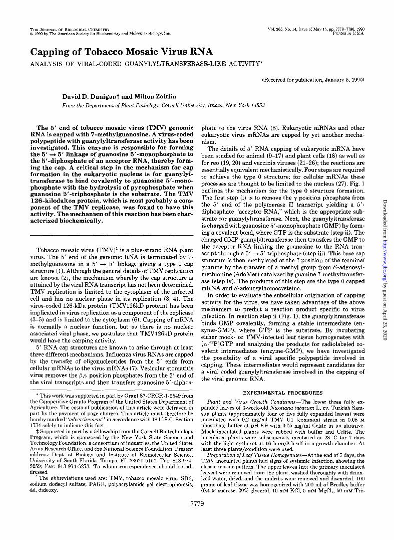
be considered a candidate as a guanylyltransferase, then the nucleotide binding should have preference for GTP as a substrate over other nucleotide 5’-triphosphates (11). The data shown in Fig. 4 and Table I indicate that there was a strong preference for GTP, but ATP was also bound, whereas CTP and UTP were not substrates for this reaction. The rate of incorporation of radioactivity into TMV126kD protein was measured when either a-““P-labeled GTP, ATP, CTP, or UTP was used in the reaction mixture. The initial rate of incorpo- ration for GTP was approximately 25 times greater than for ATP; no virus-specific products were ever observed for either CTP or UTP (Fig. 4A). Whereas the GTP reaction products were as described in Fig. 2 and specific to the TMV126kD protein, binding assays with ATP yielded a complex pattern. Initial experiments with ATP as the substrate gave confusing results in which both the mock- and TMV-infected material bound the nucleotide, and the labeled protein for the mock- infected tissue comigrated with the TMV126kD protein. How- ever, the data presented in Fig. 4B and in other experiments show that the host-associated ATP-binding protein (that is, present in both the mock- and TMV-infected material) mi- grates slightly faster than the virus-specific nucleotide-bind- ing protein. Thus, in addition to identifying TMV126kD protein as a purine nucleotide-binding protein (where GTP and possibly ATP are substrates), there is also a host-specific ATP-binding protein that migrates at approximately 120,000 molecular weight. Serendipitously, ATP binding also helps to indicate the purity of the nucleotides used because if the GTP were contaminated with ATP, then the mock-infected mate- rial would also show radiolabeling at the 120,000 molecular weight position when labeling with [o(-“‘P]GTP. However, we cannot be certain that the ATP is not contaminated with small amounts of GTP (the manufacturer) Amersham Corp., claims the nucleotides are greater than 95% pure).
The radiolabeling of the TMV126kD protein was presumed to be due to nucleotide addition, but transfer of hydrolyzed
by guest on April 25, 2020
http://ww
w.jbc.org/
Dow
nloaded from
7782 TMV-encoded Guanylyltransferase-like Activity
Time of Incubation (min)
B GTP ATP CTP UTP - - - - M I M I M I M I
160 -
116 -
A 84 -
‘
FIG. 4. Kinetic analysis and nucleotide specificity for incor- poration into TMV126kD protein. Mock- (M) and TMV-infected (I) leaf tissue homogenates were fractionated by centrifugation at 13.000 X g for 10 min at 4 “C. The supernatant fractions (S-13) were reacted at 25 “C with 380 &i/ml of either [W”“P]GTP (GTP), or [W ““P]ATP (ATP), [e-“2P]CTP (CTI’), or [w”“P]UTP (UTP), each at 3,000 Ci/mmol specific activity. Samples were taken at 2, 4, 6, 8, 10, 15,20, and 25 min after mixing the S-13 with Bradley buffer contain- ing the radioactive nucleotides; both the S-13 and Bradley buffer mixtures were equilibrated to 25 “C before mixing. The samples were quenched with SDS-PAGE sample buffer and then analyzed by SDS- PAGE and autoradiography, and the incorporation was quantified by scanning densitometry, as described under “Experimental Proce- dures.” Initial rat,es were determined by plotting the relative film densities versus the time of incubation (pane/ A). The relative rates of nucleotide incorporation into the TMV126kD protein were: GTP, 1.00; ATP, 0.04; CTP, 0.00; UTP, 0.00. Panel R shows the reaction products after 40 min of incubation. The numbers at the left of the figure represent the positions of migration of the molecular weight standards x lo-“.
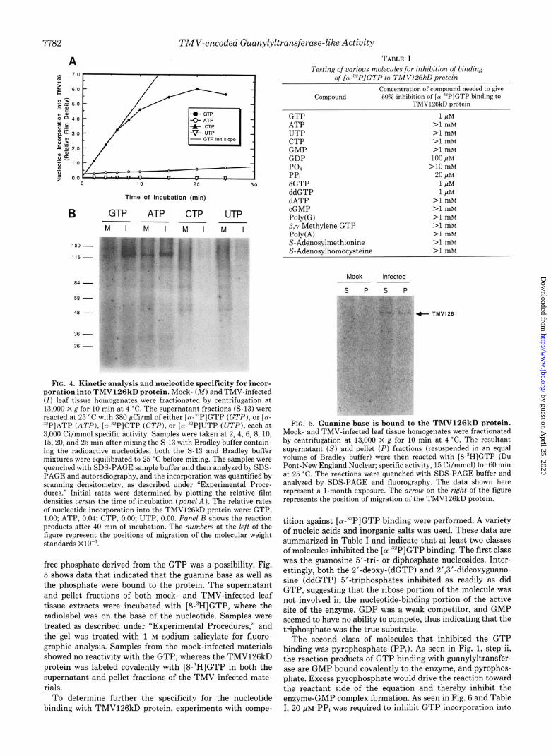
free phosphate derived from the GTP was a possibility. Fig. 5 shows data that indicated that the guanine base as well as the phosphate were bound to the protein. The supernatant and pellet fractions of both mock- and TMV-infected leaf tissue extracts were incubated with [8-“H]GTP, where the radiolabel was on the base of the nucleotide. Samples were treated as described under “Experimental Procedures,” and the gel was treated with 1 M sodium salicylate for fluoro- graphic analysis. Samples from the mock-infected materials showed no reactivity with the GTP, whereas the TMV126kD protein was labeled covalently with [8-“HIGTP in both the supernatant and pellet fractions of the TMV-infected mate- rials.
To determine further the specificity for the nucleotide binding with TMV126kD protein, experiments with compe-
TABLE I Testing of various molecules for inhibition of binding
of IPPlGTP to TMV126kD orotein
Compound Concentration of compound needed to give
W% inhibitlon of [R-Y’]GTP hmding to TMVl%kD protem
GTP ATP UTP CTP GMP GDP PO., PPi dGTP ddGTP dATP cGMP Poly(G) P,r Methylene GTP Poly(A) S-Adenosylmethionine S-Adenosilhomocysteine
1 PM >l mM >I mM >l mM >l mM
100 PM >lO mM
20 pM 1 PM 1 PM
>l mM >l mM >l mM >l mM >l mM >l mM >l mM
Mock Infected
s P s P
+ TMV126
FIG. 5. Guanine base is bound to the TMV126kD protein. Mock- and TMV-infected leaf tissue homogenates were fractionated by centrifugation at 13,000 x g for 10 min at 4 “C. The resultant supernatant (S) and pellet (I’) fractions (resuspended in an equal volume of Bradley buffer) were then reacted with [S-‘H]GTP (Du Pant-New England Nuclear; specific activity, 15 Ci/mmol) for 60 min at 25 ‘C. The reactions were quenched with SDS-PAGE buffer and analyzed by SDS-PAGE and fluorography. The data shown here represent a l-month exposure. The arrow on the right of the figure represents the position of migration of the TMV126kD protein.
tition against [ CY-“‘PI GTP binding were performed. A variety of nucleic acids and inorganic salts was used. These data are summarized in Table I and indicate that at least two classes of molecules inhibited the [(u-“‘P]GTP binding. The first class was the guanosine 5’-tri- or diphosphate nucleosides. Inter- estingly, both the 2’-deoxy-(dGTP) and 2’,3’-dideoxyguano- sine (ddGTP) 5’-triphosphates inhibited as readily as did GTP, suggesting that the ribose portion of the molecule was not involved in the nucleotide-binding portion of the active site of the enzyme. GDP was a weak competitor, and GMP seemed to have no ability to compete, thus indicating that the triphosphate was the true substrate.
The second class of molecules that inhibited the GTP binding was pyrophosphate (PPi). As seen in Fig. 1, step ii, the reaction products of GTP binding with guanylyltransfer- ase are GMP bound covalently to the enzyme, and pyrophos- phate. Excess pyrophosphate would drive the reaction toward the reactant side of the equation and thereby inhibit the enzyme-GMP complex formation. As seen in Fig. 6 and Table I, 20 PM PPi was required to inhibit GTP incorporation into
by guest on April 25, 2020
http://ww
w.jbc.org/
Dow
nloaded from

TMV-encoded Guanylyltransferase-like Activity 7783
Log WI, M FIG. 6. Pyrophosphate inhibits nucleotide binding to
TMV126kD protein. TMV-infected leaf tissue homogenate was fractionated by centrifugation at 13,000 X g for 10 min at 4 “C, and the resultant supernatant fraction (S-13) was incubated with [cx-~‘P] GTP at 250 &X/ml (specific activity, 3,000 Ci/mmol) plus PP, at various concentrations for 30 min at 25 “C. The reaction products were analyzed by SDS-PAGE and autoradiography and quantified by scanning densitometry. The extent of GTP incorporation into TMV126kD protein was determined by the relative film density. The concentration of PPi required to give a 50% inhibition of [ol-“‘P]GTP binding to TMV126kD protein was 20 PM.
TABLE II Tests of various enzyme treatnents on the stability
of the protein-nucleotide bond
Treatment Presence of “*P-TMV126kD
protein after treatment
Untreated Pl nuclease RNase T, RNase A Alkaline uhosohatase
TMV126kD protein by 50% and thus is consistent with the predictions of the reaction mechanism.
Nature of the Chemical Bond-To determine the nature of the bond between the GMP and the TMV126kD protein, both chemical and enzymatic methods have been utilized in an attempt to release the radioactive nucleotide from the protein. To date, only strong acid or strong base will efficiently release the radioactivity. The data shown in Table II demonstrate the results obtained using common nucleases and alkaline phosphatase. TMV-infected leaf tissue homogenate was frac- tionated by centrifugation, and the S-13 fraction was reacted with [w~‘P]GTP as the substrate. Aliquots of the reaction mixes were then incubated with either Pl nuclease, RNase T2, RNase A, or alkaline phosphatase or Bradley buffer as a control. The samples were then analyzed by SDS-PAGE/ autoradiography. These treatments had little or no effect on the amount of radioactivity retained by the TMV1‘26kD pro- tein, thus indicating that the phosphate moiety was inacces- sible to these enzymes.
Reaction Optimization-In order to evaluate the optimal reaction conditions for the binding of [(u-“‘P]GTP to TMV126kD protein, temperature, protein concentration, and divalent cation concentrations were investigated. 25 “C! was the measured temperature optimum (data not shown) and was close to the reported optimum (28 “C) for TMV replicase activity (28). Fig. 7 shows data in which the incorporation of GTP into either the soluble or membrane-bound form of TMV126kD protein was measured as a function of protein concentration. The incorporation was normalized to the quan- tity of TMV126kD protein present in the incubation, as estimated by Coomassie Blue staining. Incorporation of [LY- ‘l’P]GTP into TMV126kD protein was inhibited strongly
f Total Homogenate Fraction -D Supernatanf Fracl~on -.- Pelle, Fracr,on I
Fraction Concentration
FIG. 7. Optimization of reaction conditions for nucleotide binding to TMV126kD protein. TMV-infected leaf tissue homog- enate was fractionated by centrifugation at 13,000 x g for 10 min at 4 “C. The resultant fractions (S-13 and P-13) were analyzed for their ability to incorporate [a-“‘P]GTP into TMV126kD protein as a function of concentration of protein added to the incubation mixture. In determining the optimum proportion of the extracts in the incu- bation mix for [(u-“‘P]GTP incorporation into TMV126kD protein, the total homogenate, S-13 or P-13 fractions were diluted in 1:2 increments and added to the incubation mix; the volume was made up by Bradley buffer. The total homogenate was estimated by com- parison against standards from Coomassie Blue staining to be 16 mg/ ml; the S-13 fraction was estimated to he 12 mg/ml; the P-13 fraction was estimated to be 4 mg/ml. The amount of TMV126kD protein in total homogenate was estimated to be 0.5 mg/ml; the amount in S-13 was estimated to be 0.35 mg/ml; the amount in the P-13 was estimated to be 0.15 mg/ml. The nucleotide-binding activity of TMV126kD protein was calculated by dividing the integrated film density (rep- resenting the amount of [o(-3YP]GTP bound to the TMV126kD pro- tein) by the amount of TMV126kD protein present in the incubation.
when higher concentrations of either S-13, P-13, or total homogenate fraction were used in the incubation mix. These results suggested that either there was an inhibitor of the forward reaction (that is, formation of the enzyme-GMP intermediate), or there was an activation of the guanylyltrans- fer step (that is, transfer of GMP to an acceptor RNA) at relatively high concentration of the fraction added to the reaction mixture. If either of these hypotheses were dependent on the presence of soluble molecules (for example, inhibition by PPi or activation by some cofactor, or a change in the specific activity of the radionucleotide with changing amounts of nonradioactive GTP from the cell extract), then it would be predicted that the activity of the membrane-bound form in the P-13 fraction would be unresponsive to fraction con- centration when resuspended in Bradley buffer. As shown in Fig. 7, the accumulation of the radiolabeled TMV126kD pro- tein from the P-13 fraction, as well as the soluble form, was strongly reduced by a high P-13 concentration. This suggested that the low yield of GMP-TMV126kD protein at relatively high concentration was not due to a soluble component of the fraction and may be because of a rapid transfer of GMP to an acceptor RNA.
A number of divalent cations were tested to determine their dependence or inhibition of the [a-““P]GTP incorporation into TMV126kD protein. Under conditions tested, neither the rate nor the extent of the reaction could be increased by the addition of Mn*+, Mg’+, Zn”, Co’+, or Cu’+ chloride salts when tested in a range from 10 nM to 10 mM (data not shown). However, relatively high levels of Zn2+ (210 mM) or Co*+ (2100 PM) inhibited the reaction. The presence of 10 mM EDTA completely abolished the reaction, which indicated that there was a requirement for divalent cation. This require- ment must have been satisfied by endogenous ions in the
by guest on April 25, 2020
http://ww
w.jbc.org/
Dow
nloaded from

7784 TMV-encoded Guanylyltransferase-like Activity
homogenate, as the reaction would not respond to additional exogenous divalent cations.
Subcellular Fractionation-In order to characterize further the TMV126kD protein guanylyltransferase-like activity, subcellular fractionation was employed. As indicated in Figs. 5 and 7, the TMV126kD protein as well as the virus-specific guanylyltransferase-like activity partitioned in both the mem- brane-bound phase and the soluble phase. Fig. 8 shows data of a differential centrifugal fractionation of mock- and TMV- infected tobacco leaf tissue homogenates followed by incuba- tion with [(u-“‘P]GTP and analysis by SDS-PAGE/autoradi- ography. The low speed pellet fraction (P-10) of the mock- infected sample showed four major bands. The TMV-infected sample showed the same four bands and the infection-specific product, the TMV126kD protein. The high speed pellet frac- tion (P-100) contained only the TMV126kD protein in the infection-derived material, whereas the mock-infected-de- rived material had no detectable products. The high speed supernatant fraction (S-100) contained several nucleotide- binding products in both the mock- and virus-infected-derived materials, but only the virus-infected-derived samples con- tained the TMV126kD protein. Thus, although host-derived nucleotide-binding products partitioned to either the low speed pellet or the high speed supernatant fractions with centrifugation, the TMV126kD protein (with guanylyltrans- ferase-like activity) distributed rather uniformly throughout the fractions.
To fractionate the TMV126kD protein guanylyltransferase- like activity from the host-derived nucleotide binding prod- ucts further, the S-100 fraction of both the mock- and TMV- infected-derived extracts was desalted and then chromato- graphed on DEAE-cellulose using a linear KC1 gradient (lo- 500 mM) to elute bound material. The protein content was monitored by absorbance (280 nm), and the concentration of KC1 was monitored by measuring the conductivity. The DEAE fractions were then equilibrated with the KTMDP-10 buffer by dialysis and then sampled for their ability to bind
P-IO P-l 00 s-100
MIMIMI
84 -
FIG;. 8. Subcellular fractionation of TMV126kD protein nu- cleotide-binding activity. Mock- (M) and TMV-infected (I) leaf tissue homogenate was fractionated by differential centrifugation, 10,000 X fi at 4 “C for 10 min, which resulted in a pellet fraction (P- 10): the resultant supernatant fraction was subsequently centrifuged at 100,000 X :: at 4 “C for 1 h and resulted in a pellet fraction (P-100) and a supernatant fraction (S-100). The pellet fractions were resus- pended in an equal volume of Bradley buffer. The fractions were tested for their ability to hind [m-““P]GTP using the standard nucleo- tide-binding assay, as described under “Experimental Procedures.” Equal cell volumes were loaded onto the gel. The numbers to the left of the figure indicate the position of migration of the molecular weight standards ~10-:‘.
A
iii .
6
I - 30 TO
FIG. 9. DEAE chromatographic isolation of TMV126kD protein nucleotide-binding activity. Mock- (panel A) and TMV- infected (panel B) leaf tissue homogenates were fractionated by centrifugation, as described in Fig. 8. The buffers of the S-100 fractions were subsequently exchanged to KTMDP-10 buffer by passing over Sephadex G-25 (coarse). The resulting void volumes were then loaded onto Whatman DE52 cellulose; the unbound frac- tion was collected, the column was washed with the same buffer, and the bound material was eluted with a linear KCI gradient from 10 to 500 mM in KTMDP buffer (upper half of panels A and 19). The collected fractions were assayed for salt concentration by conductivity
by guest on April 25, 2020
http://ww
w.jbc.org/
Dow
nloaded from

TMV-encoded Guanylyltransferase-like Activity 7785
[cK-~‘P]GTP, as determined by SDS-PAGE/autoradiographic analysis.
Fig. 9A depicts the chromatogram (top panel) and the autoradiogram of the nucleotide-binding analysis (bottom panel) of the mock-infected-derived materials that had been fractionated on DEAE-cellulose. A large amount of ADTO ab- sorbing material eluted in the unbound fractions (fraction numbers 1-14) in two peaks. The bound fractions eluted with increasing salt concentration in three major peaks (fractions 18-23, 24-29, and 30-38). The majority of the [a-32P]GTP- binding activity of this mock-infected-derived material eluted largely in the first bound peak (fractions 18-21).
The chromatographic profile of the TMV-infected-derived material (Fig. 9B, top panel) was essentially the same as that for the mock-infected-derived material, except that the first peak of the unbound fractions was markedly increased. When samples of this material were tested for binding of [~Y-~*P] GTP (Fig. 9B, bottom panel), a peak of infection-specific activity was observed to elute in the unbound fraction (frac- tions 3-5), while the host-specific activity remained bound and then eluted in the first major peak of the salt gradient (fractions 21-24), as seen in the mock-infected-derived ma- terial. Thus, the virus-specific [a-32P]GTP-binding protein was readily separated from the majority of the host-specific activity.
Virus-specific [cY-32P]GTP-binding activity was not re- covered when chromatographed on either carboxymethyl cel- lulose or Cibacron Blue, and at this time it is not clear why the activity was lost (data not shown).
DISCUSSION
The experiments presented here were designed to test the hypothesis that TMV produces a guanylyltransferase-like protein. We have observed that the 126-kilodalton protein encoded at the 5’-proximal end of the viral genome (TMV126kD protein) had the ability to bind GMP covalently when GTP was the substrate (Figs. l-5). Nucleotide binding was specific for GTP as a substrate, although modifications of the ribose moiety were tolerated (Table I), as demonstrated by the fact that both dGTP and ddGTP competed efficiently for the [a-32P]GTP in the nucleotide-binding assays. GTP was approximately 100 times more efficient as a substrate than was GDP, whereas GMP had no measurable ability to act as a substrate. PPi acted as an inhibitor of the nucleotide binding, which is consistent with the hypothesis that elevated concentrations of PPi would drive the reaction toward the reactant side of the equation, GTP + TMV126kD protein * GMP-TMV126kD protein + PPi. However, the observed products in these studies seemed to favor the forward reaction because the effective pool of GTP was measured to be ap- proximately 1 PM in the in vitro reactions, yet a pool of 20 PM PPi was required to inhibit the GMP-protein complex for- mation (Table I). The concentration of PPi required to inhibit this reaction by 50% (20 PM) was approximately 10 times greater than that required to inhibit by 50% either HeLa cell or wheat germ capping activity in vitro using purified gua- nylyltransferase (11 and 18, respectively). Vaccinia virus gua- nylyltransferase activity, in the absence of AdoMet, also required approximately 2 pM to inhibit by 50%; but in the presence of AdoMet, approximately 20 pM PPi was required to inhibit by 50% (33). The supernatant fractions used in the studies presented here were not assayed for AdoMet content;
measurement and then dialyzed against KTMDP-10 buffer overnight. The dialyzed fractions were assayed for nucleotide-binding activity, as described under “Experimental Procedures” (lower half of panels A and B).
but assuming there were physiological concentrations of AdoMet present from the homogenate, then the concentration of PPi required to inhibit the TMV-specific GMP-protein complex formation was very similar to that found for vaccinia virus guanylyltransferase.
The TMV126kD protein was shown previously to be a nucleoside 5’-triphosphate-binding protein by radiolabeling the protein with UV light activation of either 8-N3-[y-32P] GTP or 8-N3-[y-32P]ATP (5). These data are supported by reports from Young et al. (3) that the TMV126kD protein is a component of the viral replicase complex. This type of y- labeled NTP affinity for the protein is significantly different from that measured in the experiments presented here. First, in our experiments, the nucleotide binding of the guanylyl- transferase-like activity observed required no exogenous en- ergy (e.g. UV light) to form a covalent bond between the polypeptide and the nucleotide. Second, the product of the guanylyltransferase-like activity was GMP bound in such a fashion that the phosphate moiety was not susceptible to nuclease or phosphatase digestion (Table II), nor was it sus- ceptible to mild acid or mild base, which is consistent with the suggestion that GMP binds to guanylyltransferase through a phosphoamide bond, probably at a lysine residue (17, 26). Third, the specificity of the guanylyltransferase-like activity for GTP, dGTP, or ddGTP was at least 25-fold greater than for any other nucleoside 5’-triphosphate tested (Table I), whereas specificity for labeling with the UV light-activated azido derivatives was not limited to either purine or pyrimi- dine ribonucleoside 5’-triphosphates (5). These two different results for nucleotide binding to TMV126kD protein may represent two unique binding sites for the protein and thereby represent two unique activities, RNA-dependent RNA polym- erase and guanylyltransferase.
The conditions of incubation altered significantly the rate and extent of reaction to form a stable GMP-TMV126kD protein complex, as well as the type of host-specific products formed. A variety of host-specific proteins was found which distributed to various fractions after fractionation of the total homogenate (Fig. 8). Yet, the TMV-specific guanylyltransfer- ase-like activity was found distributed nearly uniformly among the membrane fraction (P-lo), the ribosomal fraction (P-100), and the cytoplasmic fraction (S-100). Curiously, there was a distinct concentration effect for the TMV-specific guanylyltransferase-like activity, especially with the P-13 fraction (Fig. 7). The activity was enhanced by dilution of the fractions. This “dilution activation” may be due to the release of an inhibitor from the protein or may result when the intermediate of the guanylyltransferase reaction (enzyme- GMP) was diluted to such an extent that the probability of interacting with the acceptor RNA was low, and so the inter- mediate accumulated.
No significant dependence for exogenously added divalent cations could be observed for the virus-specific guanylyltrans- ferase-like activity, yet thk reaction was sensitive to EDTA, which indicated a divalent cation requirement. The require- ment may have been fulfilled in uiuo. This apparent lack of sensitivity to exogenously added divalent cations differs from guanylyltransferases from other sources. The HeLa cell en- zyme has sensitivity to both Mn2+ and M$’ for enzyme-GMP complex formation (15) and guanylyltransferase activity (10). At their optima (2 mM for Mn*+ and 2-10 mM for Mg2’), these activities are approximately 2-4-fold greater in incuba- tions with Mn*+ than with Mg2+. Optimal capping activity is found at 0.5 mM MnClz and 5 mM MgClz in wheat germ extracts with nearly equal activity at these optima (18). The vaccinia virus guanylyltransferase activity is also sensitive to
by guest on April 25, 2020
http://ww
w.jbc.org/
Dow
nloaded from

7786 TMV-encoded Guanylyltransferase-like Actiuity
divalent cations, but the optimal activity is found with Me. At 2.5 mM, guanylyltransfer is approximately a lo-fold greater rate in the presence of Mg2+ than in the presence of Mn2+ (33). Thus, at least three classes of guanylyltransferase may be distinguished by their sensitivity to divalent cations; the TMV-specific guanylyltransferase-like activity may represent yet another class.
The TMV-specific guanylyltransferase-like activity in the S-100 fraction was isolated from the host nucleotide-binding activity by anion exchange chromatography (Fig. 9). Three major host-specific products were eluted in the DEAE-bound low salt-eluting fractions (mock fractions 19-21, TMV frac- tions 21-33), whereas the TMV-specific activity was not bound to the DEAE-cellulose and eluted in fractions 3 and 4 (Fig. 9B). In both the mock and TMV samples, the unbound material eluted in two peaks, but the TMV material had an enhanced first peak (Fig. 9B, fractions l-6) that corresponded to the TMV-specific guanylyltransferase-like activity. The TMV material also had an enhanced second peak in the salt- eluting fractions (fractions 23-25). It is not clear why this second peak of the Azm absorbing material was so much stronger in the TMV samples than in the mock samples.
We believe the primary significance of these data is that for the first time, a specific enzymatic activity may be assigned to the major nonstructural protein implicated in TMV repli- cation. The coding region of the RNA which specifies the TMV126kD protein has been shown to have regions of se- quence similarity to those of several other members of the Sindbis-like viruses (34, 35) as well as to other proteins involved in nucleic acid replication (36, 37). One important structural feature common to all the members of the Sindbis- like viruses is the 5’ RNA capped genomes, and, to the best of our knowledge, all members of this group are replicated in the cytoplasm of the infected cell. Thus, we postulate that other members of this group of viruses have virus-coded guanylyltransferases, possibly associated with their nonstruc- tural proteins.
1. 2.
REFERENCES
Zimmern, D. (1975) Nucleic Acids Res. 2, 1189-1201 Palukaitis, P., and Zaitlin, M. (1986) in The Plant Viruses (van
Regenmortel, M. H. V., and Fraenkel-Conrat, H., eds) Vol. 2, pp. 105-131, Plenum Publishing Corp., New York
Young, N., Forney, J., and Zaitlin, M. (1987) J. Cell Sci. 7, (suppl.) 277-285
Wilson, T. M. A. (1988) Oxford Suru. Plant Mol. Cell. Biol. 5,89- 144
Evans, R. K., Haley, B. E., and Roth, D. A. (1985) J. Biol. Chem. 260, 7800-7804
Hills, G. J., Plaskitt, K. A., Young, N. D., Dunigan, D. D., Watts,
7.
8.
9.
10.
11.
12.
13.
14.
15. 16.
17.
18.
19.
20.
21.
22.
23.
24.
25.
26.
27.
28. 29.
30. 31.
32.
33.
34.
35. 36.
37.
J. W., Wilson, T. M. A., and Zaitlin, M. (1987) Virology 161, 488-496
Plotch, S. J., Bouloy, M., Ulmanen, I., and Klug, R. M. (1981) Cell 23,847-858
Abraham, G., Rhodes, D. P., and Banerjee, A. K. (1975) Cell 5, 51-58
Mizumoto. K.. and Lioman. F. (1979) Proc. N&l. Acad. Sci. U. S. A. 76,4961-4965 -
Venkatesan. S.. Gershowitz. A.. and Moss. B. (1980) J. Biol. Chem. 255, i829-2834 ’
Venkatesan, S., and Moss, B. (1980) J. Biol. Chem. 255, 2835- 2842
Venkatesan, S., and Moss, B. (1982) Proc. Natl. Acad. Sci. U. S. A. 79, 340-344
Mizumoto, K., Kaziro, Y., and Lipman, F. (1982) PFOC. Natl. Acad. Sci. U. S. A. 79, 1693-1697
Wang, D., Furuichi, Y., and Shatkin, A. J. (1982) Mol. Cell. Biol. 2,993-1001
Shuman, S. (1982) J. Biol. Chem. 257,7237-7245 Yagi, Y., Mizumoto, K., and Kaziro, Y. (1983) EMBO J. 2, 611-
616 Tovama. R.. Mizumoto. K.. Nakahara. Y.. Tatsuno. T.. and
Kazird, Y.‘(1983) EMBO i 2, 2195-2202 Keith. J. M.. Venkatesan. S.. Gershowitz. A.. and Moss. B. (1982)
Biodhemi&y 21,327-333 Furuichi, Y., Muthukrishnan, S., Tomasz, J., and Shatkin, A. J.
(1976) J. Biol. Chem. 251, 5043-5053 Yamakawa, M., Furuichi, Y., and Shatkin, A. J. (1982) Virology
118, 157-168 Monroy, G., Spencer, E., and Hurwitz, J. (1978) J. Biol. Chem.
253.4490-4498 Spencer, E., Loring, D., Hurwitz, J., and Monroy, G. (1978) Proc.
Natl. Acad. Sci. U. S. A. 75, 4793-4797 Venkatesan, S., Gershowitz, A., and Moss, B. (1980) J. Biol.
Chem. 255,903-908 Shuman, S., Surks, M., Furneaux, H., and Hurwitz, J. (1980) J.
Biol. Chem. 255, 11588-11598 Shuman, S., and Hurwitz, J. (1981) Proc. Natl. Acad. Sci. U. S.
A. 78, 187-191 Roth, M. J., and Hurwitz, J. (1984) J. Biol. Chem. 259, 13488-
13494 Itoh, N., Yamada, H., Kaziro, Y., and Mizumoto, K. (1987) J.
Biol. Chem. 262, 1989-1995 Bradley, D. W., and Zaitlin, M. (1971) Virology 45, 192-199 Dreyfus, G., Adam, S. A., and Choi, Y. D. (1984) Mol. Cell. Biol.
4,415-423 Laskey, R. A., and Mills, A. D. (1977) FEBS Lett. 82, 314-316 Bonner, W. M., and Laskey, R. A. (1974) Eur. J. Biochem. 46,
83-88 Sefton, B. M., Beemon, K., and Hunter, T. (1978) J. Virol. 28,
957-971 Martin, S. A., and Moss, B. (1975) J. Biol. Chem. 250, 9330-
9335 Ahlauist. P.. Strauss. E. G.. Rice. C. M.. Strauss, J. H., Haseloff,
J.,*andZimmern, fi. (1985) J. viral. g3,536-542 Goldbach. R.. and Wellink. J. (1988) IntervirologY 29, 260-267 Gorbalenya, k. E., Koonin; E. v., Donchenko, A-P., and Blinov,
V. M. (1988) Nature 333, 22 Hodgman, T. C. (1988) Nature 333, 22-23
by guest on April 25, 2020
http://ww
w.jbc.org/
Dow
nloaded from

D D Dunigan and M Zaitlinguanylyltransferase-like activity.
Capping of tobacco mosaic virus RNA. Analysis of viral-coded
1990, 265:7779-7786.J. Biol. Chem.
http://www.jbc.org/content/265/14/7779Access the most updated version of this article at
Alerts:
When a correction for this article is posted•
When this article is cited•
to choose from all of JBC's e-mail alertsClick here
http://www.jbc.org/content/265/14/7779.full.html#ref-list-1
This article cites 0 references, 0 of which can be accessed free at
by guest on April 25, 2020
http://ww
w.jbc.org/
Dow
nloaded from