Cannabidiol rather than Cannabis sativa extracts inhibit ...
16
RESEARCH ARTICLE Open Access Cannabidiol rather than Cannabis sativa extracts inhibit cell growth and induce apoptosis in cervical cancer cells Sindiswa T. Lukhele and Lesetja R. Motadi * Abstract Background: Cervical cancer remains a global health related issue among females of Sub-Saharan Africa, with over half a million new cases reported each year. Different therapeutic regimens have been suggested in various regions of Africa, however, over a quarter of a million women die of cervical cancer, annually. This makes it the most lethal cancer amongst black women and calls for urgent therapeutic strategies. In this study we compare the anti-proliferative effects of crude extract of Cannabis sativa and its main compound cannabidiol on different cervical cancer cell lines. Methods: To achieve our aim, phytochemical screening, MTT assay, cell growth analysis, flow cytometry, morphology analysis, Western blot, caspase 3/7 assay, and ATP measurement assay were conducted. Results: Results obtained indicate that both cannabidiol and Cannabis sativa extracts were able to halt cell proliferation in all cell lines at varying concentrations. They further revealed that apoptosis was induced by cannabidiol as shown by increased subG0/G1 and apoptosis through annexin V. Apoptosis was confirmed by overexpression of p53, caspase 3 and bax. Apoptosis induction was further confirmed by morphological changes, an increase in Caspase 3/7 and a decrease in the ATP levels. Conclusions: In conclusion, these data suggest that cannabidiol rather than Cannabis sativa crude extracts prevent cell growth and induce cell death in cervical cancer cell lines. Keywords: Apoptosis, Cervical cancer, Cannabidiol, Cannabis sativa Abbreviations: Bad, Bcl-2-associated death promoter; Bak-1, Bcl2-antagonist/killer 1; Bax, Bcl2-associated X protein; Bcl-2, B-cell lymphoma 2; BH, Bcl-2 homology domain; Bid, BH3 interacting-domain; Bik, Bcl-2-interacting killer; DMEM, Dulbecco’s modified eagle’s medium; DMSO, Dimethyl sulfoxide; FITC, Fluorescein isothiocyanate; P53, protein 53; HPLC, High Perfomance Liquid Chromatography; RBBP6, Retinoblastoma binding protein 6 Background Cannabis sativa is a dioecious plant that belongs to the Cannabaceae family and it originates from Central and Eastern Asia [11, 28]. It is widely distributed in countries including Morocco, South Africa, United States of America, Brazil, India, and parts of Europe [14, 28]. Cannabis sativa grows annually in tropical and warm regions around the world [11]. Different ethnic groups around the world use Cannabis sativa for smoking, preparing concoctions to treat diseases, and for various cultural purposes [17]. According to [28], it is composed of chemical constituents including cannabinoids, nitrogenous compounds, flavonoid glycosides, steroids, terpenes, hydrocarbons, non-cannabinoid phenols, vitamins, amino acids, proteins, sugars and other related compounds. Cannabinoids are a family of naturally occur- ring compounds highly abundant in Cannabis sativa plant [1, 6, 14, 24]. Screening of Cannabis sativa has led to isolation of at least 66 types of cannabinoid compounds [1, 14, 30]. These compounds are almost structurally simi- lar or possess identical pharmacological activities and offer various potential applications including the ability to inhibit cell growth, proliferation and inflammation [22]. One such compound is cannabidiol (CBD), which is among the top three most widely studied compounds, * Correspondence: [email protected] Department of Biochemistry, North-west University (Mafikeng campus), Private Bag X1290, Potchefstroom 2520, South Africa © 2016 The Author(s). Open Access This article is distributed under the terms of the Creative Commons Attribution 4.0 International License (http://creativecommons.org/licenses/by/4.0/), which permits unrestricted use, distribution, and reproduction in any medium, provided you give appropriate credit to the original author(s) and the source, provide a link to the Creative Commons license, and indicate if changes were made. The Creative Commons Public Domain Dedication waiver (http://creativecommons.org/publicdomain/zero/1.0/) applies to the data made available in this article, unless otherwise stated. Lukhele and Motadi BMC Complementary and Alternative Medicine (2016) 16:335 DOI 10.1186/s12906-016-1280-0
Transcript of Cannabidiol rather than Cannabis sativa extracts inhibit ...

Lukhele and Motadi BMC Complementary and Alternative Medicine (2016) 16:335 DOI 10.1186/s12906-016-1280-0
RESEARCH ARTICLE Open Access
Cannabidiol rather than Cannabis sativaextracts inhibit cell growth and induceapoptosis in cervical cancer cells
Sindiswa T. Lukhele and Lesetja R. Motadi*Abstract
Background: Cervical cancer remains a global health related issue among females of Sub-Saharan Africa, with overhalf a million new cases reported each year. Different therapeutic regimens have been suggested in various regionsof Africa, however, over a quarter of a million women die of cervical cancer, annually. This makes it the most lethalcancer amongst black women and calls for urgent therapeutic strategies. In this study we compare the anti-proliferativeeffects of crude extract of Cannabis sativa and its main compound cannabidiol on different cervical cancer cell lines.
Methods: To achieve our aim, phytochemical screening, MTT assay, cell growth analysis, flow cytometry,morphology analysis, Western blot, caspase 3/7 assay, and ATP measurement assay were conducted.
Results: Results obtained indicate that both cannabidiol and Cannabis sativa extracts were able to halt cellproliferation in all cell lines at varying concentrations. They further revealed that apoptosis was induced by cannabidiolas shown by increased subG0/G1 and apoptosis through annexin V. Apoptosis was confirmed by overexpression ofp53, caspase 3 and bax. Apoptosis induction was further confirmed by morphological changes, an increase in Caspase3/7 and a decrease in the ATP levels.
Conclusions: In conclusion, these data suggest that cannabidiol rather than Cannabis sativa crude extracts prevent cellgrowth and induce cell death in cervical cancer cell lines.
Keywords: Apoptosis, Cervical cancer, Cannabidiol, Cannabis sativa
Abbreviations: Bad, Bcl-2-associated death promoter; Bak-1, Bcl2-antagonist/killer 1; Bax, Bcl2-associated Xprotein; Bcl-2, B-cell lymphoma 2; BH, Bcl-2 homology domain; Bid, BH3 interacting-domain; Bik, Bcl-2-interactingkiller; DMEM, Dulbecco’s modified eagle’s medium; DMSO, Dimethyl sulfoxide; FITC, Fluorescein isothiocyanate;P53, protein 53; HPLC, High Perfomance Liquid Chromatography; RBBP6, Retinoblastoma binding protein 6
BackgroundCannabis sativa is a dioecious plant that belongs to theCannabaceae family and it originates from Central andEastern Asia [11, 28]. It is widely distributed in countriesincluding Morocco, South Africa, United States ofAmerica, Brazil, India, and parts of Europe [14, 28].Cannabis sativa grows annually in tropical and warmregions around the world [11]. Different ethnic groupsaround the world use Cannabis sativa for smoking,preparing concoctions to treat diseases, and for variouscultural purposes [17]. According to [28], it is composed
* Correspondence: [email protected] of Biochemistry, North-west University (Mafikeng campus),Private Bag X1290, Potchefstroom 2520, South Africa
© 2016 The Author(s). Open Access This articInternational License (http://creativecommonsreproduction in any medium, provided you gthe Creative Commons license, and indicate if(http://creativecommons.org/publicdomain/ze
of chemical constituents including cannabinoids,nitrogenous compounds, flavonoid glycosides, steroids,terpenes, hydrocarbons, non-cannabinoid phenols,vitamins, amino acids, proteins, sugars and other relatedcompounds. Cannabinoids are a family of naturally occur-ring compounds highly abundant in Cannabis sativa plant[1, 6, 14, 24]. Screening of Cannabis sativa has led toisolation of at least 66 types of cannabinoid compounds[1, 14, 30]. These compounds are almost structurally simi-lar or possess identical pharmacological activities and offervarious potential applications including the ability toinhibit cell growth, proliferation and inflammation [22].One such compound is cannabidiol (CBD), which isamong the top three most widely studied compounds,
le is distributed under the terms of the Creative Commons Attribution 4.0.org/licenses/by/4.0/), which permits unrestricted use, distribution, andive appropriate credit to the original author(s) and the source, provide a link tochanges were made. The Creative Commons Public Domain Dedication waiverro/1.0/) applies to the data made available in this article, unless otherwise stated.

Lukhele and Motadi BMC Complementary and Alternative Medicine (2016) 16:335 Page 2 of 16
following delta-9-tetrahydrocannabinol (Δ9-THC) [14].It has been found to be effective against a variety ofdisorders including neurodegerative disorders, auto-immune diseases, and cancer [24, 25]. In a research studyconducted by [26], it was found that CBD inhibited cellproliferation and induces apoptosis in a series of humanbreast cancer cell lines including MCF-10A, MDA-MB-231, MCF-7, SK-BR- 3, and ZR-7-1 and further studiesfound it to possess similar characteristics in PC-3 prostatecancer cell line [25]. However, to allow us to further ourstudies in clinical trials a range of cancers in vitro shouldbe tested to give us a clear mechanism before we canproceed. Cannabis sativa in particular cannabidiol, wepropose it plays important role in helping the body fightcancer through inhibition of pain and cell growth. There-fore, the aim of this study was to evaluate the cytotoxicand anti-proliferative properties of Cannabis sativa andits isolate, cannabidiol in cervical cancer cell lines.
MethodsMaterialsAn aggressive HeLa, a metastatic ME-180 and a primarySiHa cell lines were purchased from ATCC (USA, MD).Camptothecin was supplied by Calbiochem® and canna-bidiol was purchased from Sigma-Aldrich and used as astandard reference.
Plant collection and preparation of extractsFresh leaves, stem and roots of Cannabis sativa werecollected from Nhlazatshe 2, in Mpumalanga province.Air dried C. sativa plant material was powdered andsoaked for 3 days in n-hexane, ethanol and n-butanol,separately. Extracts were filtered using Whatman filterpaper and dried. Dimethyl sulfoxide was added to driedextracts to give a final concentration of a 100 mg/ml.Different concentrations (50, 100, and 150 μg/ml) of C.sativa extracts were prepared from the stock and usedin treating cells during MTT assay. HPLC-Mass spectro-photometry was performed to verify the presence ofcannabidiol in our extracts. The plant was identified byforensic specialist in a forensic laboratory in Pretoria.The laboratory number 201213/2009 and the vouchernumber is CAS239/02/2009.
Cell cultureHeLa, ME-180 and SiHa were cultured in Dulbecco’sModified Eagle Media (DMEM) supplemented with10 % Fetal Bovine Serum (FBS) (Highveld biological,)and 1 % penicillin/streptomycin (Sigma, USA). Cellswere maintained at 37 °C under 5 % of carbon dioxide(CO2) and 95 % relative humidity. After every third dayof the week, old media was removed and replaced withfresh media, to promote growth until the cells reach aconfluence of ~70–80 %.
MethodsMTT assayNinety microlitres of HeLa and SiHa cells were seededinto 96-well plates at 5×10³ cells per well and incubatedovernight at 37 °C under 5 % CO2 and 95 % relative hu-midity to promote cell attachment at the bottom of theplate. Media was changed and the cells were treated withCannabis sativa plant extracts at various concentrations(0, 50, 100, and 150 μg/ml (w/v)) for 24 h. After 24 h, thecells were treated with 10 μl of (5 mg/ml) MTT reagent(3-[4, 5-dimethylthiazol-2-yl]-2, 5-diphenyltetrazoliumbromide) for 4 h at 37 °C under 5 % CO2. Ninetymicrolitres of DMSO was added into each well in-cluding wells containing media only and serves as ablank, to dissolve formazan crystals. Camptothecinand DMSO were included as controls. Optical densitywas measured using a micro plate reader (Bio-Rad) at570 nm to determine the percentage of viable cellsand account for cell death induced according to theoutlined equation below:
% Cell viability
¼ Absorbance of treated cells−Absorbance of blankAbsorbance of untreated cells−Absorbance of blank
� 100
Cell growth analysisBefore seeding cells, a 100 μl of media was added to the16 well E-plate and placed in the incubator to recordbackground readings. A blank with media only was in-cluded to rule the possibility of media having a negativeeffect on the cells. In each well of the E-plate, 1×10⁴
cells were seeded and left in the incubator for 30 min toallow the cells to adhere to the bottom of the plate. TheE-plate was placed and locked in the RTCA machineand experiment allowed to run for 22 h prior to theaddition of the test compounds. Cells were treated withvarious concentrations (0, 50, 100, 150 μg/ml) of C.sativa hexane extract. Following treatment, the experi-ment was allowed to run for a further 22 h. Camptothe-cin (0.3 μM (v/v)) and DMSO (0.1 % (v/v)) were used ascontrols for comparative purposes. Procedure wasrepeated for C. sativa butanol extract.
Apoptosis assayCells were washed twice with 100 μl of cold Biolegend’scell staining buffer followed by resuspension in 100 μl ofAnnexin V binding buffer. A 100 μl of cell suspensionwas transferred into 15 ml falcon tube and 5 μl of FITCAnnexin V and 10 μl of Propidium iodide solution (PI)were added into untreated and treated cell suspension.The cells were gently vortexed and incubated at roomtemperature (25 °C) in the dark for 15 min. After
Lukhele and Motadi BMC Complementary and Alternative Medicine (2016) 16:335 Page 3 of 16
15 min, 400 μl of Annexin V binding buffer was addedto the cells. The stained cells were analysed using FACS-Calibur (BD Biosciences, USA).
Morphological analysisFive hundred microliter of 1×10⁴ cells was added ontoa 6-well plate containing coverslips. The plate was incu-bated overnight to allow the cells to attach. Followingattachment, media was removed and cells were washedtwice with PBS, prior to incubation with IC50 ofCannabis sativa extracts for 24 h. After 24 h, media wasremoved and cells were washed twice with PBS. Fourpercent (4 %) was added into each well and the plateincubated for 20 min at room temperature, to allowefficient fixation of cells. Cells were washed twice withPBS and once with 0.1 % BSA wash buffer and furtherstained with DAPI and Annexin V/FITC for 5 min.BX-63 Olympus microscope (Germany) was used tovisualize the cells.
Mitochondrial assay (ATP detection)Twenty five microlitres of 1×10⁴ cells per well wereplated in a white 96-well luminometer plate overnight.Cells were treated with 25 μl of IC50 concentrations ofCannabis sativa crude extracts and cannabidiol dissolvedin a glucose free media supplemented with 10 mMgalactose. The plate was incubated at 37° in a humidifiedand CO2-supplemented incubator for a period of 24 h.Fifty microlitres of ATP detection reagent was added toeach well and the plate further incubated for 30 min.Luminescence was measured using GLOMAX (Promega,USA). The assay was conducted in duplicates and ATPlevels were reported as a mean of Relative Light Units(RLU). The following formula was used to calculate theATP levels in RLU:
RLU ¼ Luminescence sampleð Þ−Luminescence blankð Þ
Caspase 3/7 activityA hundred microliters of 1×10⁴ cells were platedovernight on a 96-well luminometer plate and allowedto attach overnight. The next day, cells were treatedwith 0.3 μM camptothecin and the IC50 concentra-tions of Cannabis sativa crude extracts and furtherincubated for a period of 24 h. Caspase-Glo 3/7 assaywas performed according to manufacturer’s protocol(Promega, USA). Briefly, following treatment, mediawas replaced with caspase glo 3/7 reagent mixed witha substrate at a ratio of 1:1 v/v of DMEM: Caspase-glo 3/7 reagent and was incubated for 2 h at 37 °C in5 % CO2. Luminescence was quantified using
GLOMAX from Promega (USA). The assay wasconducted in duplicates and caspase 3/7 activity wasreported as a mean of Relative Light Units (RLU).The following formula was used to calculate caspase3/7 activity in RLU:
RLU ¼ Luminiscence sampleð Þ−Luminiscence blankð Þ
Cell cycle analysisCells were harvested with 2 ml of 0.05 % trypsin-EDTA.Ten millilitres of media was added to the cells to inacti-vate trypsin and the cell suspension was centrifuged at1500 rpm for 10 min. The supernatant was discardedand pellet was re-suspended twice in 1 ml PBS. Cellsuspension was centrifuged at 5000 rpm for 2–5 minand PBS was discarded. Seven hundred microlitres ofpre-chilled absolute ethanol was added to the cellsuspension followed by storage at −20 °C for 30 min, toallow efficient permeabilization and fixing of the cells.After 30 min, cells were centrifuged at 5000 rpm for5 min to remove ethanol. The pellet was washed twicewith PBS and centrifuged at 5000 rpm to remove PBS.Five hundred microlitres of FxCycle™ PI/RNase Stainingsolution (Life technologies, USA) was added to the cellsand vortexed for 30 s (sec). The cells were analysed withFACSCalibur (BD Biosciences, USA).
Western blotFollowing 24 h of treatment with IC50 concentrations,cells were lysed using RIPA buffer (50 mM Tris-HClpH 7.4, 150 mM NaCl, 1 % NP-40, 0.1 % SDS, 2 mMEDTA). Protein content was measured by the BCAassay and equal amounts were electrophoresed inSDS polyacrylamide gel and then transferred ontonitrocellulose membranes. Membranes were subse-quently immunoblotted with Anti-mouse monoclonalp53, Bcl-2, Bax, RBBP6, Caspase-3 and -9 antibodieswere used at 1:500–1000 dilutions as primary anti-bodies, while a goat anti-mouse horseradish peroxidise-conjugated horse IgG (Santa Cruz, USA) were used at a1:2000 dilution as a secondary antibody. The membraneswere developed using Chemiluminescence detection kit(Santa Cruz Biotechnology, CA). The membranes wereimaged using a Biorad ChemiDoc MP.
Data analysisExperiments were performed in duplicates. Statisticalanalysis of the graphical data was expressed as themean standard deviation. The p-value was analysed incomparison to the untreated using Students t-Testwherein p < 0.05 was considered as significant.
Lukhele and Motadi BMC Complementary and Alternative Medicine (2016) 16:335 Page 4 of 16
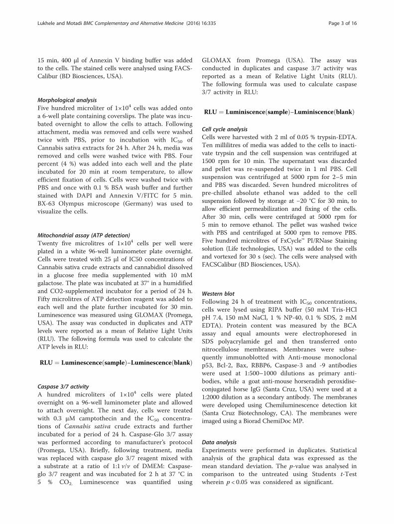
ResultsEffect of Cannabis sativa and cannabidiol on SiHa, HeLa,and ME-180 cellsTo determine half maximal inhibitory concentration(IC50) for both Cannabis sativa and cannabidiol, MTTassay was used. Camptothecin, as our positive control,significantly reduced cell viability in SiHa (40.36 %),HeLa (47.19 %), and ME-180 cells (32.25 %), respect-ively. As shown in Fig. 1a and d, the IC50 was cell typedependent rather than time dependent with SiHa at lessthan 50 μg/ml in both butanol (56 %) and hexane (48.9 %).Similarly IC50 in HeLa was at 100 μg/ml at p < 0.001(Fig. 1b). while ME-180 cells treated with butanolic extractexhibited an IC50 of a 100 μg/ml, reducing viability to48.6 % (Fig. 1c) and hexane extracts IC50 was at 50 μg/mlwith 54 % death (Fig. 1f). This was not the case in cannabi-diol with SiHa (51 %) and HeLa (50) IC50 at a much lowerdose (3.2 μg\ml) while ME-180 cells (56 %) at 1.5 μg\mlwhen compared to Cannabis sativa extracts (p < 0.001)(Fig. 1g–i). Dimethyl sulfoxide (DMSO) was included as avehicle control and it inhibited between 4 and 11 %.Whereas ethanol exhibited between 7.3 and 7.8 % sincecannabidiol was alcohol extracted.
Fig. 1 Representative cell viability bar graphs of cervical cancer cell lines. MSiHa, HeLa, and ME-180 cells with different doses of butanol extract (a, b, c), h24 h. Data was expressed as mean value ± standard deviation (SD). The level op > 0.05, ***represents p < 0.001, **represents p < 0.01, and *represents p < 0.0
Effect of Cannabis sativa extracts and cannabidiol oncell growth of cervical cancer cellsThe IC50 obtained during MTT assay was tested for theirability to alter cell viability in real time. An impedancebased system was employed to evaluate the effect of Can-nabis sativa and cannabidiol on SiHa, HeLa, and ME-180.Cells were seeded in an E-plate and allowed to attach. Cellswere further treated with IC50 for a period of 22–24 h, de-pending on their doubling time. Continuous changes inthe impedance were measured and displayed as cell index(CI). Little can be read from xCELLigence except that can-nabidiol in all cell lines has shown to reduce cell indexwhile the plant extract had mixed results sometimes show-ing reduction on the other hand remained unchanged(Fig. 3). Suggesting that cannabidiol is the most effectivecompound.
Cannabis sativa extracts and cannabidiol induceapoptosis in cervical cancer cellsFlow cytometry revealed a significant increase in SiHacells undergoing apoptosis during treatment withbutanol (from 2 to 28.5 %) and hexane (from 2 to17.2 %) as compared to camptothecin with 30.4 %. In
TT assay was conducted to determine IC50 following incubation ofexane extract (d, e, f), and cannabidiol extract (g, h, i) for a period off significance was determined using Students t-Test with nsrepresenting5

Fig. 2 xCELLigence analysis of the cell growth pattern after treatment of cervical cancer cells with Cannabis sativa extracts and cannabidiol. SiHa(a, d, g), HeLa (b, e, h), and ME-180 (c, f, i) cells were seeded for a period of 22–24 h, followed by treatment with IC50 concentration of butanol(a, b, c), hexane (d, e, f), and cannabidiol (g, h, i)
Lukhele and Motadi BMC Complementary and Alternative Medicine (2016) 16:335 Page 5 of 16
HeLa cells, apoptosis was increased to 31.9 % inbutanol extract and only 15.3 % in hexane cells(Fig. 3b). A similar events was observed followingtreatment of ME-180 cells with butanol extract were44.8 % apoptosis was recorded and 43.2 % in hexanetreated cells (Fig. 3c). Cannabidiol was also tested forits ability to induce apoptosis in all three cell lines.The results further confirmed that the type of celldeath induced was apoptosis. Figure shows that can-nabidiol induced early apoptosis in all three cell lines.Cannabidiol was more effective in inducing apoptosisIn comparison to both extracts of Cannabis sativa. InSiHa cells cannabidiol induced 51.3 % apoptosis(Fig. 3d), 43.3 % in HeLa and 28.6 % in ME-180 celllines (Fig. 3f ).
Effect of Cannabis sativa extracts and cannabidiol on themorphology of SiHa and HeLa cellsTo characterise the cell death type following treat-ment with our test compounds, cell were stainedwith DAPI and Annixin V to show if apoptosis wastaking place. Treatment of SiHa and HeLa cells with
IC50 of both butanol and hexane extracts confirmedthe type of cell death as apoptosis since they picked agreen colour from Annexin V that bind on phosphotidylmolecules that appear in early stages of apoptosis. Similarresults were also observed in cannabidiol treated cells.Another feature that is a representative of cell death is thechange in morphology. Morphological appearance oflive cells displayed a round blue nuclei followingstaining with DAPI. Exposure of SiHa and HeLa cellsto IC50 of Cannabis sativa extracts caused a change inmorphology coupled with an uptake of annexin V. Lossof shape, nuclear fragmentation, reduction in cell sizeand blebbing of the cell membrane were among theobserved morphological features implicated to beassociated with apoptosis (Fig. 6).
Effect of Cannabis sativa extracts and cannabidiol on theATP levelsSince Adenosine 5’-triphosphate acts as a biomarker forcell proliferation and cell death, an ATP assay wasconducted. This was done in order to determine whetherCannabis sativa and cannabidiol deplete ATP levels in

Fig. 3 Apoptosis assessment following treatment of cervical cancer cells with IC50 concentrations of Cannabis sativa extract and cannabidiol.These bar graphs are a representative of apoptosis induction in SiHa (a and d), HeLa (b and e), and ME-180 (c and f) cells. Cells were treated withIC50 of Cannabis sativa extracts and cannabidiol for a period of 24 h and further stained with Annexin-V/PI. Data represented as mean ± standarddeviation with ***p < 0.001, **p < 0.01 and nsp > 0.05 representing the level of significance in comparison to the untreated
Lukhele and Motadi BMC Complementary and Alternative Medicine (2016) 16:335 Page 6 of 16
cervical cancer cells. SiHa, HeLa, and ME-180 cells weretreated at different time points, between 2 and 24 h.ATP levels were first detected after 2 h. In general, ATPdepletion was cell type dependent. In HeLa cells treatedwith the crude extracts from butanol and hexane, ATPwas significantly reduced by 74 % (from 627621 to164208 RLU) and 78 % (from 627621 to 133693 RLU)respectively. While with SiHa there was reduction of 31 %(from 4719589 to 3221245 RLU) and 22.5 % (4719589 to3655730 RLU) respectively (figure). Whereas in ME-180there was no change between treatments and untreated.Similar results were observed in cannabidiol treated cells.At 2 h, treatment with IC50 led to a reduction in ATPlevels by ~ 61 % (from 4704419 to 1802508 RLU), 93 %(from 627621 to 40371 RLU), and 8 % (from 798688 to734039 RLU) in SiHa, HeLa, and ME-180 cells respect-ively (Figure). A prolonged incubation period (24 h) ofcells with IC50 led to a further decrease in the ATP levelsby ~66 % (from 4486150 to 1497648 RLU), 97 % (from
601694 to 13426 RLU), and 8.5 % (from 790757 to 723039RLU) in SiHa, HeLa, and ME-180 cells respectively. Thiscould mean that cannabidiol depletes ATP levels morethan Cannabis sativa extracts and might be the maincompound responsible for cell death in cancer cellstreated with Cannabis sativa.
Effect of Cannabis sativa and cannabidiol on caspase 3/7activity of SiHa, HeLa, and ME-180 cellsAs shown in Fig. 8a, b and c, we observed an increase incaspase 3/7 activity all three cell lines following treat-ment with 0.3 μM of camptothecin. Similar results wereobserved in crude extract treated cells by 25 % (SiHa)and 40 % (HeLa) in butanol extract and 50 % (SiHa) and100 % (HeLa) in hexane treated. There was no signifi-cant change in ME-180 cells (Figure). When cells weretreated with cannabidiol, caspase 3/7 activity increasedin all three cell lines. SiHa cells so an increase from200000 RLU to 2500000 while HeLa increased to

Fig. 4 Morphological analysis and assessment of apoptosis in SiHa cells stained with DAPI and Annexin V dye. Cells were incubated with IC50 ofCannabis sativa extracts for a period of 24 h. Cells were stained with Annexin V and counterstained with DAPI. BX63-fluorescence confocal microscopywas used to visualize the cells
Lukhele and Motadi BMC Complementary and Alternative Medicine (2016) 16:335 Page 7 of 16
900000 from 800000 RLU. ME-180 was fairly increasedalso to 230000 from 200000 RLU all increase weresignificant and in line with other increase in apoptosis asshown in Annexin V.
Effect of Cannabis sativa extracts and cannabidiol on cellcycle progressionWe further assessed the effects of Cannabis sativa ex-tracts and cannabidiol on cell cycle progression usingflow cytometry. Flow cytometry showed that in the pres-ence of Cannabis sativa crude extracts and camptothe-cin, SiHa cells exhibited a significant increase (p < 0.001)in sub-G0 population with a decrease in G0/G1, S, andG2/M phase. In butanol, sub-G0 phase was increasedfrom 4.2 to 20.1 %, while decreasing the G0/G1 (from64.0 to 48.7 %), S-phase (from 9.3 to 6.5 %), and G2/M(from 18.5 to 17 %) in SiHa population (Fig. 9a) while inhexane treated sub-G0 phase was at 39.1 % compared to4.2 % in untreated, with a decrease in G0/G1 (from 64.0to 30.4 %), S (from 9.3 to 6.5 %), and G2/M (from 18.5to 13.8 %) population (Fig. 9a). In HeLa cells, butanolextracts reduced G0/G1 to 54.9 % while the S-phase andG2/M significantly increased to 18.4 and 25.7 % whilewith hexane there was increase in the G2/M phase
(20.3 %) and a decrease in the S-phase (8.1 %). In ME-180there was insignificant increase in all cell cycle stages.Each cell line responded differently to cannabidiol treat-ment. Almost 42.2 % of SiHa cells were observed in thesub-G0 (p < 0.001) while there was reduction in cells inthe G0/G1 phase, from 57.9 to 42.8 % (Fig. 9d). A similartrend was observed in HeLa cells but much lower sub-G0
(from 5.1 to 17.4 %) and S phase (from 4.8 to 11.2 %)(Fig. 9e). A similar event was observed during treatmentof ME-180 cells. Cannabidiol significantly increased sub-G0 in ME-180 cells to 34.3 % (Fig. 9f). From this data, wecan conclude that cannabidiol induced cell death withoutcell cycle arrest.
Effect of Cannabis sativa extracts and cannabidiol on theexpression of upstream and downstream target proteinsFrom the apoptosis experiments conducted, it is clearthat the mode of cell death induced by cannabidiol andextract of Cannabis Savita was that of apoptosis.However, we needed to confirm whether the type ofapoptosis induced is it p53 dependent or independent asit is well known that p53 is mutated in many cancers.Protein expression of RBBP6, Bcl-2, Bax and p53wereperformed and results recorded. In butanol extract p53

Fig. 5 Morphological analysis and assessment of apoptosis in HeLa cells stained with DAPI and Annexin V dye. Cells were incubated with IC50 ofCannabis sativa extracts for a period of 24 h. Cells were stained with Annexin V and counterstained with DAPI. BX63-fluorescence confocal microscopywas used to visualize the cells
Lukhele and Motadi BMC Complementary and Alternative Medicine (2016) 16:335 Page 8 of 16
was significantly increased in SiHa and HeLa cells whileremaining unchanged in ME-180. Similar results wereobserved in hexane treated cells. In all cell lines the levelof p53 negative regulator in cancer development wasreduced by all treatment.Following treatment of cervical cancer cells, Bax pro-
tein was up-modulated and Bcl-2 was down-modulated.Western blot analysis revealed that cannabidiol effectivelycaused an increase in the expression of pro-apoptosis pro-teins, p53 and Bax, while simultaneously decreasing theanti-apoptosis proteins, RBBP6 and Bcl-2 in all three cer-vical cancer cell lines (SiHa, HeLa, and ME-180 cells).Caspases play an effective role in the execution of apop-tosis, an effector caspase-9 and executor capsase-3 wereincluded in our western blot to check if they played a rolein inducing apoptosis. In all Cannabis sativa extracts,caspase-3 and caspase-9 were upregulated in all cell lines.
Similar results were also observed in cannabidiol treatedcells with upregulation of both caspase-3 and -9.
DiscussionCervical cancer remains a burden for women of Sub-Saharan Africa. Half a million new cases of cervicalcancer and a quarter of a million deaths are reported an-nually due to lack of effective treatment [12]. Currently,the recommended therapeutic regimens include chemo-therapy, radiation therapy, and surgery. However, theypresent several limitations including side effects or inef-fectiveness [2]. Therefore, it is important to search fornew novel therapeutic agents that are naturally synthe-sized and cheaper, but still remain effective. Medicinalplants have been used for decades for health benefitsand to treat several different diseases [22]. In SouthAfrica, over 80 % of the population are still dependent

Fig. 6 Caspase 3/7 activity after treatment of SiHa, HeLa, and ME-180 cells with IC50 of Cannabis sativa extract and cannabidiol. Cells were treatedwith IC50 of Cannabis sativa and cannabidiol extracts for a period of 24 h. Caspase 3/7 reagent was added to the treated cells for 1 h. Luminescence–wasmeasured using GLOMAX instrument in RLU. Data represented as mean ± standard deviation with ***p < 0.001, **p < 0.01, and *p < 0.05 representing thelevel of significance in comparison to the untreated
Lukhele and Motadi BMC Complementary and Alternative Medicine (2016) 16:335 Page 9 of 16
on medicinal plants to maintain mental and physicalhealth [27]. However, some of the medicinal plants usedby these individuals are not known to be effective andtheir safety is still unclear. It is therefore important toscientifically evaluate and validate their efficacy andsafety. In the present study, cervical cancer cell lines(SiHa, HeLa, and ME-180) were exposed to differentconcentrations of Cannabis sativa extracts and that ofits compound, cannabidiol, with the aim of investigatingtheir anti-proliferative activity.We first determined whether Cannabis sativa extracts
and cannabidiol possess anti-proliferative effects usingMTT assay. MTT assay determines IC50, which repre-sents the half maximal concentration that induces 50 %cell death. Cannabis sativa extracts were able to reduce
cell viability and increase cell death in SiHa, HeLa, andME-180 cells. These results correlate with the findingsobtained by [23], whereby they reported reduced cellproliferation in colorectal cancer cell lines followingtreatment with Cannabis sativa. According to [7, 24, 25]Cannabis sativa extracts rich in cannabidiol were able toinduce cell death in prostate cancer cell lines LNCaP,DU145, and PC3 at low doses (20–70 μg/ml). It was sug-gested that cannabidiol might be responsible for the re-ported activities. Therefore, in this study, cannabidiolwas included as a reference standard in order to deter-mine whether the reported pharmacological activitiesdisplayed by Cannabis sativa extracts might have beendue to the presence of this compound. For positiveextract inhibitory activity, Camptothecin was included as
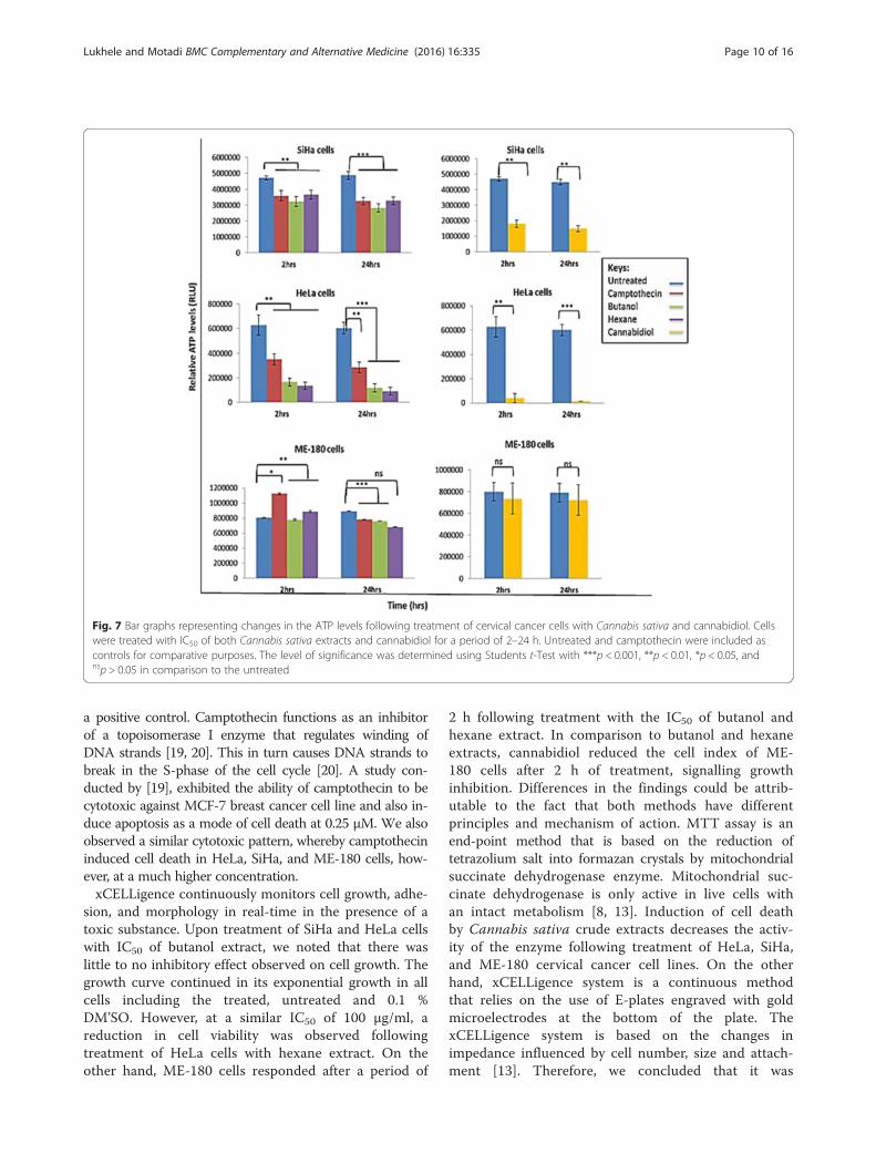
Fig. 7 Bar graphs representing changes in the ATP levels following treatment of cervical cancer cells with Cannabis sativa and cannabidiol. Cellswere treated with IC50 of both Cannabis sativa extracts and cannabidiol for a period of 2–24 h. Untreated and camptothecin were included ascontrols for comparative purposes. The level of significance was determined using Students t-Test with ***p < 0.001, **p < 0.01, *p < 0.05, andnsp > 0.05 in comparison to the untreated
Lukhele and Motadi BMC Complementary and Alternative Medicine (2016) 16:335 Page 10 of 16
a positive control. Camptothecin functions as an inhibitorof a topoisomerase I enzyme that regulates winding ofDNA strands [19, 20]. This in turn causes DNA strands tobreak in the S-phase of the cell cycle [20]. A study con-ducted by [19], exhibited the ability of camptothecin to becytotoxic against MCF-7 breast cancer cell line and also in-duce apoptosis as a mode of cell death at 0.25 μM. We alsoobserved a similar cytotoxic pattern, whereby camptothecininduced cell death in HeLa, SiHa, and ME-180 cells, how-ever, at a much higher concentration.xCELLigence continuously monitors cell growth, adhe-
sion, and morphology in real-time in the presence of atoxic substance. Upon treatment of SiHa and HeLa cellswith IC50 of butanol extract, we noted that there waslittle to no inhibitory effect observed on cell growth. Thegrowth curve continued in its exponential growth in allcells including the treated, untreated and 0.1 %DM’SO. However, at a similar IC50 of 100 μg/ml, areduction in cell viability was observed followingtreatment of HeLa cells with hexane extract. On theother hand, ME-180 cells responded after a period of
2 h following treatment with the IC50 of butanol andhexane extract. In comparison to butanol and hexaneextracts, cannabidiol reduced the cell index of ME-180 cells after 2 h of treatment, signalling growthinhibition. Differences in the findings could be attrib-utable to the fact that both methods have differentprinciples and mechanism of action. MTT assay is anend-point method that is based on the reduction oftetrazolium salt into formazan crystals by mitochondrialsuccinate dehydrogenase enzyme. Mitochondrial suc-cinate dehydrogenase is only active in live cells withan intact metabolism [8, 13]. Induction of cell deathby Cannabis sativa crude extracts decreases the activ-ity of the enzyme following treatment of HeLa, SiHa,and ME-180 cervical cancer cell lines. On the otherhand, xCELLigence system is a continuous methodthat relies on the use of E-plates engraved with goldmicroelectrodes at the bottom of the plate. ThexCELLigence system is based on the changes inimpedance influenced by cell number, size and attach-ment [13]. Therefore, we concluded that it was

Fig. 8 Representative bar graph of the cervical cancer cell cycle before and after treatment with Cannabis sativa extracts and cannabidiol. Cellswere harvested and treated with camptothecin and the IC50 concentrations of Cannabis sativa extracts and cannabidiol. Bar graph a and d representsSiHa cells, b and e represents HeLa cells, c and f represents ME-180 cells. Data represented as mean ± standard deviation with ***p < 0.001, **p < 0.01,*p < 0.05, and nsp > 0.05 representing the level of significance in comparison to the untreated
Lukhele and Motadi BMC Complementary and Alternative Medicine (2016) 16:335 Page 11 of 16
possible that dead cells might have been attached atthe bottom of the E-plate after treatment.Cell death can be characterized by a decrease in the
energy levels as a result of dysfunction of the mitochon-dria [8]. Therefore, to evaluate the effect of treatment onthe energy content of the cells, we conducted mitochon-drial assay. We only used IC50 as indicated by MTTassay only. ATP acts as determinant of both cell deathand cell proliferation [15]. Exposure of SiHa, HeLa, andME-180 cells to the IC50 of Cannabis sativa extractscaused a reduction in the ATP levels. Treatment of cellswith cannabidiol either slightly or severely depleted theATP levels. According to [16], a reduction of the ATPlevels compromises the status of cell and often leads tocell death either by apoptosis or necrosis, while anincrease is indicative of cell proliferation. Therefore, weconcluded that the reduction of ATP might have been asa result of cell death induction since the cells ATPproduction recovered.Following confirmation that Cannabis sativa and can-
nabidiol have anti-proliferative activity, we had to verify
whether both treatments have the ability to induce cellcycle arrest in all three cell lines. This method uses a PIstain and flow cytometry to measure the relative amountof DNA present in the cells. In this study, propidiumiodide (PI) was used to stain cells. Propidium iodide canonly intercalate into the DNA of fixed and permeabilizedcells with a compromised plasma membrane or cells inthe late stage of apoptosis. Viable cells with an intactplasma membrane cannot uptake the dye. The intensityof stained cells correlates with the amount of DNAwithin the cells. HeLa, SiHa, and ME-180 cervical cancercells were stained with PI and analysed using flowcytometry. Treatment of SiHa cells with butanol andhexane extracts led to the accumulation of cells in thecell death phase (sub-G0 phase), without cell cycle arrest.When compared to the S-phase and G2/M phase of un-treated cells, exposure of HeLa cells to Cannabis sativabutanol extract resulted in the accumulation of cells inthe S-phase of the cell cycle and slight cell death induc-tion. And thus, according to [3], signals DNA synthesisand cell cycle proliferation. A decrease in the S-phase

Fig. 9 Western blot analysis of the protein expression before and after 24 h treatment with IC50 of Cannabis sativa extracts and cannabidiol. SiHa(a and d), HeLa (b and e), and ME-180 (c and f) cells were treated for a period of 24 h and protein lysates were separated using SDS-PAGE gel.Untreated protein was used as a control. Antibodies against pro-apoptotic proteins (p53 and Bax) and anti-apoptotic proteins (Bcl-2 and RBBP6),Initiator caspase-9 and effecter caspase-3 were included to elucidate apoptosis induction
Lukhele and Motadi BMC Complementary and Alternative Medicine (2016) 16:335 Page 12 of 16
and an increase in the G2/M phase of HeLa cells follow-ing treatment with hexane extract, suggests a blockageof mitosis and an induction of cell cycle arrest. Interest-ing to note was that, treatment of ME-180 cells withboth extracts led to an increase of cells coupled by anincrease in the S-phase population which favoursreplication and duplication of DNA. This was not thecase following treatment of cells with cannabidiol.Cannabidiol resulted in the accumulation of cells in thecell death phase of the cell cycle. SiHa, and HeLa, andME-180 cells were committed to the cell death phase. Insummary, Cannabis sativa induces cell death with orwithout cell cycle arrest while cannabidiol induces celldeath without cell cycle arrest.Apoptosis plays a major role in determining cell sur-
vival. Annexin V/FITC and PI were used to stain the
cells to be able to distinguish between viable, apoptoticand necrotic cells. Annexin V/ FITC can only bind tophosphatidylserine residues exposed on the surface ofthe cell membrane while PI intercalates into the nucleusand binds to the fragmented DNA. Viable cells cannotuptake both dyes due to the presence of an intact cellmembrane. Since treatment caused the accumulation ofcells in the sub-G0 phase, also known as the cell deathphase, and the severe depletion of ATP levels bycannabidiol, we further conducted an apoptosis assay.Treatment of all three cell lines with camptothecin, IC50
of Cannabis sativa and cannabidiol exhibited the type ofinduced cell death as apoptosis. Sharma et al. [25] alsoshowed a similar pattern of cell death, wherebytreatment of a prostate cancer cell lines with Cannabissativa resulted in the induction of apoptosis.

Fig. 10 A densitometry analysis SiHa protein was performed using ImageJ quantification software to measure the relative band intensity. CPTrepresents camptothecin. Data represented as mean ± standard deviation with ***p < 0.001, **p < 0.01 and nsp > 0.05 representing the level ofsignificance in comparison to the untreated represent the western blot analysis of SiHa and HeLa cells. The genes analyzed are p53 and RBBP6including caspases. Equal amount of protein (conc) was loaded in each well. Note that the darker the bands increased expression of the gene
Lukhele and Motadi BMC Complementary and Alternative Medicine (2016) 16:335 Page 13 of 16
Apoptosis is characterized by morphological changesand biochemical features which include condensation ofchromatin, convolution of nuclear and cellular outlines,nuclear fragmentation, formation of apoptotic blebswithin the plasma membrane, cell shrinkage due to theleakage of organelles in the cytoplasm as well as thepresence of green stained cells at either late or earlyapoptosis [5, 17, 28]. Annexin V/FITC and DAPI wereused to visualize the cells under a fluorescence confocalmicroscopy. According to [18], an uptake of Annexin V/FITC suggests the induction of apoptosis, since it canonly bind to externalized PS residues. This also provesthat during cell growth analysis, SiHa and HeLa cellswere undergoing cell death while still attached to thesurface of the flask.
Apoptosis is known to occur via two pathways, thedeath receptor pathway and the mitochondrial pathway[30]. Cannabis sativa isolates including cannabidiol havebeen implicated in apoptosis induction via the death re-ceptor pathway, by binding to Fas receptor or throughan activated of Bax triggered by the synthesis of cer-amide in the cells [4]. However, not much has beenreported on the induction of apoptosis via activation ofp53 by Cannabis sativa. Our focus in this study was alsoto identify downstream molecular effect of extracts. Onesuch important gene is p53 which acts as a transcriptionfactor for a number of target genes [29]. Under normalconditions, p53 levels are maintained through constantdegradation MDM2 and its monomers [29]. RBBP6 isone of the monomers that helps degrade p53, due the

Fig. 11 A densitometry analysis HeLa protein was performed using ImageJ quantification software to measure the relative band intensity. CPTrepresents camptothecin. Data represented as mean ± standard deviation with ***p < 0.001, **p < 0.01 and nsp > 0.05 representing the level ofsignificance in comparison to the untreated
Lukhele and Motadi BMC Complementary and Alternative Medicine (2016) 16:335 Page 14 of 16
presence of Ring finger domain that promotes the inter-action of both proteins [14]. In response to stress stimulisuch as DNA damage, hypoxia, UV light, and radiationlight, p53 becomes activated and causes MDM2 expres-sion to decrease [10]. Mutation of p53, implicated to beassociated with 50 % of all human cancers, promote thetumorigenesis. Bax and Bcl-2 form part of the proteinsthat regulate apoptosis via the mitochondria [21].Following activation, p53 translocates into the cytosoland triggers the oligomerization of Bcl-2 with BAD,resulting in the inhibition of Bcl-2 activity [17]. This inturn allows Bax protein to be translocated to the mito-chondria and participate in the release of cytochrome cthrough poration of the outer mitochondrial membrane[9, 17]. An imbalance between Bax and Bcl-2 has beenlinked to the development and progression of tumoursthrough the resistance of apoptosis [17]. It is thereforecrucial to design drugs that would effectively target these
genes involved in the execution of apoptosis via the mito-chondrial pathway. Camptothecin, hexane extract, andcannabidiol effectively up-modulated the expression ofp53 in all three cell lines, leading to a decrease in RBBP6protein expression. Apart from SiHa and HeLa, butanolextract failed to up-modulate p53 in ME-180 cells.Interesting to note is that butanol extract reduced theexpression of RBBP6 protein in ME-180 cells. The mech-anism behind failure of butanol to up-modulate p53 whiledown-modulating RBBP6 is unclear. However, we came toa conclusion that butanol induces apoptosis independentlyof p53. We further demonstrated that Cannabis sativaextracts, cannabidiol, and camptothecin were able todown-modulate the expression of Bcl-2 protein and up-modulate Bax expression.Caspases play an effective role in the execution of
apoptosis either through the extrinsic or intrinsic path-way [9]. In this study, we wanted to validate whether

Fig. 12 A densitometry analysis ME-180 protein was performed using ImageJ quantification software to measure the relative band intensity. CPTrepresents camptothecin. Data represented as mean ± standard deviation with ***p < 0.001, **p < 0.01 and nsp > 0.05 representing the level ofsignificance in comparison to the untreated
Lukhele and Motadi BMC Complementary and Alternative Medicine (2016) 16:335 Page 15 of 16
caspase-9 and caspase-3 were involved in the initiationand execution of apoptosis. We demonstrated the abilityof Cannabis sativa to initiate apoptosis by activatingcaspase-9. However, execution of apoptosis was eitherwith or without the presence of capsase-3, depending oneach cell line. Western blot revealed that Cannabissativa hexane extract induced apoptosis via the activa-tion of caspase-9 and caspase-3 when compared tountreated cells in all three cell lines. Similar results wereobtained during treatment of all three cell lines withcamptothecin. This was not the case with butanol. Butanolextracts up-modulated caspase-9 and caspase-3 in SiHaand HeLa cells only. Caspase-3 was not up-modulated inME-180 cells. Caspase 3/7 activity assay revealed the up-modulation of caspase 3/7 following treatment of cervicalcancer cells. However on the basis of the Western blotresults, wherein butanol extract failed to up-modulatecaspase-3, we can conclude that caspase-7 was responsiblefor the reported activity. Cannabidiol effectively up-
modulated caspase-9 and caspase-3 in all three cell lines,when compared to the untreated and Cannabis sativaextract. From the results we can conclude that, apoptosisinduction was caspase dependent.
ConclusionsThe aim of this study was to evaluate for the anti-growtheffects of Cannabis sativa extracts and to also determinethe mode of cell death following treatment. The activity ofCannabis sativa extracts was compared to that of canna-bidiol, in order to verify whether the reported results weredue to the presence of the compound. The study showedthat the activity of one of the extracts might have beendue to the presence of cannabidiol. It further demon-strated the ability of Cannabis sativa to induce apoptosiswith or without cell cycle arrest and via mitochondrialpathway. More research needs to be done elucidating themechanism between the active ingredients and moleculartargets involved in the regulation of the cell cycle.

Lukhele and Motadi BMC Complementary and Alternative Medicine (2016) 16:335 Page 16 of 16
AcknowledgementsOur gratitude goes to South African MRC for funding assistance.
FundingThe work was funded by MRC.
Availability of data and materialsThe datasets supporting the conclusions of this article are included withinthe article and its additional files.
Authors’ contributionsSTL was responsible for the experimental design and LRM prepared themanuscript. Both authors read and approved the final manuscript.
Competing interestsThe authors declare that they have no competing interests.
Consent for publicationThe authors give consent for the journal to be published.
Ethics approval and consent to participateThis study was approved by the Human Research Ethics Committee (Medical):M140801.
Received: 2 June 2016 Accepted: 11 August 2016
References1. Alexander A, Smith PF, Rosengren RJ. Cannabinoids in the treatment of
cancer. Cancer Lett. 2009;285:6–12.2. Arbyn M, Castellsague X, de Sanjose S, Bruni L, Saraiya M, Bray F, Ferlay J.
Worldwide burden of cervical cancer in 2008. Ann Oncol. 2011;22:2675–86.3. Armania N, Yazan LS, Ismail IS, Foo JB, Tor YS, Ishak N, Ismail N, Ismail M.
Dillenia suffruticosa extract inhibits proliferation of human breast cancer celllines (MCF-7 and MDA-MB-231) via induction of G2/M arrest and apoptosis.Molecules. 2013;18(11):13320–39.
4. Bla’zquez C, Galve-Roperh I, Guzma’n M. De novo-synthesized ceramidesignals apoptosis in astrocytes via extracellular signal-regulated kinase.FASEB J. 2000;14:2315–22.
5. Bortner CD, Oldenburg NB, Cidlowski JA. The role of DNA fragmentation inapoptosis. Trends Cell Biol. 1995;5:21–6.
6. Caffarel MM, Andradas C, Perez-Gomez E, Guzman M, Sanchez C.Cannabinoids: Anew hope for breast cancer therapy? Cancer Treat Rev.2012;38:911–8.
7. Chen P, Yu J, Chalmers B, Drisko J, Yang J, Li B, Chen Q. Pharmacologicascorbate induces cytotoxicity in prostate cancer cells through ATPdepletion and the induction of autophagy. Anticancer Drugs Preclinical Rep.2011;23:437–44.
8. Choene M, Motadi L. Validation of the Antiproliferative Effects of Euphorbiatirucalli extracts in Breast Cancer Cell Lines. Mol Biol. 2016;50(1):115–28.
9. Chipuk TE, Kuwana T, Bouchier-Hayes L, Droin MN, Newmeyer DD, SchulerM, Green DR. Direct activation of Bax by p53 mediates MitochondrialMembrane Permeabilization and Apoptosis. Sci J. 2004;303:1010.
10. de Bruin EC, Medema JP. Apoptosis and non-apoptosis deaths in cancerdevelopment and treatment response. Cancer Treat Rev. 2008;34:737–49.
11. Flemming R, Muntendam T, Steup T, Kayser O. Chemistry and biologicalactivity of tetrahydrocannabinol and its derivatives. Top Heterocycl Chem.2007;10:1–42.
12. GLOBOCAN 2012 v1.0, Cancer Incidence and Mortality Worldwide: IARCCancer Base No. 11 [Internet]. Lyon, France: International Agency forResearch on Cancer. Available from http://globocan.iarc.fr Accessed 25 July2014.
13. Gumulec J, Balvan J, Sztalmachova M, Raudeska M, et al. Cisplatin-resistantprostate cancer model: differences in antioxidant system, apoptosis, and cellcycle. Int J Oncol. doi:10.3892/ijo.2013.2223.
14. Happyana N, Agnolet S, Muntendam R, Van Dam A, Schneider B, Kayser O.Analysis of cannabinoids in laser-micro dissected trichomes of medicinalCannabis sativa using LCMS and cryogenic NMR. Phytochemistry. 2013;87:51–9.
15. Lemasters JJ, Nieminen A, Qian T, Frost LC, et al. The mitochondrialpermeability transition in cell death: a common mechanism in necrosis,apoptosis, and autophagy. Biochim Biophys Acta. 1998;1366:177–96.
16. Ligresti A, Moriello AS, Matias I, et al. Anti-tumor activity of plantcannabinoids with the emphasis on the effect of cannabidiol on humanbreast cancer. J Pharmacol Exp Ther. 2006;318(3):1375–87.
17. Li-Weber M. Targeting apoptosis pathways in cancer by Chinese medicine.Cancer Lett. 2013;332:304–12.
18. Lozano I. The therapeutic use of Cannabis sativa in Arabic medicine. J CannabisTher. 2001;1(1):63-70.
19. Moela P, Choene MS, Motadi LR. Silencing RBBP6 (Retinoblastoma bindingprotein 6) sensitizes breast cancer cells MCF-7 to camptothecin andstaurosporine-induced cell death. Immunology. 2013;219:1–9.
20. Nobili S, Lippi D, Witort E, Donnini M, et al. Natural compounds for cancertreatment and prevention. Pharmacol Res. 2009;59(6):365–78.
21. O’Brien MA, Kirby R. Apoptosis: a review of pro-apoptotic and anti-apoptoticpathways and dysregulation in disease. J Vet Emerg Crit Care. 2008;18(6):572–85.
22. Rao GV, Kumar S, Islam M, Mansour ES. Folk medicines for anticancertherapy-a current status. Cancer Ther. 2008;6:913–22.
23. Romano B, Borrelli F, Pagano E, Cascio MG, Pertwee RG, Izzo AA. Inhibitionof colon carcinogenesis by a standardized Cannabis sativa extract with highcontent of cannabidiol. Phytomedicine. 2014;21(5):631–9.
24. Safaraz S, Adhami VM, Syed DN, Afaq, Mukhtar H. Cannabinoids for cancertreatment: Progress and promise. Cancer Res. 2008;68(2):339–44.
25. Sharma M, Hudson JB, Adomat H, Guns E, Cox ME. In Vitro AnticancerActivity of Plant-Derived Cannabidiol on Prostate Cancer Cell Line.Pharmacol Pharm. 2014;5:806–20.
26. Shrivastava A, Kuzontkoski PM, Groopman JE, Prasad A. Cannabidiol inducesprogrammed cell death by coordinating the cross-talk between apoptosisand autophagy. Mol Cancer Ther. 2011;10(7):1161–72.
27. Street RA, Prinsloo G. “Commercially Important Medicinal Plants of SouthAfrica: A Review.”. J Chem. 2013:1–16. doi:10.1155/2013/205048.
28. Thafeni M, Sayed Y, Motadi L. Euphorbia mauritanica and Kedrostis hirtellaextracts induces cell death in lung cancer cells. J Mol Biol.2012;39(12):10785–94.
29. Turner CE, Hadley KW, Holley HJ, Billets S, Mole Jr LM. Constituents ofCannabis sativa L. VIII: Possible biological application of a new method toseparate cannabidiol and cannabichromene. J Pharm Sci. 1975;64(5):810–4.
30. Yamaori S, Kushihara M, Yamamoto I, Watanabe K. Characterization of majorphytocannabinoids, cannabidiol and cannabinol, as isoform-selective andpotent inhibitors of human CYP1 enzymes. Biochem Pharmacol.2010;79:1691–8.
• We accept pre-submission inquiries
• Our selector tool helps you to find the most relevant journal
• We provide round the clock customer support
• Convenient online submission
• Thorough peer review
• Inclusion in PubMed and all major indexing services
• Maximum visibility for your research
Submit your manuscript atwww.biomedcentral.com/submit
Submit your next manuscript to BioMed Central and we will help you at every step: