Cannabidiol changes P-gp and BCRP expression in ... Eshkoli2, Boaz Sheizaf2, Eyal Sheiner2, Mahmud...
19
Submitted 27 May 2013 Accepted 15 August 2013 Published 12 September 2013 Corresponding author Valeria Feinshtein, [email protected] Academic editor Maria Deli Additional Information and Declarations can be found on page 13 DOI 10.7717/peerj.153 Copyright 2013 Feinshtein et al. Distributed under Creative Commons CC-BY 3.0 OPEN ACCESS Cannabidiol changes P-gp and BCRP expression in trophoblast cell lines Valeria Feinshtein 1 ,Offer Erez 2 , Zvi Ben-Zvi 1 , Noam Erez 1 , Tamar Eshkoli 2 , Boaz Sheizaf 2 , Eyal Sheiner 2 , Mahmud Huleihel 3 and Gershon Holcberg 2 1 Department of Clinical Biochemistry and Pharmacology, Ben-Gurion University of the Negev, Israel 2 Department of Obstetrics and Gynecology, Soroka University Medical Center, School of Medicine, Faculty of Health Sciences, Ben-Gurion University of the Negev, Israel 3 The Shraga Segal Department of Microbiology and Immunology, Ben-Gurion University of the Negev, Israel ABSTRACT Objectives. Marijuana is the most commonly used illicit drug during pregnancy. Due to high lipophilicity, cannabinoids can easily penetrate physiological barriers like the human placenta and jeopardize the developing fetus. We evaluated the impact of cannabidiol (CBD), a major non-psychoactive cannabinoid, on P-glycoprotein (P-gp) and Breast Cancer Resistance Protein (BCRP) expression, and P-gp function in a placental model, BeWo and Jar choriocarcinoma cell lines (using P-gp induced MCF7 cells (MCF7/P-gp) for comparison). Study design. Following the establishment of the basal expression of these trans- porters in the membrane fraction of all three cell lines, P-gp and BCRP protein and mRNA levels were determined following chronic (24–72 h) exposure to CBD, by Western Blot and qPCR. CBD impact on P-gp efflux function was examined by uptake of specific P-gp fluorescent substrates (calcein-AM, DiOC2(3) and rho- damine123(rh123)). Cyclosporine A (CsA) served as a positive control. Results. Chronic exposure to CBD resulted in significant changes in the protein and mRNA levels of both transporters. While P-gp was down-regulated, BCRP levels were up-regulated in the choriocarcinoma cell lines. CBD had a remarkably different influence on P-gp and BCRP expression in MCF7/P-gp cells, demonstrating that these are cell type specific effects. P-gp dependent efflux (of calcein, DiOC2(3) and rh123) was inhibited upon short-term exposure to CBD. Conclusions. Our study shows that CBD might alter P-gp and BCRP expression in the human placenta, and inhibit P-gp efflux function. We conclude that marijuana use during pregnancy may reduce placental protective functions and change its morphological and physiological characteristics. Subjects Gynecology and Obstetrics, Pharmacology Keywords Cannabidiol, Marijuana, Breast Cancer Resistance Protein (BCRP), P-glycoprotein (P-gp), BeWo and Jar cells, Human placenta INTRODUCTION Until recently it was assumed that prenatal exposure to marijuana is frequently combined with other drugs (e.g., tobacco and alcohol), making cannabis effects difficult to isolate How to cite this article Feinshtein et al. (2013), Cannabidiol changes P-gp and BCRP expression in trophoblast cell lines. PeerJ 1:e153; DOI 10.7717/peerj.153
Transcript of Cannabidiol changes P-gp and BCRP expression in ... Eshkoli2, Boaz Sheizaf2, Eyal Sheiner2, Mahmud...

Submitted 27 May 2013Accepted 15 August 2013Published 12 September 2013
Corresponding authorValeria Feinshtein,[email protected]
Academic editorMaria Deli
Additional Information andDeclarations can be found onpage 13
DOI 10.7717/peerj.153
Copyright2013 Feinshtein et al.
Distributed underCreative Commons CC-BY 3.0
OPEN ACCESS
Cannabidiol changes P-gp and BCRPexpression in trophoblast cell linesValeria Feinshtein1, Offer Erez2, Zvi Ben-Zvi1, Noam Erez1,Tamar Eshkoli2, Boaz Sheizaf2, Eyal Sheiner2, Mahmud Huleihel3 andGershon Holcberg2
1 Department of Clinical Biochemistry and Pharmacology, Ben-Gurion University of the Negev,Israel
2 Department of Obstetrics and Gynecology, Soroka University Medical Center, School ofMedicine, Faculty of Health Sciences, Ben-Gurion University of the Negev, Israel
3 The Shraga Segal Department of Microbiology and Immunology, Ben-Gurion University of theNegev, Israel
ABSTRACTObjectives. Marijuana is the most commonly used illicit drug during pregnancy. Dueto high lipophilicity, cannabinoids can easily penetrate physiological barriers likethe human placenta and jeopardize the developing fetus. We evaluated the impactof cannabidiol (CBD), a major non-psychoactive cannabinoid, on P-glycoprotein(P-gp) and Breast Cancer Resistance Protein (BCRP) expression, and P-gp functionin a placental model, BeWo and Jar choriocarcinoma cell lines (using P-gp inducedMCF7 cells (MCF7/P-gp) for comparison).Study design. Following the establishment of the basal expression of these trans-porters in the membrane fraction of all three cell lines, P-gp and BCRP proteinand mRNA levels were determined following chronic (24–72 h) exposure to CBD,by Western Blot and qPCR. CBD impact on P-gp efflux function was examinedby uptake of specific P-gp fluorescent substrates (calcein-AM, DiOC2(3) and rho-damine123(rh123)). Cyclosporine A (CsA) served as a positive control.Results. Chronic exposure to CBD resulted in significant changes in the protein andmRNA levels of both transporters. While P-gp was down-regulated, BCRP levelswere up-regulated in the choriocarcinoma cell lines. CBD had a remarkably differentinfluence on P-gp and BCRP expression in MCF7/P-gp cells, demonstrating thatthese are cell type specific effects. P-gp dependent efflux (of calcein, DiOC2(3) andrh123) was inhibited upon short-term exposure to CBD.Conclusions. Our study shows that CBD might alter P-gp and BCRP expression inthe human placenta, and inhibit P-gp efflux function. We conclude that marijuanause during pregnancy may reduce placental protective functions and change itsmorphological and physiological characteristics.
Subjects Gynecology and Obstetrics, PharmacologyKeywords Cannabidiol, Marijuana, Breast Cancer Resistance Protein (BCRP), P-glycoprotein(P-gp), BeWo and Jar cells, Human placenta
INTRODUCTIONUntil recently it was assumed that prenatal exposure to marijuana is frequently combined
with other drugs (e.g., tobacco and alcohol), making cannabis effects difficult to isolate
How to cite this article Feinshtein et al. (2013), Cannabidiol changes P-gp and BCRP expression in trophoblast cell lines. PeerJ 1:e153;DOI 10.7717/peerj.153
and assess (Kozer & Koren, 2001; Moore et al., 2010). However, the studies of Dekker et
al. and Hayatbakhsh et al. suggested that cannabis use prior to, during the first trimester,
or throughout gestation is associated with a higher risk for a low birth weight and
neonatal length, as well as preterm labor (Dekker et al., 2012; Hayatbakhsh et al., 2012).
Moreover, the presence of the endocannabinoid receptors CB1 and CB2 on placental
syncytiotrophoblast (Habayeb et al., 2008a), along with marijuana being the most popular
drug of abuse among pregnant population (Brown & Graves, 2013), raised the need to
understand the effect of cannabinoids on the placenta.
Cannabidiol (CBD) is one of the most abundant cannabinoids in the marijuana plant
(Mechoulam & Shvo, 1963; Mechoulam & Hanus, 2002; Schier et al., 2012). It is a promising
candidate for clinical utilization, due to low affinity binding to CB1 and CB2 cannabinoid
receptors and no cognitive and psychoactive activity (Zuardi, 2008; Deiana, 2012).
Preliminary data from in vitro models indicate that cannabinoids may interact
with human P-gp (ABCB1) and BCRP (ABCG2). Acute and long-term exposures to
cannabinoids were shown to alter P-gp expression and function (Holland et al., 2006;
Zhu et al., 2006). However, BCRP exposure to cannabinoids demonstrated functional
inhibition only (Holland et al., 2007).
P-gp and BCRP are both thought to be protective for the fetus. These ATP-binding
cassette (ABC) efflux transporters expressed at the apical membrane of the polarized
syncytiotrophoblast layer (Ni & Mao, 2011), and play a significant role in drug transfer
across the placental barrier (Lankas et al., 1998; Mao, 2008; Vahakangas & Myllynen,
2009; Myllynen, Kummu & Sieppi, 2010; Eshkoli et al., 2011). P-gp was found to be an
anti-apoptotic cellular agent (Smyth et al., 1998; Huls, Russel & Masereeuw, 2009) and
BCRP was shown to have a role in placental tissue and syncytial survival by protecting
cells from pro-apoptotic injuries (Evseenko et al., 2007; Evseenko, Paxton & Keelan, 2007;
Hardwick, Velamakanni & van Veen, 2007; Vahakangas & Myllynen, 2009).
We have already assessed the interaction of CBD with placental BCRP, finding that in
both, in vitro and ex vivo systems, CBD inhibited its efflux function (Feinshtein et al., 2013).
These results together with other recent findings regarding the effect of cannabinoid on
the ABC transporter led us to investigate whether CBD affects placental P-gp on functional
and placental P-gp and BCRP on expressional levels (Holland et al., 2006; Zhu et al., 2006;
Holland et al., 2007; Holland, Allen & Arnold, 2008).
In the present work the implications of CBD exposure on P-gp and BCRP expression
and P-gp function is tested in a human trophoblast-like cell lines BeWo and Jar, as placental
model.
MATERIALS AND METHODSMaterialsBeWo and Jar cells were obtained from Dr. B. Ugele, Ludwig-Maximilians University,
Munich, Germany (Feinshtein et al., 2010; Polachek et al., 2010; Feinshtein et al., 2013).
MCF7/P-gp cells (BCRP expressing and P-gp induced cells), were kindly provided by
Prof. Esther Priel (Ben Gurion University, Beer Sheva, Israel) (Feinshtein et al., 2013). All
Feinshtein et al. (2013), PeerJ, DOI 10.7717/peerj.153 2/19

materials for cell culture were purchased from Biological Industries (Israel). CBD was a
kind gift from Prof. Raphael Mechoulam (The Hebrew University of Jerusalem, Jerusalem,
Israel).Calcein-AM, DiOC2(3), rh123 and cyclosporine A (CsA) were purchased from
Sigma-Aldrich (St. Louis, MO, USA). A list of all antibodies used in the current research is
summarized in Table S1.
METHODSCell culture and drug treatmentsMCF7/P-gp cells were cultured in conditions as previously described (Golan, Schreiber &
Avissar, 2009), BeWo and Jar cells were cultured as previously described (Golan, Schreiber &
Avissar, 2009; Feinshtein et al., 2010). Briefly, 24 h after seeding in 35× 10 mm cell culture
dishes (Corning), growth medium (DMEM for MCF7/P-gp, DMEM/F-12 for BeWo and
Jar cells, supplemented with 10% fetal bovine serum, 2 mM L-glutamine, 100 µg/ml strep-
tomycin and 100 units/ml penicillin in a humidified atmosphere of 95% air and 5% CO2
at 37◦C) was replaced with fresh medium containing CBD 10 or 15 µM (initially dissolved
in DMSO) or DMSO (0.08% or 0.12%, respectively, vehicle control). In order to achieve
long-term exposure, the treatment medium was refreshed every day for 24 h, 48 h or 72 h.
Subcellular fractionationFractionation procedure was carried out as previously described (Golan, Schreiber & Avis-
sar, 2009), for the isolation of the membrane fraction. The membrane fraction was diluted
in sample buffer 1:3 (10% v/v glycerol, 20% v/v SDS 20%, 5% v/v β-mercaptoethanol,
0.05% w/v bromophenol blue, pH 6.8), boiled for 5 min at 95◦C (for BCRP determination)
or incubated for 30 min at 37◦C (for P-gp determination) and frozen at −80◦C until
assayed. Aliquots were taken for protein determination using the Lowry assay. Fractions
purity was verified using specific markers: Na+/K+ ATPase for membranes and NF/kB p65
for cytosol.
IMMUNOCYTOCHEMISTRY AND CONFOCALMICROSCOPYMCF7/P-gp, BeWo and Jar cells were seeded on cover slips and grown to 70% sub-
confluency. Cells were then fixed in PFA for 15 min and stained with mouse anti-human
CD 243 (MDR-1) antibodies, diluted in PBS, containing 3% BSA, for 1 h at room
temperature, followed by incubation with Alexa Fluor 488 goat anti-mouse antibody in
PBS containing 3% BSA, for 1/2 h at room temperature. Cover-slips were washed 3 times
and mounted onto glass slides with DAPI (4′,6-diamidino-2-phenylindole)-containing
fluorescent mounting medium (DAPIFluoromount-G; SouthernBiotech). Immunofluo-
rescence was detected by an Olympus FV-1000 Spectral confocal laserscanning microscope
with excitation at 488 nm and emission at 520 nm. Image analysis was performed using
ImageJ v. 1.40C software.
Feinshtein et al. (2013), PeerJ, DOI 10.7717/peerj.153 3/19

FACS analysis of BCRP levelsCells were grown to confluency in 6-well culture dishes, trypsinized, counted, and
0.5× 10⁶ cells were placed in light resistant vials. Cells were washed three times with
cold PBS (and each time centrifuged at 1200 g for 5 min), and incubated for 30 min with
BSA 3% in PBS. Following additional PBS wash, 50 µL of BSA 1% in PBS and antibodies
were added (1:10 anti-BCRP FITC conjugated antibody or 1:10 CBL602F FTIC conjugated
isotype negative control). Cells were incubated in the dark at 4◦C for 1 h, washed with ice
cold PBS, re-suspended (in 0.5 ml PBS) and analyzed by FACS (BD FACS Vantage).
ImmunoblottingMembrane fractions were thawed on the day of assay. Protein aliquots (60–100 µg/lane)
were taken for protein separation by SDS-PAGE as previously described (Golan, Schreiber
& Avissar, 2009). Semi-quantitative analysis was carried out using a computerized image
analysis system (EZQuant-Gel 2.11; EZQuant Biology Software Solutions Ltd., Israel).
Equal protein loading was ensured by normalization to Na+/K+ ATPase (for P-gp) or actin
(for BCRP).
RNA extraction, reverse transcription, real-time polymerase chainreaction (qPCR)Isolation and purification of total RNA from BeWo and Jar cells was carried out using
EZ-RNA Kit (Biological Industries, Israel) according to manufacturer’s instructions. 1 µg
of total RNA was used for reverse transcription using High Capacity cDNA RT kit (Applied
Biosystems, Foster City, CA), in 20 µl reaction volume. BCRP and P-gp mRNA was
measured by qRT-PCR, as indicated in the manufacturer protocol (Applied Biosystems,
Foster City, CA), and performed by the Applied Biosystems Real Time PCR system (7500
system), using TaqMan probes and primers for human BCRP, P-gp and actin (Applied
Biosystems, Foster City, CA). The cycling conditions for all primers were as follows: hold
for 10 min at 95◦C, followed by 40 cycles consisting of two steps, 15 s at 95◦C (denaturing),
and 1 min at 60◦C (annealing-extension). The threshold cycle, which correlates inversely
with the mRNA levels of target, was measured as the cycle number at which the reporter
fluorescent emission increases above a threshold level. P-gp and BCRP mRNA levels were
normalized to actin mRNA in the same samples. Results were analyzed by the 2−1−1CT
method, demonstrating the relative changes in gene expression from real-time quantitative
PCR experiments, using 7500 Software v2.0.4 (Applied Biosystems, Foster City, CA).
P-gp substrate uptake experimentsCells were pre-incubated for 30 min with CBD 10 or 25 µM (working concentration
previously published (Holland et al., 2006; Ligresti et al., 2006; Zhu et al., 2006; Holland et
al., 2007; Holland, Allen & Arnold, 2008; De Filippis et al., 2011; Arnold et al., 2012; Harvey
et al., 2012; Hill et al., 2012; Maor et al., 2012; Solinas et al., 2012; Dudasova et al., 2013;
Juknat et al., 2013; Nabissi et al., 2013), and initially dissolved in DMSO) or CsA 20 µM
(known P-gp inhibitor used as positive control (Mori et al., 2012)), dissolved in transport
buffer (TB) (pH= 7.4) (Feinshtein et al., 2010), while “control cells” were pre-incubated
Feinshtein et al. (2013), PeerJ, DOI 10.7717/peerj.153 4/19

in TB with the correlating concentration of DMSO. Following pre-incubation, P-gp
substrates were added (Calcein-AM or DiOC2(3) or rh123) (Minderman et al., 1996;
Martin et al., 2003) and cells were further incubated for 30 min. At the end of incubation
plates were treated as previously detailed (Feinshtein et al., 2010), and samples were
stored (at −20◦C) for further analysis. Intracellular fluorescence of all P-gp substrates
was quantified by Infinite M200 microplate reader (Tecan) and normalized to protein
amount (determined by Lowry method) (Lowry et al., 1951), or detected by an Olympus
FV-1000 Spectral confocal laserscanning microscope and analyzed using ImageJ v. 1.40C
software.
Statistical analysisAll statistics and graphs were carried out using GraphPad Prism5 software. Student’s t-test
or one-way ANOVA followed by appropriate Bonferroni corrections were used.
RESULTSCBD impact on BCRP and P-gp protein expressionThe changes in P-gp and BCRP protein levels in the membrane fraction of BeWo, Jar
and MCF7/Pgp cells were studied. Following determination of proper cell fractionation
(Fig. 1A), the basal expression of these two transporters in non-treated cells was
determined by Western Blot analysis (Fig. 1B). Due to unexpected P-gp expression profile
in all three cell lines we verified its basal expression by immunocytochemical fluorescent
staining (Fig. 1C). It can be seen that our BeWo, Jar and MCF7/P-gp cells express detectable
levels of P-gp. For P-gp expression, the results of the Western Blot were confirmed by
immunocytochemistry showing that P-gp expression was the highest in MCF7/P-gp and
the lowest in Jar cells. For BCRP (ABCG2), results were confirmed by FACS analysis,
showing that BCRP expression was much higher in JAR cells compared to BeWo cells (in
full accordance with Western Blot analysis) (Fig. 1D).
The changes in membrane BCRP and P-gp levels in BeWo cells following long-term
exposure to CBD are displayed in Fig. 2. Indeed, BCRP levels were significantly increased
in a concentration-dependent manner (Fig. 2B) that was not time-dependent (Fig. 2A).
Following long-term exposure to CBD, P-gp protein levels significantly decreased, in a
concentration-dependent manner (Fig. 2D), with no time-dependent effect (Fig. 2C).
Similarly, in Jar cells (Fig. 3), BCRP protein concentrations was significantly elevated
following long term exposure to CBD, in a concentration but not time-dependent effect
(Figs. 3A and 3B). Due to very low P-gp initial (baseline) expression in the Jar cell line
(Figs. 1B and 1C), the expected down-regulation in P-gp expression following long-term
exposure to CBD is demonstrated in experiments of 72 h treatment only. P-gp is vaguely
visualized in the control group while it is almost undetectable in the CBD treated group
(Fig. 3C). 48 h exposure yielded the same results (P-gp levels were almost undetectable –
data not shown), making time and concentration dependent comparisons impractical.
To examine whether CBD effects are cell specific, the same experimental routine
was applied to MCF/P-gp cells. In these cells, BCRP and P-gp behavior under CBD
Feinshtein et al. (2013), PeerJ, DOI 10.7717/peerj.153 5/19

Figure 1 Cell fractionation, P-gp and BCRP basal expression. (A) Cell fractionation: to verify properfractionation of whole cell lysate, membrane fractions and cytosolic fractions of BeWo, Jar, MCF/P-gpcells were subjected to Western Blot analysis. Na+/K+ ATPase served as membrane marker, and NF-κB(p65 subunit) served as cytosolic marker. (B) P-gp and BCRP basal expression in MCF7/P-gp, Jar andBeWo cells was determined by Western Blotting. (C) Immunocytochemistry: fluorescent staining ofBeWo, Jar and MCF7/P-gp cells with anti-P-gp antibody and DAPI. NC – negative control. (D) BCRP(ABCG2) expression in choriocarcinoma cell lines BeWo and Jar as demonstrated by FACS analysis.
Feinshtein et al. (2013), PeerJ, DOI 10.7717/peerj.153 6/19
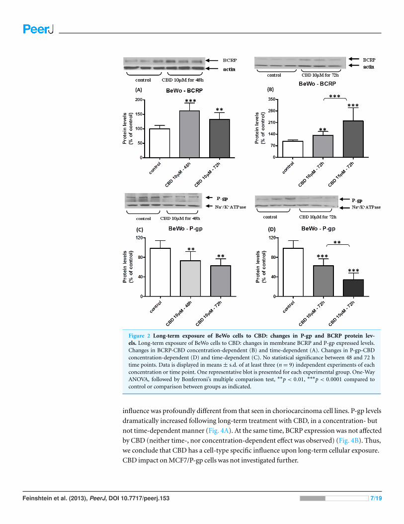
Figure 2 Long-term exposure of BeWo cells to CBD: changes in P-gp and BCRP protein lev-els. Long-term exposure of BeWo cells to CBD: changes in membrane BCRP and P-gp expressed levels.Changes in BCRP-CBD concentration-dependent (B) and time-dependent (A). Changes in P-gp-CBDconcentration-dependent (D) and time-dependent (C). No statistical significance between 48 and 72 htime points. Data is displayed in means ± s.d. of at least three (n= 9) independent experiments of eachconcentration or time point. One representative blot is presented for each experimental group. One-WayANOVA, followed by Bonferroni’s multiple comparison test, ∗∗p < 0.01, ∗∗∗p < 0.0001 compared tocontrol or comparison between groups as indicated.
influence was profoundly different from that seen in choriocarcinoma cell lines. P-gp levels
dramatically increased following long-term treatment with CBD, in a concentration- but
not time-dependent manner (Fig. 4A). At the same time, BCRP expression was not affected
by CBD (neither time-, nor concentration-dependent effect was observed) (Fig. 4B). Thus,
we conclude that CBD has a cell-type specific influence upon long-term cellular exposure.
CBD impact on MCF7/P-gp cells was not investigated further.
Feinshtein et al. (2013), PeerJ, DOI 10.7717/peerj.153 7/19

Figure 3 Long-term exposure of Jar cells to CBD: changes in P-gp and BCRP protein levels. Long-term exposure of Jar cells to CBD: changes inmembrane BCRP and P-gp expressed levels. Changes in BCRP-CBD concentration-dependent (B) and time-dependent (A). Changes in P-gp (C).Data is displayed in means± s.d. of at least three (n= 12) independent experiments of each concentration or time point. One representative blot ispresented for each experimental group. One-Way ANOVA, followed by Bonferroni’s multiple comparison test (Student’s t test for (C)), ∗p< 0.05,∗∗∗p< 0.0001 compared to control or comparison between groups as indicated.
CBD impact on BCRP and P-gp mRNATo have a glimpse into the mechanism underlying the changes seen on the protein level
in BCRP and P-gp, we further focused to track the changes that occur on mRNA level of
these two transporters. In both, BeWo and Jar cells, BCRP mRNA quantification showed
elevation following long-term exposure to CBD (Figs. 5A and 5C), and matched the
findings on protein level, supporting transcriptional up-regulation. Likewise, P-gp mRNA
expression significantly dropped following long-term treatment with CBD, matching the
outcomes seen on the protein level (Figs. 5B and 5D), indicating that the changes in P-gp
levels following long-term CBD exposure results from transcriptional down-regulation
rather than post-transcriptional changes.
CBD impact on P-gp functionOnly BeWo and MCF7/P-gp cell lines were tested for P-gp inhibition by CBD due to the
fact that Jar cells expressed very low P-gp levels. In a preliminary study we observed a
non-cell type specific P-gp inhibition by 25 µM CBD in both cell lines, as intracellular
calcein and rh123 fluorescence was significantly higher (elevation of 101 ± 50% and
70± 30%, respectively) in the presence of CBD (Figs. 6A and 6B). We further examined
whether lower concentration of CBD 10 µM also inhibits P-gp, using two different P-gp
specific substrates, calcein-AM and DiOC2(3). P-gp was inhibited following short-term
(1 h) exposure to CBD, as significantly more DiOC2(3) and calcein (elevation of 20± 8%
and 24± 9%, respectively) accumulated in the cells (Figs. 7A and 7B).
Feinshtein et al. (2013), PeerJ, DOI 10.7717/peerj.153 8/19

Figure 4 Long-term exposure of MCF/P-gp cells to CBD: changes in P-gp and BCRP protein levels.Long-term exposure of MCF/P-gp cells to CBD: changes in membrane P-gp (A) and BCRP (B) expressedlevels. Protein levels are given as percent of control levels. Data is displayed in means ± s.d. of at leastthree (n = 9) independent experiments of each concentration or time point. One representative blot ispresented for each experimental group. One-Way ANOVA, followed by Bonferroni’s multiple comparisontest. ∗∗p< 0.01, ∗∗∗p< 0.0001 compared to control or comparison between groups as indicated.
DISCUSSIONPrincipal findings of the studyThere is a dual effect of CBD on the expression of BCRP and P-gp transporters in
trophoblast-like and in MCF7/P-gp cell lines. Under long-term CBD exposure BCRP
and P-gp present cell-type specific changes in protein and mRNA levels. Occurring already
at the transcriptional level, P-gp protein expression is down-regulated, while that of BCRP
is up-regulated. Upon short-term exposure, P-gp efflux function is inhibited by CBD.
Cannabinoid effects on pregnancyCannabis is extensively used in Western society as a recreational drug. The effect of this
drug on pregnancy outcome was under constant debate; however, recently cannabis
consumption was reported to be associated with adverse pregnancy outcomes, including
preterm birth and fetal growth restriction (Dekker et al., 2012; Hayatbakhsh et al., 2012).
Although the mechanisms in which phytocannabinoids exert their effects are not well
understood, there is accumulating evidence that endocannabinoids (like anandamide)
can influence reproduction, including fertilization, implantation, angiogenesis, embryo
development and placental growth (Taylor et al., 2007; Habayeb et al., 2008a; Lewis et al.,
2012; Solinas et al., 2012; Sun & Dey, 2012). One possible route to convey the effect of
endocannabinoids is through the CB1 and CB2 receptors that are expressed by the human
Feinshtein et al. (2013), PeerJ, DOI 10.7717/peerj.153 9/19
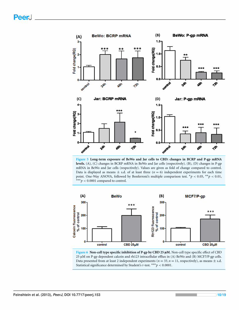
Figure 5 Long-term exposure of BeWo and Jar cells to CBD: changes in BCRP and P-gp mRNAlevels. (A), (C) changes in BCRP mRNA in BeWo and Jar cells (respectively). (B), (D) changes in P-gpmRNA in BeWo and Jar cells (respectively). Values are given as fold of change compared to control.Data is displayed as means ± s.d. of at least three (n = 6) independent experiments for each timepoint. One-Way ANOVA, followed by Bonferroni’s multiple comparison test. ∗p < 0.05, ∗∗p < 0.01,∗∗∗p< 0.0001 compared to control.
Figure 6 Non-cell type specific inhibition of P-gp by CBD 25 µM. Non-cell type specific effect of CBD25 µM on P-gp dependent calcein and rh123 intracellular efflux in (A) BeWo and (B) MCF7/P-gp cells.Data presented from at least 2 independent experiments (n = 33,n = 11, respectively), as means ± s.d.Statistical significance determined by Student’s t-test. ∗∗∗p< 0.0001.
Feinshtein et al. (2013), PeerJ, DOI 10.7717/peerj.153 10/19

Figure 7 Inhibition of P-gp by CBD 10 µM in BeWo cells. CBD 10 µM inhibition of P-gp dependentefflux of calcein (A) and DiOC2(3) (B). Data presented from at least 2 independent experiments (n= 9,n = 6, respectively), as means ± s.d. Statistical significance determined by One-Way ANOVA, followedby Bonferroni’s multiple comparison test for (A) and student’s t-test for (B). ∗∗∗p< 0.0001, ∗p< 0.05.
trophoblast and provide a direct target for cannabinoids (Kenney et al., 1999; Habayeb et
al., 2008a; Habayeb et al., 2008b). However, not all endocannabinoids exert their effect
through the classic cannabinoids receptors. For example, CBD lacks the central effects of
cannabis and works through CB1 and CB2 independent model of action (Scuderi et al.,
2009). Since our goal was to test the direct cannabinoid effect on trophoblast transporters
BCRP and P-gp, and CBD showed the most potent inhibitory effect (among major
cannabinoids) on these transporters (Holland et al., 2006; Zhu et al., 2006; Holland et
al., 2007) it made CBD the ideal cannabinoid for the present study.
BCRP and P-gp significance in pregnancyBCRP and P-gp play a key role in the transport of drugs and endogenous compounds in the
human placenta, affecting the outcome of pregnancy (Robey et al., 2009; Myllynen, Kummu
& Sieppi, 2010). They transport a broad variety of structurally diverse compounds, some of
which are congruent (Frohlich et al., 2004; Mathias, Hitti & Unadkat, 2005; Zhou, 2008). In
addition, BCRP transports a wide range of substrates, including fetal hormonal precursors
such as estrone-3-sulfate, naturally occurring carcinogens, porphyries and ceramides
(Imai et al., 2003; Krishnamurthy & Schuetz, 2005; Evseenko et al., 2007; Evseenko, Paxton
& Keelan, 2007; Mao, 2008; Dietrich et al., 2011). P-gp is expressed in the apical membrane
of syncytiotrophoblast and probably is the main placental protective transporter during
the first trimester. Its expression (both mRNA and protein) is the highest during the
first trimester and decreases with advancing gestation (Gil et al., 2005; Mathias, Hitti &
Unadkat, 2005; Sun et al., 2006). BCRP plays an important role as a survival factor in BeWo
cells as well as in the human placenta. It is thought to have a protective antiapoptotic role in
the trophoblast, regulating their survival under low oxygen conditions (Krishnamurthy &
Schuetz, 2006; Yeboah et al., 2006; Evseenko et al., 2007; Evseenko, Paxton & Keelan, 2007).
The trophoblast expression of BCRP may change in different pregnancy complications.
Indeed, placentas of women with preterm labor and intra-amniotic inflammation had
higher expressions of this transporter than those of women with preterm labor without
Feinshtein et al. (2013), PeerJ, DOI 10.7717/peerj.153 11/19

inflammation. In addition, the mRNA expression of BCRP correlated with that of
IL-8, which also increased significantly in placentas of women with preterm labor and
inflammation, suggesting that the transfer of drugs across the placenta may be altered in
cases of preterm labor with inflammation (Mason et al., 2011).
CBD impact on BCRP and P-gp upon long-term exposureIn the current study we have demonstrated for the first time that the exposure of
trophoblast-like cell lines BeWo and Jar to CBD is associated with two distinct patterns
of effects on the expression of the BCRP and P-gp transporters. The use of these cell lines,
instead of primary trophoblast cultures, results from the fact that primary trophoblasts
may rapidly differentiate in culture, continuously changing their gene and protein
expression (Evseenko, Paxton & Keelan, 2006). Comparison of these parameters with regard
to CBD influences in such a dynamic in vitro environment would be almost impossible.
Moreover, BeWo and Jar cell lines, derived from human gestational choriocarcinoma,
commonly used as an in vitro model for trophoblast toxicology studies, and they offer
a suitable model to study certain aspects of human trophoblast physiology, without the
aspect of inter-patient variability (Sullivan, 2004; Khare et al., 2006; Myren et al., 2007).
The first effect that was observed following long-term exposure is that CBD tran-
scriptionally inhibited P-gp membrane expression (as mRNA levels of P-gp were also
dramatically reduced following long-term exposure to CBD). The long-term inhibitory
effect of CBD on P-gp protein was previously reported in drug-selected human T
lymphoblastoid leukaemia cell line (CEM/VLB(100)) (Holland et al., 2006). The cell
specificity of CBD was previously reported (Khare et al., 2006; Hu, Ren & Shi, 2011),
yet our study is the first to detect this phenomenon in trophoblast-like cell lines. This
observation may have clinical implications, in light of the role of P-gp as one of the key
transport mechanisms for numerous drugs in the human placenta, especially during the
venerable period of the first trimester in which all the fetal organs are formed. Thus, our
results might be especially important to women who use cannabis on a regular basis during
the first trimester and are treated with other drugs that are P-gp substrates.
The expressional up-regulation of BCRP mRNA and protein in trophoblast-like
cell lines following long-term exposure to CBD is the second novel effect reported
in our study. This observation is similar to that reported in trophoblasts of women
with preterm labor and inflammation, suggesting that (I) long-term exposure to CBD
may elicit an inflammatory response in trophoblast-like cell lines (Mason et al., 2011);
and (II) that this phenomenon could be a direct compensational consequence of
P-gp down-regulation, providing a working defense line to the developing embryo.
Interestingly, compensation of such nature was already described in murine placentas
(Hutson, Koren & Matthews, 2010). Nonetheless, the signal transduction of CBD action in
human placenta/trophoblast/choriocarcinoma cells has not been clearly reported yet and
needs to be further elucidated.
Feinshtein et al. (2013), PeerJ, DOI 10.7717/peerj.153 12/19

CBD impact on P-gp upon acute exposureIn agreement with previously published data, we found that CBD holds inhibiting
properties over P-gp efflux function (Holland et al., 2006; Nieri et al., 2006; Zhu et al.,
2006). Moreover, our results show inhibition of P-gp efflux function in cell lines that
naturally express this transporter. It can be seen that CBD 10 µM (Fig. 7) yielded inhibition
effects lower than CBD 25 µM (Fig. 6). Similar to results we recently presented (Feinshtein
et al., 2013), this observation could imply that CBD inhibits P-gp in a concentration
dependent fashion. However, due to different quantification methods used in the present
study, this should be further elucidated.
Of note, many of the drugs that are prescribed and considered safe to use during
pregnancy are in fact P-gp or BCRP substrates, like Loratadine and H2 blockers
(i.e., Ranitidine and Cimetidine) (Collett et al., 1999; Chen et al., 2003; Li et al., 2008;
Schwarz et al., 2008; Dahan & Amidon, 2009; Gill, O’Brien & Koren, 2009; Matok et al.,
2010). Our finding may have clinical implications, suggesting that the use of cannabis
during gestation may alter drug transport through the trophoblast and lead to the absence
of a functional placental barrier during the first trimester, leaving the developing embryo
unprotected at this vulnerable period of pregnancy. Moreover, the trophoblasts of pregnant
women exposed to marijuana may exhibit some resistance to apoptotic and inflammatory
processes due to the effect of CBD on BCRP expression.
CONCLUSIONSFollowing cannabis consumption, all the drugs that are P-gp substrates can potentially
penetrate the human placental barrier at higher rates when combined with CBD, and
therefore their safety under these conditions is to be questioned. Additionally, changes in
placental BCRP expression profile might lead to altered transplacental transport of BCRP
substrates, such as medications, naturally occurring carcinogens, hormonal precursors and
apoptotic molecules, and influence pregnancy outcomes.
ACKNOWLEDGEMENTSWe gratefully thank Prof. Sofia Schreiber-Avissar from the Department of Clinical
Biochemistry and Pharmacology, Faculty of Health Sciences, Ben-Gurion University of
the Negev, Beer-Sheva, Israel, for providing writing assistance.
ADDITIONAL INFORMATION AND DECLARATIONS
FundingThe authors declare that there was no funding for this work.
Competing InterestsOffer Erez is an Academic Editor for PeerJ.
Feinshtein et al. (2013), PeerJ, DOI 10.7717/peerj.153 13/19

Author Contributions• Valeria Feinshtein conceived and designed the experiments, performed the experiments,
analyzed the data, wrote the paper.
• Offer Erez analyzed the data, wrote the paper.
• Zvi Ben-Zvi conceived and designed the experiments.
• Tamar Eshkoli, Boaz Sheizaf and Mahmud Huleihel contributed
reagents/materials/analysis tools.
• Eyal Sheiner analyzed the data, contributed reagents/materials/analysis tools, wrote the
paper.
• Gershon Holcberg conceived and designed the experiments, analyzed the data, wrote the
paper.
Supplemental InformationSupplemental information for this article can be found online at http://dx.doi.org/
10.7717/peerj.153.
REFERENCESArnold JC, Hone P, Holland ML, Allen JD. 2012. CB2 and TRPV1 receptors mediate cannabinoid
actions on MDR1 expression in multidrug resistant cells. Pharmacological Reports 64:751–757.
Brown HL, Graves CR. 2013. Smoking and marijuana use in pregnancy. Clinical Obstetrics andGynecology 56:107–113 DOI 10.1097/GRF.0b013e318282377d.
Chen C, Hanson E, Watson JW, Lee JS. 2003. P-glycoprotein limits the brain penetration ofnonsedating but not sedating H1-antagonists. Drug Metabolism and Disposition 31:312–318DOI 10.1124/dmd.31.3.312.
Collett A, Higgs NB, Sims E, Rowland M, Warhurst G. 1999. Modulation of the permeabilityof H2 receptor antagonists cimetidine and ranitidine by P-glycoprotein in rat intestine andthe human colonic cell line Caco-2. Journal of Pharmacology and Experimental Therapeutics288:171–178.
Dahan A, Amidon GL. 2009. Segmental dependent transport of low permeability compoundsalong the small intestine due to P-glycoprotein: the role of efflux transport in the oral absorptionof BCS class III drugs. Molecular Pharmaceutics 6:19–28 DOI 10.1021/mp800088f.
De Filippis D, Esposito G, Cirillo C, Cipriano M, De Winter BY, Scuderi C, Sarnelli G,Cuomo R, Steardo L, De Man JG, Iuvone T. 2011. Cannabidiol reduces intestinal inflammationthrough the control of neuroimmune axis. PLoS ONE 6:e28159DOI 10.1371/journal.pone.0028159.
Deiana S. 2012. Medical use of cannabis. Cannabidiol: A new light for schizophrenia? Drug Testingand Analysis 5(1):46–51 DOI 10.1002/dta.1425.
Dekker GA, Lee SY, North RA, McCowan LM, Simpson NA, Roberts CT. 2012. Risk factors forpreterm birth in an international prospective cohort of nulliparous women. PLoS ONE 7:e39154DOI 10.1371/journal.pone.0039154.
Dietrich CG, Vehr AK, Martin IV, Gassler N, Rath T, Roeb E, Schmitt J, Trautwein C, Geier A.2011. Downregulation of breast cancer resistance protein in colon adenomas reduces cellular
Feinshtein et al. (2013), PeerJ, DOI 10.7717/peerj.153 14/19
xenobiotic resistance and leads to accumulation of a food-derived carcinogen. InternationalJournal of Cancer 129:546–552 DOI 10.1002/ijc.25958.
Dudasova A, Keir SD, Parsons ME, Molleman A, Page CP. 2013. The effects of cannabidiol onthe antigen-induced contraction of airways smooth muscle in the guinea-pig. PulmonaryPharmacology & Therapeutics 26:373–379 DOI 10.1016/j.pupt.2013.02.002.
Eshkoli T, Sheiner E, Ben-Zvi Z, Feinshtein V, Holcberg G. 2011. Drug transport across theplacenta. Current Pharmaceutical Biotechnology 12:707–714 DOI 10.2174/138920111795470877.
Evseenko DA, Murthi P, Paxton JW, Reid G, Emerald BS, Mohankumar KM, Lobie PE,Brennecke SP, Kalionis B, Keelan JA. 2007. The ABC transporter BCRP/ABCG2 is a placentalsurvival factor, and its expression is reduced in idiopathic human fetal growth restriction. TheFASEB Journal 21:3592–3605 DOI 10.1096/fj.07-8688com.
Evseenko DA, Paxton JW, Keelan JA. 2006. ABC drug transporter expression and functionalactivity in trophoblast-like cell lines and differentiating primary trophoblast. AmericanJournal of Physiology - Regulatory, Integrative and Comparative Physiology 290:R1357–R1365DOI 10.1152/ajpregu.00630.2005.
Evseenko DA, Paxton JW, Keelan JA. 2007. The xenobiotic transporter ABCG2 plays a novelrole in differentiation of trophoblast-like BeWo cells. Placenta 28(Suppl A):S116–S120DOI 10.1016/j.placenta.2006.12.003.
Feinshtein V, Erez O, Ben-Zvi Z, Eshkoli T, Sheizaf B, Sheiner E, Holcberg G. 2013. Cannabidiolenhances xenobiotic permeability through the human placental barrier by direct inhibition ofBreast Cancer Resistance Protein (BCRP). American Journal of Obstetrics and Gynecology Epubahead of print Aug 9 2013 DOI 10.1016/j.ajog.2013.08.005.
Feinshtein V, Holcberg G, Amash A, Erez N, Rubin M, Sheiner E, Polachek H, Ben-Zvi Z. 2010.Nitrofurantoin transport by placental choriocarcinoma JAr cells: involvement of BCRP,OATP2B1 and other MDR transporters. Archives of Gynecology and Obstetrics 281:1037–1044DOI 10.1007/s00404-009-1286-7.
Frohlich M, Albermann N, Sauer A, Walter-Sack I, Haefeli WE, Weiss J. 2004. In vitro andex vivo evidence for modulation of P-glycoprotein activity by progestins. BiochemicalPharmacology 68:2409–2416 DOI 10.1016/j.bcp.2004.08.026.
Gil S, Saura R, Forestier F, Farinotti R. 2005. P-glycoprotein expression of the human placentaduring pregnancy. Placenta 26:268–270 DOI 10.1016/j.placenta.2004.05.013.
Gill SK, O’Brien L, Koren G. 2009. The safety of histamine 2 (H2) blockers in pregnancy: ameta-analysis. Digestive Diseases and Sciences 54:1835–1838 DOI 10.1007/s10620-008-0587-1.
Golan M, Schreiber G, Avissar S. 2009. Antidepressants increase beta-arrestin2 ubiquitinylationand degradation by the proteasomal pathway in C6 rat glioma cells. Journal of Pharmacologyand Experimental Therapeutics 332:970–976 DOI 10.1124/jpet.109.160218.
Habayeb OM, Taylor AH, Bell SC, Taylor DJ, Konje JC. 2008a. Expression of theendocannabinoid system in human first trimester placenta and its role in trophoblastproliferation. Endocrinology 149:5052–5060 DOI 10.1210/en.2007-1799.
Habayeb OM, Taylor AH, Finney M, Evans MD, Konje JC. 2008b. Plasma anandamideconcentration and pregnancy outcome in women with threatened miscarriage. Journal of theAmerican Medical Association 299:1135–1136 DOI 10.1001/jama.299.10.1135.
Hardwick LJ, Velamakanni S, van Veen HW. 2007. The emerging pharmacotherapeuticsignificance of the breast cancer resistance protein (ABCG2). British Journal of Pharmacology151:163–174 DOI 10.1038/sj.bjp.0707218.
Feinshtein et al. (2013), PeerJ, DOI 10.7717/peerj.153 15/19

Harvey BS, Ohlsson KS, Maag JL, Musgrave IF, Smid SD. 2012. Contrasting protective effectsof cannabinoids against oxidative stress and amyloid-β evoked neurotoxicity in vitro.Neurotoxicology 33:138–146 DOI 10.1016/j.neuro.2011.12.015.
Hayatbakhsh MR, Flenady VJ, Gibbons KS, Kingsbury AM, Hurrion E, Mamun AA,Najman JM. 2012. Birth outcomes associated with cannabis use before and during pregnancy.Pediatric Research 71:215–219 DOI 10.1038/pr.2011.25.
Hill AJ, Williams CM, Whalley BJ, Stephens GJ. 2012. Phytocannabinoids as novel therapeuticagents in CNS disorders. Pharmacology & Therapeutics 133:79–97DOI 10.1016/j.pharmthera.2011.09.002.
Holland ML, Allen JD, Arnold JC. 2008. Interaction of plant cannabinoids with themultidrug transporter ABCC1 (MRP1). European Journal of Pharmacology 591:128–131DOI 10.1016/j.ejphar.2008.06.079.
Holland ML, Lau DT, Allen JD, Arnold JC. 2007. The multidrug transporter ABCG2 (BCRP)is inhibited by plant-derived cannabinoids. British Journal of Pharmacology 152:815–824DOI 10.1038/sj.bjp.0707467.
Holland ML, Panetta JA, Hoskins JM, Bebawy M, Roufogalis BD, Allen JD, Arnold JC. 2006. Theeffects of cannabinoids on P-glycoprotein transport and expression in multidrug resistant cells.Biochemical Pharmacology 71:1146–1154 DOI 10.1016/j.bcp.2005.12.033.
Hu G, Ren G, Shi Y. 2011. The putative cannabinoid receptor GPR55 promotes cancer cellproliferation. Oncogene 30:139–141 DOI 10.1038/onc.2010.502.
Huls M, Russel FG, Masereeuw R. 2009. The role of ATP binding cassette transporters in tissuedefense and organ regeneration. The Journal of Pharmacology and Experimental Therapeutics328:3–9 DOI 10.1124/jpet.107.132225.
Hutson JR, Koren G, Matthews SG. 2010. Placental P-glycoprotein and breast cancer resistanceprotein: influence of polymorphisms on fetal drug exposure and physiology. Placenta31:351–357 DOI 10.1016/j.placenta.2010.02.010.
Imai Y, Asada S, Tsukahara S, Ishikawa E, Tsuruo T, Sugimoto Y. 2003. Breast cancer resistanceprotein exports sulfated estrogens but not free estrogens. Molecular Pharmacology 64:610–618DOI 10.1124/mol.64.3.610.
Juknat A, Pietr M, Kozela E, Rimmerman N, Levy R, Gao F, Coppola G, Geschwind D,Vogel Z. 2013. Microarray and pathway analysis reveal distinct mechanisms underlyingcannabinoid-mediated modulation of LPS-induced activation of BV-2 microglial cells. PLoSONE 8:e61462 DOI 10.1371/journal.pone.0061462.
Kenney SP, Kekuda R, Prasad PD, Leibach FH, Devoe LD, Ganapathy V. 1999. Cannabinoidreceptors and their role in the regulation of the serotonin transporter in human placenta. Amer-ican Journal of Obstetrics and Gynecology 181:491–497 DOI 10.1016/S0002-9378(99)70583-1.
Khare M, Taylor AH, Konje JC, Bell SC. 2006. 19-Tetrahydrocannabinol inhibits cytotrophoblastcell proliferation and modulates gene transcription. Molecular Human Reproduction 12:321–333DOI 10.1093/molehr/gal036.
Kozer E, Koren G. 2001. Effects of prenatal exposure to marijuana. Canadian Family Physician47:263–264.
Krishnamurthy P, Schuetz JD. 2005. The ABC transporter Abcg2/Bcrp: role in hypoxia mediatedsurvival. Biometals 18:349–358 DOI 10.1007/s10534-005-3709-7.
Krishnamurthy P, Schuetz JD. 2006. Role of ABCG2/BCRP in biology and medicine.Annual Review of Pharmacology and Toxicology 46:381–410DOI 10.1146/annurev.pharmtox.46.120604.141238.
Feinshtein et al. (2013), PeerJ, DOI 10.7717/peerj.153 16/19

Lankas GR, Wise LD, Cartwright ME, Pippert T, Umbenhauer DR. 1998. PlacentalP-glycoprotein deficiency enhances susceptibility to chemically induced birth defects in mice.Reproductive Toxicology 12:457–463 DOI 10.1016/S0890-6238(98)00027-6.
Lewis SE, Rapino C, Di Tommaso M, Pucci M, Battista N, Paro R, Simon L, Lutton D,Maccarrone M. 2012. Differences in the endocannabinoid system of sperm from fertile andinfertile men. PLoS ONE 7:e47704 DOI 10.1371/journal.pone.0047704.
Li C, Kim CS, Yang JY, Park YJ, Choi JS. 2008. Effects of roxithromycin on the pharmacokineticsof loratadine after oral and intravenous administration of loratadine in rats. European Journalof Drug Metabolism and Pharmacokinetics 33:231–236 DOI 10.1007/BF03190877.
Ligresti A, Moriello AS, Starowicz K, Matias I, Pisanti S, De Petrocellis L, Laezza C, Portella G,Bifulco M, Di Marzo V. 2006. Antitumor activity of plant cannabinoids with emphasis onthe effect of cannabidiol on human breast carcinoma. The Journal of Pharmacology andExperimental Therapeutics 318:1375–1387 DOI 10.1124/jpet.106.105247.
Lowry OH, Rosebrough NJ, Farr AL, Randall RJ. 1951. Protein measurement with the Folinphenol reagent. Journal of Biological Chemistry 193:265–275.
Mao Q. 2008. BCRP/ABCG2 in the placenta: expression, function and regulation. PharmaceuticalResearch 25:1244–1255 DOI 10.1007/s11095-008-9537-z.
Maor Y, Yu J, Kuzontkoski PM, Dezube BJ, Zhang X, Groopman JE. 2012. Cannabidiol inhibitsgrowth and induces programmed cell death in Kaposi sarcoma-associated herpesvirus-infectedendothelium. Genes & Cancer 3:512–520 DOI 10.1177/1947601912466556.
Martin C, Walker J, Rothnie A, Callaghan R. 2003. The expression of P-glycoprotein doesinfluence the distribution of novel fluorescent compounds in solid tumour models. BritishJournal of Cancer 89:1581–1589 DOI 10.1038/sj.bjc.6601300.
Mason CW, Buhimschi IA, Buhimschi CS, Dong Y, Weiner CP, Swaan PW. 2011. ATP-bindingcassette transporter expression in human placenta as a function of pregnancy condition. DrugMetabolism and Disposition 39:1000–1007 DOI 10.1124/dmd.111.038166.
Mathias AA, Hitti J, Unadkat JD. 2005. P-glycoprotein and breast cancer resistance proteinexpression in human placentae of various gestational ages. American Journal of Physiology -Regulatory, Integrative and Comparative Physiology 289:R963–R969DOI 10.1152/ajpregu.00173.2005.
Matok I, Gorodischer R, Koren G, Sheiner E, Wiznitzer A, Uziel E, Levy A. 2010. The safetyof H2-blockers use during pregnancy. The Journal of Clinical Pharmacology 50:81–87DOI 10.1177/0091270009350483.
Mechoulam R, Hanus L. 2002. Cannabidiol: an overview of some chemical andpharmacological aspects. Part I: chemical aspects. Chemistry and Physics of Lipids 121:35–43DOI 10.1016/S0009-3084(02)00144-5.
Mechoulam R, Shvo Y. 1963. Hashish. I. The structure of cannabidiol. Tetrahedron 19:2073–2078DOI 10.1016/0040-4020(63)85022-X.
Minderman H, Vanhoefer U, Toth K, Yin M-B, Minderman MD, Wrzosek C, Slovak ML,Rustum YM. 1996. DiOC2(3) is not a substrate for multidrug resistance protein(MRP)-mediated drug efflux. Cytometry 25:14–20 DOI 10.1002/(SICI)1097-0320(19960901)25:1<14::AID-CYTO2>3.0.CO;2-E.
Moore DG, Turner JD, Parrott AC, Goodwin JE, Fulton SE, Min MO, Fox HC, Braddick FM,Axelsson EL, Lynch S, Ribeiro H, Frostick CJ, Singer LT. 2010. During pregnancy, recreationaldrug-using women stop taking ecstasy (3,4-methylenedioxy-N-methylamphetamine) andreduce alcohol consumption, but continue to smoke tobacco and cannabis: initial findings
Feinshtein et al. (2013), PeerJ, DOI 10.7717/peerj.153 17/19

from the Development and Infancy Study. Journal of Psychopharmacology 24:1403–1410DOI 10.1177/0269881109348165.
Mori N, Iwamoto H, Yokooji T, Murakami T. 2012. Characterization of intestinal absorption ofquinidine, a P-glycoprotein substrate, given as a powder in rats. Pharmazie 67:384–388.
Myllynen P, Kummu M, Sieppi E. 2010. ABCB1 and ABCG2 expression in the placenta and fetus:an interspecies comparison. Expert Opinion on Drug Metabolism & Toxicology 6:1385–1398DOI 10.1517/17425255.2010.514264.
Myren M, Mose T, Mathiesen L, Knudsen LE. 2007. The human placenta – An alternative forstudying foetal exposure. Toxicology in Vitro 21:1332–1340 DOI 10.1016/j.tiv.2007.05.011.
Nabissi M, Morelli MB, Santoni M, Santoni G. 2013. Triggering of the TRPV2 channel bycannabidiol sensitizes glioblastoma cells to cytotoxic chemotherapeutic agents. Carcinogenesis34:48–57 DOI 10.1093/carcin/bgs328.
Ni Z, Mao Q. 2011. ATP-binding cassette efflux transporters in human placenta. CurrentPharmaceutical Biotechnology 12:674–685 DOI 10.2174/138920111795164057.
Nieri P, Romiti N, Adinolfi B, Chicca A, Massarelli I, Chieli E. 2006. Modulation ofP-glycoprotein activity by cannabinoid molecules in HK-2 renal cells. British Journal ofPharmacology 148:682–687 DOI 10.1038/sj.bjp.0706778.
Polachek H, Holcberg G, Polachek J, Rubin M, Feinshtein V, Sheiner E, Ben-Zvi Z. 2010.Carrier-mediated uptake of Levofloxacin by BeWo cells, a human trophoblast cell line. Archivesof Gynecology and Obstetrics 281:833–838 DOI 10.1007/s00404-009-1177-y.
Robey RW, To KK, Polgar O, Dohse M, Fetsch P, Dean M, Bates SE. 2009. ABCG2: a perspective.Advanced Drug Delivery Reviews 61:3–13 DOI 10.1016/j.addr.2008.11.003.
Schier AR, Ribeiro NP, Silva AC, Hallak JE, Crippa JA, Nardi AE, Zuardi AW. 2012. Cannabidiol,a Cannabis sativa constituent, as an anxiolytic drug. Revista Brasileira de Psiquiatria 34(Suppl1):104–110 DOI 10.1590/S1516-44462012000500008.
Schwarz EB, Moretti ME, Nayak S, Koren G. 2008. Risk of hypospadias in offspring of womenusing loratadine during pregnancy: a systematic review and meta-analysis. Drug Safety31:775–788 DOI 10.2165/00002018-200831090-00006.
Scuderi C, De Filippis D, Iuvone T, Blasio A, Steardo A, Esposito G. 2009. Cannabidiol inmedicine: a review of its therapeutic potential in CNS disorders. Phytotherapy Research23:597–602 DOI 10.1002/ptr.2625.
Smyth MJ, Krasovskis E, Sutton VR, Johnstone RW. 1998. The drug efflux protein,P-glycoprotein, additionally protects drug-resistant tumor cells from multiple forms ofcaspase-dependent apoptosis. Proceedings of the National Academy of Sciences of the UnitedStates of America 95:7024–7029 DOI 10.1073/pnas.95.12.7024.
Solinas M, Massi P, Cantelmo AR, Cattaneo MG, Cammarota R, Bartolini D, Cinquina V,Valenti M, Vicentini LM, Noonan DM, Albini A, Parolaro D. 2012. Cannabidiol inhibitsangiogenesis by multiple mechanisms. British Journal of Pharmacology 167:1218–1231DOI 10.1111/j.1476-5381.2012.02050.x.
Sullivan MH. 2004. Endocrine cell lines from the placenta. Molecular and Cellular Endocrinology228:103–119 DOI 10.1016/j.mce.2003.03.001.
Sun M, Kingdom J, Baczyk D, Lye SJ, Matthews SG, Gibb W. 2006. Expression of the multidrugresistance P-glycoprotein, (ABCB1 glycoprotein) in the human placenta decreases withadvancing gestation. Placenta 27:602–609 DOI 10.1016/j.placenta.2005.05.007.
Feinshtein et al. (2013), PeerJ, DOI 10.7717/peerj.153 18/19

Sun X, Dey SK. 2012. Endocannabinoid signaling in female reproduction. ACS ChemicalNeuroscience 3:349–355 DOI 10.1021/cn300014e.
Taylor AH, Ang C, Bell SC, Konje JC. 2007. The role of the endocannabinoid system ingametogenesis, implantation and early pregnancy. Human Reproduction Update 13:501–513DOI 10.1093/humupd/dmm018.
Vahakangas K, Myllynen P. 2009. Drug transporters in the human blood-placental barrier. BritishJournal of Pharmacology 158:665–678 DOI 10.1111/j.1476-5381.2009.00336.x.
Yeboah D, Sun M, Kingdom J, Baczyk D, Lye SJ, Matthews SG, Gibb W. 2006. Expression ofbreast cancer resistance protein (BCRP/ABCG2) in human placenta throughout gestation andat term before and after labor. Canadian Journal of Physiology and Pharmacology 84:1251–1258DOI 10.1139/y06-078.
Zhou SF. 2008. Structure, function and regulation of P-glycoprotein and its clinical relevance indrug disposition. Xenobiotica 38:802–832 DOI 10.1080/00498250701867889.
Zhu HJ, Wang JS, Markowitz JS, Donovan JL, Gibson BB, Gefroh HA, Devane CL. 2006.Characterization of P-glycoprotein inhibition by major cannabinoids from marijuana.The Journal of Pharmacology and Experimental Therapeutics 317:850–857DOI 10.1124/jpet.105.098541.
Zuardi AW. 2008. Cannabidiol: from an inactive cannabinoid to a drug with wide spectrum ofaction. Revista Brasileira de Psiquiatria 30:271–280 DOI 10.1590/S1516-44462008000300015.
Feinshtein et al. (2013), PeerJ, DOI 10.7717/peerj.153 19/19