
Canine Rehabilitation by J. E. Steiss; · PDF fileEnergy within an ultrasound ... Therapeutic...
32
In: Clinical Neurology in Small Animals - Localization, Diagnosis and Treatment, K.G. Braund (Ed.) Publisher: International Veterinary Information Service (www.ivis.org), Ithaca, New York, USA. Canine Rehabilitation (20-Feb-2003) J. E. Steiss Department of Biomedical Sciences, College of Veterinary Medicine, Nursing and Allied Health, Tuskegee University, Tuskegee, AL, USA. Introduction The field of physical therapy/rehabilitation has much to offer veterinarians in terms of its application to sporting dogs as well as orthopedic and neurologic patients. In 1978, canine physical therapy techniques were described by Ann Downer, a physical therapist on faculty at Ohio State University [1]. More recent texts on canine physical therapy are becoming available [2,64]. The purpose of this chapter is to present an overview of rehabilitation methods. The procedures described here are applicable to dogs with various neurological disorders, as well as dogs with orthopedic disorders. Some of the physical therapy methods that can be adapted to canine rehabilitation include: 1. Thermal Agents (ice, hot packs, therapeutic ultrasound, diathermy) 2. Phonophoresis and Iontophoresis for Transcutaneous Drug Delivery 3. Electrical Stimulation 4. Therapeutic Exercise 5. Aquatic Therapy 6. Goniometry 7. Orthotics (braces/splints) 8. Exercise Prescriptions a. Joint Contracture b. Thoraco-lumbar Intervertebral Disk Disease, Non-weight-bearing in Pelvic Limbs c. Thoraco-lumbar Intervertebral Disk Disease, Weight-bearing in Pelvic Limbs d. Lumbosacral Disease, Post-operative e. Anterior Cruciate Ligament Rupture, Post-operative 1. Thermal Agents Superficial heating agents penetrate to a depth of approximately one centimeter. Deep heating agents can elevate tissue temperatures at depths of three centimeters or more. Deep heating agents include ultrasound and diathermy. As a general rule, cold therapy ("cryotherapy") is indicated during acute inflammation when the lesion exhibits signs of redness, swelling, pain and/or heat. Heat is indicated during the sub-acute and chronic inflammatory phases. Ice application is also indicated to resolve swelling after exercise. In some cases, such as the treatment of muscle spasm and spasticity, the therapist may need to use a trial and error method to determine if either heat or cold yields clinical improvement. In humans, ice is usually not applied for longer than 15 minutes at one time in order to avoid reflex vasodilation. This guideline seems prudent for animals, also. The indications for alternating heat and cold ("contrast baths") include impaired venous circulation and indolent ulcers, and traumatic or inflammatory conditions during the subacute or chronic phases. The mechanism of action of contrast baths is production of alternating vasoconstriction and vasodilation of local blood vessels. This reaction is considered to stimulate blood flow to the treated area and thereby to help stimulate healing. Therapeutic Ultrasound Two physical therapy modalities utilize ultrasound. These modalities are therapeutic ultrasound, which produces deep heating of tissues, and phonophoresis, which utilizes ultrasound waves to move drugs through intact skin (Fig. 1).
Transcript of Canine Rehabilitation by J. E. Steiss; · PDF fileEnergy within an ultrasound ... Therapeutic...
In: Clinical Neurology in Small Animals - Localization, Diagnosis and Treatment, K.G. Braund (Ed.) Publisher: International Veterinary Information Service (www.ivis.org), Ithaca, New York, USA. Canine Rehabilitation (20-Feb-2003) J. E. Steiss
Department of Biomedical Sciences, College of Veterinary Medicine, Nursing and Allied Health, Tuskegee University, Tuskegee, AL, USA.
Introduction The field of physical therapy/rehabilitation has much to offer veterinarians in terms of its application to sporting dogs as well as orthopedic and neurologic patients. In 1978, canine physical therapy techniques were described by Ann Downer, a physical therapist on faculty at Ohio State University [1]. More recent texts on canine physical therapy are becoming available [2,64]. The purpose of this chapter is to present an overview of rehabilitation methods. The procedures described here are applicable to dogs with various neurological disorders, as well as dogs with orthopedic disorders.
Some of the physical therapy methods that can be adapted to canine rehabilitation include:
1. Thermal Agents (ice, hot packs, therapeutic ultrasound, diathermy) 2. Phonophoresis and Iontophoresis for Transcutaneous Drug Delivery 3. Electrical Stimulation 4. Therapeutic Exercise 5. Aquatic Therapy 6. Goniometry 7. Orthotics (braces/splints) 8. Exercise Prescriptions
a. Joint Contracture b. Thoraco-lumbar Intervertebral Disk Disease, Non-weight-bearing in Pelvic Limbs c. Thoraco-lumbar Intervertebral Disk Disease, Weight-bearing in Pelvic Limbs d. Lumbosacral Disease, Post-operative e. Anterior Cruciate Ligament Rupture, Post-operative
1. Thermal Agents Superficial heating agents penetrate to a depth of approximately one centimeter. Deep heating agents can elevate tissue temperatures at depths of three centimeters or more. Deep heating agents include ultrasound and diathermy. As a general rule, cold therapy ("cryotherapy") is indicated during acute inflammation when the lesion exhibits signs of redness, swelling, pain and/or heat. Heat is indicated during the sub-acute and chronic inflammatory phases. Ice application is also indicated to resolve swelling after exercise. In some cases, such as the treatment of muscle spasm and spasticity, the therapist may need to use a trial and error method to determine if either heat or cold yields clinical improvement. In humans, ice is usually not applied for longer than 15 minutes at one time in order to avoid reflex vasodilation. This guideline seems prudent for animals, also.
The indications for alternating heat and cold ("contrast baths") include impaired venous circulation and indolent ulcers, and traumatic or inflammatory conditions during the subacute or chronic phases. The mechanism of action of contrast baths is production of alternating vasoconstriction and vasodilation of local blood vessels. This reaction is considered to stimulate blood flow to the treated area and thereby to help stimulate healing.
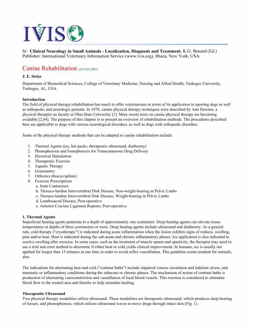
Therapeutic Ultrasound Two physical therapy modalities utilize ultrasound. These modalities are therapeutic ultrasound, which produces deep heating of tissues, and phonophoresis, which utilizes ultrasound waves to move drugs through intact skin (Fig. 1).
Therapeutic ultrasound delivers ultrasound waves through a transducer head comparable in size to the heads used in diagnostic ultrasound (Fig. 1). As with diagnostic ultrasound, the transducer must be in contact with the skin surface. With therapeutic ultrasound, however, the ultrasound waves increase tissue temperatures at depths up to 3 - 5 cm or more. Energy within an ultrasound beam decreases due to scatter and absorption as the waves travel through tissue. Scattering is the deflection of sound when the beam strikes a reflecting surface. In contrast, absorption is the transfer of energy from the sound beam to the tissues. Absorption is high in tissues with a high proportion of protein [3,4] and minimal in adipose tissue [3,5]. This means that ultrasound penetrates subcutaneous fat with little attenuation, whereas tissues with a high collagen content absorb more of the energy.
Figure 1. Portable ultrasound equipment (Courtesy of Chattanooga Medical Supply). - To view this image in full size go to the IVIS website at www.ivis.org . -
Therapeutic ultrasound has both thermal and non-thermal effects. The non-thermal effects have been studied primarily in wound healing. The thermal effect of therapeutic ultrasound is a major indication for its use. Increases in tissue temperatures of 1 to 4ºC are associated with increases in collagen extensibility, blood flow, pain threshold, and enzyme activity. In humans, the rate of increase in tissue temperature is directly related to the frequency of the ultrasound waves (MHz) and the intensity (watts/ cm2). Experimentally, when tissue temperatures were compared at various depths (0.8 to 5.0 cm) using thermistors inserted into muscles, the rate of temperature increase per minute ranged from 0.04ºC at 0.5 W/cm2 to 0.38ºC at 2.0 W/cm2 for 1 MHz treatment and from 0.30ºC at 0.5 W/cm2 to 1.4ºC at 2.0 W/cm2 for 3 MHz treatment [6]. Clearly, the 3 MHz treatment heated the tissues faster than the 1 MHz treatment at all intensities.
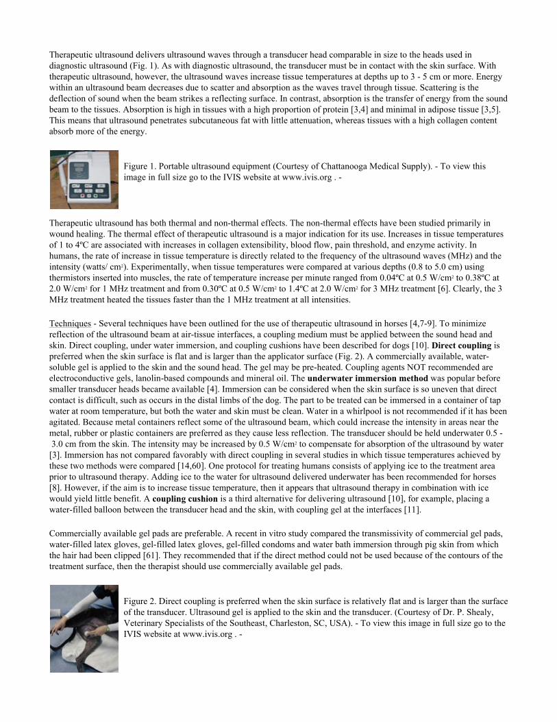
Techniques - Several techniques have been outlined for the use of therapeutic ultrasound in horses [4,7-9]. To minimize reflection of the ultrasound beam at air-tissue interfaces, a coupling medium must be applied between the sound head and skin. Direct coupling, under water immersion, and coupling cushions have been described for dogs [10]. Direct coupling is preferred when the skin surface is flat and is larger than the applicator surface (Fig. 2). A commercially available, water-soluble gel is applied to the skin and the sound head. The gel may be pre-heated. Coupling agents NOT recommended are electroconductive gels, lanolin-based compounds and mineral oil. The underwater immersion method was popular before smaller transducer heads became available [4]. Immersion can be considered when the skin surface is so uneven that direct contact is difficult, such as occurs in the distal limbs of the dog. The part to be treated can be immersed in a container of tap water at room temperature, but both the water and skin must be clean. Water in a whirlpool is not recommended if it has been agitated. Because metal containers reflect some of the ultrasound beam, which could increase the intensity in areas near the metal, rubber or plastic containers are preferred as they cause less reflection. The transducer should be held underwater 0.5 - 3.0 cm from the skin. The intensity may be increased by 0.5 W/cm2 to compensate for absorption of the ultrasound by water [3]. Immersion has not compared favorably with direct coupling in several studies in which tissue temperatures achieved by these two methods were compared [14,60]. One protocol for treating humans consists of applying ice to the treatment area prior to ultrasound therapy. Adding ice to the water for ultrasound delivered underwater has been recommended for horses [8]. However, if the aim is to increase tissue temperature, then it appears that ultrasound therapy in combination with ice would yield little benefit. A coupling cushion is a third alternative for delivering ultrasound [10], for example, placing a water-filled balloon between the transducer head and the skin, with coupling gel at the interfaces [11].
Commercially available gel pads are preferable. A recent in vitro study compared the transmissivity of commercial gel pads, water-filled latex gloves, gel-filled latex gloves, gel-filled condoms and water bath immersion through pig skin from which the hair had been clipped [61]. They recommended that if the direct method could not be used because of the contours of the treatment surface, then the therapist should use commercially available gel pads.
Figure 2. Direct coupling is preferred when the skin surface is relatively flat and is larger than the surface of the transducer. Ultrasound gel is applied to the skin and the transducer. (Courtesy of Dr. P. Shealy, Veterinary Specialists of the Southeast, Charleston, SC, USA). - To view this image in full size go to the IVIS website at www.ivis.org . -

What about the hair coat? - The presence of the hair coat may be a major drawback to ultrasound treatment and phonophoresis in dogs. Because ultrasound energy is absorbed by tissues with a high protein content, and deflection of the ultrasound beam occurs at tissue interfaces, it would be expected that ultrasound penetration through the hair coat into underlying tissues would be poor. For horses, one investigator [7] has specifically recommended clipping the hair and using adequate coupling gel, or standing the horse in water to reduce the air interface, although immersion will not prevent the scattering due to the hair coat. We conducted a study in dogs to quantify temperatures in underlying tissues when ultrasound was delivered through intact hair coats, using commercial ultrasound gel at room temperature [12]. In that study, ultrasound delivered through either short or long hair coats produced only minimal temperature increases in underlying tissues, compared to the responses achieved when the hair had been clipped. Furthermore, there was considerable warming within the hair coat. Even hair coats as short as those of Greyhound dogs inhibited tissue heating. Future research is needed to determine if modifications such as warming the ultrasound gel might enhance ultrasound penetration through the intact hair coat.
Treatment Variables - Tissue heating depends on frequency, intensity, duration, treatment area and the characteristics of the tissue. Ultrasound beams are considered collimated such that frequencies in the megahertz (MHz) range expose a limited target area. Frequency determines depth of penetration. As the frequency of the ultrasound waves increases, penetration decreases. One MHz heats at depths around 2 - 5 cm [12,13]. Three MHz heat at depths around 0.5 - 2 cm [4,13,59]. Consequently, treatment of superficial lesions can be problematic if the unit delivers only 1 MHz. If there is underlying bone, then most of the ultrasound will hit the bone and the intensity cannot be turned up very high to heat the underlying soft tissue before the patient feels periosteal pain [13]. An area of study which is currently receiving attention is the use of low frequency (long wave) ultrasound (< 1 MHz frequencies). Intensity refers to the rate of energy delivery per unit area. The power meter in an ultrasound unit indicates both watts and watts/cm2. Intensities typically range from 0.25 to 3.0 W/cm2. The greatest intensity within the beam is termed "spatial peak intensity". The "beam non-uniformity ratio" compares the maximal intensity from the transducer to the average intensity, and should be low (2:1 to 6:1) to indicate that energy distribution is relatively uniform, with minimal risk of tissue damage from areas of concentrated ultrasound energy ("hot spots"). The higher the intensity, the larger and faster the temperature increases. Generally, intensities required to elevate tissue temperature to the range of 40 - 45ºC vary from 1.0 - 2.0 W/cm2 with continuous wave mode for 5 - 10 min. If there is less soft tissue and/or bone close to the surface, lower intensity and higher frequency are appropriate. In any case, the patient’s tolerance is the final determinant [3].
Several therapists have observed that dogs sometimes whine or seem uncomfortable 5 - 10 min after commencing therapeutic ultrasound. Any distress that animals show should be assumed to be due to pain and the therapist should either reduce the intensity or terminate the session. Most therapists use intensities that produce no detectable sensation in human patients. However, some experts are advising that the intensity be increased until the person feels vigorous heating of the tissues and then be lowered [13]. This difference in opinion has probably come about because the heating effects in the underlying tissues are difficult to monitor. Therefore, although tissue damage can result from excessive ultrasound exposure, there is also the possibility of insufficient exposure. Duty cycle refers to the fraction of time that the sound is emitted during one pulse period. In continuous mode, the ultrasound intensity is constant, whereas a pulsed wave is interrupted in an on-off manner. Typical duty cycles range from 0.05 to 0.5. Pulsing may be used when the desired effect is based on a non-thermal mechanism or when heating is to be minimal, such as treating near bone [4]. The duration of treatment is short, typically 10 minutes or less. Duration of a session can be estimated as 5 min/25 in2 treatment area [13]. Five to ten minutes are necessary for adequate tissue heating. For instance, in one study it took nearly 8 minutes for the temperature to reach therapeutic levels at a depth of 3 cm in the gastrocnemius muscle in humans [14]. In that study, a direct coupling technique was used, with continuous mode, at 1.5 W/cm2 and the sound head was moved at 4 cm per sec. At an intensity of 1.5 W/cm2 and a treatment area twice the size of the effective radiating area of the transducer, it has been recommended that treatment duration be approximately 3 - 4 minutes at 3 MHz or 10 minutes at 1 MHz [13]. Our findings with 1 MHz in dogs support these recommendations [12]. Treatment area should be 2 - 3 times the size of the effective radiating area of the transducer head [13]. Increasing the treatment area decreases the effective heating of the tissues. Speed of the sound head over the skin is usually recommended to be approximately 4 cm per sec [11] to achieve uniform distribution of energy. Moving the transducer too quickly diminishes heating and makes the therapist prone to cover too large an area. The transducer should NEVER be stationary. Because the ultrasound beam is non-uniform, some target areas could receive a large amount of energy with the risk of causing "hot spots" and cavitation. Treatment schedules may be daily initially, followed by less frequent sessions as the condition improves [3]. Bromiley has indicated that treatment can be daily for up to 10 days, but should not exceed two 10 day courses without a 3 week rest [4]. Grant has made similar recommendations for horses [7].
Precautions and Contraindications - Tissue burns can occur if the intensity is too high or the transducer is held stationary. These factors can put the patient at risk for cavitation, a phenomenon whereby bubbles of dissolved gas form and grow

during each rarefaction phase. Also, if the transducer is held in the air while emitting ultrasound, the face of the transducer will overheat. The end result could be damage to the ultrasound transducer or tissue damage if the transducer contacted the animal's skin. Some units have a built-in system to prevent the transducer from overheating. Some ultrasound energy is unavoidably emitted through the housing of the sound head to the therapist's hand, but the effects of this "parasitic" exposure are unknown.
It is important to avoid direct ultrasound exposure to the following: cardiac pacemakers, carotid sinus or cervical ganglia, eyes, gravid uterus, heart, injured areas immediately after exercise [7,9], malignancy, spinal cord if a laminectomy has been performed, testes and contaminated wounds. One should exert caution in considering ultrasound therapy in the following situations: tissue near a bone fracture or bony prominences, tissue that has been treated with cold packs or ice, decreased circulation, decreased pain/temperature sensation (animals should not be overly sedated, restrained or under local anesthesia) and epiphyseal areas in immature animals [4,11]. Also, there is evidence that intracapsular heating may accelerate destruction of articular cartilage in acute inflammatory joint disease [3]. Although metal implants are not necessarily a contraindication, the effects of ultrasound on cementing compounds such as methyl methacrylate are unknown [11].
Clinical Applications - In human rehabilitation, ultrasound therapy has been used for many years, but it is only quite recently that this modality is being evaluated with controlled clinical trials. In some regards, the jury is still out regarding the role of ultrasound therapy. Clinical trials and outcome assessments remain to be done in veterinary medicine. In horses, ultrasound therapy has been suggested for the treatment of tendinitis, desmitis, sprains, joint lesions, lacerations, scar tissue reduction, edema, exostosis and myositis [7,8]. In human athletes, some experts consider that the most beneficial results from ultrasound are in treating tendonitis [13]. In chronic tendinitis, recommended therapy includes heating with ultrasound, followed by cross-frictional massage. Another indication is treatment of limited range of motion associated with joint contracture, for which patients receive ultrasound therapy before passive range of motion or joint mobilization techniques. A third indication is pain relief prior to activity, such as for an athlete with tendinitis which is mild enough that the person can continue training; ultrasound treatment is administered prior to activity to assist in the warm-up and provide some pain relief. Tendinitis and bursitis are treated with ultrasound to increase blood flow, increase temperature to reduce pain and drive anti-inflammatory drugs across the skin by means of phonophoresis. In humans, lateral epicondylitis, sub-acromial bursitis, and bicipital tendinitis are typical indications for ultrasound therapy. Animal studies suggest that the stage of healing at which ultrasound therapy is administered is important, as ultrasound during early tendon repair could be detrimental [11].
The principle of "heat and stretch" can be applied in cases of joint contracture and scar tissue in an effort to increase range of motion. Tissues are first heated by ultrasound and then passively or actively stretched by the therapist. The effects of heat on ligament extensibility were studied in healthy humans [15] who underwent knee joint displacement tests before and after continuous ultrasound therapy (1 MHz, 1.5 W/cm2 for 8 minutes). The investigators reported only minimal increases in the extensibility of some knee ligaments after this form of treatment. Because scar tissue is denser than surrounding tissues, it can be heated selectively. However, more research is needed to determine optimal intensities and durations needed to affect scar tissue. Pain threshold usually increases after ultrasound therapy, although the physiological mechanisms underlying pain reduction remain speculative. Heating could increase the activation threshold of free nerve endings which mediate pain sensation, produce counter-irritation, or activate large diameter nerve fibers [11]. Non-thermal mechanisms also may play a role in pain relief. The mechanism of action for reduction of skeletal muscle spasm may rely on thermal effects that alter the skeletal muscle contractile process, reduce muscle spindle activity, or break the pain-spasm-pain cycle [11]. Additional research is needed to determine the mechanism of action of ultrasound therapy on the different stages of wound healing and the optimal treatment parameters. The results in wound healing appear to depend on the intensity and duration of treatment and the time after injury. Low intensities appear to enhance healing whereas high intensities may have pro-inflammatory effects. Similarly, ultrasound therapy initiated within the first week after injury may compromise tissue repair whereas the same treatment initiated after 2 weeks may be beneficial. Dyson reported that ultrasound enhanced growth of tissue in experimental wounds in rabbit ears [16]. Because the temperature increases were small, the investigators suggested that the mechanism involved acoustic streaming, a biophysical response to ultrasound energy.
Figure 3. Diathermy is another method to obtain deep heating of tissues. In this figure, a single drum unit is applied over the hamstring muscles in a German Shepherd dog with fibrotic myopathy. The layer of towelling is used as spacing when the drum is positioned over the limb. However, research remains to be done to document the heating effects of diathermy in small and large animals. - To view this image in full size go to the IVIS website at www.ivis.org . -
Other Conditions - Claims have been made that ultrasound treatment causes calcium resorption. A recent study confirmed
that ultrasound therapy was associated with an increased rate of calcium resorption in humans with calcific tendonitis of the shoulder [17]. The ultrasound-induced excitation of calcium bound to proteins may promote the fragmentation and resorption of calcified masses within soft tissue [4]. Ultrasound also may have a role in reduction of swelling, but this remains to be validated.
2. Phonophoresis Phonophoresis refers to the use of ultrasound to enhance the delivery of topically applied drugs to the underlying tissues. In humans, the most common use of phonophoresis is for treatment of localized musculoskeletal inflammatory conditions. Hydrocortisone [18], dexamethasone [19,20], salicylates, indomethacin [21,22] or lidocaine [11] are incorporated into a vehicle such as glycerol, cream, oil or water for skin application. Ultrasound is then applied over the area in an effort to drive these substances into the tissues. The major limiting factor has been low skin permeability [23]. Current research focuses on maximizing absorption of drugs and determining which drugs can be applied with this method [20]. Low-frequency phonophoresis can enhance the transdermal delivery of proteins such as insulin and interferon [23,24]. Research in animals should be directed toward documenting drug penetration through the skin, and the effect of the hair coat.
The section on Therapeutic Ultrasound and Phonophoresis was adapted with permission from Steiss JE. Physical therapy in veterinary medicine: Therapeutic ultrasound and phonophoresis. Comp Cont Edu Pract Vet 2000; 22:690-693 [55].
Iontophoresis Introduction - Methods of transdermal drug delivery include iontophoresis and phonophoresis. Iontophoresis ("ion transfer") is a form of electrotherapy. Certain drugs which ionize in solution can be driven into the skin and underlying tissues by direct current applied through surface electrodes. Since an electrode will repel similarly charged ions, positively charged ions can be introduced into the tissues by the positive electrode (anode); ions with a negative charge can be introduced by the cathode.
Effective skin penetration of a variety of drugs has been documented in humans and laboratory animals. Therefore, it is reasonable to expect that this technique could be applied in veterinary medicine. The potential uses for iontophoresis could be of particular interest to veterinarians dealing with musculoskeletal conditions in performance animals and to anesthesiologists. The information presented here is taken from basic research articles and literature on the treatment of humans, primarily athletes.
Equipment and Techniques - The procedure and instrumentation are relatively straightforward [25]. Direct current (DC) is required to ensure the unidirectional flow of ions during the procedure. A typical battery-operated unit incorporates a control for adjusting the current output, an ammeter to measure the current, a voltage control knob and meter, and a timer. The size of units is small enough to make them easily portable (Fig. 4). In the United States, several companies market iontophoresis units. The prices vary from approximately $500 - $1000. New iontophoresis units ("IontoPatch"), which are currently marketed for human use, have adhesive electrodes with a built-in battery for continuous drug delivery.
The factors which determine the amount of drug introduced into the tissues include the polarity, intensity and duration of the current, the electrode size, skin resistance, ionization potential, and nature of the solvents. Typical current intensity is in the range of 3 - 5 milliamps (mA) with treatment duration of 10 - 20 min. For humans, the sensation of the stimulation can be used to gauge intensity. When initiating treatment, the current intensity usually is increased slowly until the person reports feeling a tingling sensation [25]. A guideline which could be followed in treating animals is to set current amplitude to deliver a current density between 0.1 and 0.5 mA/cm2 of the active electrode surface [25]. There seem to be differing opinions on the effect of the stimulus intensity. However, many authors state that low intensity currents appear to be more effective as a driving force than currents with higher intensities [25]. Therefore, iontophoresis in animals likely could be effective at intensities which would not cause pain or discomfort.
The two electrodes, termed the active and dispersive electrodes, are applied to the skin surface. Electrode systems range from simple electrodes fabricated in the clinic to commercially available electrodes specifically made for iontophoresis. Commercially available disposable iontophoresis electrodes include a well that contacts the skin with a semipermeable membrane. Electrode size and shape alter current density and affect the size of the area treated. As a rule, the smaller the electrode, the larger the current density. Choice of electrode size depends on the lesion. When a larger or poorly localized area is to be treated, larger electrodes are indicated.
Frequency of Treatment - This needs to be determined based on the drug chosen and the patient's response.

What About the Hair Coat? - An intact hair coat impedes the penetration of ultrasound waves into the tissues [12]. Whether iontophoresis can be delivered efficiently through an intact hair coat remains to be studied. For human patients, authors typically caution that the skin should be shaved and cleaned in order to ensure maximum contact of the electrodes [25]. In published animal experiments, the hair has been removed and the skin cleaned [26,27]. However, there is reason to be optimistic that iontophoresis could be performed with an intact hair coat. Proper electrical conductivity can be established despite the hair coat for other techniques involving electrical stimulation. For instance, the hair can be parted but does not need to be clipped when applying surface stimulating electrodes for nerve conduction velocity studies in domestic animals. And, electrical impedance is low enough that electroencephalograms can be recorded in dogs and other species with surface recording electrodes without clipping the hair.
Precautions - The most severe complication in humans is an adverse drug reaction. Patients allergic to a medication should not receive this treatment. The second adverse reaction is a skin burn. Burns are more common under the cathode. The incidence of burns has decreased after current-regulated generators became available. Safety features are available that automatically terminate the treatment if impedance rises too quickly or too high. Caution should be used if the skin is damaged (since damaged skin has a lower resistance to the current and a burn may occur more easily) or if the patient has a sensory deficit [25]. Banta pointed out that iontophoresis provides an excellent complication and side-effect profile compared with other methods of delivering dexamethasone [28]. In his study, no complications occurred, including no significant elevation of serum glucose in insulin-dependent diabetics.
Clinical Indications - Currently, iontophoresis is used by physical therapists primarily for (1) the treatment of musculoskeletal inflammatory conditions (bursitis, tendonitis, etc), edema, and, (2) the production of local anesthesia of the skin [29]. Most of the published clinical information on iontophoresis involves the treatment of inflammatory conditions. The goal is to use iontophoresis to concentrate the medication directly into the problem area to achieve more rapid recovery. Lidocaine iontophoresis can produce a local anesthesia of longer duration than topical application but shorter duration than infiltration, which is sufficient to enable suture placement. Lidocaine iontophoresis also has been used for performing myringotomies, where anesthesia of the ear canal and ear drum were obtained [29].
A list of indications for iontophoresis in humans includes [25,30]:
Allergic rhinitis Analgesia Burns Calcium deposits Edema Fungi Gout Herpes infection Hyperhidrosis Inflammation Ischemia Muscle spasm Open skin lesions Reflex sympathetic dystrophy Scar tissue Tumors
Drugs which have been Administered by Iontophoresis - The candidate drug must be both water and lipid soluble [30]. It must be water soluble to remain ionized in solution and it must be lipid soluble to permeate cell membranes. Direct current will transfer any ions of the appropriate polarity. The drug should be in a solution that contains a limited number of extraneous ions that might compete with it [30]. The list of drugs which can be delivered by iontophoresis includes anti-inflammatories, antibiotics, antivirals, antifungals, sclerolytic agents, local anesthetics, and drugs which promote edema reduction, vasodilation, muscle relaxation, wound healing and calcium resorption.

The positive ions include [25,29-31]:
5-fluorouracil Acyclovir Ara-AMP Cefazolin Copper (copper sulfate as source) Hyaluronidase Idoxuridine Lidocaine Magnesium (magnesium sulfate as source) Ticarcillin Zinc (zinc oxide as source)
The negative ions include:
6-hydroxydopamine Acetate (to enhance calcium absorption; acetic acid as source) Alpha-methylparatyrosine Chloride (sodium chloride as source) Ciprofloxacin Dexamethasone Epinephrine Gentamycin Iodine Ketoconazole Salicylate Tobramycin Vancomycin
Research Findings - Anderson has pointed out that a number of methods for local drug delivery to musculoskeletal tissues in horses are available, but that research is required to document the disposition of the drugs delivered by such methods and to correlate the information with efficacy [32]. In order to determine the role of iontophoresis in clinical medicine, both human and veterinary, there is a need for outcome assessments comparing iontophoresis to parenteral, oral and topical routes of administration. Unfortunately, many clinical trials published on the use of iontophoresis for conditions in humans have not had optimal experimental design. Clinical trials have tended to lack adequate controls, random assignment to treatment groups, or else the relevant treatment parameters have not been reported. In some studies, patients received additional modalities such as therapeutic exercise, heating, electrotherapy, ultrasound, etc, or the patients had various musculoskeletal diagnoses and did not represent a uniform population [30].
In an animal study, iontophoresis was performed (4 - 5 mA, 20 min) on two Rhesus monkeys and there was significant penetration of dexamethasone (but not hydrocortisone) compared to controls [26]. Local tissue concentrations of dexamethasone were higher than would be obtained by systemic therapy and lower than would be obtained by local injection. The authors concluded that the concentrations of steroid recovered in the various tissues were sufficient for clinical anti-inflammatory effects. In sites treated with dexamethasone and lidocaine hydrochloride, the depth of penetration was approximately 1.7 cm. They also found evidence that iontophoresis was concentration independent; electrodes containing 8 mg of drug delivered the same amount as electrodes containing 4 mg of drug. Several studies have been conducted on cases of tendonitis, with varying results. In 30 patients with infrapatellar tendinitis, iontophoresis (dexamethasone and lidocaine) was compared to an established protocol consisting of modalities and transverse friction massage [33]. Patients were assessed with a visual analog pain scale, a functional index questionnaire, rating of tenderness on palpation and number of step-ups needed to elicit pain. The authors concluded that iontophoresis may be more effective in decreasing pain, reducing inflammation and promoting healing. Perron et al., [34] studied acetic acid iontophoresis and ultrasound for the treatment of calcifying tendinitis of the shoulder using a randomized control trial and stratified patients according to the type of lesions seen on radiographs. No significant difference was found between groups. Gudeman et al., [35] investigated whether iontophoresis of dexamethasone in conjunction with other traditional modalities provided more rapid pain relief than traditional modalities alone in patients suffering from plantar fasciitis. He used a
randomized, double blind, placebo-controlled study with treatments given 6 times over 2 weeks. Iontophoresis plus traditional modalities showed greater improvement than traditional modalities alone at the end of the treatment period. However, follow-up at one month indicated that there was no significant difference between the groups. Those results suggested that iontophoresis in conjunction with traditional modalities provides immediate reduction in symptoms and should be considered when more immediate results are needed, for example, in treating athletes.
Kaneps et al., recently investigated the iontophoretic administration of dexamethasone into the tarsocrural joint in horses [62]. The hair was clipped at the sites of electrode attachment. They found that the drug concentration in the synovial fluid was detectable but did not reach therapeutic concentrations. They did not evaluate dexamethasone concentrations in the soft tissues underlying the active electrode.
The section on Iontophoresis was adapted with permission from Steiss JE. Physical therapy in veterinary medicine: Iontophoresis in horses. Comp Cont Edu Pract Vet 2001; 23:95-99 [56].
Figure 4. Battery-powered iontophoresis unit with electrodes which are available in a variety of shapes and sizes. (Courtesy of Empi Corporation). - To view this image in full size go to the IVIS website at www.ivis.org . -
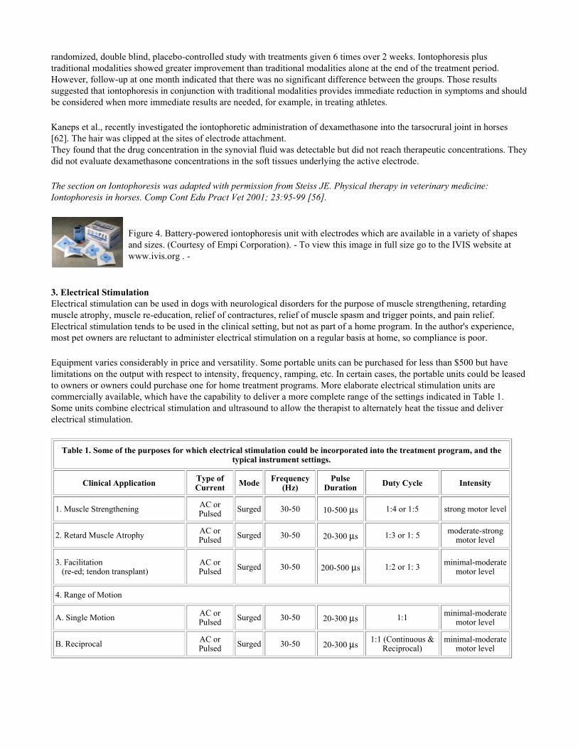
3. Electrical Stimulation Electrical stimulation can be used in dogs with neurological disorders for the purpose of muscle strengthening, retarding muscle atrophy, muscle re-education, relief of contractures, relief of muscle spasm and trigger points, and pain relief. Electrical stimulation tends to be used in the clinical setting, but not as part of a home program. In the author's experience, most pet owners are reluctant to administer electrical stimulation on a regular basis at home, so compliance is poor.
Equipment varies considerably in price and versatility. Some portable units can be purchased for less than $500 but have limitations on the output with respect to intensity, frequency, ramping, etc. In certain cases, the portable units could be leased to owners or owners could purchase one for home treatment programs. More elaborate electrical stimulation units are commercially available, which have the capability to deliver a more complete range of the settings indicated in Table 1. Some units combine electrical stimulation and ultrasound to allow the therapist to alternately heat the tissue and deliver electrical stimulation.
Table 1. Some of the purposes for which electrical stimulation could be incorporated into the treatment program, and the typical instrument settings.
Clinical Application Type of Current Mode Frequency
(Hz)Pulse
Duration Duty Cycle Intensity
1. Muscle Strengthening AC or Pulsed Surged 30-50 10-500 µs 1:4 or 1:5 strong motor level
2. Retard Muscle Atrophy AC or Pulsed Surged 30-50 20-300 µs 1:3 or 1: 5 moderate-strong
motor level
3. Facilitation (re-ed; tendon transplant)
AC or Pulsed Surged 30-50 200-500 µs 1:2 or 1: 3 minimal-moderate
motor level
4. Range of Motion
A. Single Motion AC or Pulsed Surged 30-50 20-300 µs 1:1 minimal-moderate
motor level
B. Reciprocal AC or Pulsed Surged 30-50 20-300 µs 1:1 (Continuous &
Reciprocal) minimal-moderate
motor level

TENS = Transcutaneous Electrical Nerve Stimulation Modified from D. Lions, PT, University of Alabama, Birmingham, AL, USA.
Figure 5. Electrostimulation for pain relief post-operatively in an orthopedic patient. This dog was treated with high frequency, low intensity stimulation (conventional TENS). (Courtesy of Dr. P. Shealy, Veterinary Specialists of the Southeast, Charleston, SC, USA). - To view this image in full size go to the IVIS website at www.ivis.org . -
Figure 6. Dual channel (or multichannel) electrical stimulators allow more than one pair of stimulating electrodes to be applied to the dog. The purposes include: (1) Production of interferential current (amplitude modulated beats) for treatment of deep pain; and, (2) Reciprocal or alternating patterns of stimulation in cases where range of motion is restricted in two directions (e.g., flexion and extension). (Courtesy of Dr. P. Shealy, Veterinary Specialists of the Southeast, Charleston, SC, USA). - To view this
image in full size go to the IVIS website at www.ivis.org . -
5. Relieve Contractures
AC or Pulsed
Surged (slow ramp up &
down) 30-50 100-500 µs 4:1 or 3:1 moderate-strong motor
level
6. Relieve Spasticity
A. Fatigue AC or Pulsed Surged 30-50 200-500 µs 1:1, 2:1
or 3:1 moderate-strong motor
level
B. Antagonist (Reciprocal Inhibition)
AC or Pulsed Surged 30-50 200-500 µs 1:2 or 1:3 moderate-strong motor
level
7. Circulatory Effect
A. Muscle Pump AC or Pulsed
Surged or Interrupted/Burst
30-50 or 2-4 20-300 µs 1:1 or 1:3 moderate motor level
B. Medical Galvanism DC Continuous N/A N/A N/A low sensory level
(Polarity should be 50% of Rx "+" & 50% of Rx "-" with both pads)
8. Iontophoresis DC Continuous N/A N/A N/A low/dependent on formula
(Polarity is critical and is determined by polarity of the substance used)
9. Wound Healing
A. Continuous DC DC Continuous N/A N/A N/A low sensory or sub-sensory level
(Polarity "-" for bactericidal effect and "+" for stimulation of granulation tissue)
B. High Volt Pulsed monophasic Unknown 100 Preset N/A sensory level
10. Pain Relief
A. Conventional TENS
AC or Pulsed Continuous 50-100 50-100 µs
comfortable N/A low sensory level
B. Acupuncture-like TENS
AC or Pulsed Interrupted/Burst 2-4 150-500 µs N/A strong motor level
C. Brief Intense TENS
AC or Pulsed Interrupted/Burst
Variable 1-4 or >100
300-500 µs N/A noxious-little or no motor response
4. Therapeutic Exercise Some of the techniques employed for training agility dogs, dressage horses and human patients can be adapted for therapeutic exercise in rehabilitation programs. Because each animal is an individual, no "cook-book" recommendations can be made. However, the exercise program may include some of the following exercises, for use either in the clinic or as part of a home exercise program after the owners or handler have been instructed on how to perform the exercises.
Weight shifting Step up and step down Step over Serpentines Circles Figure 8’s Transitions (slow-fast-slow walk, walk-trot-walk, etc) Leash walking on level and uneven surfaces
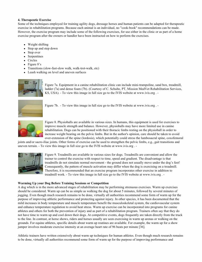
Figure 7a. Equipment in a canine rehabilitation clinic can include mini-trampoline, sand box, treadmill, ladder (7a) and dense foam (7b). (Courtesy of C. Schulte, PT, Mission MedVet Rehabilitation Services, KS, USA). - To view this image in full size go to the IVIS website at www.ivis.org . -
Figure 7b. - To view this image in full size go to the IVIS website at www.ivis.org . -
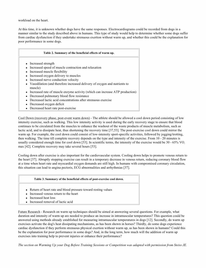
Figure 8. Physioballs are available in various sizes. In humans, this equipment is used for exercises to improve muscle strength and balance. However, physioballs may have more limited use in canine rehabilitation. Dogs can be positioned with their thoracic limbs resting on the physioball in order to increase weight bearing on the pelvic limbs. But in the author's opinion, care should be taken to avoid over-extension of the spine (lordosis), which potentially could stress the lumbosacral spine, coxofemoral
joints and/or sacro-iliac joints. Other forms of exercise can be used to strengthen the pelvic limbs, e.g., gait transitions and uneven terrain. - To view this image in full size go to the IVIS website at www.ivis.org . -
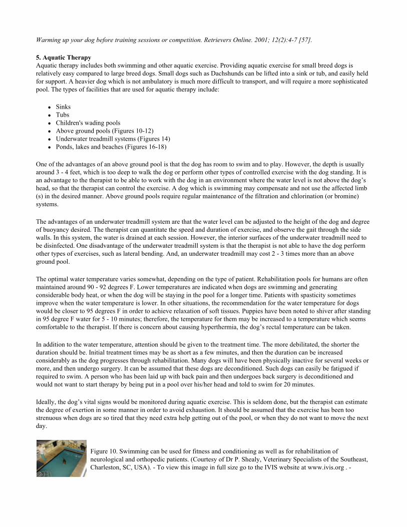
Figure 9. Treadmills are available in various sizes for dogs. Treadmills are convenient and allow the trainer to control the exercise with respect to time, speed and gradient. The disadvantage is that treadmills do not simulate normal movement - the ground does not usually move under the dog’s feet! Consequently, the pattern of muscle activation may differ when the dog is exercising on a treadmill. Therefore, it is recommended that an exercise program incorporates other exercise in addition to treadmill work. - To view this image in full size go to the IVIS website at www.ivis.org . -
Warming Up your Dog Before Training Sessions or Competition A dog which is in the more advanced stages of rehabilitation may be performing strenuous exercises. Warm up exercises should be considered. Warm up can be as simple as walking the dog for about 5 minutes, followed by several minutes of jogging. Even though much research remains to be done, virtually all authorities recommend some form of warm up for the purpose of improving athletic performance and protecting against injury. In other species, it has been documented that the mild increases in body temperature and muscle temperature benefit the musculoskeletal system, the cardiovascular system and enhance temperature regulation to avoid heat stress. Warm up exercise can be incorporated into programs for canine athletes and others for both the prevention of injury and as part of a rehabilitation program. Trainers often say that they do not have time to warm up and cool down their dogs. At competitive events, dogs frequently are taken directly from the truck to the line. In contrast, at horse shows, riders and horses usually are seen exercising in warm up arenas or walking on the grounds. For equine athletes, specific details about warm up routines are available. For example, the warm up for a show jumper involves moderate exercise intensity at an average heart rate of 96 beats per minute [36].
Athletic trainers have written extensively about warm up techniques for human athletes. Even though much research remains to be done, virtually all authorities recommend some form of warm up for the purpose of improving performance and
protecting against injury [37,38]. The purpose of this article is to describe the potential benefits of warm up and cool down for canine athletes, based on what has been written for human and equine athletes and for racing Greyhounds [39].
What Is a Warm Up? - The warm up is a preparation phase before exercise. The purpose of the warm up is to produce a mild increase in body temperature of approximately 1 - 2 degrees F, and thereby improve performance and protect against injury. In human athletes, warming up can include walking, jogging, swimming, and/or mild-to-moderate resistance cycle ergometry [37]. Warm up exercise should not be confused with preliminary cardiovascular conditioning [40], such as roading in Pointers early in the season to increase their level of fitness.
Warm up can be classified as passive and active. In passive warm up, an increase in body temperature is achieved by techniques such as massage, diathermy, ultrasound and heat application [41]. However, this does not significantly increase the blood flow to the working muscles [42], and passive warm up is not used to a great extent.
Active warm up is the easiest way to increase muscle temperature. It is divided into two phases, general and specific [43]. General warm up is a form of loosening up, such as walking followed by jogging/trotting. The effect is to raise core temperature (which can be estimated from measuring rectal temperature), heart rate and respiration rate [43]. Some of the procedures used for horses could be adapted for dogs. Typically, the horse is walked for several minutes, followed by trotting. For horses, warm up is often done on a lunge line or under saddle, or occasionally with a treadmill or hot walker if such equipment is available. For racing Greyhounds, 5 - 10 minutes of brisk walking or jogging prior to racing is recommended [39]. Specific (neuromuscular) warm up is a rehearsal for the activity. This phase mimics the anticipated activity and brings about full range of motion of the joints [41]. Specific warm up is thought to improve skill and coordination. Sports that require accuracy, timing and precise movements tend to benefit from some type of specific preliminary practice [41]. Dogs competing in agility would be candidates for including this phase of the warm up.
Stretching exercises have been part of the traditional warm up [37,43,44]. However, in a recent study, investigators have found that muscle stretching performed during the pre-exercise warm up in adult men did not significantly reduce the risk of exercise-related injury [45]. Instead, they concluded that fitness may be an important, modifiable risk factor. For trainers, the warm up for dogs would certainly be simplified if additional stretching procedures did not need to be incorporated.
Guidelines for Warm Up - The ideal warm up prepares the athlete for subsequent activity without creating fatigue [42,43]. Some trainers state that the ideal warm up for any endurance activity is the same activity but at a lower intensity [37]. More precisely, the goal of warm up is to exercise at a relatively low intensity which is less than 60% VO2 max or 70% of the maximal heart rate for less than 15 minutes [42].
For people, the aim of the warm up is to increase their body core temperature 1 - 2 degrees F and the muscle temperature up to 5 degrees F [42]. Experimentally, muscle temperatures can be measured using a needle thermistor inserted into the muscle tissue. However, in the field, a guideline for trainers might be that the rectal temperature should increase by 1 - 2 degrees F in order for the muscle temperature to reach a satisfactory level [44]. Or, the trainer or athlete may simply consider that 5 - 15 minutes of low to moderate intensity exercise should be adequate [43]. If a human athlete is sweating freely in normal climatic conditions, it is assumed that this temperature has been reached [44]. Unfortunately, this particular guideline cannot be applied to dogs.
The intensity and duration of the warm up depend on factors such as the individual athlete, the event, the facilities, and the ambient temperature [42,43]. If the warm up exercise is too strenuous or the duration too long, performance can be impaired by fatigue [42]. Excessive warm up depletes energy stores, causes lactic acid build up, and/or raises body temperature too high [43]. In cold weather, a slightly longer warm up may be required, whereas in hot weather, a shorter or less intense warm up may be appropriate [43]. The level of fitness of the athlete is another factor. An elite human athlete may require 20 - 30 minutes of relatively intense exercise to achieve maximal potential, whereas this would be excessive for an unconditioned novice and would result in exhaustion [44]. Even among individuals near the same level of training, variation exists and needs to be taken into account. For instance, individual horses were found to have different increases in rectal temperature after the same duration of trotting and cantering [43].
The effects of warming up soon wear off [37]. After the warm up is completed, the activity or event should begin within several minutes [41]. This allows recovery from temporary fatigue without losing the beneficial effects of the warm up. For human athletes, it is recommended that the time interval be no longer than 10 minutes [37].

Why should Athletes Warm Up? - A. Musculoskeletal Effects - Numerous beneficial effects of warm up have been documented for the musculoskeletal system. Greater forces are needed to injure a warm muscle than a cold muscle [41]. With an increase in tissue temperature, collagen and the muscle-tendon junctions are more able to stretch, thereby minimizing trauma [46]. When muscles from rabbits were stretched at 35 degrees C, the muscles could be lengthened approximately 31% before tearing whereas warming to 39 degrees C (103 degrees F) allowed the muscles to be lengthened 35% before tearing [47].
In addition, blood saturation can affect the elasticity of muscle. Cold muscles have low blood saturation and tend to be more susceptible to tears than warm muscles [48]. Increased temperature within muscles promotes vasodilation. A muscle achieves maximum endurance performance when all its blood vessels are maximally dilated [37]. Vasodilation increases blood flow and therefore increases the delivery of oxygen and nutrients to muscle and the removal of waste products [43]. At rest, 15 - 20% of the blood flow supplies the skeletal muscles; after 10 minutes of exercise, the percentage may increase to 70 - 75% [37].
No one has conducted a study to measure the limb temperatures in Retrievers working in cold conditions, such as sub-zero ambient temperatures or ponds covered with ice. The degree of thermal insulation provided by the hair coat is not known. If the temperatures of the limbs do decrease while dogs work in these cold environments, then warm up exercise might be important to minimize the tissue cooling.
Another very important result of increased tissue temperature is increased oxygen delivery to muscles. As temperature increases, hemoglobin releases oxygen from the red blood cells to the tissues more readily. Or, stated another way, the oxy-hemoglobin dissociation curve is shifted to the right [38]. Although considerable effort and expense are spent on finding supplements and other methods to increase the oxygen carrying capacity of hemoglobin and therefore enhance aerobic metabolism, here is a built-in mechanism to achieve this result.
The warm up appears to have the most benefit when athletes compete in high intensity-short duration activities, such as sprinting [43]. This could also be relevant to dogs participating in events such as Schutzhund and agility and field trials, where they may be at risk for muscle strain.
Other effects of warm up on the musculoskeletal system include increased speed of muscle contraction and relaxation [41,42]. In contrast, reaction times are prolonged and muscle excitability is reduced when the tissue temperatures are below normal. Warm up is also associated with an increase in muscle strength as well as speed [48]. Warm up has been reported to improve swimming speed and running speed [48].
B. Temperature Regulation - Warm up exercises activate the body’s heat dissipating mechanisms. When horses were warmed up before brief high intensity exercise, their temperatures did not rise as high during the subsequent high intensity work and they recovered faster [49]. The investigators in that study speculated that the warm up activated blood flow to the skin, causing an earlier onset of sweating and improved heat removal. Dogs rely on panting and heat loss through the respiratory system rather than sweating. Therefore, separate studies need to be conducted for dogs to determine if the mild elevations in body temperature achieved during warm up can enhance heat loss through the lungs or nasal cavity. Heat stress is a problem which is becoming more widely recognized in sporting dogs. Retrievers [50] and Pointers [51] sometimes have rectal temperatures approaching 105 - 107 degrees F during exercise, representing a rise in body temperature of 3 degrees F or more. Although most of these dogs appear clinically normal and their rectal temperature declines to normal soon after the activity ceases, some dogs seem to experience heat stress to the extent that their training is impaired.
C. Cardiovascular Effects - Sudden strenuous exertion can provoke adverse effects on heart function [37]. In one study, healthy men (n = 44) ran on a treadmill for 10 - 15 seconds without warming up [52]. Electrocardiograms (ECG) were recorded immediately after exercise. Seventy per cent of the men had ECG abnormalities that were indicative of insufficient oxygen supply to the heart muscle due to inadequate coronary blood flow. The abnormalities were not related to their age or fitness level. When the men (n = 22) warmed up by jogging for 2 minutes before treadmill running, only 2 had significant ECG changes.
In addition, blood pressure rises higher when there is no warm up. In the study described above, the average systolic pressure was 168 mmHg after the treadmill run. After the 2 minute warm up, the value was 140. Therefore, warm up reduces the
workload on the heart.
At this time, it is unknown whether dogs have the same responses. Electrocardiograms could be recorded from dogs in a manner similar to the study described above in humans. This type of study would help to determine whether some dogs suffer from cardiac dysfunction if they undertake strenuous exertion without warm up, and whether this could be the explanation for poor performance in some dogs.
Cool Down (recovery phase, post-event warm down) - The athlete should be allowed a cool down period consisting of low intensity exercise, such as walking. This low intensity activity is used during the early recovery stage to ensure that blood continues to be circulated from the muscles to enhance the washout of the waste products of muscle metabolism, such as lactic acid, and to dissipate heat, thus shortening the recovery time [37,53]. The post-exercise cool down could mirror the warm up. For example, the cool down could consist of low-intensity sport-specific activities, followed by jogging/trotting, then walking. The time till complete recovery depends on the type and intensity of the exercise. From 10 - 20 minutes is usually considered enough time for cool down [53]. In scientific terms, the intensity of the exercise would be 30 - 65% VO2 max [42]. Complete recovery may take several hours [53].
Cooling down after exercise is also important for the cardiovascular system. Cooling down helps to promote venous return to the heart [37]. Abruptly stopping exercise can result in a temporary decrease in venous return, reducing coronary blood flow at a time when heart rate and myocardial oxygen demands are still high. In humans with compromised coronary circulation, this situation can lead to angina pectoris, ECG abnormalities and arrhythmias [37].
Future Research - Research on warm up techniques should be aimed at answering several questions. For example, what duration and intensity of warm up are needed to produce an increase in intramuscular temperatures? This question could be answered using methods already established for measuring intramuscular temperatures in dogs [12]. Secondly, do warm up exercises activate the dog's heat dissipating mechanisms, as has been shown in horses? Thirdly, do some dogs experience cardiac dysfunction if they perform strenuous physical exertion without warm up, as has been shown in humans? Could this be the explanation for poor performance in some dogs? And, in the long term, how much will the addition of warm up exercises into training help to prevent injuries or enhance their performance?
The section on Warming Up your Dog Before Training Sessions or Competition was adapted with permission from Steiss JE.
Table 2. Summary of the beneficial effects of warm up.
Increased strength Increased speed of muscle contraction and relaxation Increased muscle flexibility Increased oxygen delivery to muscles Increased nerve conduction velocity Vasodilation (and therefore increased delivery of oxygen and nutrients to muscle) Increased rate of muscle enzyme activity (which can increase ATP production) Decreased pulmonary blood flow resistance Decreased lactic acid concentrations after strenuous exercise Decreased oxygen deficit Decreased heart rate post-exercise
Table 3. Summary of the beneficial effects of post-exercise cool down.
Return of heart rate and blood pressure toward resting values Increased venous return to the heart Increased heat loss Increased removal of lactic acid
Warming up your dog before training sessions or competition. Retrievers Online. 2001; 12(2):4-7 [57].
5. Aquatic Therapy Aquatic therapy includes both swimming and other aquatic exercise. Providing aquatic exercise for small breed dogs is relatively easy compared to large breed dogs. Small dogs such as Dachshunds can be lifted into a sink or tub, and easily held for support. A heavier dog which is not ambulatory is much more difficult to transport, and will require a more sophisticated pool. The types of facilities that are used for aquatic therapy include:
Sinks Tubs Children's wading pools Above ground pools (Figures 10-12) Underwater treadmill systems (Figures 14) Ponds, lakes and beaches (Figures 16-18)
One of the advantages of an above ground pool is that the dog has room to swim and to play. However, the depth is usually around 3 - 4 feet, which is too deep to walk the dog or perform other types of controlled exercise with the dog standing. It is an advantage to the therapist to be able to work with the dog in an environment where the water level is not above the dog’s head, so that the therapist can control the exercise. A dog which is swimming may compensate and not use the affected limb(s) in the desired manner. Above ground pools require regular maintenance of the filtration and chlorination (or bromine) systems.
The advantages of an underwater treadmill system are that the water level can be adjusted to the height of the dog and degree of buoyancy desired. The therapist can quantitate the speed and duration of exercise, and observe the gait through the side walls. In this system, the water is drained at each session. However, the interior surfaces of the underwater treadmill need to be disinfected. One disadvantage of the underwater treadmill system is that the therapist is not able to have the dog perform other types of exercises, such as lateral bending. And, an underwater treadmill may cost 2 - 3 times more than an above ground pool.
The optimal water temperature varies somewhat, depending on the type of patient. Rehabilitation pools for humans are often maintained around 90 - 92 degrees F. Lower temperatures are indicated when dogs are swimming and generating considerable body heat, or when the dog will be staying in the pool for a longer time. Patients with spasticity sometimes improve when the water temperature is lower. In other situations, the recommendation for the water temperature for dogs would be closer to 95 degrees F in order to achieve relaxation of soft tissues. Puppies have been noted to shiver after standing in 95 degree F water for 5 - 10 minutes; therefore, the temperature for them may be increased to a temperature which seems comfortable to the therapist. If there is concern about causing hyperthermia, the dog’s rectal temperature can be taken.
In addition to the water temperature, attention should be given to the treatment time. The more debilitated, the shorter the duration should be. Initial treatment times may be as short as a few minutes, and then the duration can be increased considerably as the dog progresses through rehabilitation. Many dogs will have been physically inactive for several weeks or more, and then undergo surgery. It can be assumed that these dogs are deconditioned. Such dogs can easily be fatigued if required to swim. A person who has been laid up with back pain and then undergoes back surgery is deconditioned and would not want to start therapy by being put in a pool over his/her head and told to swim for 20 minutes.
Ideally, the dog’s vital signs would be monitored during aquatic exercise. This is seldom done, but the therapist can estimate the degree of exertion in some manner in order to avoid exhaustion. It should be assumed that the exercise has been too strenuous when dogs are so tired that they need extra help getting out of the pool, or when they do not want to move the next day.
Figure 10. Swimming can be used for fitness and conditioning as well as for rehabilitation of neurological and orthopedic patients. (Courtesy of Dr P. Shealy, Veterinary Specialists of the Southeast, Charleston, SC, USA). - To view this image in full size go to the IVIS website at www.ivis.org . -
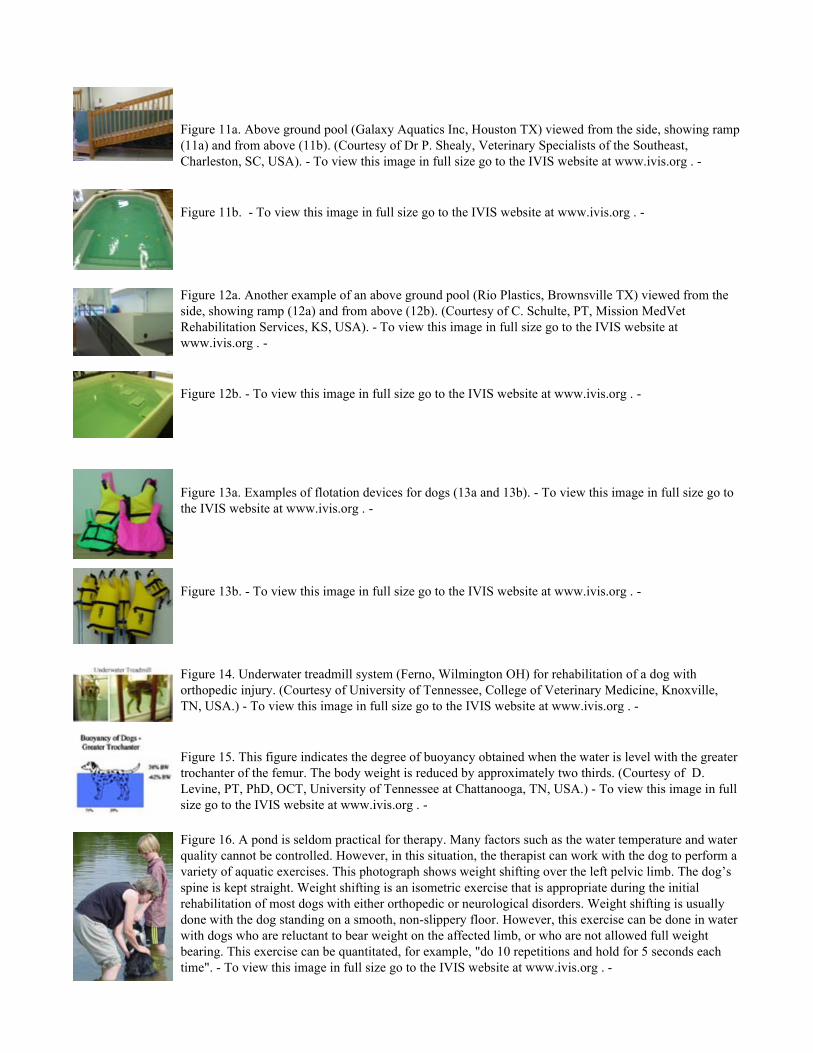
Figure 11a. Above ground pool (Galaxy Aquatics Inc, Houston TX) viewed from the side, showing ramp (11a) and from above (11b). (Courtesy of Dr P. Shealy, Veterinary Specialists of the Southeast, Charleston, SC, USA). - To view this image in full size go to the IVIS website at www.ivis.org . -
Figure 11b. - To view this image in full size go to the IVIS website at www.ivis.org . -
Figure 12a. Another example of an above ground pool (Rio Plastics, Brownsville TX) viewed from the side, showing ramp (12a) and from above (12b). (Courtesy of C. Schulte, PT, Mission MedVet Rehabilitation Services, KS, USA). - To view this image in full size go to the IVIS website at www.ivis.org . -
Figure 12b. - To view this image in full size go to the IVIS website at www.ivis.org . -
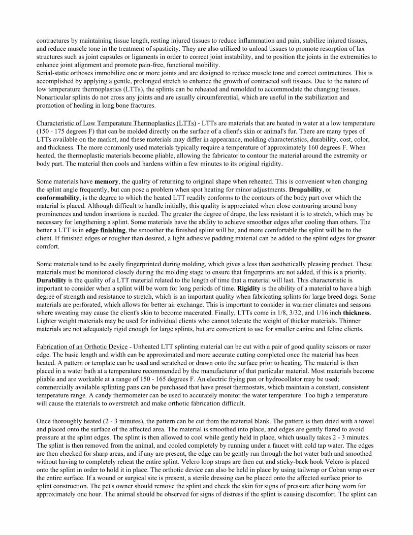
Figure 13a. Examples of flotation devices for dogs (13a and 13b). - To view this image in full size go to the IVIS website at www.ivis.org . -
Figure 13b. - To view this image in full size go to the IVIS website at www.ivis.org . -
Figure 14. Underwater treadmill system (Ferno, Wilmington OH) for rehabilitation of a dog with orthopedic injury. (Courtesy of University of Tennessee, College of Veterinary Medicine, Knoxville, TN, USA.) - To view this image in full size go to the IVIS website at www.ivis.org . -
Figure 15. This figure indicates the degree of buoyancy obtained when the water is level with the greater trochanter of the femur. The body weight is reduced by approximately two thirds. (Courtesy of D. Levine, PT, PhD, OCT, University of Tennessee at Chattanooga, TN, USA.) - To view this image in full size go to the IVIS website at www.ivis.org . -
Figure 16. A pond is seldom practical for therapy. Many factors such as the water temperature and water quality cannot be controlled. However, in this situation, the therapist can work with the dog to perform a variety of aquatic exercises. This photograph shows weight shifting over the left pelvic limb. The dog’s spine is kept straight. Weight shifting is an isometric exercise that is appropriate during the initial rehabilitation of most dogs with either orthopedic or neurological disorders. Weight shifting is usually done with the dog standing on a smooth, non-slippery floor. However, this exercise can be done in water with dogs who are reluctant to bear weight on the affected limb, or who are not allowed full weight bearing. This exercise can be quantitated, for example, "do 10 repetitions and hold for 5 seconds each time". - To view this image in full size go to the IVIS website at www.ivis.org . -

Figure 17. In addition to weight shifting side to side, as discussed in Figure 16, the dog’s center of gravity can be shifted cranially and caudally. This exercise seems to activate the paraspinal muscles as well as the appendicular muscles. - To view this image in full size go to the IVIS website at www.ivis.org . -
Figure 18. Exercise in water allows unloading of the joints for orthopedic rehabilitation. With neurological disorders, a dog with severe weakness may be able to achieve weight bearing when in an aquatic environment. Where available, a pond or beach with a level bottom allows the dog to be exercised in various heights of water to achieve varying amounts of buoyancy. In addition, equipment such as plastic benches can be used to perform step up exercises in the water (see Figure 19). - To view this image in full size go to the IVIS website at www.ivis.org . -
Figure 19. Although much of the equipment available for people for aquatic therapy is not readily adapted to dogs, the benches which are seen stacked in this illustration can be placed in water to allow the dog to step up and step down. The degree of weight bearing and difficulty of this exercise will depend on the level of the water and the height of the bench compared to the size of the dog. (Courtesy of Cheryl Fuller, MS, ATC, Aquatics Director, Spain Rehabilitation Center, The University of Alabama at Birmingham). - To view this image in full size go to the IVIS website at www.ivis.org . -
What about the possibility of having access to rehabilitation pools such as the one depicted in (Fig. 20) for the purpose of treating canine patients? In many locations, county health regulations prohibit animals in or around these pools (Cheryl Fuller, Director of Aquatic Therapy, Spain Rehabilitation Hospital, UAB, Birmingham AL, personal communication, 2001). Many county health departments allow seeing-eye or care dogs on public pool decks, but do not allow them in the water with their owners (Alison Osinski, PhD, Aquatic Consulting Services, San Diego, CA, 2001).
This question was posed to Dr. Bill Johnston, State Public Health Veterinarian for the Alabama Department of Public Health. He stated that in a properly chlorinated pool, E. coli and Giardia would be eliminated but Cryptosporidium would be a potential concern (personal communication, 2001). Dr. Johnston recommended that the dog be bathed and brushed with a medicated shampoo containing antifungal agents, within 24 hours before entering the pool. The dog should be free of ectoparasites. The veterinarian could perform a fecal screen and even provide a signed health certificate, somewhat similar to the workup for therapy dogs performing hospital visitations.
Dr. Johnston posed this question to members of the National Association of State Public Health Veterinarians, and received the following comments:
If in contact with the dogs, patients should not have any open or healing wounds. The more people in the pool at one time with the dog, the greater the risk of some form of microbe transmission. A clear policy is needed on handling fecal and urine contamination of the pool by both dogs and humans. Care must be taken to maintain the chlorine concentration at an acceptable level (pool inspection requirements by county health officials state the exact levels of chlorine required to eliminate E. coli). There might be concern about patients colonized with multi-drug resistant organisms who may transmit the organism to the dog, which then becomes a source of infection for other patients. Even for chlorine sensitive organisms, a certain amount of contact time is needed, since inactivation is not instantaneous. Cryptosporidium is especially a concern. One should also consider leptospirosis. Although Cryptosporidium might survive and be infectious if ingested by a person sharing a pool with an infected dog, the degree of risk would not exceed the risk of swimming with a child, for example, a group in which the prevalence of asymptomatic Cryptosporidiosis probably exceeds that in most dog populations. Humans excreting Cryptosporidium and Giardia in pools pose more of a potential problem than a healthy clean groomed dog would.
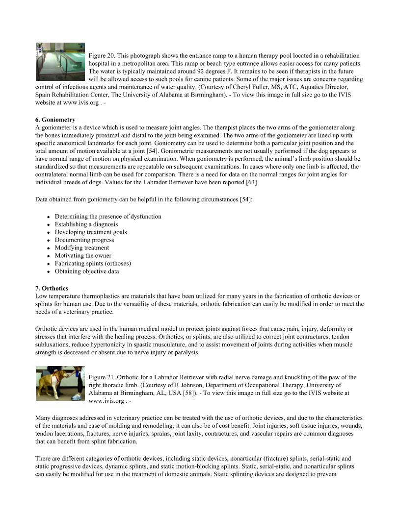
Figure 20. This photograph shows the entrance ramp to a human therapy pool located in a rehabilitation hospital in a metropolitan area. This ramp or beach-type entrance allows easier access for many patients. The water is typically maintained around 92 degrees F. It remains to be seen if therapists in the future will be allowed access to such pools for canine patients. Some of the major issues are concerns regarding
control of infectious agents and maintenance of water quality. (Courtesy of Cheryl Fuller, MS, ATC, Aquatics Director, Spain Rehabilitation Center, The University of Alabama at Birmingham). - To view this image in full size go to the IVIS website at www.ivis.org . -
6. Goniometry A goniometer is a device which is used to measure joint angles. The therapist places the two arms of the goniometer along the bones immediately proximal and distal to the joint being examined. The two arms of the goniometer are lined up with specific anatomical landmarks for each joint. Goniometry can be used to determine both a particular joint position and the total amount of motion available at a joint [54]. Goniometric measurements are not usually performed if the dog appears to have normal range of motion on physical examination. When goniometry is performed, the animal’s limb position should be standardized so that measurements are repeatable on subsequent examinations. In cases where only one limb is affected, the contralateral normal limb can be used for comparison. There is a need for data on the normal ranges for joint angles for individual breeds of dogs. Values for the Labrador Retriever have been reported [63].
Data obtained from goniometry can be helpful in the following circumstances [54]:
Determining the presence of dysfunction Establishing a diagnosis Developing treatment goals Documenting progress Modifying treatment Motivating the owner Fabricating splints (orthoses) Obtaining objective data
7. Orthotics Low temperature thermoplastics are materials that have been utilized for many years in the fabrication of orthotic devices or splints for human use. Due to the versatility of these materials, orthotic fabrication can easily be modified in order to meet the needs of a veterinary practice.
Orthotic devices are used in the human medical model to protect joints against forces that cause pain, injury, deformity or stresses that interfere with the healing process. Orthotics, or splints, are also utilized to correct joint contractures, tendon subluxations, reduce hypertonicity in spastic musculature, and to assist movement of joints during activities when muscle strength is decreased or absent due to nerve injury or paralysis.
Figure 21. Orthotic for a Labrador Retriever with radial nerve damage and knuckling of the paw of the right thoracic limb. (Courtesy of R Johnson, Department of Occupational Therapy, University of Alabama at Birmingham, AL, USA [58]). - To view this image in full size go to the IVIS website at www.ivis.org . -
Many diagnoses addressed in veterinary practice can be treated with the use of orthotic devices, and due to the characteristics of the materials and ease of molding and remodeling; it can also be of cost benefit. Joint injuries, soft tissue injuries, wounds, tendon lacerations, fractures, nerve injuries, sprains, joint laxity, contractures, and vascular repairs are common diagnoses that can benefit from splint fabrication.
There are different categories of orthotic devices, including static devices, nonarticular (fracture) splints, serial-static and static progressive devices, dynamic splints, and static motion-blocking splints. Static, serial-static, and nonarticular splints can easily be modified for use in the treatment of domestic animals. Static splinting devices are designed to prevent
contractures by maintaining tissue length, resting injured tissues to reduce inflammation and pain, stabilize injured tissues, and reduce muscle tone in the treatment of spasticity. They are also utilized to unload tissues to promote resorption of lax structures such as joint capsules or ligaments in order to correct joint instability, and to position the joints in the extremities to enhance joint alignment and promote pain-free, functional mobility. Serial-static orthoses immobilize one or more joints and are designed to reduce muscle tone and correct contractures. This is accomplished by applying a gentle, prolonged stretch to enhance the growth of contracted soft tissues. Due to the nature of low temperature thermoplastics (LTTs), the splints can be reheated and remolded to accommodate the changing tissues. Nonarticular splints do not cross any joints and are usually circumferential, which are useful in the stabilization and promotion of healing in long bone fractures.
Characteristic of Low Temperature Thermoplastics (LTTs) - LTTs are materials that are heated in water at a low temperature (150 - 175 degrees F) that can be molded directly on the surface of a client's skin or animal's fur. There are many types of LTTs available on the market, and these materials may differ in appearance, molding characteristics, durability, cost, color, and thickness. The more commonly used materials typically require a temperature of approximately 160 degrees F. When heated, the thermoplastic materials become pliable, allowing the fabricator to contour the material around the extremity or body part. The material then cools and hardens within a few minutes to its original rigidity.
Some materials have memory, the quality of returning to original shape when reheated. This is convenient when changing the splint angle frequently, but can pose a problem when spot heating for minor adjustments. Drapability, or conformability, is the degree to which the heated LTT readily conforms to the contours of the body part over which the material is placed. Although difficult to handle initially, this quality is appreciated when close contouring around bony prominences and tendon insertions is needed. The greater the degree of drape, the less resistant it is to stretch, which may be necessary for lengthening a splint. Some materials have the ability to achieve smoother edges after cooling than others. The better a LTT is in edge finishing, the smoother the finished splint will be, and more comfortable the splint will be to the client. If finished edges or rougher than desired, a light adhesive padding material can be added to the splint edges for greater comfort.
Some materials tend to be easily fingerprinted during molding, which gives a less than aesthetically pleasing product. These materials must be monitored closely during the molding stage to ensure that fingerprints are not added, if this is a priority. Durability is the quality of a LTT material related to the length of time that a material will last. This characteristic is important to consider when a splint will be worn for long periods of time. Rigidity is the ability of a material to have a high degree of strength and resistance to stretch, which is an important quality when fabricating splints for large breed dogs. Some materials are perforated, which allows for better air exchange. This is important to consider in warmer climates and seasons where sweating may cause the client's skin to become macerated. Finally, LTTs come in 1/8, 3/32, and 1/16 inch thickness. Lighter weight materials may be used for individual clients who cannot tolerate the weight of thicker materials. Thinner materials are not adequately rigid enough for large splints, but are convenient to use for smaller canine and feline clients.
Fabrication of an Orthotic Device - Unheated LTT splinting material can be cut with a pair of good quality scissors or razor edge. The basic length and width can be approximated and more accurate cutting completed once the material has been heated. A pattern or template can be used and scratched or drawn onto the surface prior to heating. The material is then placed in a water bath at a temperature recommended by the manufacturer of that particular material. Most materials become pliable and are workable at a range of 150 - 165 degrees F. An electric frying pan or hydrocollator may be used; commercially available splinting pans can be purchased that have preset thermostats, which maintain a constant, consistent temperature range. A candy thermometer can be used to accurately monitor the water temperature. Too high a temperature will cause the materials to overstretch and make orthotic fabrication difficult.
Once thoroughly heated (2 - 3 minutes), the pattern can be cut from the material blank. The pattern is then dried with a towel and placed onto the surface of the affected area. The material is smoothed into place, and edges are gently flared to avoid pressure at the splint edges. The splint is then allowed to cool while gently held in place, which usually takes 2 - 3 minutes. The splint is then removed from the animal, and cooled completely by running under a faucet with cold tap water. The edges are then checked for sharp areas, and if any are present, the edge can be gently run through the hot water bath and smoothed without having to completely reheat the entire splint. Velcro loop straps are then cut and sticky-back hook Velcro is placed onto the splint in order to hold it in place. The orthotic device can also be held in place by using tailwrap or Coban wrap over the entire surface. If a wound or surgical site is present, a sterile dressing can be placed onto the affected surface prior to splint construction. The pet's owner should remove the splint and check the skin for signs of pressure after being worn for approximately one hour. The animal should be observed for signs of distress if the splint is causing discomfort. The splint can
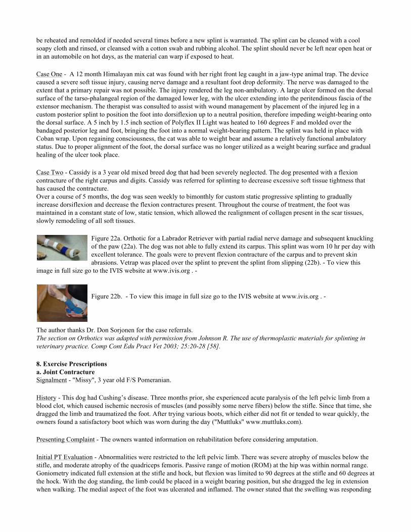
be reheated and remolded if needed several times before a new splint is warranted. The splint can be cleaned with a cool soapy cloth and rinsed, or cleansed with a cotton swab and rubbing alcohol. The splint should never be left near open heat or in an automobile on hot days, as the material can warp if exposed to heat.
Case One - A 12 month Himalayan mix cat was found with her right front leg caught in a jaw-type animal trap. The device caused a severe soft tissue injury, causing nerve damage and a resultant foot drop deformity. The nerve was damaged to the extent that a primary repair was not possible. The injury rendered the leg non-ambulatory. A large ulcer formed on the dorsal surface of the tarso-phalangeal region of the damaged lower leg, with the ulcer extending into the peritendinous fascia of the extensor mechanism. The therapist was consulted to assist with wound management by placement of the injured leg in a custom posterior splint to position the foot into dorsiflexion up to a neutral position, therefore impeding weight-bearing onto the dorsal surface. A 5 inch by 1.5 inch section of Polyflex II Light was heated to 160 degrees F and molded over the bandaged posterior leg and foot, bringing the foot into a normal weight-bearing pattern. The splint was held in place with Coban wrap. Upon regaining consciousness, the cat was able to weight bear and assume a relatively functional ambulatory status. Due to proper alignment of the foot, the dorsal surface was no longer utilized as a weight bearing surface and gradual healing of the ulcer took place.
Case Two - Cassidy is a 3 year old mixed breed dog that had been severely neglected. The dog presented with a flexion contracture of the right carpus and digits. Cassidy was referred for splinting to decrease excessive soft tissue tightness that has caused the contracture. Over a course of 5 months, the dog was seen weekly to bimonthly for custom static progressive splinting to gradually increase dorsiflexion and decrease the flexion contractures present. Throughout the course of treatment, the foot was maintained in a constant state of low, static tension, which allowed the realignment of collagen present in the scar tissues, slowly remodeling of all soft tissues.
Figure 22a. Orthotic for a Labrador Retriever with partial radial nerve damage and subsequent knuckling of the paw (22a). The dog was not able to fully extend its carpus. This splint was worn 10 hr per day with excellent tolerance. The goals were to prevent flexion contracture of the carpus and to prevent skin abrasions. Vetrap was placed over the splint to prevent the splint from slipping (22b). - To view this
image in full size go to the IVIS website at www.ivis.org . -
Figure 22b. - To view this image in full size go to the IVIS website at www.ivis.org . -
The author thanks Dr. Don Sorjonen for the case referrals. The section on Orthotics was adapted with permission from Johnson R. The use of thermoplastic materials for splinting in veterinary practice. Comp Cont Edu Pract Vet 2003; 25:20-28 [58].
8. Exercise Prescriptions a. Joint Contracture Signalment - "Missy", 3 year old F/S Pomeranian.
History - This dog had Cushing’s disease. Three months prior, she experienced acute paralysis of the left pelvic limb from a blood clot, which caused ischemic necrosis of muscles (and possibly some nerve fibers) below the stifle. Since that time, she dragged the limb and traumatized the foot. After trying various boots, which either did not fit or tended to wear quickly, the owners found a satisfactory boot which was worn during the day ("Muttluks" www.muttluks.com).
Presenting Complaint - The owners wanted information on rehabilitation before considering amputation.
Initial PT Evaluation - Abnormalities were restricted to the left pelvic limb. There was severe atrophy of muscles below the stifle, and moderate atrophy of the quadriceps femoris. Passive range of motion (ROM) at the hip was within normal range. Goniometry indicated full extension at the stifle and hock, but flexion was limited to 90 degrees at the stifle and 60 degrees at the hock. With the dog standing, the limb could be placed in a weight bearing position, but she dragged the leg in extension when walking. The medial aspect of the foot was ulcerated and inflamed. The owner stated that the swelling was responding
to antibiotic therapy. Sensation to the limb was intact. The patellar reflex appeared decreased.
Short Term Goals -
1. Increase ROM of stifle and hock, left pelvic limb; 2. Strengthen muscles, left pelvic limb; 3. Instruct owners in a home exercise program (HEP).
Long Term Goal - Regain normal function of affected limb.
Treatment - The owners were given the following HEP to be done 4x per day. Steps 2 - 4 were demonstrated, including hand placement for the stretching exercises, and the owners practiced under supervision before leaving the clinic.
1. Warming the tissues: Apply moist heat to the LH limb for 5 minutes (towel soaked in warm water or a hot pack, at a temperature which you find tolerable). Alternatively, leash walk x 5 minutes, with boot on, since exercise can also warm up the tissues. Step 1 can be omitted if time does not permit.
2. Stretching: With your dog relaxed in lateral recumbency, affected limb uppermost, bend the stifle to the point of resistance and hold for 20 - 30 sec. If you feel any relaxation or "give" while you are holding, increase the bend till you encounter the new point of resistance. Relax for a few seconds and repeat 2x. Treat the hock joint similarly.
3. Exercise (weight shifting): With your dog standing with all 4 feet positioned normally, her spine straight, place your hands on her left and right flank and shift her weight onto her affected limb. Hold for 10 sec. Repeat 10x.
4. Leash walk for 5 minutes at a speed slow enough that she is able to put weight on her affected leg and place it in the correct position.
Re-evaluation - By 2 weeks, the owners reported that Missy had started jumping onto furniture and occasionally put weight on her affected limb when running. At the 3-week recheck, passive ROM at the stifle was 0 - 120 deg (flexion increased 30 deg) and ROM at the hock was 0 - 85 deg (flexion increased 25 deg). Swelling was absent from the foot. The owners were advised to continue the HEP until ROM in the stifle and hock appeared to be the same as in the unaffected limb. The boot was gradually worn for shorter times each day.
Follow-up - By 10 weeks, the owners estimated that ROM at the stifle had further increased to 150 deg of flexion. She continued to hold the affected limb up when running at top speed. By 20 weeks, the owners stated that she was "back to normal", and consistently ran with "4 on the floor". They continued to perform the stretching exercises once per day. Missy continued to receive Warfarin and Lysodren medication.
Comments - It can be difficult to predict how rapidly contractures will respond to treatment. This dog benefited from the excellent compliance on the part of the family members. For instance, they divided up the therapy so that the person who disliked performing the stretching did the leash walking. Goniometry was helpful to document increased ROM and to provide positive feedback for the owners. For this dog, the Muttluks boot gave enough support and protection to the distal limb so that we did not think that it was necessary to make a custom-fitted splint.
b. Thoraco-lumbar Intervertebral Disk Disease, Non-weight-bearing in Pelvic Limbs Signalment - "Rosebud", 7 year old Dachshund, F/S
History - This dog had previously undergone surgery for a herniated intervertebral disk at T13 - L1. The dog had made an uneventful recovery and was ambulatory. Eight months later, the dog experienced a second onset of paraplegia associated with intervertebral disk herniations in the caudal thoracic spine as determined on myelogram. The owner was reluctant to undertake additional surgery. The orthopedic surgeon who had done the initial surgery suggested that the owner pursue physical therapy.
Presenting Complaint - The dog has been unable to bear weight on the pelvic limbs for 7 days.
Physical Therapy Evaluation - Abnormalities were restricted to the pelvic limbs. The dog had pain sensation in both pelvic limbs. The muscle mass was not decreased. Patellar reflexes were mildly increased. The withdrawal response to toe pinching
was poor in both pelvic limbs. When lifted to a standing position, the dog was not able to bear weight on the pelvic limbs. Joint range of motion was within normal range for both pelvic limbs. The dog moved around by using the thoracic limbs and dragging the pelvic limbs.
Short term goals (within 4 weeks) -
1. Strengthen the pelvic limbs so that dog is able to bear weight for a minimum of 10 seconds without assistance when standing in water.
2. Dog will be able to voluntarily flex each pelvic limb in response to toe pinch while standing with assistance. 3. Maintain range of motion within normal limits in pelvic limbs.
Long term goals - Restore function in pelvic limbs so that dog can ambulate independently in the home environment.
Treatment - The owner was provided with a home exercise program that was demonstrated to her in the clinic, and which she practiced under supervision before leaving the clinic. She was instructed to repeat the following exercises 2 - 3 times each day if possible (but only 1 - 2 times for the exercises in water).
1. Place a rubber-backed mat (or non-slippery mat) on your table or counter top. Support her under her abdomen with one hand and support her under her ribs with the other hand. Place her hind feet so that they are in a normal position under her body. For the first exercise, use one hand to gently lift her up under her abdomen so that her feet are just barely touching the mat and then gently lower her very slightly (approximately a centimeter) so that she gets the sensation of bearing some weight on her hind legs. Repeat 10 times.
2. Continue to support Rosebud in the same manner with her feet placed under her body, and gently shift her weight from side-to-side so that she has the sensation of bearing some of her weight on her left side, and then on her right side. Repeat this 10 times also.
3. Have Rosebud lie on her side while you pinch a toe (on the leg that is uppermost) just enough to get her to pull back that leg. Tell her "good girl" in order to encourage her to do this without having to pinch a lot. Repeat 5 times. Place her on her other side and repeat. Or, simply do this during the day when she is lying on her side. When she gets stronger, you can do the toe pinches with her standing because working against gravity is more difficult and will strengthen the limbs more.
4. Repeat the first 2 exercises in water once or twice per day. Have the water level near the point of her shoulder, about 95 degrees F so it is comfortably warm. After the exercises, continue to support her under her abdomen and chest with your hands so that she can stand in the water for 5 - 10 minutes. Encourage her to take several steps in the water by helping her to advance her feet. Take her out of the water earlier if she seems to become fatigued.
Follow-up - This dog slowly progressed over 4 months to the point where she could walk, with mild ataxia, on level surfaces in the house.
Comments - The rehabilitation program for this dog was based on a combination of partial weight bearing with assisted stepping and aquatic therapy. Standing and walking with assistance in water helped to give the dog proprioceptive sensory feedback from the pelvic limbs. In addition, seeing her dog able to stand in the water provided motivation for the owner at the start of the program. When thinking about rehabilitation, people often think about dogs swimming. However, this dog was deconditioned after the previous surgery and lack of exercise, and would have become easily fatigued if she had been forced to swim at this early stage of the rehabilitation. In addition, she might have compensated in the water by using her thoracic limbs and not necessarily activating the muscles of the pelvic limbs. By having the dog stand in water, instead of swimming, the therapist is able to control the activity in the limbs. Swimming could be considered later in the program when the goal is to promote cardiovascular conditioning. Water temperature is important. Cooler water temperatures are indicated when dogs are swimming vigorously and generating body heat. In the author’s opinion, water temperatures should be around 95 degrees F for therapy. In cases where the dog is not exercising in the water, but just standing, some dogs will start shivering after 5 - 10 minutes. In this case, the water temperature can be increased closer to 100 degrees F (or what feels comfortable to the therapist). If in doubt, the dog’s rectal temperature can be measured. These procedures are quite easy to carry out on a small breed dog. In this instance, the owner was elderly but she was able to perform the home program. The exercises were done with the dog standing on a table and in a utility sink. Working at this height prevented the owner from having to lean over and potentially injure her own back while doing the therapy, as might
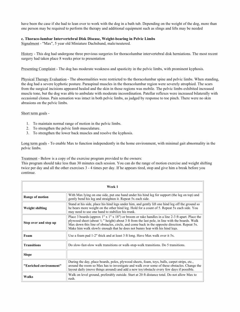
have been the case if she had to lean over to work with the dog in a bath tub. Depending on the weight of the dog, more than one person may be required to perform the therapy and additional equipment such as slings and lifts may be needed
c. Thoraco-lumbar Intervertebral Disk Disease, Weight-bearing in Pelvic Limbs Signalment - "Max", 5 year old Miniature Dachshund, male/neutered.
History - This dog had undergone three previous surgeries for thoracolumbar intervertebral disk herniations. The most recent surgery had taken place 8 weeks prior to presentation
Presenting Complaint - The dog has moderate weakness and spasticity in the pelvic limbs, with prominent kyphosis.
Physical Therapy Evaluation - The abnormalities were restricted to the thoracolumbar spine and pelvic limbs. When standing, the dog had a severe kyphotic posture. Paraspinal muscles in the thoracolumbar region were severely atrophied. The scars from the surgical incisions appeared healed and the skin in those regions was mobile. The pelvic limbs exhibited increased muscle tone, but the dog was able to ambulate with moderate incoordination. Patellar reflexes were increased bilaterally with occasional clonus. Pain sensation was intact in both pelvic limbs, as judged by response to toe pinch. There were no skin abrasions on the pelvic limbs.
Short term goals -
1. To maintain normal range of motion in the pelvic limbs. 2. To strengthen the pelvic limb musculature. 3. To strengthen the lower back muscles and resolve the kyphosis.
Long term goals - To enable Max to function independently in the home environment, with minimal gait abnormality in the pelvic limbs.
Treatment - Below is a copy of the exercise program provided to the owners: This program should take less than 30 minutes each session. You can do the range of motion exercise and weight shifting twice per day and all the other exercises 3 - 4 times per day. If he appears tired, stop and give him a break before you continue.
Week 1
Range of motion With Max lying on one side, put one hand under his hind leg for support (the leg on top) and gently bend his leg and straighten it. Repeat 5x each side.
Weight shiftingStand at his side, place his hind legs under him, and gently lift one hind leg off the ground so he bears more weight on the other hind leg. Hold for a count of 5. Repeat 5x each side. You may need to use one hand to stabilize his trunk.
Step over and step upPlace 3 boards (approx 1" x 1" x 18") or broom or rake handles in a line 2-3 ft apart. Place the plywood sheet (about ½ " height) about 3 ft from the last pole, in line with the boards. Walk Max down this line of obstacles, circle, and come back in the opposite direction. Repeat 5x. Make him walk slowly enough that he does not bunny hop with his hind legs.
Foam Use a foam pad 1-2" thick and at least 3 ft long. Have Max walk over it 5x.
Transitions Do slow-fast-slow walk transitions or walk-stop-walk transitions. Do 5 transitions.
Slope
"Enriched environment"During the day, place boards, poles, plywood sheets, foam, toys, balls, carpet strips, etc., around the room so Max has to investigate and walk over some of these obstacles. Change the layout daily (move things around) and add a new toy/obstacle every few days if possible.
Walks Walk on level ground, preferably outside. Start at 20 ft distance total. Do not allow Max to rush.

Week 2
Range of motion With Max lying on one side, put one hand under his hind leg for support (the leg on top) and gently bend his leg and straighten it. Repeat 5x each side.
Weight shiftingStand at his side, place his hind legs under him, and gently lift one hind leg off the ground so he bears more weight on the other hind leg. Hold for a count of 5. Repeat 5x each side. You may need to use one hand to stabilize his trunk.
Step over and step up Repeat 10x.
Foam Repeat 5x.
Transitions Repeat 10 transitions.
Slope
"Enriched environment"
During the day, place boards, poles, plywood sheets, foam, toys, balls, carpet strips, etc., around the room so Max has to investigate and walk over some of these obstacles. Change the layout daily (move things around) and add a new toy/obstacle every few days if possible.
Walks Walk 25 ft.
Week 3
Range of motion With Max lying on one side, put one hand under his hind leg for support (the leg on top) and gently bend his leg and straighten it. Repeat 5x each side.
Weight shiftingStand at his side, place his hind legs under him, and gently lift one hind leg off the ground so he bears more weight on the other hind leg. Hold for a count of 5. Repeat 5x each side. You may need to use one hand to stabilize his trunk.
Step over and step up Repeat 10x and add a second plywood sheet on top of the first one, so height is about 1"
Foam Repeat 10x.
Transitions Repeat 10 transitions.
Slope Do at walk only. Start on week 3. Walk up and down a gentle gradient, about 10 ft length. Repeat 2x.
"Enriched environment"
During the day, place boards, poles, plywood sheets, foam, toys, balls, carpet strips, etc., around the room so Max has to investigate and walk over some of these obstacles. Change the layout daily (move things around) and add a new toy/obstacle every few days if possible.
Walks Walk 50 ft.
Week 4
Range of motion With Max lying on one side, put one hand under his hind leg for support (the leg on top) and gently bend his leg and straighten it. Repeat 5x each side.
Weight shiftingStand at his side, place his hind legs under him, and gently lift one hind leg off the ground so he bears more weight on the other hind leg. Hold for a count of 5. Repeat 5x each side. You may need to use one hand to stabilize his trunk.
Step over and step up Repeat 10x.
Foam Repeat 10x.
Transitions Repeat 10 transitions.
Slope Repeat 4x.

Follow-up - This dog gradually improved so that at 4 months follow-up, he was judged to be functioning at a level similar to his condition before his last surgery. The spasticity resolved to the point where it did not interfere significantly with ambulation. Within the first few weeks, his posture improved and the kyphosis had resolved at the time of follow-up.
Comments - The program for this dog was based on a progressive exercise program for muscle strengthening plus providing an enriched environment. Aquatic therapy was not instituted for this dog, but would also have been an appropriate addition. Exercises in water could have included standing, walking, step ups (onto plastic benches, for example) and step overs, if sufficient room were available in the tub or pool for such equipment.
"Enriched environment"
During the day, place boards, poles, plywood sheets, foam, toys, balls, carpet strips, etc., around the room so Max has to investigate and walk over some of these obstacles. Change the layout daily (move things around) and add a new toy/obstacle every few days if possible.
Walks Walk 50 ft.
Week 5
Range of motion With Max lying on one side, put one hand under his hind leg for support (the leg on top) and gently bend his leg and straighten it. Repeat 5x each side.
Weight shiftingStand at his side, place his hind legs under him, and gently lift one hind leg off the ground so he bears more weight on the other hind leg. Hold for a count of 5. Repeat 5x each side. You may need to use one hand to stabilize his trunk.
Step over and step up Repeat 10x.
Foam Repeat 10x. Change to a thicker foam if this becomes too easy.
Transitions Repeat 10 transitions.
Slope Repeat 5x.
"Enriched environment"
During the day, place boards, poles, plywood sheets, foam, toys, balls, carpet strips, etc., around the room so Max has to investigate and walk over some of these obstacles. Change the layout daily (move things around) and add a new toy/obstacle every few days if possible.
Walks Walk 100 ft.
Week 6
Range of motion With Max lying on one side, put one hand under his hind leg for support (the leg on top) and gently bend his leg and straighten it. Repeat 5x each side.
Weight shiftingStand at his side, place his hind legs under him, and gently lift one hind leg off the ground so he bears more weight on the other hind leg. Hold for a count of 5. Repeat 5x each side. You may need to use one hand to stabilize his trunk.
Step over and step up Repeat 10x.
Foam Repeat 10x. Change to a thicker foam if this becomes too easy.
Transitions Repeat 10 transitions.
Slope Repeat 5x.
"Enriched environment"
During the day, place boards, poles, plywood sheets, foam, toys, balls, carpet strips, etc., around the room so Max has to investigate and walk over some of these obstacles. Change the layout daily (move things around) and add a new toy/obstacle every few days if possible.
Walks Gradually increase distance to tolerance.
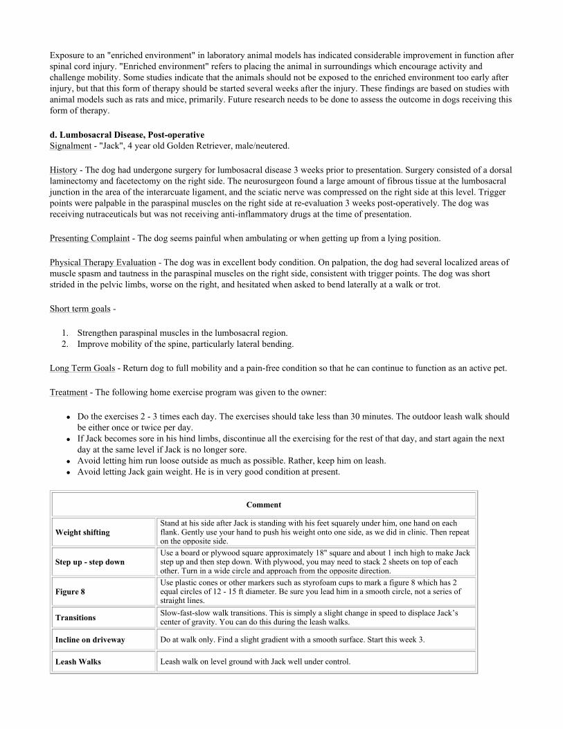
Exposure to an "enriched environment" in laboratory animal models has indicated considerable improvement in function after spinal cord injury. "Enriched environment" refers to placing the animal in surroundings which encourage activity and challenge mobility. Some studies indicate that the animals should not be exposed to the enriched environment too early after injury, but that this form of therapy should be started several weeks after the injury. These findings are based on studies with animal models such as rats and mice, primarily. Future research needs to be done to assess the outcome in dogs receiving this form of therapy.
d. Lumbosacral Disease, Post-operative Signalment - "Jack", 4 year old Golden Retriever, male/neutered.
History - The dog had undergone surgery for lumbosacral disease 3 weeks prior to presentation. Surgery consisted of a dorsal laminectomy and facetectomy on the right side. The neurosurgeon found a large amount of fibrous tissue at the lumbosacral junction in the area of the interarcuate ligament, and the sciatic nerve was compressed on the right side at this level. Trigger points were palpable in the paraspinal muscles on the right side at re-evaluation 3 weeks post-operatively. The dog was receiving nutraceuticals but was not receiving anti-inflammatory drugs at the time of presentation.
Presenting Complaint - The dog seems painful when ambulating or when getting up from a lying position.
Physical Therapy Evaluation - The dog was in excellent body condition. On palpation, the dog had several localized areas of muscle spasm and tautness in the paraspinal muscles on the right side, consistent with trigger points. The dog was short strided in the pelvic limbs, worse on the right, and hesitated when asked to bend laterally at a walk or trot.
Short term goals -
1. Strengthen paraspinal muscles in the lumbosacral region. 2. Improve mobility of the spine, particularly lateral bending.
Long Term Goals - Return dog to full mobility and a pain-free condition so that he can continue to function as an active pet.
Treatment - The following home exercise program was given to the owner:
Do the exercises 2 - 3 times each day. The exercises should take less than 30 minutes. The outdoor leash walk should be either once or twice per day. If Jack becomes sore in his hind limbs, discontinue all the exercising for the rest of that day, and start again the next day at the same level if Jack is no longer sore. Avoid letting him run loose outside as much as possible. Rather, keep him on leash. Avoid letting Jack gain weight. He is in very good condition at present.
Comment
Weight shiftingStand at his side after Jack is standing with his feet squarely under him, one hand on each flank. Gently use your hand to push his weight onto one side, as we did in clinic. Then repeat on the opposite side.
Step up - step downUse a board or plywood square approximately 18" square and about 1 inch high to make Jack step up and then step down. With plywood, you may need to stack 2 sheets on top of each other. Turn in a wide circle and approach from the opposite direction.
Figure 8Use plastic cones or other markers such as styrofoam cups to mark a figure 8 which has 2 equal circles of 12 - 15 ft diameter. Be sure you lead him in a smooth circle, not a series of straight lines.
Transitions Slow-fast-slow walk transitions. This is simply a slight change in speed to displace Jack’s center of gravity. You can do this during the leash walks.
Incline on driveway Do at walk only. Find a slight gradient with a smooth surface. Start this week 3.
Leash Walks Leash walk on level ground with Jack well under control.
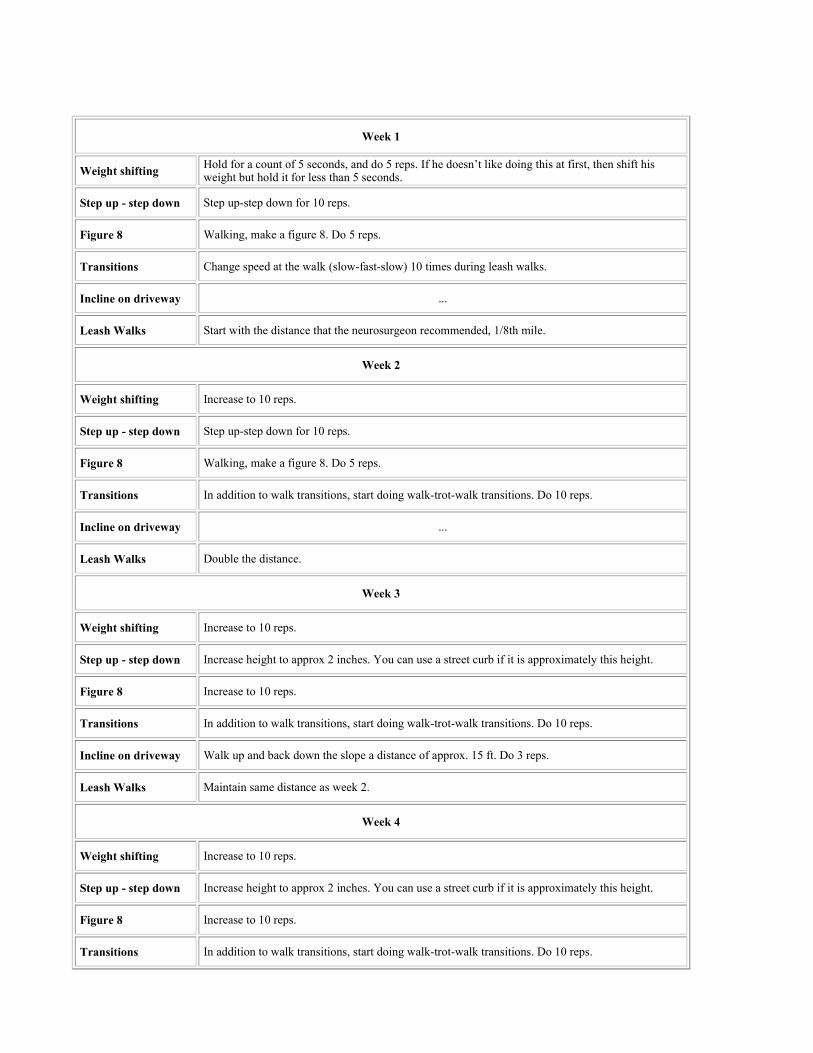
Week 1
Weight shifting Hold for a count of 5 seconds, and do 5 reps. If he doesn’t like doing this at first, then shift his weight but hold it for less than 5 seconds.
Step up - step down Step up-step down for 10 reps.
Figure 8 Walking, make a figure 8. Do 5 reps.
Transitions Change speed at the walk (slow-fast-slow) 10 times during leash walks.
Incline on driveway ...
Leash Walks Start with the distance that the neurosurgeon recommended, 1/8th mile.
Week 2
Weight shifting Increase to 10 reps.
Step up - step down Step up-step down for 10 reps.
Figure 8 Walking, make a figure 8. Do 5 reps.
Transitions In addition to walk transitions, start doing walk-trot-walk transitions. Do 10 reps.
Incline on driveway ...
Leash Walks Double the distance.
Week 3
Weight shifting Increase to 10 reps.
Step up - step down Increase height to approx 2 inches. You can use a street curb if it is approximately this height.
Figure 8 Increase to 10 reps.
Transitions In addition to walk transitions, start doing walk-trot-walk transitions. Do 10 reps.
Incline on driveway Walk up and back down the slope a distance of approx. 15 ft. Do 3 reps.
Leash Walks Maintain same distance as week 2.
Week 4
Weight shifting Increase to 10 reps.
Step up - step down Increase height to approx 2 inches. You can use a street curb if it is approximately this height.
Figure 8 Increase to 10 reps.
Transitions In addition to walk transitions, start doing walk-trot-walk transitions. Do 10 reps.

Follow-up - At follow-up at 3 months, the owner stated that this dog had returned to normal function and appeared to be pain-free.
Comments - The rehabilitation in this dog was based entirely on progressive exercise. The weight shifting allowed relatively gentle isometric exercise, which was followed with other exercises on a straight line, such as leash walking, step up-step down, and gait transitions. Lateral bending was achieved with figure eight exercises. If the dog had exhibited more discomfort, lateral bending could have been introduced with serpentines, to achieve mild lateral bending, before asking the dog to walk full circles or figure eight’s. In addition, the dog was challenged to accommodate for changes in his center of gravity by working him on leash doing transitions, then short distances on a gradient and later introducing uneven ground. The presence of trigger points in the paraspinal muscles of the lumbosacral region was consistent with chronic pain in that area. Trigger points can serve as diagnostic points. A decision can be made of whether to treat them, using techniques such as manual therapy, needling, or electrostimulation. In this case, the therapeutic exercise program was aimed at treating the primary problem. If the dog had not improved, then the trigger points could have been treated directly. This protocol is an example of how the rehabilitation program can comply with the instructions that the owners receive from the referring veterinarian. In this case, the neurosurgeon had instructed them to start leash walking, and this was incorporated as part of the exercise program.
e. Anterior Cruciate Ligament Rupture, Post-operative Signalment - "Sydney", 2 year old, Australian Shepherd, male/neutered.
Incline on driveway Walk up and back down the slope a distance of approx. 15 ft. Do 3 reps.
Leash Walks Increase the distance to approximately 3/8th mile.
Week 5
Weight shifting Increase to 10 reps
Step up - step down Increase height to approx 2 inches. You can use a street curb if it is approximately this height.
Figure 8 Decrease circle diameter to approximately 10 ft. Do 10 reps.
Transitions In addition to walk transitions, start doing walk-trot-walk transitions. Do 10 reps.
Incline on driveway Increase distance to 20 ft. Do 5 reps.
Leash Walks Maintain approximately same distance but include some light jogging during the walk.
Week 6
Weight shifting Increase to 10 reps.
Step up - step down Increase height to approx 2 inches. You can use a street curb if it is approximately this height.
Figure 8 Increase to 10 reps.
Transitions In addition to walk transitions, start doing walk-trot-walk transitions. Do 10 reps.
Incline on driveway Walk up and back down the slope a distance of approx. 15 ft. Do 3 reps.
Leash Walks Vary the walk by including more uneven terrain.

History - This dog suffered a ruptured ACL when hit by a car on 4/21. His first surgical repair, which was an extracapsular repair, done on 4/30. After discharge, the dog jumped over a barricade at home. The repair was noted to have failed soon afterward. A second surgical repair was performed on 5/24 and the owner kept the dog kenneled at the orthopedic clinic for the following 2 weeks. She presented her dog for physical therapy evaluation on 6/25.
Presenting Complaint - The owner stated that her dog was not putting weight on his operated limb when he walked, instead just touching down with his toes. The owner stated that the only thing she knew to do when her dog returned home was to keep him in a crate and pen, and that she did not know how much activity was too much or not enough!
Physical Therapy Evaluation - The dog was overweight. Physical therapy abnormalities were restricted to the operated right pelvic limb. The dog had moderate - severe muscle atrophy in all major muscle groups of the operated limb. He avoided full weight bearing on that limb, tending to just touch down with his toes. Range of motion of all joints appeared to be within normal limits. Flexion at the stifle appeared mildly restricted due to soft tissue swelling around the joint rather than soft tissue contracture. Goniometry was not performed.
Short Term Goals
1. 1. Increase amount of weight bearing on operated limb using progressive exercise. 2. 2. Strengthen operated limb and reverse muscle atrophy associated with disuse. 3. 3. Provide a rehabilitation program that can be performed by the owner at home and which fits within the budget
constraints of the owner.
Long Term Goals - Restore mobility, strength and coordination in pelvic limb to enable dog to function as a physically active pet.
Treatment - A home exercise program was explained to the owner and practiced under supervision in the clinic. The exercises were done 2 - 3 times per day. Exercises were done on leash, under control, on level, non-slippery surfaces in the house or outside on pavement. The owner was also advised regarding weight loss in this dog and the additional orthopedic stress associated with obesity.
Comment
Weight shifting Stand at his side, one hand over each flank, and gently use your hand to push his weight onto the side of the operated leg, as we practiced in the clinic.
Step up - step down Use a plywood square about ½ inch high x 24" x 24".
Figure 8 Use markers (cones, Styrofoam cups, etc) to mark a figure eight having 2 circles of equal size (approx 15 ft diameter). Start week 3.
Transitions Slow-fast-slow walk transitions. This is simply a slight change in speed. You can do this during his leash walks, starting at week 2.
Incline on driveway Do at walk only. Find a slight gradient with a smooth surface (paved driveway or sidewalk). Start this at week 4.
Leash Walks Walk on level ground. Avoid lawns for first few weeks since the surface is uneven.
Week 1
Weight shifting Hold for 5 sec, x 5 reps. If he doesn’t like doing this at first, shift his weight but do not hold it for a count of 5.
Step up - step down Put plywood on a level surface. Walk straight toward the obstacle. Step up-step down onto plywood. Circle around and repeat 10 x.
Figure 8 ...
Transitions ...
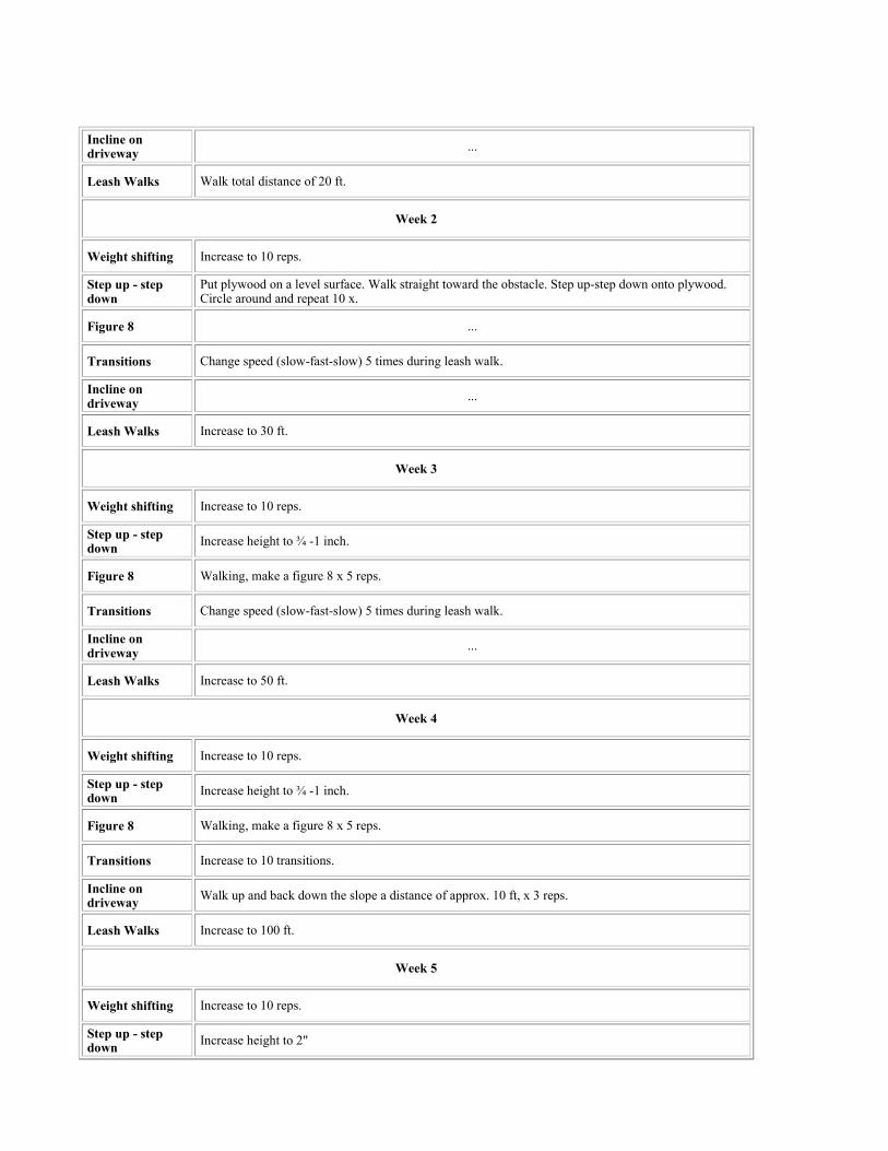
Incline on driveway ...
Leash Walks Walk total distance of 20 ft.
Week 2
Weight shifting Increase to 10 reps.
Step up - step down
Put plywood on a level surface. Walk straight toward the obstacle. Step up-step down onto plywood. Circle around and repeat 10 x.
Figure 8 ...
Transitions Change speed (slow-fast-slow) 5 times during leash walk.
Incline on driveway ...
Leash Walks Increase to 30 ft.
Week 3
Weight shifting Increase to 10 reps.
Step up - step down Increase height to ¾ -1 inch.
Figure 8 Walking, make a figure 8 x 5 reps.
Transitions Change speed (slow-fast-slow) 5 times during leash walk.
Incline on driveway ...
Leash Walks Increase to 50 ft.
Week 4
Weight shifting Increase to 10 reps.
Step up - step down Increase height to ¾ -1 inch.
Figure 8 Walking, make a figure 8 x 5 reps.
Transitions Increase to 10 transitions.
Incline on driveway Walk up and back down the slope a distance of approx. 10 ft, x 3 reps.
Leash Walks Increase to 100 ft.
Week 5
Weight shifting Increase to 10 reps.
Step up - step down Increase height to 2"
Follow-up - Three weeks later, the owner stated that the weight shifting exercise was difficult because Sydney continued to avoid putting weight on the operated limb while standing. However, he did bear weight on the limb when performing step ups and during leash walks. The owner also noted that her dog seemed worn out by the time they were through the exercises, which is consistent with the deconditioning that would have occurred after the length of time that the dog was required to rest. Follow-up at 6 months indicated that the dog was physically active and the lameness had resolved. Comments - This program is one example of a method for post-operative rehabilitation of dogs after anterior cruciate ligament repair. Rehabilitation can be started within the first few days after surgery, in the clinic, and may incorporate modalities such as therapeutic ultrasound, and electrical stimulation, and underwater treadmill work. The rehabilitation program described here is very conservative, but it was tailored to the client’s circumstances. The client wanted to pursue physical therapy after her dog was discharged by the orthopedic surgeon. In the area where the client lived, there were no veterinary rehabilitation facilities available. This owner was very compliant and was happy to be actively involved in her dog’s recovery. According to some rehabilitation specialists, exercises to strengthen the knee extensors have shifted away from open kinetic chain (ie, the distal segment of the limb is not fixated) and toward closed kinetic chain exercises (ie, the distal segment of the limb is fixated) in the rehabilitation of ACL reconstruction in humans. The exercise program for this dog consisted of functional movements which would be classified as closed chain exercises. The goals in ACL rehabilitation include restoration of: the optimal muscle firing pattern, joint stability, and the ability to perform complex movements and skills, while minimizing joint forces. The level of sophistication of the rehabilitation program depends in part on the dog and owner. For performance dogs, additional rehabilitation in the form of a neuromuscular training program could be instituted, for example. In such cases, the rehabilitation could include balance exercises, dynamic joint stability exercises, jumping, plyometrics, and agility exercises followed by specific training in the sport or work to which the dog is returning. The weight shifting exercise is a relatively gentle isometric exercise, but was the most difficult part of the program for this dog. Initially, the dog would not put full weight on the operated limb during this exercise. Within a few weeks, the dog would put weight on the limb, but only if allowed to put the foot in the position he chose, not where the owner placed the foot. During step up’s and leash walks outside, he was more willing to bear weight on the limb. To encourage him, the owner occasionally used treats to coax him to perform the step up’s.
References
1. Downer AH. Physical therapy for Animals. Selected Techniques. Springfield: Charles C. Thomas, 1978. 2. Taylor R, Levine D, Millis D. Canine Physical Therapy and Rehabilitation. In press. 3. Hayes KW. Physical Agents. 4th ed. Norwalk: Appleton & Lange, 1993. 4. Bromiley MW. Physiotherapy in Veterinary Medicine. Oxford: Blackwell Scientific Publications, 1991. 5. Draper DO, Sunderland MS. Examination of the law of Grotthus-Draper: Does ultrasound penetrate subcutaneous fat in
Figure 8 Decrease circle diameter to approx 10 ft. Do 5 reps.
Transitions Increase to 10 transitions.
Incline on driveway Increase distance to 15 ft, x 5 reps.
Leash Walks Increase to 5-10 min, or what seems comfortable to him.
Week 6
Weight shifting Increase to 10 reps.
Step up - step down Increase height to 2"
Figure 8 Decrease circle diameter to approx 10 ft. Do 5 reps.
Transitions Add walk-trot-walk transitions, 5 times, during leash walk.
Incline on driveway Increase distance to 15 ft, x 5 reps.
Leash Walks Increase to 5-10 min, or what seems comfortable to him.

humans? J Athl Train 1993; 28:246-250. 6. Draper DO, Castel JC, Castel D. Rate of temperature increase in human muscle during 1 MHz and 3 MHz continuous ultrasound. J Orthop Sports Phys Ther 1995; 22:142-150. 7. Grant BD. Joint Disease in the Horse. In: McIlwraith CW, Trotter G (ed). Philadelphia: WB Saunders, 1996. 8. Denoix J, Pailloux J. Physical Therapy and Massage for the Horse. North Pomfret: Trafalgar Square Publishing, 1996. 9. Porter M. The New Equine Sports Therapy. Lexington KY: Eclipse Press, 1998. 10. Downer AH. Physical Therapy for Animals. Selected Techniques. Springfield: Charles C. Thomas, 1978; 63-74. 11. Michlovitz SL. Thermal Agents in Rehabilitation. 2nd ed. Philadelphia: FA Davis Co., 1990. 12. Steiss JE, Adams CC. Effect of coat on rate of temperature increase in muscle during ultrasound treatment of dogs. Am J Vet Res 1999; 60(1):76-80. 13. Schulthies SS. Interview with Dr. David O. Draper. Sports Physical Therapy Section Newsletter. Am Phy Ther Assoc 1995;12-13. 14. Draper DO, Sunderland S, Kirkendall DT et al. A comparison of temperature rise in human calf muscles following applications of underwater and topical gel ultrasound. J Orthop Sports Phys Ther 1993; 17:247-251. 15. Reed B, Ashikaga T. The effects of heating with ultrasound on knee joint displacement. J Orthop Sports Phys Ther 1997; 26:131-137. 16. Dyson M, Pond JB, Joseph J et al. The stimulation of tissue regeneration by means of ultrasound. Clin Sci 1968; 35:273-285. 17. Ebenbichler GR, Erdogmus CB, Resch KL et al. Ultrasound therapy for calcific tendinitis of the shoulder. New Eng J Med 1999; 340:1533-1538. 18. Bare AC, McAnaw MB, Pritchard AE, et al. Phonophoretic delivery of 10% hydrocortisone through the epidermis of humans as determined by serum cortisol concentrations. Phys Ther 1996; 76:738-745. 19. Franklin ME, Smith ST, Chenier TC et al. Effect of phonophoresis with dexamethasone adrenal function. J Orthop Sports Phys Ther 1995; 22:103-107. 20. Byl NN. The use of ultrasound as an enhancer for transcutaneous drug delivery: Phonophoresis. Phys Ther 1995; 75:539-553. 21. Asano J, Suisha F, Takada M et al. Effect of pulsed output ultrasound on the transdermal absorption of indomethacin from an ointment in rats. Biol Pharmaceut Bull 1997; 20:288-291. 22. Shin SM, Choi JK. Effect of indomethacin phonophoresis on the relief of temporomandibular joint pain. Cranio 1997; 15:345-348. 23. Mitragorti S, Blankschtein D, Langer R. Ultrasound-mediated transdermal protein delivery. Science 1995; 269:850-853. 24. Singer AJ, Homan CS, Church AL et al. Low-frequency sonophoresis: Pathologic and thermal effects in dogs. Acad Emerg Med 1998; 5:35-40. 25. Prentice WE. Iontophoresis. In: Prentice WE (ed). Therapeutic Modalities in Sports Medicine. 4th ed, Boston: WCB/McGraw-Hill, 1999; 118-130. 26. Glass JM, Stephen RL, Jacobson SC. The quantity and distribution of radiolabeled dexamethasone delivered to tissue by iontophoresis. Int J Derm 1980; 19:519-525. 27. Li LC, Vu NT, Allen LV Jr. Iontophoretic permeation of sodium cromoglycate through synthetic membrane and excised hairless mouse skin. J Pharm & Pharmacol 1992; 44:444-446. 28. Banta CA. A prospective, nonrandomized study of into, wrist splinting, and antiinflammatory medication in the treatment of early-mild carpal tunnel syndrome. J Occupational Med 1994; 36:166-168. 29. Cummings JP. Additional therapeutic uses of electricity. In: Gersh MR (ed). Electrotherapy in Rehabilitation. Philadelphia: FA Davis Co, 1992; 337-339. 30. Glick E, Snyder-Mackler L. Iontophoresis. In: Snyder-Mackler L, Robinson AJ (eds). Clinical Electrophysiology. Baltimore: Williams & Wilkins, 1989; 247-260. 31. Hill JM, O’Callaghan RJ, Hobden JA. Ocular iontophoresis. In: Mitra AK (ed). Ophthalmic Drug Delivery Systems. New York: Marcel Dekker Inc, 1993; 331-354. 32. Anderson BH, Ethell MT. Modes of local drug delivery to the musculoskeletal system. Vet Clin North Am Equine Pract 1999; 15:603-622. 33. Pellecchia GL, Hamel H, Behnke P. Treatment of infrapatellar tendinitis: a combination of modalities and transverse friction massage versus iontophoresis. J Sport Rehab 1994; 3:135-145. 34. Perron M, Malouin F. Acetic acid iontophoresis and ultrasound for the treatment of calcifying tendinitis of the shoulder: a randomized control trial. Arch Physical Med and Rehab 1997; 78:379-384. 35. Gudeman SD, Eisele SA, Heidt RS et al. Treatment of plantar fasciitis by iontophoresis of 0.4% dexamethasone: a randomized, double-blind, placebo-controlled study. Amer J Sports Med 1997; 25:312-316. 36. Lekeux P, Art T, Linden A, et al. Heart rate, hematological and serum biochemical responses to show jumping. In:

Persson SGB, Lindholm A, Jeffcott LB (eds). Equine Exercise Physiology 3. Davis: ICEEP Publ, 1991; 385. 37. Franklin BA, Buchal M, Hollingsworth V, et al. Exercise prescription. In: Strauss RH (ed). Sports Medicine (2nd ed). Philadelphia: WB Saunders, 1991. 38. Bar-Or O. Pediatric Sports Medicine for the Practitioner. New York: Springer-Verlag, 1983; 34-38. 39. Blythe LL, Gannon JR, Craig AM. Care of the Racing Greyhound. Am Greyhound Council, Inc, 1994; 263. 40. Clayton HM. Training show jumpers. In: Hodgson DR, Rose RJ (eds). The Athletic Horse: Principles and Practice of Equine Sports Medicine. Philadelphia: WB Saunders, 1994; 434. 41. McArdle WD, Katch FI, Katch VL. Exercise Physiology (3rd ed). Philadelphia: Lea & Febiger, 1991; 511-513. 42. Wenger HA, McFadyen PF, McFadyen RA. Physiological principles of conditioning. In: Zachazewski JE, Magee DJ, Quillen WS (ed). Athletic Injuries and Rehabilitation. Philadelphia: WB Saunders, 1996; 202-204. 43. Lawrence L. The benefits of warming up. World Equ Vet Rev 1999; 4(2):6-11. 44. Ray S, Irvin R. Sports Medicine. Englewood Cliffs: Prentice-Hall, 1983: 3. 45. Pope RP, Herbert RD, Kirwan JD, et al. A randomized trial of preexercise stretching for prevention of lower-limb injury. Med Sci Sports Exerc 2000; 32:271-277. 46. Malone TR, Garrett WE, Zachazewski JE. Muscle: Deformation, injury and repair. In: Zachazewski JE, Magee DJ, Quillen WS (ed). Athletic Injuries and Rehabilitation. Philadelphia: WB Saunders, 1996; 86-89. 47. Strickler T, Malone T, Garrett W. The effects of passive warming on muscle injury. Amer J Sports Med 1990; 18:141-145. 48. Jensen CR, Fisher AG. Scientific Basis of Athletic Conditioning (2nd ed). Philadelphia: Lea & Febiger, 1979: 267-271. 49. Lund RJ, Guthrie AJ, Mostert HJ, et al. Effect of three different warm up regimens on heat balance and oxygen consumption of thoroughbred horses. J Appl Physiol 1996; 80:2190-2197. 50. Matwichuk CL, Taylor S, Shmon CL, et al. Changes in rectal temperature and hematologic, biochemical, blood gas, and acid-base values in healthy Labrador Retrievers before and after strenuous exercise. Am J Vet Res 1999; 60:88-92. 51. Steiss JE, Spano J. Roading versus running: What is the difference? Am Field 1996; 246(32):17. 52. Barnard RJ, Gardner GW, Diaco NV, et al. Cardiovascular responses to sudden strenuous exercise: heart rate, blood pressure and ECG. J Appl Physiol 1973; 34:833-837. 53. Fisher AG, Jensen CR. Scientific Basis of Athletic Conditioning (3rd ed). Philadelphia: Lea & Febiger, 1990; 177-178. 54. Norkin CC, White DJ. Measurement of Joint Motion. A Guide to Goniometry (2nd ed). Philadelphia: FA Davis, 1995. 55. Steiss JE. Physical therapy in veterinary medicine: Therapeutic ultrasound and phonophoresis. Comp Cont Edu Pract Vet 2000; 22:690-693. 56. Steiss JE. Physical therapy in veterinary medicine: Iontophoresis in horses. Comp Cont Edu Pract Vet 2001; 23:95-99. 57. Steiss JE. Warming up your dog before training sessions or competition. Retrievers Online. 2001; 12(2):4-7. 58. Johnson R, Steiss J and Sorjonen D. The use of thermoplastic materials for splinting in veterinary practice. Comp Cont Edu Pract Vet 2003; 25:20-28. 59. Levine D, Millis DL, Mynatt T. Effects of 3.3 MHz ultrasound on caudal thigh muscle temperature in dogs. Vet Surg 2001; 30:170-174. 60. Forrest G, Rosen K. Ultrasound: Effectiveness of treatments given under water. Arch Phys Med Rehab 1989; 70: 28-29. 61. Klucinec B, Scheidler M, Denegen C, et al. Transmissivity of coupling agents used to deliver ultrasound through indirect methods. J Orthop Sports Phys Ther 2000; 30: 263-269. 62. Kaneps AJ, Craig AM, Walker IC, et al. Iontophoretic administration of dexamethasone into the tarsocrural joint in horses. Am J Vet Res 2002; 63: 11-14. 63. Jaegger G, Marcellin-Little DJ, Levine D. Reliability of goniometry in Labrador Retrievers. Amer J Vet Res 63: 979-986, 2002. 64. Gross DM. Canine Physical Therapy: Orthopedic Physical Therapy. Wizard of Paws: East Lyme, Connecticut, 2002; 315 pp.
All rights reserved. This document is available on-line at www.ivis.org. Document No. B0238.0203.


![High Intensity Focused Ultrasound Facial Skin Lifting · HIFU Mechanism HIFU [High Intensity Focused Ultra-sound] • Non-invasive treatment with thermal energy that focused ultrasound](https://static.fdocuments.us/doc/165x107/5fd8347ce1f6a6277a18bb54/high-intensity-focused-ultrasound-facial-skin-hifu-mechanism-hifu-high-intensity.jpg)

