Candida Infections of the Genitourinary Tract · Definition and Diagnosis of Candidal Balanitis...
21
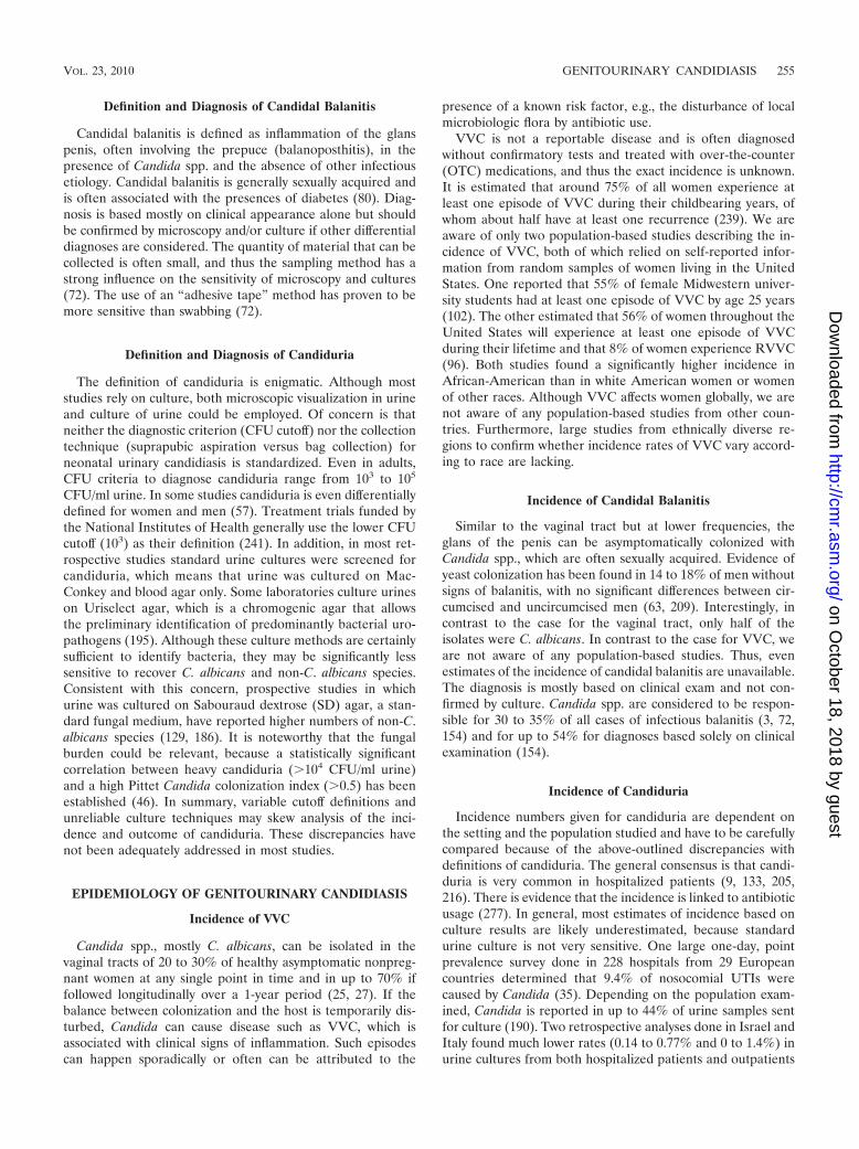
CLINICAL MICROBIOLOGY REVIEWS, Apr. 2010, p. 253–273 Vol. 23, No. 2 0893-8512/10/$12.00 doi:10.1128/CMR.00076-09 Copyright © 2010, American Society for Microbiology. All Rights Reserved. Candida Infections of the Genitourinary Tract Jacqueline M. Achkar 1 and Bettina C. Fries 1,2 * Departments of Medicine 1 and Microbiology and Immunology, 2 Albert Einstein College of Medicine, Bronx, New York INTRODUCTION .......................................................................................................................................................254 DEFINITIONS AND DIAGNOSIS OF GENITOURINARY DISEASES CAUSED BY CANDIDA SPECIES ..............................................................................................................................................................254 Definition and Diagnosis of VVC and RVVC......................................................................................................254 Definition and Diagnosis of Candidal Balanitis ................................................................................................255 Definition and Diagnosis of Candiduria .............................................................................................................255 EPIDEMIOLOGY OF GENITOURINARY CANDIDIASIS .................................................................................255 Incidence of VVC ....................................................................................................................................................255 Incidence of Candidal Balanitis ...........................................................................................................................255 Incidence of Candiduria ........................................................................................................................................255 MICROBIOLOGY ......................................................................................................................................................256 Distribution of Candida spp. in VVC ...................................................................................................................256 Distribution of Candida spp. in Candidal Balanitis ..........................................................................................256 Distribution of Candida spp. in Candiduria .......................................................................................................257 Antifungal Resistance in Candida Isolates from Genitourinary Infections ....................................................257 PATIENT POPULATIONS AT RISK AND PREDISPOSING RISK FACTORS FOR GENITOURINARY CANDIDIASIS.....................................................................................................................................................258 Risk Factors for VVC .............................................................................................................................................258 Risk Factors for Candidal Balanitis ....................................................................................................................258 Risk Factors for Candiduria .................................................................................................................................258 CLINICAL PRESENTATION OF GENITOURINARY CANDIDIASIS .............................................................259 Clinical Presentation of VVC ................................................................................................................................259 Clinical Presentation of Candidal Balanitis .......................................................................................................259 Clinical Presentation of Candiduria ....................................................................................................................259 MORBIDITY, MORTALITY, AND ASSOCIATED COSTS .................................................................................259 PATHOGENESIS OF GENITOURINARY CANDIDIASIS ..................................................................................260 Animal and Other Models for Genitourinary Candidiasis ...............................................................................260 Rodent models for VVC .....................................................................................................................................260 Primate models for VVC ....................................................................................................................................260 Vaginal tissue models for VVC .........................................................................................................................260 Rodent models for candiduria ..........................................................................................................................261 Rabbit models for candiduria ...........................................................................................................................261 Primary human cells, cell lines, and reconstituted epithelium as models for candiduria.......................261 CANDIDA VIRULENCE TRAITS RELEVANT FOR GENITOURINARY DISEASE .......................................261 Bud-Hypha Formation in Candida spp. ...............................................................................................................261 Aspartic Proteinases and Phospholipases in Candida spp. ..............................................................................262 Adhesion Proteins in Candida spp. ......................................................................................................................262 Biofilm Formation in Candida spp. ......................................................................................................................262 Phenotypic Switching in Diverse Candida spp....................................................................................................262 HOST IMMUNE RESPONSES IN GENITOURINARY CANDIDIASIS ...........................................................263 Cell-Mediated Immune Responses to VVC .........................................................................................................263 Humoral Immune Responses to VVC ..................................................................................................................263 Hypersensitivity Reactions to VVC.......................................................................................................................263 Innate Immune Responses to VVC.......................................................................................................................263 Immune Responses to Candiduria .......................................................................................................................264 TREATMENT OF GENITOURINARY CANDIDIASIS ........................................................................................264 Treatment of VVC ...................................................................................................................................................264 Treatment of uncomplicated VVC ....................................................................................................................264 Treatment of complicated VVC and RVVC .....................................................................................................265 Probiotics and other alternative methods for treatment of RVVC ..............................................................265 * Corresponding author. Mailing address: Medicine, Infectious Dis- ease, and Microbiology and Immunology, Albert Einstein College of Medicine, 1300 Morris Park Avenue, Ullmann 1223, Bronx, NY 10461. Phone: (718) 430-2365. Fax: (718) 430-8968. E-mail: Bettina.fries @einstein.yu.edu. 253 on October 18, 2018 by guest http://cmr.asm.org/ Downloaded from
Transcript of Candida Infections of the Genitourinary Tract · Definition and Diagnosis of Candidal Balanitis...
CLINICAL MICROBIOLOGY REVIEWS, Apr. 2010, p. 253–273 Vol. 23, No. 20893-8512/10/$12.00 doi:10.1128/CMR.00076-09Copyright © 2010, American Society for Microbiology. All Rights Reserved.
Candida Infections of the Genitourinary TractJacqueline M. Achkar1 and Bettina C. Fries1,2*
Departments of Medicine1 and Microbiology and Immunology,2 Albert Einstein College ofMedicine, Bronx, New York
INTRODUCTION .......................................................................................................................................................254DEFINITIONS AND DIAGNOSIS OF GENITOURINARY DISEASES CAUSED BY CANDIDA
SPECIES ..............................................................................................................................................................254Definition and Diagnosis of VVC and RVVC......................................................................................................254Definition and Diagnosis of Candidal Balanitis ................................................................................................255Definition and Diagnosis of Candiduria .............................................................................................................255
EPIDEMIOLOGY OF GENITOURINARY CANDIDIASIS .................................................................................255Incidence of VVC ....................................................................................................................................................255Incidence of Candidal Balanitis ...........................................................................................................................255Incidence of Candiduria ........................................................................................................................................255
MICROBIOLOGY ......................................................................................................................................................256Distribution of Candida spp. in VVC ...................................................................................................................256Distribution of Candida spp. in Candidal Balanitis ..........................................................................................256Distribution of Candida spp. in Candiduria .......................................................................................................257Antifungal Resistance in Candida Isolates from Genitourinary Infections ....................................................257
PATIENT POPULATIONS AT RISK AND PREDISPOSING RISK FACTORS FOR GENITOURINARYCANDIDIASIS.....................................................................................................................................................258
Risk Factors for VVC .............................................................................................................................................258Risk Factors for Candidal Balanitis ....................................................................................................................258Risk Factors for Candiduria .................................................................................................................................258
CLINICAL PRESENTATION OF GENITOURINARY CANDIDIASIS .............................................................259Clinical Presentation of VVC ................................................................................................................................259Clinical Presentation of Candidal Balanitis .......................................................................................................259Clinical Presentation of Candiduria ....................................................................................................................259
MORBIDITY, MORTALITY, AND ASSOCIATED COSTS .................................................................................259PATHOGENESIS OF GENITOURINARY CANDIDIASIS..................................................................................260
Animal and Other Models for Genitourinary Candidiasis...............................................................................260Rodent models for VVC .....................................................................................................................................260Primate models for VVC....................................................................................................................................260Vaginal tissue models for VVC .........................................................................................................................260Rodent models for candiduria ..........................................................................................................................261Rabbit models for candiduria ...........................................................................................................................261Primary human cells, cell lines, and reconstituted epithelium as models for candiduria.......................261
CANDIDA VIRULENCE TRAITS RELEVANT FOR GENITOURINARY DISEASE.......................................261Bud-Hypha Formation in Candida spp. ...............................................................................................................261Aspartic Proteinases and Phospholipases in Candida spp. ..............................................................................262Adhesion Proteins in Candida spp. ......................................................................................................................262Biofilm Formation in Candida spp. ......................................................................................................................262Phenotypic Switching in Diverse Candida spp....................................................................................................262
HOST IMMUNE RESPONSES IN GENITOURINARY CANDIDIASIS ...........................................................263Cell-Mediated Immune Responses to VVC .........................................................................................................263Humoral Immune Responses to VVC ..................................................................................................................263Hypersensitivity Reactions to VVC.......................................................................................................................263Innate Immune Responses to VVC.......................................................................................................................263Immune Responses to Candiduria .......................................................................................................................264
TREATMENT OF GENITOURINARY CANDIDIASIS ........................................................................................264Treatment of VVC...................................................................................................................................................264
Treatment of uncomplicated VVC ....................................................................................................................264Treatment of complicated VVC and RVVC.....................................................................................................265Probiotics and other alternative methods for treatment of RVVC..............................................................265
* Corresponding author. Mailing address: Medicine, Infectious Dis-ease, and Microbiology and Immunology, Albert Einstein College ofMedicine, 1300 Morris Park Avenue, Ullmann 1223, Bronx, NY 10461.Phone: (718) 430-2365. Fax: (718) 430-8968. E-mail: [email protected].
253
on October 18, 2018 by guest
http://cmr.asm
.org/D
ownloaded from

Treatment of Candiduria.......................................................................................................................................265Treatment of asymptomatic candiduria...........................................................................................................266Treatment of symptomatic candiduria.............................................................................................................266Treatment of other causes of candiduria ........................................................................................................266
CONCLUSIONS .........................................................................................................................................................266ACKNOWLEDGMENTS ...........................................................................................................................................266REFERENCES ............................................................................................................................................................267
INTRODUCTION
Candida spp. are the most common cause of fungal infec-tions (205), leading to a range of life-threatening invasive tonon-life-threatening mucocutaneous diseases. Among Can-dida spp., Candida albicans is the most common infectiousagent. This dimorphic yeast is a commensal that colonizesskin, the gastrointestinal and the reproductive tracts.Non-C. albicans species are emerging pathogens and canalso colonize human mucocutaneous surfaces (231). Conse-quently, they are also isolated in the setting of candidiasis,albeit at a lower frequency. The pathogenesis and prognosisof candidial infections are affected by the host immunestatus and also differ greatly according to disease presenta-tions. Therefore, diagnosis, management, and treatmentchoices vary and need to be considered in the overall settingof the affected human host.
Mucocutaneous candidiasis can be divided into nongeni-tal disease and genitourinary disease. Among nongenitouri-nary candidiasis, oropharyngeal manifestations are the mostcommon and usually are diagnosed in immunocompromisedpatients, such as human immunodeficiency virus (HIV)-in-fected persons (extensively reviewed in reference 71). Themost frequent manifestations of genitourinary candidiasisinclude vulvovaginal candidiasis (VVC) in women, balanitisand balanoposthitis in men, and candiduria in both sexes.These diseases are remarkably common but occur in differ-ent populations, immunocompetent as well as immunocom-promised. While VVC affects mostly healthy women, candi-duria is commonly diagnosed in immunocompromisedpatients or neonates. In the majority of women, a diagnosisof VVC is made at least once during their childbearing years(239). Among the many causes of vaginitis, VVC is thesecond most common after bacterial vaginosis and is diag-nosed in up to 40% of women with vaginal complaints in theprimary care setting (13). Candida is also the most commoninfectious agent causing inflammation of the glans penis(80). Both these diseases are usually diagnosed and treatedin the outpatient setting. In contrast to genital manifesta-tions of candidiasis, candiduria is usually diagnosed in el-derly hospitalized patients, and Candida is the most fre-quently isolated pathogen in nosocomial urinary tractinfections (UTIs). A second patient group at risk is neo-nates, especially those who receive prolonged antibiotictherapy. We here review the epidemiology, microbiology,risk factors, clinical features, pathogenesis, and treatmentstrategies for the diverse manifestations of genitourinarycandidiasis. This review focuses on the most common man-ifestations of genitourinary candidiasis and concentrates onVVC and candiduria because few studies on candidal bala-nitis are published.
DEFINITIONS AND DIAGNOSIS OF GENITOURINARYDISEASES CAUSED BY CANDIDA SPECIES
Definition and Diagnosis of VVC and RVVC
The presence of Candida in the vagina, in the absence ofimmunosuppression or damaged mucosa, is usually not asso-ciated with any signs of disease and is thus referred to ascolonization. In contrast to asymptomatic colonization, VVC isdefined as signs and symptoms of inflammation in the presenceof Candida spp. and in the absence of other infectious etiology.Over a decade ago, VVC was classified into uncomplicated andcomplicated cases, a classification that has been internationallyaccepted and adapted (189, 239, 287). Uncomplicated VVC ischaracterized by sporadic or infrequent occurrence of mild tomoderate disease caused by C. albicans in immunocompetentwomen. Complicated VVC includes cases of severe VVC,VVC caused by non-C. albicans species, VVC associated withpregnancy or other concurrent conditions such as uncontrolleddiabetes or immunosuppression, and recurrent VVC (RVVC)in immunocompetent women. RVVC is defined as at least fourepisodes of VVC during 1 year (239). Women who suffer fromRVVC are a separate population of otherwise healthy womenand are distinct from women who experience sporadic acuteVVC. Long-term suppressive antifungal therapy is commonlyrequired to control RVVC, and recurrence rates of up to 40%to 50% occur after discontinuation of suppressive therapy(242). Compared to the case for women with other chronicvaginal symptoms, symptoms of women with RVVC are re-ported to have the greatest negative impact on work and sociallife (183).
About 50% of patients have positive microscopy of a wetmount or saline preparation, where yeast cells and hyphalelements can be seen. A 10% potassium hydroxide (KOH)preparation is more sensitive than a saline preparation in iden-tifying yeast cells or hyphae (235). The vaginal pH is oftenmeasured to exclude other infections such as bacterial vagino-sis or trichomoniasis in which it is high (�4.5), while it isnormal (4.0 to 4.5) in VVC. Vaginal culture is the most accu-rate method for the diagnosis of VVC and is indicated ifmicroscopy is negative but VVC is suspected or in cases of highrisk for non-C. albicans VVC. Among the various culturemethods, there appears to be no difference between Sab-ouraud agar, Nickerson’s medium, or Microstix-candida me-dium (235). CHROMagar Candida is a selective fungal me-dium that includes chromogenic substances allowing for quickidentification of several different Candida spp. based on theircolor, which also facilitates the detection of mixed infectionswith more than one species of Candida (185, 196). Antigendetection or serologic tests as well PCR-based diagnosis areeither not yet reliable or not clinically useful because they aretoo sensitive (235).
254 ACHKAR AND FRIES CLIN. MICROBIOL. REV.
on October 18, 2018 by guest
http://cmr.asm
.org/D
ownloaded from
Definition and Diagnosis of Candidal Balanitis
Candidal balanitis is defined as inflammation of the glanspenis, often involving the prepuce (balanoposthitis), in thepresence of Candida spp. and the absence of other infectiousetiology. Candidal balanitis is generally sexually acquired andis often associated with the presences of diabetes (80). Diag-nosis is based mostly on clinical appearance alone but shouldbe confirmed by microscopy and/or culture if other differentialdiagnoses are considered. The quantity of material that can becollected is often small, and thus the sampling method has astrong influence on the sensitivity of microscopy and cultures(72). The use of an “adhesive tape” method has proven to bemore sensitive than swabbing (72).
Definition and Diagnosis of Candiduria
The definition of candiduria is enigmatic. Although moststudies rely on culture, both microscopic visualization in urineand culture of urine could be employed. Of concern is thatneither the diagnostic criterion (CFU cutoff) nor the collectiontechnique (suprapubic aspiration versus bag collection) forneonatal urinary candidiasis is standardized. Even in adults,CFU criteria to diagnose candiduria range from 103 to 105
CFU/ml urine. In some studies candiduria is even differentiallydefined for women and men (57). Treatment trials funded bythe National Institutes of Health generally use the lower CFUcutoff (103) as their definition (241). In addition, in most ret-rospective studies standard urine cultures were screened forcandiduria, which means that urine was cultured on Mac-Conkey and blood agar only. Some laboratories culture urineson Uriselect agar, which is a chromogenic agar that allowsthe preliminary identification of predominantly bacterial uro-pathogens (195). Although these culture methods are certainlysufficient to identify bacteria, they may be significantly lesssensitive to recover C. albicans and non-C. albicans species.Consistent with this concern, prospective studies in whichurine was cultured on Sabouraud dextrose (SD) agar, a stan-dard fungal medium, have reported higher numbers of non-C.albicans species (129, 186). It is noteworthy that the fungalburden could be relevant, because a statistically significantcorrelation between heavy candiduria (�104 CFU/ml urine)and a high Pittet Candida colonization index (�0.5) has beenestablished (46). In summary, variable cutoff definitions andunreliable culture techniques may skew analysis of the inci-dence and outcome of candiduria. These discrepancies havenot been adequately addressed in most studies.
EPIDEMIOLOGY OF GENITOURINARY CANDIDIASIS
Incidence of VVC
Candida spp., mostly C. albicans, can be isolated in thevaginal tracts of 20 to 30% of healthy asymptomatic nonpreg-nant women at any single point in time and in up to 70% iffollowed longitudinally over a 1-year period (25, 27). If thebalance between colonization and the host is temporarily dis-turbed, Candida can cause disease such as VVC, which isassociated with clinical signs of inflammation. Such episodescan happen sporadically or often can be attributed to the
presence of a known risk factor, e.g., the disturbance of localmicrobiologic flora by antibiotic use.
VVC is not a reportable disease and is often diagnosedwithout confirmatory tests and treated with over-the-counter(OTC) medications, and thus the exact incidence is unknown.It is estimated that around 75% of all women experience atleast one episode of VVC during their childbearing years, ofwhom about half have at least one recurrence (239). We areaware of only two population-based studies describing the in-cidence of VVC, both of which relied on self-reported infor-mation from random samples of women living in the UnitedStates. One reported that 55% of female Midwestern univer-sity students had at least one episode of VVC by age 25 years(102). The other estimated that 56% of women throughout theUnited States will experience at least one episode of VVCduring their lifetime and that 8% of women experience RVVC(96). Both studies found a significantly higher incidence inAfrican-American than in white American women or womenof other races. Although VVC affects women globally, we arenot aware of any population-based studies from other coun-tries. Furthermore, large studies from ethnically diverse re-gions to confirm whether incidence rates of VVC vary accord-ing to race are lacking.
Incidence of Candidal Balanitis
Similar to the vaginal tract but at lower frequencies, theglans of the penis can be asymptomatically colonized withCandida spp., which are often sexually acquired. Evidence ofyeast colonization has been found in 14 to 18% of men withoutsigns of balanitis, with no significant differences between cir-cumcised and uncircumcised men (63, 209). Interestingly, incontrast to the case for the vaginal tract, only half of theisolates were C. albicans. In contrast to the case for VVC, weare not aware of any population-based studies. Thus, evenestimates of the incidence of candidal balanitis are unavailable.The diagnosis is mostly based on clinical exam and not con-firmed by culture. Candida spp. are considered to be respon-sible for 30 to 35% of all cases of infectious balanitis (3, 72,154) and for up to 54% for diagnoses based solely on clinicalexamination (154).
Incidence of Candiduria
Incidence numbers given for candiduria are dependent onthe setting and the population studied and have to be carefullycompared because of the above-outlined discrepancies withdefinitions of candiduria. The general consensus is that candi-duria is very common in hospitalized patients (9, 133, 205,216). There is evidence that the incidence is linked to antibioticusage (277). In general, most estimates of incidence based onculture results are likely underestimated, because standardurine culture is not very sensitive. One large one-day, pointprevalence survey done in 228 hospitals from 29 Europeancountries determined that 9.4% of nosocomial UTIs werecaused by Candida (35). Depending on the population exam-ined, Candida is reported in up to 44% of urine samples sentfor culture (190). Two retrospective analyses done in Israel andItaly found much lower rates (0.14 to 0.77% and 0 to 1.4%) inurine cultures from both hospitalized patients and outpatients
VOL. 23, 2010 GENITOURINARY CANDIDIASIS 255
on October 18, 2018 by guest
http://cmr.asm
.org/D
ownloaded from

(57, 68). The incidence of candiduria also varies with hospitalsetting, being most common in intensive care units (ICUs)(216) and among those in burn units (34). Other studies reportthat 11 to 30% of nosocomial urinary tract infections (UTIs)are caused by Candida (87, 161, 205). One large study reportsthat 11% of 1,738 renal transplant patients had at least oneepisode of candiduria within 54 days of transplant (214). In theNational Nosocomial Infection surveillance done between1992 to 1997 in 112 ICUs across the United States, C. albicanswas the most commonly reported pathogen (including bacte-ria) from urine (21%), constituting more than half of the fun-gal isolates. Also, C. albicans was more commonly reported incatheter-associated UTIs than in non-catheter-associated in-fections (21% versus 13%, P � 0.009). Fungal urinary infec-tions occurred more frequently in patients with urinary cathe-ters than in those without urinary catheters (40% versus 22%,P � 0.001) (205). In pediatric ICUs the percentage of candi-duria was lower (14.3%) (206). In ICUs, preterm neonateswith low birth weight are now routinely treated with flucon-azole prophylaxis, which has dramatically decreased the inci-dence in those patients (112, 126, 134, 136).
MICROBIOLOGY
Distribution of Candida spp. in VVC
The distribution of Candida spp. identified in women withVVC varies widely depending on the locations as well as thepopulations studied (Table 1). Typically, a single species isidentified, but two or more species have been found in thesame vaginal culture in a minority of women (2 to 5%) withcomplicated as well as uncomplicated VVC (86, 208). In theUnited States, Europe, and Australia, C. albicans is the mostcommon species identified in women with VVC (76 to 89%),followed by C. glabrata (7 to 16%) (59, 116, 208, 249, 271)(Table 1). The overall percentage of non-C. albicans speciesassociated with VVC in these countries/continents ranges from24% to 11%. Some studies have reported an increasing trendin the occurrence of non-C. albicans species over time (47,249), while a recent U.S. study of over 90,000 samples found aconsistent yearly distribution from 2003 to 2007 (271). In con-trast to the case in the United States, Europe, and Australia,non-C. albicans species, in particular C. glabrata, appear to be
more commonly associated with VVC in some Asian and Af-rican countries (Table 1). In Turkey, India, and Nigeria, casesdue to C. glabrata range between 30 to 37%, while the Candidasp. distribution in China resembles closely the one in theUnited States. With higher resistance levels of most non-C.albicans species to the commonly used azole-based treatmentsand limited possibilities for yeast identification and suscepti-bility testing in some of these settings, the consequences forwomen affected by these strains might be incapacitating.
Higher percentages of non-C. albicans species have beenisolated in women with RVVC than in women with VVC (40 to32% versus 20 to 11%, respectively) (116, 184, 208, 249). Otherpopulations with higher rates of non-C. albicans species in-clude HIV-infected women (94, 249), women after menopause,and especially women with uncontrolled diabetes irrespectiveof HIV infection (70, 105). Interestingly, higher percentages ofnon-C. albicans species are associated with increasing age inwomen with VVC (116, 271). In all of these populations, C.glabrata is the most common among the non-C. albicansspecies. These data highlight the importance of determiningCandida spp. and susceptibilities in women at high risk fornon-C. albicans VVC in order to provide effective therapy.
Genetic relatedness of Candida strains in women withRVVC has been studied, and three scenarios have been enter-tained. They include VVC strain maintenance without geneticvariation, strain maintenance with minor genetic variation, andstrain replacement (157). In 18/18 patients with recurrent in-fections, the same strain was responsible for sequential infec-tions, suggesting that the predominant scenario is strain main-tenance. However, 56% of the strains exhibited minor stablegenetic variations in sequential isolates. A recent study con-firmed strain maintenance with microevolution in the majorityof women with recurrent episodes of VVC but also reportedstrain replacement in 4/24 (14%) (54).
Distribution of Candida spp. in Candidal Balanitis
There is a lack of studies about the distribution of Candidaspp. in candidal balanitis. Considering the fact that this diseaseis often sexually transmitted, the distribution of Candida spe-cies is likely to be similar to that in VVC.
TABLE 1. Candida spp. identified in women with VVC
Region No. ofsubjects
% With:
ReferenceC. albicans C. glabrata C. tropicalis C. parapsilosis C. krusei Other non-
C. albicans
US, all states 93,775 89 8 1 2 271US, Iowa 429 76 16 1 4 4 2 208Australia 1,087 89 7 1 1 1 1 116Italy 410 84 9 1 1 5 249Italy 909 77 15 2 1 4 1 59Austria 3,184 88 3 �1 1 �1 7 193China 1,070 90 8 1 1 1 86India 215 47 37 6 10 1 2 8Turkey 240 44 30 6 19a 45Nigeria 517 20 34 18 5 24b 187
a C. keyfir, 6%.b C. guilliermondii, 18%.
256 ACHKAR AND FRIES CLIN. MICROBIOL. REV.
on October 18, 2018 by guest
http://cmr.asm
.org/D
ownloaded from
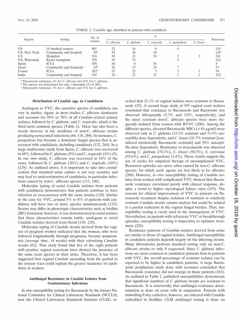
Distribution of Candida spp. in Candiduria
Analogous to VVC, the causative species of candiduria canvary in studies. Again, in most studies C. albicans dominatesand accounts for 50% to 70% of all Candida-related urinaryisolates, followed by C. glabrata, and C. tropicalis, which is thethird most common species (Table 2). There has also been asteady increase in the incidence of non-C. albicans strainsproducing nosocomial infections (68, 118, 288). In neonates, C.parapsilosis has become a dominant fungal species that is as-sociated with candidiasis, including candiduria (152, 264). In alarge multicenter study from Spain, C. albicans was recoveredin 68%, followed by C. glabrata (8%) and C. tropicalis (4%) (9).In our own study, C. albicans was recovered in 54% of thecases, followed by C. glabrata (36%) and C. tropicalis (10%)(129). As outlined above, it is important to take into consid-eration that standard urine culture is not very sensitive andmay lead to underestimation of candiduria, in particular infec-tions caused by non-C. albicans species (113, 186).
Molecular typing of serial Candida isolates from patientswith candiduria demonstrates that patients continue to haveinfection or reoccurrence with the same strains (129). Similarto the case for VVC, around 5% to 8% of patients with can-diduria will have two or more species simultaneously (133).Strains may differ in phenotypic characteristics such as biofilm(BF) formation; however, it was demonstrated in serial isolatesthat these characteristics remain stable, analogous to serialCandida isolates derived from blood (110, 129).
Molecular typing of Candida strains derived from the vagi-nas of pregnant women indicated that the women, who werefollowed longitudinally through pregnancy, became symptom-atic (average time, 14 weeks) with their colonizing Candidastrains (62). That study found that five of the eight patientswith positive vaginal secretions later showed the presence ofthe same yeast species in their urine. Therefore, it has beensuggested that vaginal Candida ascending from the genital tothe urinary tract could explain the greater incidence of candi-duria in women.
Antifungal Resistance in Candida Isolates fromGenitourinary Infections
In vitro susceptibility testing for fluconazole by the former Na-tional Committee for Clinical Laboratory Standards (NCCLS),now the Clinical Laboratory Standards Institute (CLSI), re-
vealed that 21.1% of vaginal isolates were resistant to flucon-azole (25). A second large study of 593 vaginal yeast isolatesconcluded that resistance to fluconazole and flucytosine wasobserved infrequently (3.7% and 3.0%, respectively), andthe more resistant non-C. albicans species were more fre-quently isolated from women with RVVC (208). Among thedifferent species, elevated fluconazole MICs (�16 �g/ml) wereobserved only in C. glabrata (15.2% resistant and 51.8% sus-ceptible-dose dependent), and C. krusei (41.7% resistant [con-sidered intrinsically fluconazole resistant] and 50% suscepti-ble-dose dependent). Resistance to itraconazole was observedamong C. glabrata (74.1%), C. krusei (58.3%), S. cerevisiae(55.6%), and C. parapsilosis (3.4%). These results support theuse of azoles for empirical therapy of uncomplicated VVC.Recurrent episodes are more often caused by non-C. albicansspecies, for which azole agents are less likely to be effective(208). However, in vitro susceptibility testing of Candida iso-lates from women with complicated VVC showed that flucon-azole resistance correlated poorly with clinical response, de-spite a trend to higher mycological failure rates (243). Thereasons for clinical improvement of VVC in patients on flu-conazole treatment despite isolation of resistant or relativelyresistant Candida strains remain unclear but could be relatedto a partial reduction in the vaginal fungal burden. Thus, sus-ceptibility testing is rarely used in the management of VVC.Nevertheless, in patients with refractory VVC or breakthroughepisodes, susceptibility testing is imperative to optimize treat-ment (226).
Resistance patterns of Candida isolates derived from urineare similar to those of vaginal isolates. Antifungal susceptibilityin candiduric patients depends largely on the infecting strains.Many laboratories perform standard testing only on non-C.albicans strains or only if requested. Since C. glabrata infec-tions are more common in candiduric patients than in patientswith VVC, the overall percentage of resistant isolates can beexpected to be higher in candiduric patients. A large flucon-azole prophylaxis study done with neonates concluded thatfluconazole resistance did not emerge in those patients (163).As outlined in Table 2, published susceptibilities demonstratethat significant numbers of C. glabrata strains are resistant tofluconazole. It is noteworthy that antifungal resistance deter-mination is done on yeast cells in suspension. Patients withindwelling Foley catheters, however, are infected with Candidaembedded in biofilms. CLSI antifungal testing is done on
TABLE 2. Candida spp. identified in patients with candiduria
Region Setting No. ofisolates
%Reference
C. albicans C. glabrata C. tropicalis C. parapsilosis C. krusei
US 10 medical centers 861 52 16 8 4 133US, New York Community and hospital 55a 54 36 10 129US Hospitals 316 50 21 10 2 2 241US, Wisconsin Renal transplant 276 35 53 4 214Spain ICUs 389 68 8 36 9Israel Community and hospitals 52b 35 15 19 7 57France ICUs 262c 67 22 3 7 2 34India Community and hospital 145 24 21 31 1 1 192
a Fluconazole resistance, 4% for C. albicans and 25% for C. glabrata.b The species was determined for only a subsample (52 of 283).c Fluconazole resistance, 1% for C. albicans and 15% for C. glabrata.
VOL. 23, 2010 GENITOURINARY CANDIDIASIS 257
on October 18, 2018 by guest
http://cmr.asm
.org/D
ownloaded from

planktonic logarithmically growing yeast cell populations. Inmodified antifungal susceptibility testing with Candida cellsthat are biofilm associated and attached to a plastic surface,one can demonstrate that these biofilms are usually resistant toazoles, and a high percentage are even resistant to amphoter-icin B (AmB), whereas the planktonic phenotype of the samestrain remains sensitive to azoles. This may explain persistentor relapsing candiduria in patients without the presence oremergence of antifungal resistance (241).
In summary, due to the increased antifungal resistance ofnon-C. albicans species, their emergence remains a concern.The reason for the considerable higher frequency of non-C.albicans species in some countries is unclear. Genetic, im-mune-based, behavioral, and nutritional factors, among others,have to be taken into consideration.
PATIENT POPULATIONS AT RISK AND PREDISPOSINGRISK FACTORS FOR GENITOURINARY CANDIDIASIS
Genitourinary candidiasis is more commonly diagnosed inwomen than in men. With respect to patient populations af-fected as well as predisposing risk factors, candiduria and VVCdiffer (Table 3).
Risk Factors for VVC
Although many healthy women develop VVC sporadically,several behavioral and host-related risk factors have been as-sociated with VVC and recurrent episodes. These episodes arecaused by Candida overgrowth from the gastrointestinal and/orthe vaginal tract or through sexual transmission (201). Behav-ioral risk factors that have been significantly associated with ahigher incidence of VVC include frequent sexual intercourseand receptive oral sex, as well as the use of high-estrogen (notlow-dose) oral contraceptives, condoms, and spermicides (45,79, 96, 101). Among university students, tight clothing and typeof underwear was not associated with VVC (96), while amongwomen with RVVC the use of panty liners or pantyhose waspositively associated with symptomatic recurrence (191).
Host-related risk factors that have been significantly associ-ated with VVC and RVVC include antibiotic use, uncontrolleddiabetes, conditions with high reproductive hormone levels,and genetic predispositions (105, 235). Antibiotics alter thebacterial microflora of the vaginal and gastrointestinal tractsand thus allow for overgrowth of Candida spp. After antibioticuse, the increase in vaginal colonization with Candida spp.,mostly C. albicans, is estimated to range from 10 to 30%, andVVC occurs in 28 to 33% of cases (235). It is commonlyhypothesized that the reduction of lactobacilli in the vaginaltract predisposes women to VVC. Lactobacilli play a key rolein the vaginal flora through the production of hydrogen per-oxide, bacteriocins, and lactic acid, which protect against inva-sion or overgrowth of pathogenic species (82, 212). However,studies have failed to provide evidence that an altered or ab-normal vaginal bacterial flora predisposes women to recurrentepisodes of VVC in the absence of antibiotic intake (237, 272,295). In fact, a recent prospective study demonstrated thatvaginal Lactobacillus colonization was associated with a nearly4-fold increase in the likelihood of symptomatic VVC (168).Episodes of VVC occur mostly during childbearing years andare rare in premenarchal and postmenopausal women. Anincreased frequency of VVC has been reported during thepremenstrual week (79) and during pregnancy (60). Somestudies suggest that there might be a genetic predisposition inwomen who experience sporadic or frequent episodes of VVC.An increased incidence of VVC was found in African-Ameri-can compared to white American women in two different pop-ulation-based studies (97, 102). Furthermore, increased inci-dence of blood group ABO-Lewis nonsecretor phenotype wasfound in women with RVVC compared to controls (48), andmore recently, polymorphism in the mannose-binding lectingene was found to be associated with RVVC (18, 104). HIV�
women have higher rates of vaginal colonization with Candida,often non-C. albicans species, than HIV� women (76, 94, 223,249). However, in a multicenter cohort study no difference infrequency of VVC between HIV� women not receiving anti-retroviral therapy and HIV� women was found (223). Similarto other genital diseases that cause damage of the mucosa,VVC has been associated with increased vaginal HIV shedding(274) and in a recent meta-analysis was found to be associatedwith a 2-fold increase in the risk of HIV acquisition (213).
Risk Factors for Candidal Balanitis
Predisposing risk factors include diabetes mellitus, immuno-suppression, and being uncircumcised (80, 154). Furthermore,being uncircumcised is considered to be a major predisposingfactor for candidal balanoposthitis when associated with poorhygiene and buildup of smegma.
Risk Factors for Candiduria
Most risk factors associated with candiduria differ consider-ably from the ones associated with VVC, although a few, suchas prior antibiotic use and uncontrolled diabetes, are similar(Table 3). The majority of clinical studies have identified sim-ilar risk factors associated with candiduria in adults. They in-clude anatomic urinary tract abnormalities, comorbidities, in-dwelling urinary drainage devices, abdominal surgery, ICU
TABLE 3. Comparison of risk factors associated withVVC and candiduria
Risk factorAssociation with:
VVC Candiduria
Age Childbearing years Elderly and neonates
Female gender Yes Yes
BehavioralSexual practices Yes No
HostAntibiotic usage Yes YesDiabetes Yes YesHIV Only if advanced Not knownNeutropenia No YesRenal transplant No YesRenal obstruction No YesIndwelling Foley
catheterNo Yes
Abdominal surgery No Yes
258 ACHKAR AND FRIES CLIN. MICROBIOL. REV.
on October 18, 2018 by guest
http://cmr.asm
.org/D
ownloaded from
admission, broad-spectrum antibiotics, diabetes mellitus, in-creased age, and female sex (9, 87, 108, 133, 161). Even in thesubgroup of renal transplant patients, who compromise only asmall fraction (0.9 to 3%) of patients with candiduria, theserisk factors prevail (214). Only one large study has comparedrisk factors in community-acquired versus nosocomial candi-duria. They report that patients who present from the commu-nity with candiduria are younger, more commonly female,pregnant, and, interestingly, more likely to have dysuria. Asecond patient population at risk are prematurely born neo-nates, especially those who received antibiotic treatment andhave indwelling lines (111, 135, 159, 164, 165). In pediatricpatients the mean rate of urinary catheter utilization in pedi-atric ICUs is lower than that in medical ICUs, which may inpart explain the lower number of candiduria cases in thesepatients (206).
Most studies do not differentiate among the different speciesthat can cause candiduria, in part because the species of thenon-C. albicans species are often not determined. In two case-control studies designed to compare risk factors for catheterrelated nosocomial candiduria caused by C. albicans versus C.glabrata at a tertiary hospital in Boston, it was found that riskfactors were similar except that fluconazole and quinolone usewas associated more with C. glabrata-mediated candiduria(109). In other studies, one in renal transplant patients and onein ICU patients, this association of quinolone and fluconazoleusage has not been documented as a risk factor for C. glabrata-mediated disease (9, 214).
CLINICAL PRESENTATION OF GENITOURINARYCANDIDIASIS
Despite the vicinity of the two niches (bladder and vagina)the clinical presentations of the diseases differ greatly.
Clinical Presentation of VVC
The clinical symptoms of VVC are nonspecific and can beassociated with a variety of other vaginal diseases and infec-tions, such as bacterial vaginosis, trichomoniasis, Chlamydiainfection, and gonorrhea. Vulvar pruritus and burning are thehallmark symptoms in most women with VVC, frequently ac-companied by soreness and irritation leading to dyspareuniaand dysuria (13). On physical exam, vulvar and vaginal ery-thema, edema, fissures, and a thick curdy vaginal discharge arecommonly found (79).
Clinical Presentation of Candidal Balanitis
As with VVC, the clinical signs and symptoms of candidalbalanitis are often nonspecific and could be due to a variety ofother causes, such as infections with bacteria or noninfectiouscauses (80). Patients commonly complain of local burning andpruritus, and the clinical features include mild glazed erythemaand papules with or without satellite-eroded pustules (154).
Clinical Presentation of Candiduria
Fungal urinary tract infections are mostly asymptomatic(205). This means the majority of patients have neither fever,
dysuria, nor any other urinary tract-related complaint. Leuko-cyturia, which is also not part of the definition criteria ofasymptomatic bacteriuria, is mostly not present in candiduricpatients. C. albicans UTIs are commonly catheter associated(205, 206). Candiduria occurs late in the hospital stay. In alarge prospective study done in French ICUs, the mean inter-val between ICU admission and candiduria was 17.2 � 1.1days, and similar numbers were reported in studies from Spain(9, 34). In renal transplant patients, the first episode occurreda median of 54 days after transplantation (range, 0 to 2,922days) (214). In these patients candiduria is also mostly asymp-tomatic. Candiduria can be the result of uncomplicated cystitisand/or pyelonephritis, analogous to bacteriuria. In contrast tobacteriuria, the majority of candiduric patients do not presentwith concomitant septicemia. Studies report that a low per-centage (1 to 8%) of candiduric patients develop candidemiaand that ICU patients with candiduria carry the highest risk tobecome candidemic (34, 35, 133, 214).
The differentiation between upper and lower urinary tractinfections has been inherently difficult to make. A small imag-ing study using white blood cells labeled with indium-111 con-cluded that 50% of the studied patients (n � 8) with candiduriashowed renal uptake in 111In-labeled leukocyte scintigraphy,with uptake persisting after antifungal treatment (107). Thisstudy excluded patients with concomitant bacteriuria, patientson antifungal treatment, and patients in the ICU setting. Thisfinding should be confirmed in a larger study, as it raises theconcern that subclinical pyelonephritis may be more frequentin patients with candiduria than thought. The diagnostic valueof single amphotericin washout in candiduria to predict kidneyinfection or invasive candidiasis is low, as the positive predic-tive value is only 44% (95). Data from experiments in rabbitssuggests that detection of renal Candida casts may be a usefuldiagnostic marker in distinguishing upper versus lower urinarytract candidiasis (178), but the frequency of this finding isunknown (16, 106). One study suggests that the D-arabinitol/creatinine ratio could be used to differentiate between Candidapyelonephritis and colonization (261). However, in that studythe clinical distinction between pyelonephritis and colonizationwas poorly documented.
Prostatitis and epididymitis can also lead to candiduria(282). They are more common in older or immunocompro-mised men and should be evident by careful clinical examina-tion. In some cases these patients develop an abscess in thetissue.
Candiduria rarely is associated with pneumaturia, which isthe result of emphysematous tissue invasion or perinephricabscess formation (254). These complicated urinary tract in-fections are observed predominantly in diabetic patients (74,114, 211, 252) and can also occur in the setting of prostatitisand epididymitis. In summary, most patients with candiduriahave few or no symptoms, which complicates treatment deci-sions, as outlined below.
MORBIDITY, MORTALITY, AND ASSOCIATED COSTS
Although not associated with any mortality, VVC andRVVC are associated with considerable morbidity. Symptomsof vaginitis can cause substantial distress, resulting in time lostfrom work and altered self-esteem (77). Thus, it is not surpris-
VOL. 23, 2010 GENITOURINARY CANDIDIASIS 259
on October 18, 2018 by guest
http://cmr.asm
.org/D
ownloaded from
ing that vaginal complaints are the most common reason forgynecological consultation. Among the many causes of vagini-tis, VVC is the second most common after bacterial vaginosis,and it is diagnosed in up to 40% of women with vaginal com-plaints in the primary care setting (13). In the United States,prior to the availability of OTC treatment, approximately 13million cases of VVC annually accounted for 10 million visitsto the gynecologist (138). In 1990, the first topical treatmentfor VCC was approved by the Food and Drug Administrationfor OTC use, and since then the combined antifungal prescrip-tion and OTC sales have almost doubled (153). In 1995 alone,the annual cost of VVC was estimated to be $1.8 billion, withapproximately half of this amount consisting of charges fordoctor visits (97). Furthermore, industry sources report that in1995 OTC sales of vaginal antifungals were the largest com-ponent of the feminine health care sales in drugstores, gener-ating nearly 60% of the category’s sales and resulting in ap-proximately $290 million (75a).
Despite the lack of symptoms, most studies that comparemortality of candiduric patients with that of an appropriatecontrol patient cohort demonstrate increased mortality in can-diduric patients relative to the control population (9, 133, 241).Given that these patients are usually very sick, it remains dif-ficult to determine to what extend candiduria contributes tothe death of these patients. Despite controlled retrospectivestudies, confounding factors cannot be ruled out. An increasedcolonization burden may predispose them to invasive candidi-asis. Eight percent of candiduric patients developed candi-demia with the same species in one prospective multicenterstudy (which included 24 ICUs) from France (34). In a largeSpanish study, in-hospital mortality was 48.8% in patients withcandiduria compared to 36.6% in those without candiduria(P � 0.001) (9). Significant differences were also found forICU mortality (38% versus 28.1%, P � 0.001) in that study. Astudy done in French ICUs also concluded that the crudemortality was 31.3% for candiduric patients (34). In a similarmanner, increased mortality is found in candiduric renaltransplant patients (2, 214). In contrast, Sobel et al. did notobserve complications of fungal urinary tract infection inover 330 hospitalized patients (not only ICU patients), in-cluding pyelonephritis, candidemia, systemic candidiasis, andfungus-related death (241). They concluded that asymptomaticor minimally symptomatic candiduria in itself was usually be-nign. In a large review of candidemic subjects, Ang et al.concluded that candidemia was rarely the consequence of can-diduria and usually occurred only in the presence of upperurinary tract obstruction (14).
Candiduria is one of the most common nosocomial infec-tions. The cost that it inflicts upon the health care system is notknown. Conservative estimates calculate a rise of the meanhospital cost by $1,955 for nosocomial UTIs (52). In caseswhere candiduria leads to candidemia, the costs would risedramatically, since candidemia results in an increase of per-patient hospital charges of up to $39,331 for adults and $92,266for children (204, 289). Given new developments where hos-pitals are not to be reimbursed for nosocomial infections, it willbecome essential to define asymptomatic and symptomaticcandiduria better so that unnecessary treatment, catheterchanges, and hospital prolongation can be avoided, especiallysince these costs will have to be absorbed by hospitals.
PATHOGENESIS OF GENITOURINARY CANDIDIASIS
Animal and Other Models for Genitourinary Candidiasis
Rodent models for VVC. Murine and rat models have beenvaluable in advancing our understanding of Candida pathoge-nicity, antifungal pharmacokinetics, and the immune responsesto Candida, including Candida vaginitis (reviewed in refer-ences 43, 88, and 177). Infection with C. albicans in theserodents is a de novo event in a naïve host which differs signif-icantly from humans, who develop immune responses to Can-dida early in life, as C. albicans is a commensal organism of thegastrointestinal tract and often of the genital tract (43, 88, 198).
Experimental Candida vaginitis in rodents typically requiresa large intravaginal inoculum of 105 to 106 organisms, while thenumber of Candida cells that cause disease in humans is notknown (43, 88, 198). In humans, VVC often results in theinability to control fungal growth of the resident Candidastrains from the vaginal and/or intestinal reservoir and thustriggers the disruption of the fine balance between commen-salisms without tissue damage and disease. Furthermore, ex-perimental infection in rodents is either self-healing or persis-tent with a low fungus burden, and in both cases it effectivelyimmunizes the animals against any secondary challenge (42,92, 93). Thus, there are no rodent models that allow for thestudy of RVVC, the clinically most relevant and difficult-to-manage condition. In addition, experimental Candida vaginitisin rodents requires estrogen induction to mimic human condi-tions. This estrogen administration results in a vaginal epithe-lium that is thicker and fully keratinized and thus allows forfungal attachment, growth, and biofilm formation (49, 140).The keratin layer enhances hypha formation, adherence, andSap production, and the estrogens down-modulate cell-medi-ated immunity and reduce leukocyte infiltration, which helpsthe development of disease (124, 140). The facts that rodents’susceptibility to Candida infection strongly correlates with es-trogen sensitivity (56) and that VVC in humans is commonlyassociated with phases in life that are influenced by estrogens,such as childbearing years and pregnancy (77, 235), supportsome validity of using rodent models.
Primate models for VVC. In contrast to rodents, healthynonhuman primates have shown to be colonized with Candidaspp. and to develop mucocutaneous candidiasis and thus mightbe better models than rodents (253). Intravaginal inoculationat dosages of 5 � 106 C. albicans blastoconidia with and with-out intravenous concurrent estrogen administration resulted insuccessful vaginal infection of one type of macaque species(rhesus) but not another, a finding which was unexpected, withthe different susceptibility remaining unexplained. However,despite successful C. albicans infection with demonstrable vag-inal cytokine responses, the rhesus macaques did not show anysigns or symptoms of vaginitis, the hallmark of human disease.Therefore, despite fewer limitations than with rodent models,primate models still have important limitations regarding theunderstanding of the pathogenesis and immunity associatedwith human VVC.
Vaginal tissue models for VVC. Efforts have also been madeto reproduce the vaginal infection in in vitro studies by usingreconstituted human vaginal epithelial tissue (217–219) or vag-inal tissue sections (66). Valuable information has been ob-
260 ACHKAR AND FRIES CLIN. MICROBIOL. REV.
on October 18, 2018 by guest
http://cmr.asm
.org/D
ownloaded from

tained, but the limitations of in vitro results under well-con-trolled conditions, in the absence of estrogenic conditioningand immune control, have to be kept in mind when makingcomparisons to in vivo conditions.
Rodent models for candiduria. Candiduria has been infre-quently studied in animal models. Bacterial UTI has beenstudied in rodents, dogs, and pigs, but candiduria has beenstudied predominantly in rats, mice, and rabbits. In these an-imal experiments, renal candidiasis is achieved mostly by in-travenous infection of the yeast (181). This route of infectionmay not mimic the pathogenesis adequately, because naturalinfection, especially in the setting of an indwelling catheter, ismost often the result of an ascending infection.
In mice, large doses of C. albicans injected intravenouslycause generalized candidiasis and multiple organ involvement,whereas smaller doses produce transient candidemia and onlyisolated renal involvement (50, 123). These studies suggest alsothat renal involvement starts as an acute pyelonephritis leadingto multiple cortical abscesses and scarring within 9 to 11 daysafter inoculation. At that stage candidal mycelia can be iden-tified within the tubular lumen of the renal medulla, and localproliferation leads to papillary necrosis, bezoar formation, andobstructive uropathy (50, 123). Alternatively, it has been sug-gested that isolated renal involvement may represent an as-cending infection. Murine models have also been used toexamine the effects of treatment with antifungals and temper-ature on the course of infection (139, 263). In rats, the effect ofureteral obstruction on the course of renal candidiasis has beendemonstrated, using both normal and diabetic Sprague-Daw-ley rats. Mean CFU were similar in obstructed and control ratsbut higher in diabetic rats regardless of the presence of ure-teral obstruction. An effect of amphotericin B treatment couldalso be demonstrated, and in general rat models have beenhelpful to determine adequate drug levels (65, 259).
Rabbit models for candiduria. Rabbit models are an alter-native to rodent models, as they allow more easily the analysisof candiduria and catheter placement (178, 179). In studies oncandiduria, rabbits were usually injected intravenously withCandida, in contrast to ascending UTI models with bacteriawhich were described almost 100 years ago. Thus, rabbits haveoften been used to draw conclusions on candiduria as a resultof hematogenous spread, which represents the minority ofcases. With this model, e.g., it was established that urinaryconcentrations of C. albicans were not predictive of the fungalburden in the kidney and that negative urine cultures in rabbitsdid not rule out renal candidiasis. Also, rabbits have been usedto examine the relevance of casts and quantitative urine cul-tures (178, 179). Finally, rabbit models can be more convenientfor pharmacodynamic studies.
Primary human cells, cell lines, and reconstituted epithe-lium as models for candiduria. Human renal epithelial anduroendothelial cells can be isolated from normal human tissue,cryopreserved immediately, and delivered frozen. Human re-nal epithelial cells are capable of internalizing bacteria (188).They can produce cytokines and chemokines and actively par-ticipate in acute inflammatory processes by affecting, e.g., leu-kocyte chemotaxis via the production of interleukin-8 (IL-8)(222). Human urothelial cells also release inflammatory medi-ators. In addition, the urothelial cell line TEU-1 was estab-lished from a healthy human ureter, and followed by immor-
talization, it can be used to address specific questions (141). Inthe last few years more sophisticated in vitro systems have beendeveloped and neobladders have been reconstructed in vitro byculturing urothelial cells on collagen matrix to reproduce nor-mal bladder mucosa. This culture technique leads to the for-mation of wide pluristratified epithelial sheets, resembling thenormal transitional epithelium (64). In summary, these in vitrocells provide a useful in vitro model to study the relationshipbetween renal epithelial cells, uroendothelial cells, and recon-stituted transitional epithelium with pathogens.
In summary, results from rodent models allow for under-standing of some aspects of pathogenesis of human disease.However, due to the lack of prior colonization, these animalsusually have to be immunosuppressed to achieve persistentinfection (273). As Candida is not a commensal, the host re-sponse will be different and involve more immune sensitiza-tion. Therefore, rodent models must be viewed in light of theseimportant limitations. Rabbits are valuable, especially forpharmacokinetic studies, because longer courses of antifungaltherapy can be administered. However, due to high costs, onlylimited numbers of animals can be studied. In vitro models canbe used for effective high-throughput screening for novel an-tifungal agents and to study the inflammatory response.
CANDIDA VIRULENCE TRAITS RELEVANT FORGENITOURINARY DISEASE
Using the above-discussed infection models in addition tomolecular tools and microarrays, distinct stages of infections ofboth superficial and invasive Candida disease have been inves-tigated (220, 221). There is a paucity of studies done on can-diduria. However, virulence attributes have been investigatedin other mucosal candidiasis models, including VVC. Impor-tantly, recent studies suggest that the presence of vaginal Can-dida strains with enhanced virulence and tropism for the vaginacorrelates with the severity of VVC in humans (149). Fromthese studies we have learned of Candida’s exceptional adapt-ability by rapid alterations in gene expression in response tovarious environmental stimuli. Many attributes contribute toC. albicans virulence, among them adhesion, hyphal formation,phenotypic switching (PS), extracellular hydrolytic enzymeproduction, and biofilm formation (Table 4).
Bud-Hypha Formation in Candida spp.
C. albicans is both a commensal and a pathogen that canexhibit yeast, hyphal, or pseudohyphal morphology. Thesemorphological transitions promote colonization and invasionat different anatomical sites. They also occur in other Candida
TABLE 4. Virulence traits of Candida spp.
Virulence traitPresence in:
C. albicans C. glabrata
Bud-hypha transition Present PresentProteases Sap, Plb YpsAdhesion proteins Als EpaBiofilm formation Present PresentPhenotypic switching White to opaque Core switching
VOL. 23, 2010 GENITOURINARY CANDIDIASIS 261
on October 18, 2018 by guest
http://cmr.asm
.org/D
ownloaded from

spp. The yeast form is associated with dissemination and thehyphal form with adhesion, tissue invasion, and proteolyticactivity (281). Accordingly, genes involved in these functions(ALS3, SAP4 to -6, HWP1, HYR1, and ECE1) are differentiallyexpressed (20, 28, 120, 215, 251). The mitogen-activated pro-tein (MAP) kinase, cyclic AMP (cAMP), and pH-sensingRim101 signal transduction pathways regulate cellular mor-phology and expression of hypha-associated genes (281). Somealternative therapy regimens have been investigated becausethey claim to inhibit the bud-hypha transition (4). The bud-hypha transition may also contribute to virulence of otherCandida species such as C. glabrata (144). Even though C.glabrata strains do not exhibit germ tube formation in classicalmycological assays (210), the majority of clinical strains un-dergo coreswitching and in that process exhibit pseudohyphaformation and tube formation, demonstrating that these de-velopmental programs are general characteristics of moststrains of C. glabrata (144).
Aspartic Proteinases and Phospholipases in Candida spp.
The extracellular hydrolytic enzymes, including the secretedaspartyl proteinase (SAP) and phospholipase (PLB) geneproducts, have been shown to directly contribute to C. albicansvirulence (31, 32, 40, 61, 173). C. albicans possesses at least 10members of a SAP gene family, all of which have been se-quenced and extensively characterized. Saps contribute to thevirulence of C. albicans in animal models of infection. Fourtypes of phospholipases have been reported in C. albicans, i.e.,phospholipases A (15), B (22, 117) C (200), and D (122), butonly the PLB1 and PLB2 gene products have been detectedextracellularly. Although PLB1 is thought to account for mostof the secreted phospholipase B activity in C. albicans, PLB2contributes in a minor way, because a PLB1-deficient strainstill produces residual amounts of phospholipase B activity(103). The in vivo expression of the C. albicans SAP1 to SAP8and PLB1 and -2 genes was analyzed in 137 human subjectswith oral or vaginal candidiasis or Candida carriage by reversetranscription-PCR (RT-PCR) using specific primer sets. Aspectrum of SAP gene expression profiles was obtained fromdifferent C. albicans strains during symptomatic disease andasymptomatic carriage. During both disease and carriage SAP2and SAP5 were the most commonly expressed genes. Also, itwas found that SAP1, SAP3, SAP4, SAP7, SAP8, and PLB1expression was correlated with oral disease. Furthermore,SAP1, SAP3, and SAP8 were preferentially expressed in vaginalrather than oral disease. Other human studies also reporteddifferential expression of Saps in patients with VVC andRVVC or asymptomatic carriers (150, 151, 176). Antibodies(Abs) that bind to Sap2 have shown to be protective in certainmodels for VVC (66). In C. glabrata the glycosylphosphati-dylinositol-linked aspartyl proteases (Yps) is linked to viru-lence (137).
Adhesion Proteins in Candida spp.
Many genes are implicated in participation in adhesion toepithelial cells (39, 99, 175, 268). The C. albicans agglutinin-like sequence (ALS) family includes eight genes that encodelarge cell surface glycoproteins. Although adhesive function
has been demonstrated for several Als proteins, the dissectionof their role in C. albicans pathogenesis is very complex be-cause of extensive allelic variation, strain differences (162),different models, and complex interplay (119). The role ofAls1p has been evaluated most thoroughly. Mice injected witha C. albicans Als1/Als1 strain lived longer, and the diseasedeveloped slower in the initial 28 h after intravenous (i.v.)injection, than mice injected with the wild-type control strain(98, 160). Decreased virulence was also demonstrated in theearly stages in a model of oral candidiasis (130). Also, loss ofAls3p was noted to affect adhesion more than loss of Als1p(291). Other Als proteins have in part given conflicting results(291–294). Importantly, an anti-C. albicans vaccine composedof the recombinant N terminus of Als1p (rAls1p-N) reducesCFU and improves survival in both immunocompetent andimmunocompromised mice (248). Newer trials report that vac-cination of mice with rAls3p-N induces a broader Ab responsethan rAls1p-N and a similar cell-mediated immune response(247). This vaccine was especially more effective than rAls1p-Nagainst oropharyngeal or vaginal candidiasis.
In C. glabrata, EPA1 encodes a glycosylphosphatidylinositol(GPI)-anchored cell wall protein (Epa1), which is importantfor adhesion to uroepithelial cells in a murine model (73). In C.glabrata strains, 21 paralogues of EPA1 are characterized, allencoding proteins highly related to Epa1. Most of these EPAgenes are located in subtelomeric position where they aretranscriptionally silenced (44). Transcription of some subtelo-meric EPA genes can be derepressed by limitation of NAD�
precursors (73). Genetically engineered strains that have eitherSIR3, SIR4, or RIF1 deletions overexpress many EPA genes,resulting in a hyperadherent phenotype of the C. glabratastrain (44, 69, 125).
Biofilm Formation in Candida spp.
Surface-associated Candida can grow embedded in extracel-lular matrix that is composed of carbohydrates and proteinsand is referred to as a biofilm. Biofilms (BFs) form readily onplastic surfaces such as Foley catheters and intrauterine de-vices (IUDs), and they render the embedded Candida isolatesresistant to antifungal reagents, especially azoles. BF can alsoform on the mucosal surfaces and promote persistence of fun-gal infection (228). Formation of BF among individual Can-dida strains can differ greatly, and urinary isolates can be dif-ferentiated into low and high BF formers. Furthermore, BFappears to be a strain-specific characteristic that does notchange during chronic infection (129). Other Candida spp.(110) can also form BF with extracellular matrix, althoughBF-associated gene regulation is studied predominantly in C.albicans, which is reviewed extensively elsewhere (29). Twotranscription factors, Tec1 and Bcr1 (182, 225), regulate hy-pha-specific genes and genes downstream of hyphal differ-entiation. Candida genes that control adherence, attach-ment hyphal formation, and quorum-sensing molecules alsoregulate BF.
Phenotypic Switching in Diverse Candida spp.
Phenotypic switching (PS) alters virulence and was first de-scribed in Candida strains 20 years ago (229, 230). In C. albi-
262 ACHKAR AND FRIES CLIN. MICROBIOL. REV.
on October 18, 2018 by guest
http://cmr.asm
.org/D
ownloaded from

cans isolates derived from VVC, phenotypic switching could bedemonstrated and also occurred during treatment relapses(245). The WO-1 strain is a model strain for reversible white-to-opaque switching (WOS). White-phase cells are round, andopaque-phase cells are elongated (12). Opaque-phase cells aremating competent, whereas white-phase cells survive betterwithin the mammalian host but can switch to mating-compe-tent cells when needed (172). Clinical isolates can undergoWOS if they are homozygous (a/a or /), whereas heterozy-gous (a/) strains cannot switch (147, 156). The precise con-tribution to pathogenesis of VVC and recurrent VVC is stillnot clear (244, 246). Wor1 has been identified as a masterregulator of WOS, as its deletion blocks WOS (121). The genesMTLa1, MTL 2, WOR1, CZF1, WOR2, and EFG1 constitutea circuit that regulates WOS (296, 297). White-phase cells aremore virulent in i.v. infection (143), and opaque-phase cellscolonize skin better (144). WOS also affects other virulencetraits, including the bud-hypha transition (10), sensitivity toneutrophils and oxidants (142), antigenicity (11), adhesion,secretion of proteinase (174, 270), drug susceptibility, andphagocytosis by macrophages (158). All of these altered traitscan potentially affect survival in specific niches of the mamma-lian host and promote chronic infection.
The relevance of phenotypic switching for pathogenesis inother Candida species is less well understood. The majority ofclinical C. glabrata strains undergo “core switching” on agarcontaining CuSO4 (144–146). Core switching results in white(W), light brown (LB), dark brown (DB), very dark brown(vDB), and irregular wrinkle (IWr) colonies. DB predominatesamong natural isolates and in mice has a colonization advan-tage over other colony types (250). Phenotypic switching oc-curred in C. glabrata strains of patients with VVC, and al-though the DB colonies predominated, different switchphenotypes could simultaneously dominate different body lo-cations in the same host (38). Mating type switching demon-strated at both the genetic and transcription levels occurred inone host. In C. lusitaniae, a rare pathogen in candiduria thatcan undergo phenotypic switching, a high amphotericin resis-tance is associated with W, whereas low amphotericin resis-tance and filamentation are associated with LB and DB colo-nies (171). Hence, it is proposed that phenotypic switching mayconfer a selective advantage in a host that is treated withamphotericin.
HOST IMMUNE RESPONSES IN GENITOURINARYCANDIDIASIS
All manifestations of candidiasis (whether superficial or in-vasive infections) are dependent on the host, and therefore thehost’s immune response is a crucial element of pathogenesisand host-pathogen interaction (207). The state of commensal-isms is temporarily disturbed when Candida causes diseasesuch as VVC, while in RVVC this balance may be more per-manently disturbed. With respect to genitourinary candidiasis,there is an impressive body of literature on host immune re-sponses to VVC (most recently reviewed in references 43 and88). In contrast, there are virtually no studies done on the hostresponse to candiduria, and therefore the distinction betweendisease and commensalism in this disease has remained enig-matic.
Cell-Mediated Immune Responses to VVC
Despite the early hypotheses that cell-mediated immunityplays a major role in all forms of mucosal candidiasis, thegeneral consensus today is that women with RVVC do nothave a defect in their systemic cell-mediated immunity and thatthe immune deficiencies reside locally (88). T cells can bedemonstrated in large numbers in the human vagina, and theircharacteristics suggest that they migrate to the vaginal epithe-lium in response to local antigenic stimuli and/or inflammatorychemokines (43, 199). However, their exact role in VVC andRVVC is not clear.
Humoral Immune Responses to VVC
Similar to T cells, B cells and immunoglobulin (Ig)-secretingplasma cells, which are normally absent in the healthy vagina,are recruited into the vaginal tissue following antigenic stimu-lation (124, 199). Anti-Candida IgG and IgA have been iden-tified in the sera and cervicovaginal secretions of women withand without RVVC (30, 67, 166, 170). While serum anti-Can-dida IgA and IgG levels appear to be similar in women regard-less of a history of RVVC, some studies have shown higherlevels of vaginal anti-Candida IgA and IgG in women withRVVC than in those without RVVC (67), whereas others havenot found such differences (30, 91, 170). Thus, a potential roleof local Abs in RVVC is dismissed by most authors. However,neither the specific types of Candida antigens that these vagi-nal Abs respond to nor their affinity or function has beenstudied, and thus their potential role in the pathogenesis ofRVVC remains unclear (43). This is important to keep inmind, as a number of various Abs have shown to be protectiveagainst Candida-associated disease, systemic as well as mucosal(43), and human domain Abs against virulence traits of C.albicans have recently been shown to inhibit fungal adherenceto vaginal epithelium and protect against experimental vagini-tis in rats (66).
Hypersensitivity Reactions to VVC
Several studies suggest that about a quarter, or even more,of women with RVVC could have an allergic component con-tributing to the etiology and/or severity of their disease (85,202, 278, 283, 284). These studies have shown higher levels ofeosinophils, Candida-specific IgE, and/or prostaglandin E2 inthe vaginal secretions of women with RVVC compared towomen without RVVC. This is further supported by the clin-ical response of 30% of women with RVVC to an antiallergictreatment with the leukotriene receptor antagonist zafirlukast(280). However, all of these studies had some methodologicallimitations.
Innate Immune Responses to VVC
Studies by Fidel and coworkers suggest that vaginal epithe-lial cells play a crucial role in the defense mechanisms againstVVC (21, 88, 89). Based on data from a human life challengemodel, these authors hypothesize that following the interactionof Candida with vaginal epithelial cells, VVC is associated withsignals that promote a nonprotective inflammatory leukocyteresponse and concomitant clinical symptoms (21, 88, 90). They
VOL. 23, 2010 GENITOURINARY CANDIDIASIS 263
on October 18, 2018 by guest
http://cmr.asm
.org/D
ownloaded from

suggest that resistance to VVC is associated with a lack of suchsignals and/or antifungal activity of vaginal epithelial cells.Furthermore, these studies indicate that neutrophils contrib-ute to the pathogenesis and local inflammation in VVC. Suchconclusions are supported by the fact that neutropenia, a majorrisk factor for disseminated candidiasis, is not a risk factor forVVC (Table 3).
Mannose-binding lectin (MBL) is an epithelial cell-associ-ated host protein, binds to Candida mannan, activates comple-ment, and thus inhibits Candida growth. Reduced levels ofMBL and genetic polymorphism in the MBL gene were foundin Chinese and Latvian women with RVVC (18, 104, 155).
Immune Responses to Candiduria
Although candiduria and VVC affect neighboring mucosalsurfaces, the anatomy of the mucosal surfaces and their localmicroenvironments are very different. For example, the vaginalmicroenvironment is an estrogen-controlled environment,while the bladder milieu has a high urea content. To whatextent the host’s presiding defense mechanisms differ greatlyneeds to be further investigated. As opposed to the vagina, theurinary tract is sterile. Most candiduria manifests as cystitis orpyelonephritis as a result of an ascending infection. In anascending UTI, the first line of defense that the pathogenencounters is the one provided through the mucosa of theurinary tract. However, the response of the urinary tract mu-cosa to Candida has not been studied. Abs can inhibit micro-bial adherence to mucosal surfaces (255, 256). Immunizationeliciting such Abs has been shown to protect against bacterialUTIs in monkeys (267). In adults, a vaginally administeredvaccine has proven to be protective against reinfections inphase II trials (266). All of these studies are done in bacterialUTI models, but Epa proteins of C. glabrata have also beendemonstrated to mediate adherence and could potentially beblocked by Abs (73). Another focus in research of bacterialUTIs has been the identification and quantification of Ab-
secreting cells, including their lymphocyte homing receptors, inthe blood of patients with pyelonephritis as an indicator oflocal immune response (131, 132). In candiduria the serologi-cal response has been investigated, but clear correlations withinvasive disease were not established (262). Another elementof the host response in UTI are defensins (290). The protectiverole of -defensins against oral microbes, including Candida,has been studied only in oral epithelia (5). The role of otherimportant proteins that have immune-modulatory capacity inbacterial UTIs, such as the Tamm-Horsfall, is not known forcandiduria (5, 115, 290). As Candida is a commensal in thevaginal but not in the urinary tract, mechanisms of local mu-cosal immunity may not be the same.
TREATMENT OF GENITOURINARY CANDIDIASIS
Treatment of VVC
Treatment recommendations for VVC are separated intotreatment of uncomplicated VVC caused by C. albicans and ofcomplicated VVC, which includes RVVC, severe VVC, VVCcaused by non-C. albicans species, and VVC in immunocom-promised hosts (189, 287). In cases of complicated VVC, con-tributing factors such as diabetes or behavioral factors shouldbe controlled or avoided. Treatment should not differ on thebasis of HIV infection (189).
Treatment of uncomplicated VVC. Successful treatment ofuncomplicated VVC is achieved with single-dose or short-course therapy in over 90% of cases. Several topical and oraldrugs are available, without evidence for superiority of anyagent or route of administration (189), although among thetopically applied drugs, azoles are more effective than nystatin(287). Table 5 summarizes the intravaginal treatment optionsrecommended by the Centers for Disease Control and Preven-tion (CDC) and the Infectious Diseases Society of America(IDSA). As an oral agent, fluconazole at 150 mg as a one-timedose is recommended for uncomplicated VVC (189, 287). Be-
TABLE 5. Intravaginal agents for treatment of uncomplicated VVC
Drug Formulation Dose Duration(days)
Prescriptionstatus
Butoconazole 2% cream 5 g daily 3 OTC2% sustained release cream 5 g 1 Prescription
Clotrimazole 1% cream 5 g daily 7 OTC2% cream 5 g daily 3 OTC100-mg vaginal tablet 1 tablet 7 OTC100-mg vaginal tablet 2 tablets 3 OTC
Miconazole 2% cream 5 g daily 7 OTC100-mg vaginal suppository 1 suppository 7 OTC200-mg vaginal suppository 1 suppository 3 OTC1,200-mg vaginal suppository 1 suppository 1 OTC
Nystatin 100,000-unit vaginal tablet 1 tablet 14 Prescription
Tioconazole 6.5% ointment 5 g 1 OTC
Terconazole 0.8% cream 5 g 7 Prescription0.8% cream 5 g 3 Prescription80-mg vaginal suppository 1 suppository 3 Prescription
264 ACHKAR AND FRIES CLIN. MICROBIOL. REV.
on October 18, 2018 by guest
http://cmr.asm
.org/D
ownloaded from

cause oral and topical antimycotics have shown to achieveequivalent results for the treatment of VVC (276), both flu-conazole given orally and topical agents have received thesame recommendation in the IDSA guidelines (A-1), and nopreference is given to either treatment (189).
Treatment of complicated VVC and RVVC. ComplicatedVVC with azole-susceptible strains requires topical therapyadministered intravaginally daily for at least 7 days or multipledoses of oral fluconazole (150 mg every 72 h for three doses)(234, 240). In cases of RVVC, this regimen followed by long-term weekly treatment with fluconazole at 150 mg orally hasshown to significantly reduce recurrence rates compared tothree doses of fluconazole alone (242). Long-term suppressivetherapy with oral fluconazole is the most convenient and well-tolerated regimen among other options and was shown to beeffective in over 90% of patients with RVVC. Against expec-tations, patients on suppressive therapy with fluconazole haveshown little evidence of developing fluconazole resistance in C.albicans isolates or superinfection with non-C. albicans species(226, 242). However, species identification and MIC testingshould be performed in women experiencing breakthrough orrefractory infection. Other oral treatment options that havebeen shown to be effective for RVVC with azole-susceptiblestrains include suppressive therapy with ketoconazole (100 mgdaily) (232) and with itraconazole (200 mg twice daily for oneday each month) (286). However, because of liver toxicityassociated with oral ketoconazole (148), other regimens arenow preferred as maintenance therapy. For women withRVVC who prefer topical to oral drugs, clotrimazole (500-mgsuppositories weekly or 200-mg suppositories twice weekly) isrecommended (189), although the 500-mg formulation is nolonger available in the United States. Alternatively, otherforms of topical maintenance therapy can be considered, with-out data supporting the use of a specific topical formulation.Patients on no maintenance therapy have a recurrence rate ofover 70% within the first 6 months after successful treatment ofVVC (233), while a 40% to 50% recurrence rate after a 12-month cessation of maintenance therapy has to be anticipated(242). Non-C. albicans-related disease is less likely to respondto azole therapy (184). Vaginal boric acid, administered in agelatin capsule at a dosage of 600 mg daily for 14 days, cures upto 70% of C. glabrata infections (238). Treatment with AmBsuppositories (50 mg nightly for 14 days) is another option withminimal side effects that has shown to be successful in 70% ofwomen with non-C. albicans VVC, mostly due to C. glabrata,that did not respond to azole treatment (197). Other alterna-tives include topical 17% flucytosine cream alone or in com-bination with 3% AmB cream administered daily for 14 days,albeit at considerable expense due to the high cost of flucy-tosine (189). Of note is that all the treatment options fornon-C. albicans VVC need to be compounded.
Probiotics and other alternative methods for treatment ofRVVC. Many women with RVVC turn to probiotics or alter-native methods as adjunct or sole therapy to control theirrecurrences. Most probiotics, in oral or topical formulation,contain lactobacilli, which are felt to inhibit or reduce thegrowth of Candida in the vaginal tract. While some clinicaltrials support the effectiveness of certain lactobacilli, others donot, and most are limited by methodological problems (83).There are a number of other nonconventional methods avail-
able, but these have not been assessed in well-designed ran-domized clinical trials (275). One recently published random-ized clinical trial showed that monthly oral itraconazoletreatment was significantly more effective in preventing recur-rent episodes of VVC than a homeopathic regimen and that anL. acidophilus-containing agent applied intravaginally did notadd any benefit to itraconazole treatment alone (286).
Treatment of Candiduria
The presence of yeast in the urine, whether microscopicallyvisualized or grown in culture, must be evaluated in the contextof the clinical setting to determine its relevance and make anappropriate decision about the need for antifungal therapy.Similar to the case for asymptomatic bacteriuria, there hasbeen a revolving debate on whether and how to treat patientswith candiduria (180). Several facts have to be taken intoconsideration. First, as outlined above, decreased survival ofcandiduric patients compared to matched control populationshas been consistently reported. Second, large treatment trialswith fluconazole have demonstrated that treatment clears onlyhalf of the patients successfully and has no impact on survival(241). Lastly, asymptomatic candiduria can resolve spontane-ously or with catheter removal alone (241). High-risk patientscan be treated with antifungal medication prophylactically. Inneonatal ICU patients, especially those with low birth weight,fluconazole prophylaxis may prevent invasive candidiasis (111,112), although another study concluded that mortality wassimilar among treated and untreated groups despite lowerrates of colonization (135). In adult ICU patients, a recentlarge multicenter study could not exclude a relative benefit offluconazole in up to a 32% difference in success (defined by thecomposite endpoint) between fluconazole and placebo recipi-ents (224). However, the rates of invasive fungal infection inthat study were low, and confidence limits were wide. In addi-tion, their inclusion criteria required that the patients werefebrile, and their outcome measurement constituted of fourfactors that made up a composite endpoint: resolution of theindex fever, no emergent invasive fungal infection identified,no study medication treatment stopped for toxicity, and no useof alternative systemic antifungal medication. Other studieshave reached different conclusions, but not all of them aregeneralizable to most ICU patients because they involve pa-tients with very specific predisposing risk factors (100, 194).
Importantly, these studies did not report emergence of re-sistance. A concern with respect to overtreatment is that ex-cessive treatment and prophylaxis may affect the overall mi-crobiotome.
Fluconazole is the main drug used in candiduric adults andin neonates. Oral fluconazole therapy gives bioavailability of90%, diffuses readily into all body sites, and is concentrated inthe urine and skin (36, 37). The echinocandins have very fewside effects but are poorly renally excreted (33), although thereare a few animal studies and human case reports of successfultreatment of persistent UTIs with resistant C. glabrata (6, 236).IDSA guidelines, however, do not currently recommend echi-nocandins for treatment of non-C. albicans candiduria becauseof very limited clinical data (189). Flucytosine, at a dosage of25 mg/kg (rounded to the nearest 250 mg) orally four timesdaily, can also be used to treat cystitis (260). Because of its
VOL. 23, 2010 GENITOURINARY CANDIDIASIS 265
on October 18, 2018 by guest
http://cmr.asm
.org/D
ownloaded from
marrow suppressive effects, patients have to be monitored.Concerns about the development of treatment-emergent resis-tance reported for flucytosine may be less relevant for candi-duria because of the high concentrations of the drug in theurine and the relatively short duration of therapy.
Bladder irrigation with a suspension of amphotericin B israrely considered. This treatment is used mostly in patientswith refractory cystitis due to azole-resistant organisms, such asC. glabrata and C. krusei. Although irrigation resolves candi-duria in 90% of patients (127), relapse is frequent and expertsadvise against this treatment modality (75). A recent meta-analysis on amphotericin B bladder irrigation, which included213 studies and 377 patients, demonstrated a higher clearanceof candiduria at 24 h after amphotericin B bladder irrigationthan with fluconazole treatment (265). This finding stands incontrast to two randomized studies on 53 and 106 elderlypatients, which concluded that both treatments had similarpercentages of clearance (86a, 128). The latter study, however,found a lower overall survival in patients treated with ampho-tericin B irrigation (128). It is noteworthy that IDSA recom-mendations advise against the usage of liposomal ampho-tericin B as a first choice in pyelonephritis because ofpresumed low drug levels in renal tissue and treatment fail-ure of Candida pyelonephritis in animals as well as a fewtreated patients (7, 269).
Treatment of asymptomatic candiduria. New updated IDSAguidelines (189) divide between asymptomatic and symptom-atic candiduria, a distinction that as outlined could be chal-lenging to make in certain clinical settings. If the candiduria iscatheter associated, we often do not find the patients to besymptomatic or to exhibit leukocyturia (257, 258). The currentguidelines recommend observation of asymptomatic patientsand elimination of predisposing factors if feasible (133, 257).Specifically, removal of an indwelling catheter may be sufficientto eliminate candiduria without antifungal therapy. In contrast,asymptomatic patients with a high risk of dissemination, suchas neutropenic patients and infants with low birth weight,should be treated with prolonged high-dose antifungal intra-venous therapy. Also, urologic procedures enhance the risk fordissemination, and therefore candiduric patients should betreated before and after the procedure with fluconazole (dos-age of 200 to 400 mg [3 to 6 mg/kg] daily). In selected cases(fluconazole-resistant non-C. albicans species), amphotericincan be administered (0.3 to 0.6 mg/kg daily). If indicated,imaging of the kidneys and collecting system to exclude ab-scess, fungus ball, or urologic abnormality is advised.
Treatment of symptomatic candiduria. Treatment dependson the disease manifestation. Candida cystitis can be treatedorally (200 mg per day for 14 days). Pyelonephritis is alsotreated with 200 to 400 mg per day for 2 weeks. As outlined,other azoles or echinocandins may not be useful because ofminimal excretion of active drug into the urine. Azole-resistantCandida strains, such as C. glabrata, C. tropicalis, or C. krusei,are treated with alternative regimens. Standard amphotericinat a dosage of 0.5 to 0.7 mg/kg daily can be used with or withoutflucytosine at a dosage of 25 mg/kg four times daily. Compli-cated emphysematous UTIs have been described, especially indiabetic patients. These infections require aggressive manage-ment and often surgical intervention, as they can be life-threat-ening (58, 74, 211, 252, 254, 279). Also, successful treatment of
perinephric abscess usually consists of percutaneous or surgicaldrainage of the abscess (114).
Treatment of other causes of candiduria. Candida prostatitisand epididymo-orchitis are rare (23, 282) and usually requiresurgical drainage or other surgical debridement, in addition toantifungal therapy. Fluconazole is the agent of choice, buttreatment recommendations are based on anecdotal data. Can-didal bezoars or “fungus balls” in the urinary collecting systemare described mainly in neonates (203). Aggressive surgicaldebridement is required in addition to systemic treatment withamphotericin (with or without flucytosine) or fluconazole (24,55, 167). Debulking of the fungal mass and aggressive localirrigation with amphotericin and even streptokinase could beconsidered as an adjunct to systemic antifungal therapy if apercutaneous device provides direct access (17, 53, 227). Per-cutaneous nephrostomies can relieve the obstruction initiallyand also permit irrigation with antifungals (19). In some caseswhere irrigation via nephrostomy tubes failed, subsequentflank exploration and bilateral cutaneous calicostomies wereperformed and successfully eradicated persistent fungal dis-ease (41). The patient’s urinary tract was successfully divertedand the fungal balls eradicated. It is noteworthy that nonsur-gical management of neonatal obstructive uropathy can alsobe successful and may constitute an alternative, especially inneonates (169).
Lastly, it is important to note that these recommendationsare based largely on expert opinions and committees and areonly scarcely backed by randomized controlled studies. In ad-dition, these guidelines are not followed consistently by treat-ing physicians (51). Especially since new data now indicate thatcandiduria is underdiagnosed, it is our opinion that treatmentregimens and outcome should be revisited, and studies thatdefine asymptomatic and symptomatic candiduria are war-ranted.
CONCLUSIONS
As outlined in this review, genitourinary candidiasis is ex-ceedingly common. Although C. albicans remains the mainresponsible species, non-C. albicans species are emerging,highlighting the importance of improved and reliable culturetechniques. The different disease manifestations are distinctfrom each other and affect different risk groups, and thustreatment regimens and indications differ also. For candiduriathe benefit and the indication of treatment is disputed. Despiteits high incidence and clinical relevance, genitourinary candi-diasis is understudied, and therefore important questionsabout pathogenesis and treatment guidelines remain to beresolved. Candiduria is associated with increased mortality,and genital candidiasis is associated with considerable morbid-ity. Given how many patients are affected, the psychological,medical, and socioeconomic impact of these diseases is likelygreatly underestimated.
ACKNOWLEDGMENTS
This work was supported in parts by grants from the National Insti-tute of Allergy and Infectious Diseases (AI-059681 to B.C.F. andAI-067665 to J.M.A.) and by the Center for AIDS Research at theAlbert Einstein College of Medicine and Montefiore Medical Center,funded by the National Institutes of Health (grant AI-51519).
Neither author has potential conflicts of interest.
266 ACHKAR AND FRIES CLIN. MICROBIOL. REV.
on October 18, 2018 by guest
http://cmr.asm
.org/D
ownloaded from

REFERENCES
1. Reference deleted.2. Abbott, K. C., I. Hypolite, R. K. Poropatich, P. Hshieh, D. Cruess, C. A.
Hawkes, L. Y. Agodoa, and R. A. Keller. 2001. Hospitalizations for fungalinfections after renal transplantation in the United States. Transpl. Infect.Dis. 3:203–211.
3. Abdullah, A. N., S. M. Drake, A. A. Wade, and M. Walzman. 1992. Balanitis(balanoposthitis) in patients attending a department of genitourinary med-icine. Int. J. STD AIDS. 3:128–129.
4. Abe, S., Y. Sato, S. Inoue, H. Ishibashi, N. Maruyama, T. Takizawa, H.Oshima, and H. Yamaguchi. 2003. Anti-Candida albicans activity of essen-tial oils including Lemongrass (Cymbopogon citratus) oil and its compo-nent, citral. Nippon Ishinkin Gakkai Zasshi 44:285–291.
5. Abiko, Y., M. Saitoh, M. Nishimura, M. Yamazaki, D. Sawamura, and T.Kaku. 2007. Role of beta-defensins in oral epithelial health and disease.Med. Mol. Morphol. 40:179–184.
6. Abruzzo, G. K., C. J. Gill, A. M. Flattery, L. Kong, C. Leighton, J. G. Smith,V. B. Pikounis, K. Bartizal, and H. Rosen. 2000. Efficacy of the echinocan-din caspofungin against disseminated aspergillosis and candidiasis in cyclo-phosphamide-induced immunosuppressed mice. Antimicrob. Agents Che-mother. 44:2310–2318.
7. Agustin, J., S. Lacson, J. Raffalli, M. E. Aguero-Rosenfeld, and G. P.Wormser. 1999. Failure of a lipid amphotericin B preparation to eradicatecandiduria: preliminary findings based on three cases. Clin. Infect. Dis.29:686–687.
8. Ahmad, A., and A. U. Khan. 2009. Prevalence of Candida species andpotential risk factors for vulvovaginal candidiasis in Aligarh, India. Eur. J.Obstet. Gynecol. Reprod. Biol. 144:68–71.
9. Alvarez-Lerma, F., J. Nolla-Salas, C. Leon, M. Palomar, R. Jorda, N.Carrasco, and F. Bobillo. 2003. Candiduria in critically ill patients admittedto intensive care medical units. Intensive Care Med. 29:1069–1076.
10. Anderson, J., L. Cundiff, B. Schnars, M. X. Gao, I. Mackenzie, and D. R.Soll. 1989. Hypha formation in the white-opaque transition of Candidaalbicans. Infect. Immun. 57:458–467.
11. Anderson, J., R. Mihalik, and D. R. Soll. 1990. Ultrastructure and antige-nicity of the unique cell wall pimple of the Candida opaque phenotype. J.Bacteriol. 172:224–235.
12. Anderson, J. M., and D. R. Soll. 1987. Unique phenotype of opaque cells inthe white-opaque transition of Candida albicans. J. Bacteriol. 169:5579–5588.
13. Anderson, M. R., K. Klink, and A. Cohrssen. 2004. Evaluation of vaginalcomplaints. JAMA 291:1368–1379.
14. Ang, B. S., A. Telenti, B. King, J. M. Steckelberg, and W. R. Wilson. 1993.Candidemia from a urinary tract source: microbiological aspects and clin-ical significance. Clin. Infect. Dis. 17:662–666.
15. Arai, H., K. Inoue, Y. Natori, Y. Banno, Y. Nozawa, and S. Nojima. 1985.Intracellular phospholipase activities of Tetrahymena pyriformis. J. Bio-chem. 97:1525–1532.
16. Argyle, C., G. B. Schumann, L. Genack, and M. Gregory. 1984. Identifica-tion of fungal casts in a patient with renal candidiasis. Hum. Pathol. 15:480–481.
17. Babu, R., and K. A. Hutton. 2004. Renal fungal balls and pelvi-uretericjunction obstruction in a very low birth weight infant: treatment with strep-tokinase. Pediatr. Surg. Int. 20:804–805.
18. Babula, O., G. Lazdane, J. Kroica, W. J. Ledger, and S. S. Witkin. 2003.Relation between recurrent vulvovaginal candidiasis, vaginal concentra-tions of mannose-binding lectin, and a mannose-binding lectin gene poly-morphism in Latvian women. Clin. Infect. Dis. 37:733–737.
19. Babut, J. M., P. Coeurdacier, F. Bawab, C. Treguier, and B. Fremond. 1995.Urinary fungal bezoars in children—report of two cases. Eur. J. Pediatr.Surg. 5:248–252.
20. Bailey, D. A., P. J. Feldmann, M. Bovey, N. A. Gow, and A. J. Brown. 1996.The Candida albicans HYR1 gene, which is activated in response to hyphaldevelopment, belongs to a gene family encoding yeast cell wall proteins. J.Bacteriol. 178:5353–5360.
21. Barousse, M. M., T. Espinosa, K. Dunlap, and P. L. Fidel, Jr. 2005. Vaginalepithelial cell anti-Candida albicans activity is associated with protectionagainst symptomatic vaginal candidiasis. Infect. Immun. 73:7765–7767.
22. Barrett-Bee, K., Y. Hayes, R. G. Wilson, and J. F. Ryley. 1985. A compar-ison of phospholipase activity, cellular adherence and pathogenicity ofyeasts. J. Gen. Microbiol. 131:1217–1221.
23. Bartkowski, D. P., and J. R. Lanesky. 1988. Emphysematous prostatitis andcystitis secondary to Candida albicans. J. Urol. 139:1063–1065.
24. Bartone, F. F., R. S. Hurwitz, E. L. Rojas, E. Steinberg, and R. France-schini. 1988. The role of percutaneous nephrostomy in the management ofobstructing candidiasis of the urinary tract in infants. J. Urol. 140:338–341.
25. Bauters, T. G., M. A. Dhont, M. I. Temmerman, and H. J. Nelis. 2002.Prevalence of vulvovaginal candidiasis and susceptibility to fluconazole inwomen. Am. J. Obstet. Gynecol. 187:569–574.
26. Reference deleted.27. Beigi, R. H., L. A. Meyn, D. M. Moore, M. A. Krohn, and S. L. Hillier. 2004.
Vaginal yeast colonization in nonpregnant women: A longitudinal study.Obstet. Gynecol. 104:926–930.
28. Birse, C. E., M. Y. Irwin, W. A. Fonzi, and P. S. Sypherd. 1993. Cloning andcharacterization of ECE1, a gene expressed in association with cell elon-gation of the dimorphic pathogen Candida albicans. Infect. Immun. 61:3648–3655.
29. Blankenship, J. R., and A. P. Mitchell. 2006. How to build a biofilm: afungal perspective. Curr. Opin. Microbiol. 9:588–594.
30. Bohler, K., H. Klade, C. Poitschek, and A. Reinthaller. 1994. Immunohis-tochemical study of in vivo and in vitro IgA coating of candida species invulvovaginal candidiasis. Genitourin. Med. 70:182–186.
31. Borg-von Zepelin, M., T. Niederhaus, U. Gross, M. Seibold, M. Monod, andK. Tintelnot. 2002. Adherence of different Candida dubliniensis isolates inthe presence of fluconazole. AIDS 16:1237–1244.
32. Borg-von Zepelin, M., K. Zaschke, U. Gross, M. Monod, and F. M. Muller.2002. Effect of Micafungin (FK463) on Candida albicans adherence toepithelial cells. Chemotherapy 48:148–153.
33. Boucher, H. W., A. H. Groll, C. C. Chiou, and T. J. Walsh. 2004. Newersystemic antifungal agents: pharmacokinetics, safety and efficacy. Drugs64:1997–2020.
34. Bougnoux, M. E., G. Kac, P. Aegerter, C. d’Enfert, and J. Y. Fagon. 2008.Candidemia and candiduria in critically ill patients admitted to intensivecare units in France: incidence, molecular diversity, management and out-come. Intensive Care Med. 34:292–299.
35. Bouza, E., R. San Juan, P. Munoz, A. Voss, and J. Kluytmans. 2001. AEuropean perspective on nosocomial urinary tract infections II. Report onincidence, clinical characteristics and outcome (ESGNI-004 study). Euro-pean Study Group on Nosocomial Infection. Clin. Microbiol. Infect. 7:532–542.
36. Brammer, K. W., and P. E. Coates. 1994. Pharmacokinetics of fluconazolein pediatric patients. Eur. J. Clin. Microbiol. Infect. Dis. 13:325–329.
37. Brammer, K. W., P. R. Farrow, and J. K. Faulkner. 1990. Pharmacokineticsand tissue penetration of fluconazole in humans. Rev. Infect. Dis. 12(Suppl.3):S318–S326.
38. Brockert, P. J., S. A. Lachke, T. Srikantha, C. Pujol, R. Galask, and D. R.Soll. 2003. Phenotypic switching and mating type switching of Candidaglabrata at sites of colonization. Infect. Immun. 71:7109–7118.
39. Buurman, E. T., C. Westwater, B. Hube, A. J. Brown, F. C. Odds, and N. A.Gow. 1998. Molecular analysis of CaMnt1p, a mannosyl transferase impor-tant for adhesion and virulence of Candida albicans. Proc. Natl. Acad. Sci.U. S. A. 95:7670–7675.
40. Calderone, R. A., and W. A. Fonzi. 2001. Virulence factors of Candidaalbicans. Trends Microbiol. 9:327–335.
41. Canon, S. J., and V. R. Jayanthi. 2008. Cutaneous calicostomy for salvageurinary diversion. J. Pediatr. Urol. 4:239–242.
42. Cassone, A., M. Boccanera, D. Adriani, G. Santoni, and F. De Bernardis.1995. Rats clearing a vaginal infection by Candida albicans acquire specific,antibody-mediated resistance to vaginal reinfection. Infect. Immun. 63:2619–2624.
43. Cassone, A., F. De Bernardis, and G. Santoni. 2007. Anticandidal immunityand vaginitis: novel opportunities for immune intervention. Infect. Immun.75:4675–4686.
44. Castano, I., S. J. Pan, M. Zupancic, C. Hennequin, B. Dujon, and B. P.Cormack. 2005. Telomere length control and transcriptional regulation ofsubtelomeric adhesins in Candida glabrata. Mol. Microbiol. 55:1246–1258.
45. Cetin, M., S. Ocak, A. Gungoren, and A. U. Hakverdi. 2007. Distribution ofCandida species in women with vulvovaginal symptoms and their associa-tion with different ages and contraceptive methods. Scand. J. Infect. Dis.39:584–588.
46. Chabasse, D. 2001. Yeast count in urine. Review of the literature andpreliminary results of a multicenter prospective study carried out in 15hospital centers. Ann. Fr. Anesth. Reanim. 20:400–406.
47. Chaim, W. 1997. Fungal vaginitis caused by nonalbicans species. Am. J.Obstet Gynecol. 177:485–486.
48. Chaim, W., B. Foxman, and J. D. Sobel. 1997. Association of recurrentvaginal candidiasis and secretory ABO and Lewis phenotype. J. Infect. Dis.176:828–830.
49. Chandra, J., D. M. Kuhn, P. K. Mukherjee, L. L. Hoyer, T. McCormick,and M. A. Ghannoum. 2001. Biofilm formation by the fungal pathogenCandida albicans: development, architecture, and drug resistance. J. Bac-teriol. 183:5385–5394.
50. Chen, C. G., Y. L. Yang, H. H. Cheng, C. L. Su, S. F. Huang, C. T. Chen,Y. T. Liu, I. J. Su, and H. J. Lo. 2006. Non-lethal Candida albicans cph1/cph1 efg1/efg1 transcription factor mutant establishing restricted zone ofinfection in a mouse model of systemic infection. Int. J. Immunopathol.Pharmacol. 19:561–565.
51. Chen, S. C., Z. S. Tong, O. C. Lee, C. Halliday, E. G. Playford, F. Widmer,F. R. Kong, C. Wu, and T. C. Sorrell. 2008. Clinician response to Candidaorganisms in the urine of patients attending hospital. Eur. J. Clin. Micro-biol. Infect. Dis. 27:201–208.
52. Chen, Y. Y., F. D. Wang, C. Y. Liu, and P. Chou. 2009. Incidence rate and
VOL. 23, 2010 GENITOURINARY CANDIDIASIS 267
on October 18, 2018 by guest
http://cmr.asm
.org/D
ownloaded from

variable cost of nosocomial infections in different types of intensive careunits. Infect. Control Hosp. Epidemiol. 30:39–46.
53. Chitale, S. V., N. Shaida, G. Burtt, and N. Burgess. 2004. Endoscopicmanagement of renal candidiasis. J. Endourol. 18:865–866.
54. Chong, P. P., Y. L. Lee, B. C. Tan, and K. P. Ng. 2003. Genetic relatednessof Candida strains isolated from women with vaginal candidiasis in Malay-sia. J. Med. Microbiol. 52:657–666.
55. Chung, B. H., S. Y. Chang, S. I. Kim, and H. S. Choi. 2001. Successfullytreated renal fungal ball with continuous irrigation of fluconazole. J. Urol.166:1835–1836.
56. Clemons, K. V., J. L. Spearow, R. Parmar, M. Espiritu, and D. A. Stevens.2004. Genetic susceptibility of mice to Candida albicans vaginitis correlateswith host estrogen sensitivity. Infect. Immun. 72:4878–4880.
57. Colodner, R., Y. Nuri, B. Chazan, and R. Raz. 2008. Community-acquiredand hospital-acquired candiduria: comparison of prevalence and clinicalcharacteristics. Eur. J. Clin. Microbiol. Infect. Dis. 27:301–305.
58. Comiter, C. V., M. McDonald, J. Minton, and S. V. Yalla. 1996. Fungalbezoar and bladder rupture secondary to candida tropicalis. Urology 47:439–441.
59. Corsello, S., A. Spinillo, G. Osnengo, C. Penna, S. Guaschino, A. Beltrame,N. Blasi, and A. Festa. 2003. An epidemiological survey of vulvovaginalcandidiasis in Italy. Eur. J. Obstet. Gynecol. Reprod. Biol. 110:66–72.
60. Cotch, M. F., S. L. Hillier, R. S. Gibbs, and D. A. Eschenbach. 1998.Epidemiology and outcomes associated with moderate to heavy Candidacolonization during pregnancy. Vaginal Infections and Prematurity StudyGroup. Am. J. Obstet Gynecol. 178:374–380.
61. Cutler, J. E. 1991. Putative virulence factors of Candida albicans. Annu.Rev. Microbiol. 45:187–218.
62. Daniels, W., D. D. Glover, M. Essmann, and B. Larsen. 2001. Candidiasisduring pregnancy may result from isogenic commensal strains. Infect. Dis.Obstet Gynecol. 9:65–73.
63. Davidson, F. 1977. Yeasts and circumcision in the male. Br. J. Vener. Dis.53:121–122.
64. De Angeli, S., L. Del Pup, A. Fandella, R. Di Liddo, G. Anselmo, C. Macchi,M. T. Conconi, P. P. Parnigotto, and G. G. Nussdorfer. 2004. New exper-imental models for the in vitro reconstitution of human bladder mucosa.Int. J. Mol. Med. 14:367–372.
65. de Araujo, B. V., C. F. da Silva, S. E. Haas, and T. Dalla Costa. 2009. Freerenal levels of voriconazole determined by microdialysis in healthy andCandida sp.-infected Wistar rats. Int. J. Antimicrob. Agents 33:154–159.
66. De Bernardis, F., H. Liu, R. O’Mahony, R. La Valle, S. Bartollino, S.Sandini, S. Grant, N. Brewis, I. Tomlinson, R. C. Basset, J. Holton, I. M.Roitt, and A. Cassone. 2007. Human domain antibodies against virulencetraits of Candida albicans inhibit fungus adherence to vaginal epitheliumand protect against experimental vaginal candidiasis. J. Infect. Dis. 195:149–157.
67. de Carvalho, R. J., C. M. Cunha, D. A. Silva, M. C. Sopelete, J. E. Urzedo,T. A. Moreira, S. Moraes Pde, and E. A. Taketomi. 2003. IgA, IgE and IgGsubclasses to Candida albicans in serum and vaginal fluid from patients withvulvovaginal candidiasis. Rev. Assoc. Med. Bras. 49:434–438.
68. De Francesco, M. A., G. Ravizzola, L. Peroni, R. Negrini, and N. Manca.2007. Urinary tract infections in Brescia, Italy: etiology of uropathogens andantimicrobial resistance of common uropathogens. Med. Sci. Monit. 13:BR136–BR144.
69. De Las Penas, A., S. J. Pan, I. Castano, J. Alder, R. Cregg, and B. P.Cormack. 2003. Virulence-related surface glycoproteins in the yeast patho-gen Candida glabrata are encoded in subtelomeric clusters and subject toRAP1- and SIR-dependent transcriptional silencing. Genes Dev. 17:2245–2258.
70. de Leon, E. M., S. J. Jacober, J. D. Sobel, and B. Foxman. 2002. Prevalenceand risk factors for vaginal Candida colonization in women with type 1 andtype 2 diabetes. BMC Infect. Dis. 2:1.
71. de Repentigny, L., D. Lewandowski, and P. Jolicoeur. 2004. Immunopatho-genesis of oropharyngeal candidiasis in human immunodeficiency virusinfection. Clin. Microbiol. Rev. 17:729–759.
72. Dockerty, W. G., and C. Sonnex. 1995. Candidal balano-posthitis: a study ofdiagnostic methods. Genitourin. Med. 71:407–409.
73. Domergue, R., I. Castano, A. De Las Penas, M. Zupancic, V. Lockatell, J. R.Hebel, D. Johnson, and B. P. Cormack. 2005. Nicotinic acid limitationregulates silencing of Candida adhesins during UTI. Science 308:866–870.
74. Donders, G. G. 2002. Lower genital tract infections in diabetic women.Curr. Infect. Dis. Rep. 4:536–539.
75. Drew, R. H., R. R. Arthur, and J. R. Perfect. 2005. Is it time to abandon theuse of amphotericin B bladder irrigation? Clin. Infect. Dis. 40:1465–1470.
75a.Drug Topics. 1996. Feminine health care: a changing market share. DrugTopics 140:42–43.
76. Duerr, A., C. M. Heilig, S. F. Meikle, S. Cu-Uvin, R. S. Klein, A. Rompalo,and J. D. Sobel. 2003. Incident and persistent vulvovaginal candidiasisamong human immunodeficiency virus-infected women: risk factors andseverity. Obstet. Gynecol. 101:548–556.
77. Eckert, L. O. 2006. Acute vulvovaginitis. N. Engl. J. Med. 355:1244–1252.78. Reference deleted.
79. Eckert, L. O., S. E. Hawes, and C. E. Stevens. 1998. Vulvovaginal candidi-asis: clinical manifestations, risk factors, management algorithm. Obstet.Gynecol. 92:757–765.
80. Edwards, S. 1996. Balanitis and balanoposthitis: a review. Genitourin. Med.72:155–159.
81. Reference deleted.82. Eschenbach, D. A., P. R. Davick, B. L. Williams, S. J. Klebanoff, K. Young-
Smith, C. M. Critchlow, and K. K. Holmes. 1989. Prevalence of hydrogenperoxide-producing Lactobacillus species in normal women and womenwith bacterial vaginosis. J. Clin. Microbiol. 27:251–256.
83. Falagas, M. E., G. I. Betsi, and S. Athanasiou. 2006. Probiotics for preven-tion of recurrent vulvovaginal candidiasis: a review. J. Antimicrob. Che-mother. 58:266–272.
84. Reference deleted.85. Fan, S. R., Q. P. Liao, X. P. Liu, Z. H. Liu, and D. Zhang. 2008. Vaginal
allergic response in women with vulvovaginal candidiasis. Int. J. Gynecol.Obstet. 101:27–30.
86. Fan, S. R., X. P. Liu, and J. W. Li. 2008. Clinical characteristics of vulvo-vaginal candidiasis and antifungal susceptibilities of Candida species iso-lates among patients in southern China from 2003 to 2006. J. Obstet.Gynaecol. Res. 34:561–566.
86a.Fan-Havard, P., C. O’Donovan, S. M. Smith, J. Oh, M. Bamberger, andR. H. Eng. 1995. Oral fluconazole versus amphotericin B bladder irrigationfor treatment of candidal funguria. Clin. Infect. Dis. 21:960–965.
87. Febre, N., V. Silva, E. A. Medeiros, S. B. Wey, A. L. Colombo, and O.Fischman. 1999. Microbiological characteristics of yeasts isolated fromurinary tracts of intensive care unit patients undergoing urinary catheter-ization. J. Clin. Microbiol. 37:1584–1586.
88. Fidel, P. L. 2007. History and update on host defense against vaginalcandidiasis. Am. J. Reprod. Immunol. 57:2–12.
89. Fidel, P. L., M. Barousse, V. Lounev, T. Espinosa, R. R. Chesson, and K.Dunlap. 2003. Local immune responsiveness following intravaginal chal-lenge with Candida antigen in adult women at different stages of themenstrual cycle. Med. Mycol. 41:97–109.
90. Fidel, P. L., Jr., M. Barousse, T. Espinosa, M. Ficarra, J. Sturtevant, D. H.Martin, A. J. Quayle, and K. Dunlap. 2004. An intravaginal live Candidachallenge in humans leads to new hypotheses for the immunopathogenesisof vulvovaginal candidiasis. Infect. Immun. 72:2939–2946.
91. Fidel, P. L., Jr., K. A. Ginsburg, J. L. Cutright, N. A. Wolf, D. Leaman, K.Dunlap, and J. D. Sobel. 1997. Vaginal-associated immunity in women withrecurrent vulvovaginal candidiasis: evidence for vaginal Th1-type responsesfollowing intravaginal challenge with Candida antigen. J. Infect. Dis. 176:728–739.
92. Fidel, P. L., Jr., M. E. Lynch, D. H. Conaway, L. Tait, and J. D. Sobel. 1995.Mice immunized by primary vaginal Candida albicans infection developacquired vaginal mucosal immunity. Infect. Immun. 63:547–553.
93. Fidel, P. L., Jr., M. E. Lynch, and J. D. Sobel. 1994. Effects of preinducedCandida-specific systemic cell-mediated immunity on experimental vaginalcandidiasis. Infect. Immun. 62:1032–1038.
94. Fidel, P. L., Jr., J. A. Vazquez, and J. D. Sobel. 1999. Candida glabrata:review of epidemiology, pathogenesis, and clinical disease with comparisonto C. albicans. Clin. Microbiol. Rev. 12:80–96.
95. Fong, I. W. 1995. The value of a single amphotericin B bladder washout incandiduria. J. Antimicrob. Chemother. 36:1067–1071.
96. Foxman, B. 1990. The epidemiology of vulvovaginal candidiasis: risk fac-tors. Am. J. Public Health 80:329–331.
97. Foxman, B., R. Barlow, H. D’Arcy, B. Gillespie, and J. D. Sobel. 2000.Candida vaginitis—self-reported incidence and associated costs. Sex.Transm. Dis. 27:230–235.
98. Fu, Y., A. S. Ibrahim, D. C. Sheppard, Y. C. Chen, S. W. French, J. E.Cutler, S. G. Filler, and J. E. Edwards, Jr. 2002. Candida albicans Als1p: anadhesin that is a downstream effector of the EFG1 filamentation pathway.Mol. Microbiol. 44:61–72.
99. Gale, C. A., C. M. Bendel, M. McClellan, M. Hauser, J. M. Becker, J.Berman, and M. K. Hostetter. 1998. Linkage of adhesion, filamentousgrowth, and virulence in Candida albicans to a single gene, INT1. Science279:1355–1358.
100. Garbino, J., D. P. Lew, J. A. Romand, S. Hugonnet, R. Auckenthaler, andD. Pittet. 2002. Prevention of severe Candida infections in nonneutropenic,high-risk, critically ill patients: a randomized, double-blind, placebo-con-trolled trial in patients treated by selective digestive decontamination. In-tensive Care Med. 28:1708–1717.
101. Geiger, A. M., and B. Foxman. 1996. Risk factors for vulvovaginal candi-diasis: a case-control study among university students. Epidemiology 7:182–187.
102. Geiger, A. M., B. Foxman, and B. W. Gillespie. 1995. The epidemiology ofvulvovaginal candidiasis among university students. Am. J. Public Health85:1146–1148.
103. Ghannoum, M. A. 2000. Potential role of phospholipases in virulence andfungal pathogenesis. Clin. Microbiol. Rev. 13:122–143, table of contents.
104. Giraldo, P. C., O. Babula, A. K. S. Goncalves, I. M. Linhares, R. L.Amaral, W. J. Ledger, and S. S. Witkin. 2007. Mannose-binding lectin
268 ACHKAR AND FRIES CLIN. MICROBIOL. REV.
on October 18, 2018 by guest
http://cmr.asm
.org/D
ownloaded from

gene polymorphism, vulvovaginal candidiasis, and bacterial vaginosis.Obstet. Gynecol. 109:1123–1128.
105. Goswami, R., V. Dadhwal, S. Tejaswi, K. Datta, A. Paul, R. N. Haricharan,U. Banerjee, and N. P. Kochupillai. 2000. Species-specific prevalence ofvaginal candidiasis among patients with diabetes mellitus and its relation totheir glycaemic status. J. Infect. 41:162–166.
106. Gregory, M. C., G. B. Schumann, J. L. Schumann, and J. C. Argyle. 1984.The clinical significance of candidal casts. Am. J. Kidney Dis. 4:179–184.
107. Gutierrez-Cuadra, M., J. Horcajada, and I. Martinez. 2007. 111-Indiumlabelled leukocyte renal scintigraphy in patients with candiduria: prelimi-nary results of a prospective study. Int. J. Antimicrobial Agents 29(Suppl.2):261.
108. Hamory, B. H., and R. P. Wenzel. 1978. Hospital-associated candiduria:predisposing factors and review of the literature. J. Urol. 120:444–448.
109. Harris, A. D., J. Castro, D. C. Sheppard, Y. Carmeli, and M. H. Samore.1999. Risk factors for nosocomial candiduria due to Candida glabrata andCandida albicans. Clin. Infect. Dis. 29:926–928.
110. Hasan, F., I. Xess, X. Wang, N. Jain, and B. C. Fries. 2009. Biofilmformation in clinical Candida isolates and its association with virulence.Microbes Infect. 11:753–761.
111. Healy, C. M., C. J. Baker, E. Zaccaria, and J. R. Campbell. 2005. Impact offluconazole prophylaxis on incidence and outcome of invasive candidiasis ina neonatal intensive care unit. J. Pediatr. 147:166–171.
112. Healy, C. M., J. R. Campbell, E. Zaccaria, and C. J. Baker. 2008. Flucon-azole prophylaxis in extremely low birth weight neonates reduces invasivecandidiasis mortality rates without emergence of fluconazole-resistant Can-dida species. Pediatrics 121:703–710.
113. Helbig, S., Q. He, N. Jain, X. Wang, R. Kohli, P. Gialanella, M. Levi, andB. Fries. 2009. Diagnosis of candiduria in the clinical setting. IDSA, Phil-adelphia, PA.
114. High, K. P., and V. J. Quagliarello. 1992. Yeast perinephric abscess: reportof a case and review. Clin. Infect. Dis. 15:128–133.
115. Hiratsuka, K., M. Hayakawa, M. Kiyama-Kishikawa, Y. Sasaki, T. Hirai,and Y. Abiko. 2008. Role of the hemin-binding protein 35 (HBP35) ofPorphyromonas gingivalis in coaggregation. Microb. Pathog. 44:320–328.
116. Holland, J., M. L. Young, O. Lee, and C. A. C. S. 2003. Vulvovaginalcarriage of yeasts other than Candida albicans. Sex. Transm. Infect. 79:249–250.
117. Hoover, C. I., M. J. Jantapour, G. Newport, N. Agabian, and S. J. Fisher.1998. Cloning and regulated expression of the Candida albicans phospho-lipase B (PLB1) gene. FEMS Microbiol. Lett. 167:163–169.
118. Horn, D. L., D. Neofytos, E. J. Anaissie, J. A. Fishman, W. J. Steinbach,A. J. Olyaei, K. A. Marr, M. A. Pfaller, C. H. Chang, and K. M. Webster.2009. Epidemiology and outcomes of candidemia in 2019 patients: datafrom the prospective antifungal therapy alliance registry. Clin. Infect. Dis.48:1695–1703.
119. Hoyer, L. L., C. B. Green, S. H. Oh, and X. Zhao. 2008. Discovering thesecrets of the Candida albicans agglutinin-like sequence (ALS) gene fam-ily—a sticky pursuit. Med. Mycol. 46:1–15.
120. Hoyer, L. L., T. L. Payne, and J. E. Hecht. 1998. Identification of Candidaalbicans ALS2 and ALS4 and localization of als proteins to the fungal cellsurface. J. Bacteriol. 180:5334–5343.
121. Huang, G., H. Wang, S. Chou, X. Nie, J. Chen, and H. Liu. 2006. Bistableexpression of WOR1, a master regulator of white-opaque switching inCandida albicans. Proc. Natl. Acad. Sci. U. S. A. 103:12813–12818.
122. Hube, B., D. Hess, C. A. Baker, M. Schaller, W. Schafer, and J. W. Dolan.2001. The role and relevance of phospholipase D1 during growth anddimorphism of Candida albicans. Microbiology 147:879–889.
123. Hurley, R., and H. I. Winner. 1963. Experimental renal moniliasis in themouse. J. Pathol. Bacteriol. 86:75–82.
124. Ildgruben, A. K., I. M. Sjoberg, and M. L. Hammarstrom. 2003. Influenceof hormonal contraceptives on the immune cells and thickness of humanvaginal epithelium. Obstet. Gynecol. 102:571–582.
125. Iraqui, I., S. Garcia-Sanchez, S. Aubert, F. Dromer, J. M. Ghigo, C.d’Enfert, and G. Janbon. 2005. The Yak1p kinase controls expression ofadhesins and biofilm formation in Candida glabrata in a Sir4p-dependentpathway. Mol. Microbiol. 55:1259–1271.
126. Isaacs, D. 2008. Fungal prophylaxis in very low birth weight neonates:nystatin, fluconazole or nothing? Curr. Opin. Infect. Dis. 21:246–250.
127. Izadnegahdar, R., S. Correia, B. Ohata, A. Kittler, S. Ter Kuile, S. Vail-lancourt, N. Saba, and T. F. Brewer. 2008. Global health in Canadianmedical education: current practices and opportunities. Acad. Med. 83:192–198.
128. Jacobs, L. G., E. A. Skidmore, K. Freeman, D. Lipschultz, and N. Fox. 1996.Oral fluconazole compared with bladder irrigation with amphotericin B fortreatment of fungal urinary tract infections in elderly patients. Clin. Infect.Dis. 22:30–35.
129. Jain, N., R. Kohli, E. Cook, P. Gialanella, T. Chang, and B. C. Fries. 2007.Biofilm formation by and antifungal susceptibility of Candida isolates fromurine. Appl. Environ. Microbiol. 73:1697–1703.
130. Kamai, Y., M. Kubota, Y. Kamai, T. Hosokawa, T. Fukuoka, and S. G.
Filler. 2002. Contribution of Candida albicans ALS1 to the pathogenesis ofexperimental oropharyngeal candidiasis. Infect. Immun. 70:5256–5258.
131. Kantele, A., N. Palkola, H. Arvilommi, O. Honkinen, T. Jahnukainen, J.Mertsola, and J. M. Kantele. 2008. Local immune response to upper uri-nary tract infections in children. Clin. Vaccine Immunol. 15:412–417.
132. Kantele, A. M., N. V. Palkola, H. S. Arvilommi, and J. M. Kantele. 2008.Distinctive homing profile of pathogen-specific activated lymphocytes inhuman urinary tract infection. Clin. Immunol. 128:427–434.
133. Kauffman, C. A., J. A. Vazquez, J. D. Sobel, H. A. Gallis, D. S. McKinsey,A. W. Karchmer, A. M. Sugar, P. K. Sharkey, G. J. Wise, R. Mangi, A.Mosher, J. Y. Lee, and W. E. Dismukes. 2000. Prospective multicentersurveillance study of funguria in hospitalized patients. The National Insti-tute for Allergy and Infectious Diseases (NIAID) Mycoses Study Group.Clin. Infect. Dis. 30:14–18.
134. Kaufman, D. 2008. Fluconazole prophylaxis decreases the combined out-come of invasive Candida infections or mortality in preterm infants. Pedi-atrics 122:1158–1159. (Author reply, 122:1159.)
135. Kaufman, D., R. Boyle, K. C. Hazen, J. T. Patrie, M. Robinson, and L. G.Donowitz. 2001. Fluconazole prophylaxis against fungal colonization andinfection in preterm infants. N. Engl. J. Med. 345:1660–1666.
136. Kaufman, D. A. 2008. Fluconazole prophylaxis: can we eliminate invasiveCandida infections in the neonatal ICU? Curr. Opin. Pediatr. 20:332–340.
137. Kaur, R., R. Domergue, M. L. Zupancic, and B. P. Cormack. 2005. A yeastby any other name: Candida glabrata and its interaction with the host. Curr.Opin. Microbiol. 8:378–384.
138. Kent, H. L. 1991. Epidemiology of vaginitis. Am. J. Obstet. Gynecol. 165:1168–1176.
139. Khan, M. A., and M. Owais. 2006. Toxicity, stability and pharmacokineticsof amphotericin B in immunomodulator tuftsin-bearing liposomes in amurine model. J. Antimicrob. Chemother. 58:125–132.
140. Kinsman, O. S., and A. E. Collard. 1986. Hormonal factors in vaginalcandidiasis in rats. Infect. Immun. 53:498–504.
141. Klumpp, D. J., A. C. Weiser, S. Sengupta, S. G. Forrestal, R. A. Batler, andA. J. Schaeffer. 2001. Uropathogenic Escherichia coli potentiates type 1pilus-induced apoptosis by suppressing NF-kappaB. Infect. Immun. 69:6689–6695.
142. Kolotila, M. P., and R. D. Diamond. 1990. Effects of neutrophils and in vitrooxidants on survival and phenotypic switching of Candida albicans WO-1.Infect. Immun. 58:1174–1179.
143. Kvaal, C., S. A. Lachke, T. Srikantha, K. Daniels, J. McCoy, and D. R. Soll.1999. Misexpression of the opaque-phase-specific gene PEP1 (SAP1) in thewhite phase of Candida albicans confers increased virulence in a mousemodel of cutaneous infection. Infect. Immun. 67:6652–6662.
144. Lachke, S. A., S. Joly, K. Daniels, and D. R. Soll. 2002. Phenotypic switch-ing and filamentation in Candida glabrata. Microbiology 148:2661–2674.
145. Lachke, S. A., S. R. Lockhart, K. J. Daniels, and D. R. Soll. 2003. Skinfacilitates Candida albicans mating. Infect. Immun. 71:4970–4976.
146. Lachke, S. A., T. Srikantha, L. K. Tsai, K. Daniels, and D. R. Soll. 2000.Phenotypic switching in Candida glabrata involves phase-specific regulationof the metallothionein gene MT-II and the newly discovered hemolysingene HLP. Infect. Immun. 68:884–895.
147. Legrand, M., P. Lephart, A. Forche, F. M. Mueller, T. Walsh, P. T. Magee,and B. B. Magee. 2004. Homozygosity at the MTL locus in clinical strainsof Candida albicans: karyotypic rearrangements and tetraploid formation.Mol. Microbiol. 52:1451–1462.
148. Lewis, J. H., H. J. Zimmerman, G. D. Benson, and K. G. Ishak. 1984.Hepatic injury associated with ketoconazole therapy. Analysis of 33 cases.Gastroenterology 86:503–513.
149. Li, J., S. R. Fan, X. P. Liu, D. M. Li, Z. H. Nie, F. Li, H. Lin, W. M. Huang,L. L. Zong, J. G. Jin, H. Lei, and F. Y. Bai. 2008. Biased genotype distri-butions of Candida albicans strains associated with vulvovaginal candidosisand candidal balanoposthitis in China. Clin. Infect. Dis. 47:1119–1125.
150. Lian, C. H., and W. D. Liu. 2007. Differential expression of Candida albi-cans secreted aspartyl proteinase in human vulvovaginal candidiasis. My-coses 50:383–390.
151. Lin, N., J. Feng, Y. Tu, and A. Feng. 2007. Expression of Candida albicanssecreted aspartyl proteinase in acute vaginal candidiasis. J. Huazhong Univ.Sci. Technol. Med. Sci. 27:333–335.
152. Linder, N., G. Klinger, I. Shalit, I. Levy, S. Ashkenazi, G. Haski, O. Levit,and L. Sirota. 2003. Treatment of candidaemia in premature infants: com-parison of three amphotericin B preparations. J. Antimicrob. Chemother.52:663–667.
153. Lipsky, M. S., and T. Waters. 1999. The “prescription-to-OTC switch”movement—its effects on antifungal vaginitis preparations. Arch. FamilyMed. 8:297–300.
154. Lisboa, C., A. Ferreira, C. Resende, and A. G. Rodrigues. 2009. Infectiousbalanoposthitis: management, clinical and laboratory features. Int. J. Der-matol. 48:121–124.
155. Liu, F., Q. P. Liao, and Z. H. Liu. 2006. Mannose-binding lectin andvulvovaginal candidiasis. Int. J. Gynecol. Obstet. 92:43–47.
156. Lockhart, S. R., C. Pujol, K. J. Daniels, M. G. Miller, A. D. Johnson, M. A.
VOL. 23, 2010 GENITOURINARY CANDIDIASIS 269
on October 18, 2018 by guest
http://cmr.asm
.org/D
ownloaded from
Pfaller, and D. R. Soll. 2002. In Candida albicans, white-opaque switchersare homozygous for mating type. Genetics 162:737–745.
157. Lockhart, S. R., B. D. Reed, C. L. Pierson, and D. R. Soll. 1996. Mostfrequent scenario for recurrent Candida vaginitis is strain maintenance with“substrain shuffling”: demonstration by sequential DNA fingerprinting withprobes Ca3, C1, and CARE2. J. Clin. Microbiol. 34:767–777.
158. Lohse, M. B., and A. D. Johnson. 2008. Differential phagocytosis of whiteversus opaque Candida albicans by Drosophila and mouse phagocytes.PLoS One 3:e1473.
159. Long, S. S., and D. K. Stevenson. 2005. Reducing Candida infections duringneonatal intensive care: management choices, infection control, and flu-conazole prophylaxis. J. Pediatr. 147:135–141.
160. Loza, L., Y. Fu, A. S. Ibrahim, D. C. Sheppard, S. G. Filler, and J. E.Edwards, Jr. 2004. Functional analysis of the Candida albicans ALS1 geneproduct. Yeast 21:473–482.
161. Lundstrom, T., and J. Sobel. 2001. Nosocomial candiduria: a review. Clin.Infect. Dis. 32:1602–1607.
162. MacCallum, D. M., L. Castillo, K. Nather, C. A. Munro, A. J. Brown, N. A.Gow, and F. C. Odds. 2009. Property differences among the four majorCandida albicans strain clades. Eukaryot. Cell 8:373–387.
163. Manzoni, P., M. Leonessa, P. Galletto, M. A. Latino, R. Arisio, M. Maule,G. Agriesti, L. Gastaldo, E. Gallo, M. Mostert, and D. Farina. 2008. Rou-tine use of fluconazole prophylaxis in a neonatal intensive care unit doesnot select natively fluconazole-resistant Candida subspecies. Pediatr. Infect.Dis. J. 27:731–737.
164. Manzoni, P., M. Monstert, G. Agriesti, C. Priolo, P. Galletto, and D.Farina. 2007. Neonatal fungal infections: the state of the art. J. Chemother.19(Suppl. 2):42–45.
165. Manzoni, P., I. Stolfi, L. Pugni, L. Decembrino, C. Magnani, G. Vetrano, E.Tridapalli, G. Corona, C. Giovannozzi, D. Farina, R. Arisio, F. Merletti, M.Maule, F. Mosca, R. Pedicino, M. Stronati, M. Mostert, and G. Gomirato.2007. A multicenter, randomized trial of prophylactic fluconazole in pre-term neonates. N. Engl. J. Med. 356:2483–2495.
166. Mathur, S., G. V. Koistinen, E. O. Horger III, T. A. Mahvi, and H. H.Fudenberg. 1977. Humoral immunity in vaginal candidiasis. Infect. Immun.15:287–294.
167. Mazer, J. M., and F. F. Bartone. 1982. Percutaneous antegrade diagnosisand management of candidiasis of the upper urinary tract. Urol. Clin. NorthAm. 9:157–164.
168. McClelland, R. S., B. A. Richardson, W. M. Hassan, S. M. Graham, J.Kiarie, J. M. Baeten, K. Mandaliya, W. Jaoko, J. O. Ndinya-Achola, andK. K. Holmes. 2009. Prospective study of vaginal bacterial flora and otherrisk factors for vulvovaginal candidiasis. J. Infect. Dis. 199:1883–1890.
169. McDonnell, M., A. H. Lam, and D. Isaacs. 1995. Nonsurgical managementof neonatal obstructive uropathy due to Candida albicans. Clin. Infect. Dis.21:1349–1350.
170. Mendling, W., and U. Koldovsky. 1996. Immunological investigations invaginal mycoses. Mycoses 39:177–183.
171. Merz, W. G. 1984. Candida lusitaniae: frequency of recovery, colonization,infection, and amphotericin B resistance. J. Clin. Microbiol. 20:1194–1195.
172. Miller, M. G., and A. D. Johnson. 2002. White-opaque switching in Candidaalbicans is controlled by mating-type locus homeodomain proteins andallows efficient mating. Cell 110:293–302.
173. Monod, M., and M. Borg-von Zepelin. 2002. Secreted proteinases and othervirulence mechanisms of Candida albicans. Chem. Immunol. 81:114–128.
174. Morrow, B., T. Srikantha, and D. R. Soll. 1992. Transcription of the genefor a pepsinogen, PEP1, is regulated by white-opaque switching in Candidaalbicans. Mol. Cell. Biol. 12:2997–3005.
175. Munro, C. A., S. Bates, E. T. Buurman, H. B. Hughes, D. M. Maccallum, G.Bertram, A. Atrih, M. A. Ferguson, J. M. Bain, A. Brand, S. Hamilton, C.Westwater, L. M. Thomson, A. J. Brown, F. C. Odds, and N. A. Gow. 2005.Mnt1p and Mnt2p of Candida albicans are partially redundant alpha-1,2-mannosyltransferases that participate in O-linked mannosylation and arerequired for adhesion and virulence. J. Biol. Chem. 280:1051–1060.
176. Naglik, J. R., S. J. Challacombe, and B. Hube. 2003. Candida albicanssecreted aspartyl proteinases in virulence and pathogenesis. Microbiol.Mol. Biol. Rev. 67:400–428.
177. Naglik, J. R., P. L. Fidel, and F. C. Odds. 2008. Animal models of mucosalCandida infection. FEMS Microbiol. Lett. 283:129–139.
178. Navarro, E. E., J. S. Almario, C. King, J. Bacher, P. A. Pizzo, and T. J.Walsh. 1994. Detection of Candida casts in experimental renal candidiasis:implications for the diagnosis and pathogenesis of upper urinary tractinfection. J. Med. Vet. Mycol. 32:415–426.
179. Navarro, E. E., J. S. Almario, R. L. Schaufele, J. Bacher, and T. J. Walsh.1997. Quantitative urine cultures do not reliably detect renal candidiasis inrabbits. J. Clin. Microbiol. 35:3292–3297.
180. Nicolle, L. E. 2006. Asymptomatic bacteriuria: review and discussion of theIDSA guidelines. Int. J. Antimicrob. Agents 28(Suppl. 1):S42–S48.
181. Nishikawa, T., S. Tokunaga, F. Fuse, M. Takashima, T. Noda, M. Ohkawa,S. Nakamura, and M. Namiki. 1997. Experimental study of ascendingCandida albicans pyelonephritis focusing on the hyphal form and oxidantinjury. Urol. Int. 58:131–136.
182. Nobile, C. J., D. R. Andes, J. E. Nett, F. J. Smith, F. Yue, Q. T. Phan, J. E.Edwards, S. G. Filler, and A. P. Mitchell. 2006. Critical role of Bcr1-dependent adhesins in C. albicans biofilm formation in vitro and in vivo.PLoS Pathog. 2:e63.
183. Nyirjesy, P., C. Peyton, M. V. Weitz, L. Mathew, and J. F. Culhane. 2006.Causes of chronic vaginitis: analysis of a prospective database of affectedwomen. Obstet. Gynecol. 108:1185–1191.
184. Nyirjesy, P., S. M. Seeney, M. H. Grody, C. A. Jordan, and H. R.Buckley. 1995. Chronic fungal vaginitis: the value of cultures. Am. J.Obstet. Gynecol. 173:820–823.
185. Odds, F. C., and R. Bernaerts. 1994. CHROMagar Candida, a new differ-ential isolation medium for presumptive identification of clinically impor-tant Candida species. J. Clin. Microbiol. 32:1923–1929.
186. Okulicz, J. F., R. G. Rivard, N. G. Conger, M. X. Nguyen, and D. R.Hospenthal. 2008. Primary isolation of Candida species from urine speci-mens using chromogenic medium. Mycoses 51:141–146.
187. Okungbowa, F. I., O. S. Isikhuemhen, and A. P. Dede. 2003. The distribu-tion frequency of Candida species in the genitourinary tract among symp-tomatic individuals in Nigerian cities. Rev. Iberoam. Micol. 20:60–63.
188. Palmer, L. M., T. J. Reilly, S. J. Utsalo, and M. S. Donnenberg. 1997.Internalization of Escherichia coli by human renal epithelial cells is asso-ciated with tyrosine phosphorylation of specific host cell proteins. Infect.Immun. 65:2570–2575.
189. Pappas, P. G., C. A. Kauffman, D. Andes, D. K. Benjamin, Jr., T. F.Calandra, J. E. Edwards, Jr., S. G. Filler, J. F. Fisher, B. J. Kullberg, L.Ostrosky-Zeichner, A. C. Reboli, J. H. Rex, T. J. Walsh, and J. D. Sobel.2009. Clinical practice guidelines for the management of candidiasis: 2009update by the Infectious Diseases Society of America. Clin. Infect. Dis.48:503–535.
190. Passos, X. S., W. S. Sales, P. J. Maciel, C. R. Costa, K. C. Miranda, A.Lemos Jde, A. Batista Mde, and R. Silva Mdo. 2005. Candida colonizationin intensive care unit patients’ urine. Mem. Inst. Oswaldo Cruz 100:925–928.
191. Patel, D. A., B. Gillespie, J. D. Sobel, D. Leaman, P. Nyirjesy, M. V. Weitz,and B. Foxman. 2004. Risk factors for recurrent vulvovaginal candidiasis inwomen receiving maintenance antifungal therapy: results of a prospectivecohort study. Am. J. Obstet. Gynecol. 190:644–653.
192. Paul, N., E. Mathai, O. C. Abraham, J. S. Michael, and D. Mathai. 2007.Factors associated with candiduria and related mortality. J. Infect. 55:450–455.
193. Paulitsch, A., W. Weger, G. Ginter-Hanselmayer, E. Marth, and W. Buzina.2006. A 5-year (2000–2004) epidemiological survey of Candida and non-Candida yeast species causing vulvovaginal candidiasis in Graz, Austria.Mycoses 49:471–475.
194. Pelz, R. K., C. W. Hendrix, S. M. Swoboda, M. Diener-West, W. G. Merz,J. Hammond, and P. A. Lipsett. 2001. Double-blind placebo-controlled trialof fluconazole to prevent candidal infections in critically ill surgical patients.Ann. Surg. 233:542–548.
195. Perry, J. D., L. A. Butterworth, A. Nicholson, M. R. Appleby, and K. E. Orr.2003. Evaluation of a new chromogenic medium, Uriselect 4, for the iso-lation and identification of urinary tract pathogens. J. Clin. Pathol. 56:528–531.
196. Pfaller, M. A., A. Houston, and S. Coffmann. 1996. Application ofCHROMagar Candida for rapid screening of clinical specimens for Can-dida albicans, Candida tropicalis, Candida krusei, and Candida (Torulop-sis) glabrata. J. Clin. Microbiol. 34:58–61.
197. Phillips, A. J. 2005. Treatment of non-albicans Candida vaginitis withamphotericin B vaginal suppositories. Am. J. Obstet Gynecol. 192:2009–2013.
198. Pirofski, L. A., and A. Casadevall. 2009. Rethinking T cell immunity inoropharyngeal candidiasis. J. Exp. Med. 206:269–273.
199. Pudney, J., A. J. Quayle, and D. J. Anderson. 2005. Immunological micro-environments in the human vagina and cervix: mediators of cellular immu-nity are concentrated in the cervical transformation zone. Biol. Reprod.73:1253–1263.
200. Pugh, D., and R. A. Cawson. 1977. The cytochemical localization of phos-pholipase in Candida albicans infecting the chick chorio-allantoic mem-brane. Sabouraudia 15:29–35.
201. Reed, B. D., P. Zazove, C. L. Pierson, D. W. Gorenflo, and J. Horrocks.2003. Candida transmission and sexual behaviors as risks for a repeatepisode of Candida vulvovaginitis. J. Womens Health (Larchmt.) 12:979–989.
202. Regulez, P., J. F. Garcia Fernandez, M. D. Moragues, J. Schneider, G.Quindos, and J. Ponton. 1994. Detection of anti-Candida albicans IgEantibodies in vaginal washes from patients with acute vulvovaginal candi-diasis. Gynecol. Obstet. Invest. 37:110–114.
203. Rehan, V. K., and D. C. Davidson. 1992. Neonatal renal candidal bezoar.Arch. Dis. Child. 67:63–64.
204. Rentz, A. M., M. T. Halpern, and R. Bowden. 1998. The impact of candi-demia on length of hospital stay, outcome, and overall cost of illness. Clin.Infect. Dis. 27:781–788.
205. Richards, M. J., J. R. Edwards, D. H. Culver, and R. P. Gaynes. 2000.
270 ACHKAR AND FRIES CLIN. MICROBIOL. REV.
on October 18, 2018 by guest
http://cmr.asm
.org/D
ownloaded from

Nosocomial infections in combined medical-surgical intensive care units inthe United States. Infect. Control Hosp. Epidemiol. 21:510–515.
206. Richards, M. J., J. R. Edwards, D. H. Culver, and R. P. Gaynes. 1999.Nosocomial infections in pediatric intensive care units in the United States.National Nosocomial Infections Surveillance System. Pediatrics 103:e39.
207. Richardson, M., and R. Rautemaa. 2009. How the host fights against Can-dida infections. Front. Biosci. 14:4363–4375.
208. Richter, S. S., R. P. Galask, S. A. Messer, R. J. Hollis, D. J. Diekema, andM. A. Pfaller. 2005. Antifungal susceptibilities of Candida species causingvulvovaginitis and epidemiology of recurrent cases. J. Clin. Microbiol. 43:2155–2162.
209. Rodin, P., and B. Kolator. 1976. Carriage of yeasts on the penis. Br. Med. J.1:1123–1124.
210. Rodrigues, A. G., P. A. Mardh, C. Pina-Vaz, J. Martinez-de-Oliveira, andA. F. Fonseca. 1999. Germ tube formation changes surface hydrophobicityof Candida cells. Infect. Dis. Obstet. Gynecol 7:222–226.
211. Ronald, A., and E. Ludwig. 2001. Urinary tract infections in adults withdiabetes. Int. J. Antimicrob. Agents 17:287–292.
212. Ronnqvist, P. D., U. B. Forsgren-Brusk, and E. E. Grahn-Hakansson. 2006.Lactobacilli in the female genital tract in relation to other genital microbesand vaginal pH. Acta Obstet. Gynecol. Scand. 85:726–735.
213. Rottingen, J. A., D. W. Cameron, and G. P. Garnett. 2001. A systematicreview of the epidemiologic interactions between classic sexually transmit-ted diseases and HIV: how much really is known? Sex. Transm. Dis. 28:579–597.
214. Safdar, N., W. R. Slattery, V. Knasinski, R. E. Gangnon, Z. Li, J. D. Pirsch,and D. Andes. 2005. Predictors and outcomes of candiduria in renal trans-plant recipients. Clin. Infect. Dis. 40:1413–1421.
215. Sanglard, D., B. Hube, M. Monod, F. C. Odds, and N. A. Gow. 1997. Atriple deletion of the secreted aspartyl proteinase genes SAP4, SAP5, andSAP6 of Candida albicans causes attenuated virulence. Infect. Immun.65:3539–3546.
216. Schaberg, D. R., D. H. Culver, and R. P. Gaynes. 1991. Major trends in themicrobial etiology of nosocomial infection. Am. J. Med. 91:72S–75S.
217. Schaller, M., M. Bein, H. C. Korting, S. Baur, G. Hamm, M. Monod, S.Beinhauer, and B. Hube. 2003. The secreted aspartyl proteinases Sap1 andSap2 cause tissue damage in an in vitro model of vaginal candidiasis basedon reconstituted human vaginal epithelium. Infect. Immun. 71:3227–3234.
218. Schaller, M., C. Borelli, H. C. Korting, and B. Hube. 2005. Hydrolyticenzymes as virulence factors of Candida albicans. Mycoses 48:365–377.
219. Schaller, M., H. C. Korting, C. Borelli, G. Hamm, and B. Hube. 2005.Candida albicans-secreted aspartic proteinases modify the epithelial cyto-kine response in an in vitro model of vaginal candidiasis. Infect. Immun.73:2758–2765.
220. Schaller, M., and G. Weindl. 2009. Models of oral and vaginal candidiasisbased on in vitro reconstituted human epithelia for the study of host-pathogen interactions. Methods Mol. Biol. 470:327–345.
221. Schaller, M., K. Zakikhany, J. R. Naglik, G. Weindl, and B. Hube. 2006.Models of oral and vaginal candidiasis based on in vitro reconstitutedhuman epithelia. Nat. Protoc. 1:2767–2773.
222. Schmouder, R. L., R. M. Strieter, R. C. Wiggins, S. W. Chensue, and S. L.Kunkel. 1992. In vitro and in vivo interleukin-8 production in human renalcortical epithelia. Kidney Int. 41:191–198.
223. Schuman, P., J. D. Sobel, S. E. Ohmit, K. H. Mayer, C. C. Carpenter, A.Rompalo, A. Duerr, D. K. Smith, D. Warren, and R. S. Klein. 1998. Mucosalcandidal colonization and candidiasis in women with or at risk for humanimmunodeficiency virus infection. HIV Epidemiology Research Study(HERS) Group. Clin. Infect. Dis. 27:1161–1167.
224. Schuster, M. G., J. E. Edwards, Jr., J. D. Sobel, R. O. Darouiche, A. W.Karchmer, S. Hadley, G. Slotman, H. Panzer, P. Biswas, and J. H. Rex.2008. Empirical fluconazole versus placebo for intensive care unit patients:a randomized trial. Ann. Intern. Med. 149:83–90.
225. Schweizer, A., S. Rupp, B. N. Taylor, M. Rollinghoff, and K. Schroppel.2000. The TEA/ATTS transcription factor CaTec1p regulates hyphal de-velopment and virulence in Candida albicans. Mol. Microbiol. 38:435–445.
226. Shahid, Z., and J. D. Sobel. 2009. Reduced fluconazole susceptibility ofCandida albicans isolates in women with recurrent vulvovaginal candidiasis:effects of long-term fluconazole therapy. Diagn. Microbiol. Infect. Dis.64:354–356.
227. Shih, M. C., D. A. Leung, J. A. Roth, and K. D. Hagspiel. 2005. Percuta-neous extraction of bilateral renal mycetomas in premature infant usingmechanical thrombectomy device. Urology 65:1226.
228. Sivasubramanian, G., and J. D. Sobel. 2009. Refractory urinary tract andvulvovaginal infection caused by Candida krusei. Int. Urogynecol. J. PelvicFloor Dysfunct. 20:1379–1381.
229. Slutsky, B., J. Buffo, and D. R. Soll. 1985. High-frequency switching ofcolony morphology in Candida albicans. Science 230:666–669.
230. Slutsky, B., M. Staebell, J. Anderson, L. Risen, M. Pfaller, and D. R. Soll.1987. “White-opaque transition”: a second high-frequency switching systemin Candida albicans. J. Bacteriol. 169:189–197.
231. Sobel, J. D. 2006. The emergence of non-albicans Candida species as causesof invasive candidiasis and candidemia. Curr. Infect. Dis. Rep. 8:427–433.
232. Sobel, J. D. 1985. Management of recurrent vulvovaginal candidiasis withintermittent ketoconazole prophylaxis. Obstet. Gynecol. 65:435–440.
233. Sobel, J. D. 1986. Recurrent vulvovaginal candidiasis. A prospective studyof the efficacy of maintenance ketoconazole therapy. N. Engl. J. Med.315:1455–1458.
234. Sobel, J. D. 1997. Vaginitis. N. Engl. J. Med. 337:1896–1903.235. Sobel, J. D. 2007. Vulvovaginal candidosis. Lancet 369:1961–1971.236. Sobel, J. D., S. K. Bradshaw, C. J. Lipka, and N. A. Kartsonis. 2007.
Caspofungin in the treatment of symptomatic candiduria. Clin. Infect. Dis.44:e46–e49.
237. Sobel, J. D., and W. Chaim. 1996. Vaginal microbiology of women withacute recurrent vulvovaginal candidiasis. J. Clin. Microbiol. 34:2497–2499.
238. Sobel, J. D., W. Chaim, V. Nagappan, and D. Leaman. 2003. Treatment ofvaginitis caused by Candida glabrata: use of topical boric acid and flucy-tosine. Am. J. Obstet. Gynecol. 189:1297–1300.
239. Sobel, J. D., S. Faro, R. W. Force, B. Foxman, W. J. Ledger, P. R. Nyirjesy,B. D. Reed, and P. R. Summers. 1998. Vulvovaginal candidiasis: epidemi-ologic, diagnostic, and therapeutic considerations. Am. J. Obstet. Gynecol.178:203–211.
240. Sobel, J. D., P. S. Kapernick, M. Zervos, B. D. Reed, T. Hooton, D. Soper,P. Nyirjesy, M. W. Heine, J. Willems, H. Panzer, and H. Wittes. 2001.Treatment of complicated Candida vaginitis: comparison of single andsequential doses of fluconazole. Am. J. Obstet. Gynecol. 185:363–369.
241. Sobel, J. D., C. A. Kauffman, D. McKinsey, M. Zervos, J. A. Vazquez, A. W.Karchmer, J. Lee, C. Thomas, H. Panzer, and W. E. Dismukes. 2000.Candiduria: a randomized, double-blind study of treatment with flucon-azole and placebo. The National Institute of Allergy and Infectious Dis-eases (NIAID) Mycoses Study Group. Clin. Infect. Dis. 30:19–24.
242. Sobel, J. D., H. C. Wiesenfeld, M. Martens, P. Danna, T. M. Hooton, A.Rompalo, M. Sperling, C. I. Livengood, B. Horowitz, J. Von Thron, L.Edwards, H. Panzer, and T. Chu. 2004. Maintenance fluconazole therapyfor recurrent vulvovaginal candidiasis. N. Engl. J. Med. 351:876–883.
243. Sobel, J. D., M. Zervos, B. D. Reed, T. Hooton, D. Soper, P. Nyirjesy, M. W.Heine, J. Willems, and H. Panzer. 2003. Fluconazole susceptibility of vag-inal isolates obtained from women with complicated Candida vaginitis:clinical implications. Antimicrob. Agents Chemother. 47:34–38.
244. Soll, D. R. 1988. High-frequency switching in Candida albicans and itsrelations to vaginal candidiasis. Am. J. Obstet. Gynecol. 158:997–1001.
245. Soll, D. R., R. Galask, S. Isley, T. V. Rao, D. Stone, J. Hicks, J. Schmid, K.Mac, and C. Hanna. 1989. Switching of Candida albicans during successiveepisodes of recurrent vaginitis. J. Clin. Microbiol. 27:681–690.
246. Soll, D. R., C. J. Langtimm, J. McDowell, J. Hicks, and R. Galask. 1987.High-frequency switching in Candida strains isolated from vaginitis pa-tients. J. Clin. Microbiol. 25:1611–1622.
247. Spellberg, B. J., A. S. Ibrahim, V. Avanesian, Y. Fu, C. Myers, Q. T. Phan,S. G. Filler, M. R. Yeaman, and J. E. Edwards, Jr. 2006. Efficacy of theanti-Candida rAls3p-N or rAls1p-N vaccines against disseminated and mu-cosal candidiasis. J. Infect. Dis. 194:256–260.
248. Spellberg, B. J., A. S. Ibrahim, V. Avenissian, S. G. Filler, C. L. Myers, Y.Fu, and J. E. Edwards, Jr. 2005. The anti-Candida albicans vaccine com-posed of the recombinant N terminus of Als1p reduces fungal burden andimproves survival in both immunocompetent and immunocompromisedmice. Infect. Immun. 73:6191–6193.
249. Spinillo, A., E. Capuzzo, R. Gulminetti, P. Marone, L. Colonna, and G.Piazzi. 1997. Prevalence of and risk factors for fungal vaginitis caused bynon-albicans species. Am. J. Obstet. Gynecol. 176:138–141.
250. Srikantha, T., K. J. Daniels, W. Wu, S. R. Lockhart, S. Yi, N. Sahni, N. Ma,and D. R. Soll. 2008. Dark brown is the more virulent of the switch phe-notypes of Candida glabrata. Microbiology 154:3309–3318.
251. Staab, J. F., and P. Sundstrom. 1998. Genetic organization and sequenceanalysis of the hypha-specific cell wall protein gene HWP1 of Candidaalbicans. Yeast 14:681–686.
252. Stapleton, A. 2002. Urinary tract infections in patients with diabetes. Am. J.Med. 113(Suppl. 1A):80S–84S.
253. Steele, C., M. Ratterree, and P. L. Fidel, Jr. 1999. Differential susceptibilityof two species of macaques to experimental vaginal candidiasis. J. Infect.Dis. 180:802–810.
254. Sultana, S. R., S. A. McNeill, G. Phillips, and D. J. Byrne. 1998. Candidalurinary tract infection as a cause of pneumaturia. J. R. Coll. Surg. Edinb.43:198–199.
255. Svanborg-Eden, C., and A. M. Svennerholm. 1978. Secretory immunoglob-ulin A and G antibodies prevent adhesion of Escherichia coli to humanurinary tract epithelial cells. Infect. Immun. 22:790–797.
256. Svanborg Eden, C., B. Andersson, L. Hagberg, L. A. Hanson, H. Leffler, G.Magnusson, G. Noori, J. Dahmen, and T. Soderstrom. 1983. Receptoranalogues and anti-pili antibodies as inhibitors of bacterial attachment invivo and in vitro. Ann. N. Y. Acad. Sci. 409:580–592.
257. Tambyah, P. A., and D. G. Maki. 2000. Catheter-associated urinary tractinfection is rarely symptomatic: a prospective study of 1,497 catheterizedpatients. Arch. Intern. Med. 160:678–682.
258. Tambyah, P. A., and D. G. Maki. 2000. The relationship between pyuria and
VOL. 23, 2010 GENITOURINARY CANDIDIASIS 271
on October 18, 2018 by guest
http://cmr.asm
.org/D
ownloaded from
infection in patients with indwelling urinary catheters: a prospective studyof 761 patients. Arch. Intern. Med. 160:673–677.
259. Tarry, W. F., T. S. Kowalkowski, and M. A. Fisher. 1989. Renal candidiasisin the rat: effects of ureteral obstruction and diabetes. Urol. Res. 17:367–370.
260. Tokunaga, S., M. Ohkawa, T. Nakashima, K. Yamaguchi, T. Nishikawa, T.Matsushita, I. Mikawa, S. Hirano, S. Kawaguchi, T. Miyagi, et al. 1992.Clinical evaluation of flucytosine in patients with urinary fungal infections.Jpn. J. Antibiot. 45:1060–1064.
261. Tokunaga, S., M. Ohkawa, M. Takashima, and H. Hisazumi. 1992. Clinicalsignificance of measurement of serum D-arabinitol levels in candiduriapatients. Urol. Int. 48:195–199.
262. Torres-Rodriguez, J. M., N. Madrenys-Brunet, J. Nolla-Salas, A. Carceller,and C. Tur. 1997. Candiduria in non-neutropenic critically-ill surgical pa-tients. Detection of IgA, IgG and IgM antibodies to Candida albicans bygerm tube immunofluorescence. Mycoses 40:439–444.
263. Toth, L. A., and L. F. Hughes. 2006. Sleep and temperature responses ofinbred mice with Candida albicans-induced pyelonephritis. Comp. Med.56:252–261.
264. Trofa, D., A. Gacser, and J. D. Nosanchuk. 2008. Candida parapsilosis, anemerging fungal pathogen. Clin. Microbiol. Rev. 21:606–625.
265. Tuon, F. F., V. S. Amato, and S. R. Filho. 2009. Bladder irrigation withamphotericin B and fungal urinary tract infection-systematic review withmeta-analysis. Int. J. Infect. Dis.
266. Uehling, D. T., W. J. Hopkins, E. Balish, Y. Xing, and D. M. Heisey. 1997.Vaginal mucosal immunization for recurrent urinary tract infection: phaseII clinical trial. J. Urol. 157:2049–2052.
267. Uehling, D. T., L. J. James, W. J. Hopkins, and E. Balish. 1991. Immuni-zation against urinary tract infection with a multi-valent vaginal vaccine.J. Urol. 146:223–226.
268. Umeyama, T., A. Kaneko, H. Watanabe, A. Hirai, Y. Uehara, M. Niimi, andM. Azuma. 2006. Deletion of the CaBIG1 gene reduces beta-1,6-glucansynthesis, filamentation, adhesion, and virulence in Candida albicans. In-fect. Immun. 74:2373–2381.
269. van Etten, E. W., C. van den Heuvel-de Groot, and I. A. Bakker-Wouden-berg. 1993. Efficacies of amphotericin B-desoxycholate (Fungizone), lipo-somal amphotericin B (AmBisome) and fluconazole in the treatment ofsystemic candidosis in immunocompetent and leucopenic mice. J. Antimi-crob. Chemother. 32:723–739.
270. Vargas, K., S. A. Messer, M. Pfaller, S. R. Lockhart, J. T. Stapleton,J. Hellstein, and D. R. Soll. 2000. Elevated phenotypic switching and drugresistance of Candida albicans from human immunodeficiency virus-posi-tive individuals prior to first thrush episode. J. Clin. Microbiol. 38:3595–3607.
271. Vermitsky, J. P., M. J. Self, S. G. Chadwick, J. P. Trama, M. E. Adelson, E.Mordechai, and S. E. Gygax. 2008. Survey of vaginal-flora Candida speciesisolates from women of different age groups by use of species-specific PCRdetection. J. Clin. Microbiol. 46:1501–1503.
272. Vitali, B., C. Pugliese, E. Biagi, M. Candela, S. Turroni, G. Bellen, G. G. G.Donders, and P. Brigidi. 2007. Dynamics of vaginal bacterial communitiesin women developing bacterial vaginosis, candidiasis, or no infection, ana-lyzed by PCR-denaturing gradient gel electrophoresis and real-time PCR.Appl. Environ. Microbiol. 73:5731–5741.
273. Walsh, T. J., and P. A. Pizzo. 1992. Experimental gastrointestinal anddisseminated candidiasis in immunocompromised animals. Eur. J. Epide-miol. 8:477–483.
274. Wang, C. C., R. S. McClelland, M. Reilly, J. Overbaugh, S. R. Emery, K.Mandaliya, B. Chohan, J. Ndinya-Achola, J. Bwayo, and J. K. Kreiss. 2001.The effect of treatment of vaginal infections on shedding of human immu-nodeficiency virus type 1. J. Infect. Dis. 183:1017–1022.
275. Watson, C., and H. Calabretto. 2007. Comprehensive review of conven-tional and non-conventional methods of management of recurrent vulvo-vaginal candidiasis. Aust. N. Z. J. Obstet. Gynaecol. 47:262–272.
276. Watson, M. C., J. M. Grimshaw, C. M. Bond, J. Mollison, and A. Ludbrook.2002. Oral versus intra-vaginal imidazole and triazole anti-fungal agents for
the treatment of uncomplicated vulvovaginal candidiasis (thrush): a system-atic review. BJOG 109:85–95.
277. Weinberger, M., S. Sweet, L. Leibovici, S. D. Pitlik, and Z. Samra. 2003.Correlation between candiduria and departmental antibiotic use. J. Hosp.Infect. 53:183–186.
278. Weissenbacher, T. M., S. S. Witkin, A. Gingelmaier, C. Scholz, K. Friese,and I. Mylonas. 2009. Relationship between recurrent vulvovaginal can-didosis and immune mediators in vaginal fluid. Eur. J. Obstet. Gynecol.Reprod. Biol. 144:59–63.
279. Wheat, L. J. 1980. Infection and diabetes mellitus. Diabetes Care 3:187–197.
280. White, D. J., A. Vanthuyne, P. M. Wood, and J. G. Ayres. 2004. Zafirlukastfor severe recurrent vulvovaginal candidiasis: an open label pilot study. Sex.Transm. Infect. 80:219–222.
281. Whiteway, M., and C. Bachewich. 2007. Morphogenesis in Candida albi-cans. Annu. Rev. Microbiol. 61:529–553.
282. Wise, G. J., and A. Shteynshlyuger. 2006. How to diagnose and treat fungalinfections in chronic prostatitis. Curr. Urol Rep. 7:320–328.
283. Witkin, S. S., J. Jeremias, and W. J. Ledger. 1988. A localized vaginalallergic response in women with recurrent vaginitis. J. Allergy Clin. Immu-nol. 81:412–416.
284. Witkin, S. S., J. Jeremias, and W. J. Ledger. 1989. Vaginal eosinophils andIgE antibodies to Candida albicans in women with recurrent vaginitis.J. Med. Vet. Mycol. 27:57–58.
285. Reference deleted.286. Witt, A., U. Kaufmann, M. Bitschnau, C. Tempfer, A. Ozbal, E. Haytouglu,
H. Gregor, and H. Kiss. 2009. Monthly itraconazole versus classic ho-meopathy for the treatment of recurrent vulvovaginal candidiasis: a ran-domised trial. BJOG 116:1499–1505.
287. Workowski, K. A., and S. M. Berman. 2006. Sexually transmitted diseasestreatment guidelines, 2006. MMWR Recommend. Rep. 55:1–94.
288. Xess, I., N. Jain, F. Hasan, P. Mandal, and U. Banerjee. 2007. Epidemiol-ogy of candidemia in a tertiary care centre of north India: 5-year study.Infection 35:256–259.
289. Zaoutis, T. E., J. Argon, J. Chu, J. A. Berlin, T. J. Walsh, and C. Feudtner.2005. The epidemiology and attributable outcomes of candidemia in adultsand children hospitalized in the United States: a propensity analysis. Clin.Infect. Dis. 41:1232–1239.
290. Zasloff, M. 2007. Antimicrobial peptides, innate immunity, and the nor-mally sterile urinary tract. J. Am. Soc. Nephrol. 18:2810–2816.
291. Zhao, X., S. H. Oh, G. Cheng, C. B. Green, J. A. Nuessen, K. Yeater, R. P.Leng, A. J. Brown, and L. L. Hoyer. 2004. ALS3 and ALS8 represent asingle locus that encodes a Candida albicans adhesin; functional compari-sons between Als3p and Als1p. Microbiology 150:2415–2428.
292. Zhao, X., S. H. Oh, and L. L. Hoyer. 2007. Unequal contribution of ALS9alleles to adhesion between Candida albicans and human vascular endo-thelial cells. Microbiology 153:2342–2350.
293. Zhao, X., S. H. Oh, R. Jajko, D. J. Diekema, M. A. Pfaller, C. Pujol, D. R.Soll, and L. L. Hoyer. 2007. Analysis of ALS5 and ALS6 allelic variabilityin a geographically diverse collection of Candida albicans isolates. FungalGenet. Biol. 44:1298–1309.
294. Zhao, X., S. H. Oh, K. M. Yeater, and L. L. Hoyer. 2005. Analysis of theCandida albicans Als2p and Als4p adhesins suggests the potential for com-pensatory function within the Als family. Microbiology 151:1619–1630.
295. Zhou, X., R. Westman, R. Hickey, M. A. Hansmann, C. Kennedy, T. W.Osborn, and L. J. Forney. 2009. Vaginal microbiota of women with fre-quent vulvovaginal candidiasis. Infect. Immun. 77:4130–4135.
296. Zordan, R. E., D. J. Galgoczy, and A. D. Johnson. 2006. Epigenetic prop-erties of white-opaque switching in Candida albicans are based on a self-sustaining transcriptional feedback loop. Proc. Natl. Acad. Sci. U. S. A.103:12807–12812.
297. Zordan, R. E., M. G. Miller, D. J. Galgoczy, B. B. Tuch, and A. D. Johnson.2007. Interlocking transcriptional feedback loops control white-opaqueswitching in Candida albicans. PLoS Biol. 5:e256.
272 ACHKAR AND FRIES CLIN. MICROBIOL. REV.
on October 18, 2018 by guest
http://cmr.asm
.org/D
ownloaded from

Bettina C. Fries, M.D., is currently an As-sociate Professor in the Departments ofMedicine and of Microbiology and Immu-nology at Albert-Einstein College of Medi-cine, New York, NY, where she teaches andmentors both medical and graduate stu-dents as well as postdoctoral fellows, andshe attends on the Infectious Diseases con-sult service at the Montefiore Medical Cen-ter, Bronx, NY. She received her medicaldiploma and doctoral degree from the Uni-versity of Freiburg, Germany, and completed her medical residencyand infectious diseases fellowship at the Albert Einstein College ofMedicine. She is board certified in internal medicine and infectiousdiseases and is a fellow of the Infectious Diseases Society of Americaand a member of the American Society for Microbiology and the NewYork Society of Infectious Diseases. Her laboratory works on differentaspects of fungal pathogenesis and on staphylococcal enterotoxins.
Jacqueline M. Achkar, M.D., M.S., is cur-rently an Assistant Professor in the Depart-ment of Medicine, Division of InfectiousDiseases, at the Albert Einstein College ofMedicine, Bronx, New York, NY, and at-tends on the Infectious Diseases consult ser-vice at the Montefiore Medical Center. Shereceived her medical diploma and doctoraldegree from the Free University of Berlin,Germany, and her master of science degreein clinical research methods from the AlbertEinstein College of Medicine, Bronx, NY. She completed her medicalresidency and infectious diseases fellowship at the New York Univer-sity School of Medicine, New York, NY, and is board certified ininternal medicine and infectious diseases and a member of the Infec-tious Diseases Society of America, the American Society for Microbi-ology, the International Society of Tuberculosis and Lung Diseases,and the New York Society of Infectious Diseases. Her research inter-ests include the study of immune responses in tuberculosis patients andin women affected by recurrent vulvovaginal candidiasis, as well as theepidemiology, clinical presentation, and diagnostic challenges of thesediseases.
VOL. 23, 2010 GENITOURINARY CANDIDIASIS 273
on October 18, 2018 by guest
http://cmr.asm
.org/D
ownloaded from