Cancers on the Rise: Changing Concepts Trends in … Concepts Thyroid Pathology Sylvia L. Asa, M.D.,...
12
Dr. S. L. Asa Thyroid Pathology Changing Concepts Changing Concepts Thyroid Pathology Thyroid Pathology Sylvia L. Asa, M.D., Ph.D. Professor Department of Laboratory Medicine and Pathobiology University of Toronto Pathologist-in-Chief Medical Director, Laboratory Medicine Program University Health Network Toronto General Hospital/ Toronto Western Hospital/Princess Margaret Hospital Cancers on the Rise: Cancers on the Rise: Trends in SEER Incidence Rates Trends in SEER Incidence Rates by Primary Cancer Site 1992 by Primary Cancer Site 1992- 2002 2002 Underlying rates are per 100,000 and age-adjusted to the 2000 U.S. standard population * The APC is significantly different from zero (p<0.5). Graph excludes data for cancers on the decline. Source: National Cancer Institute. Seer Cancer Statistics Review 1975-2002. * * * All Races, Males 3.1 3.0 2.5 0 1 2 3 4 5 Thyroid Liver & IBD Melanoma of the Skin Annual Percentage Change Over Time * * * All Races, Females 3.3 2.3 0 1 2 3 4 5 Thyroid Liver & IBD Melanoma of the Skin Annual Percentage Change Over Time 4.8 Thyroid Cancer Mortality Thyroid Cancer Mortality in the United States in the United States • It is estimated that 1,530 americans (880 women, 650 men) will die of thyroid cancer in 2007 • Thyroid cancer is the fastest-rising cause of cancer-related death in men • ¾ of annual deaths are from well-differentiated thyroid cancers 1. American Cancer Society, Cancer Facts and Figures. 2007 2. National Cancer Institute, SEER Cancer Statistics Review, 1975-2002 3. Robbins et al. Adv Intern Med. 2001; 46: 277-294 4. Hundahl et al. Cancer. 1998; 83: 2638-2648 Estimated Thyroid Estimated Thyroid Cancer Cancer-Related Deaths Related Deaths Anaplastic 11% Papillary 50% Follicular 27% Hurthle 12% Thyroid Histology: Thyroid Histology: The The “Gold Standard Gold Standard” Hyperplasia vs Neoplasia Benign vs Malignant Indolent vs Aggressive Malignancy • Observer-dependent • Inconsistent • Lack scientific criteria Lloyd RV et al, Am J Surg Pathol 2004;28:1336-40 Elsheikh T et al, Am J Clin Pathol 2008;130:736-44 Follicular Adenoma or Follicular Adenoma or Papillary Carcinoma? Papillary Carcinoma? The Answer: The Answer: 5 Years Later 5 Years Later • Do we overcall many to catch this one? • Do we undercall many and miss this one? • Do we find scientific markers to predict behavior?
Transcript of Cancers on the Rise: Changing Concepts Trends in … Concepts Thyroid Pathology Sylvia L. Asa, M.D.,...

Dr. S. L. Asa Thyroid Pathology
Changing Concepts Changing Concepts Thyroid PathologyThyroid Pathology
Sylvia L. Asa, M.D., Ph.D.
Professor Department of Laboratory Medicine and Pathobiology
University of TorontoPathologist-in-Chief
Medical Director, Laboratory Medicine ProgramUniversity Health Network
Toronto General Hospital/ Toronto Western Hospital/Princess Margaret Hospital
Cancers on the Rise:Cancers on the Rise:Trends in SEER Incidence Rates Trends in SEER Incidence Rates by Primary Cancer Site 1992by Primary Cancer Site 1992--20022002
Underlying rates are per 100,000 and age-adjusted to the 2000 U.S. standard population* The APC is significantly different from zero (p<0.5). Graph excludes data for cancers on the decline.Source: National Cancer Institute. Seer Cancer Statistics Review 1975-2002.
*
*
*
All Races, Males
3.1
3.0
2.5
0 1 2 3 4 5
Thyroid
Liver& IBD
Melanoma ofthe Skin
Annual Percentage Change Over Time
*
*
*
All Races, Females
3.3
2.3
0 1 2 3 4 5
Thyroid
Liver& IBD
Melanoma ofthe Skin
Annual Percentage Change Over Time
4.8
Thyroid Cancer Mortality Thyroid Cancer Mortality in the United Statesin the United States
• It is estimated that 1,530 americans (880 women, 650 men) will die of thyroid cancer in 2007
• Thyroid cancer is the fastest-rising cause of cancer-related death in men
• ¾ of annual deaths are from well-differentiated thyroid cancers
1. American Cancer Society, Cancer Facts and Figures. 20072. National Cancer Institute, SEER Cancer Statistics Review, 1975-20023. Robbins et al. Adv Intern Med. 2001; 46: 277-2944. Hundahl et al. Cancer. 1998; 83: 2638-2648
Estimated Thyroid Estimated Thyroid CancerCancer--Related DeathsRelated Deaths
Anaplastic11%
Papillary50%
Follicular27%
Hurthle12%
Thyroid Histology:Thyroid Histology:The The ““Gold StandardGold Standard””
Hyperplasia vs NeoplasiaBenign vs MalignantIndolent vs Aggressive Malignancy
• Observer-dependent• Inconsistent• Lack scientific criteriaLloyd RV et al, Am J Surg Pathol 2004;28:1336-40Elsheikh T et al, Am J Clin Pathol 2008;130:736-44
Follicular Adenoma orFollicular Adenoma orPapillary Carcinoma?Papillary Carcinoma?
The Answer:The Answer:5 Years Later5 Years Later
• Do we overcall many to catch this one?
• Do we undercall many and miss this one?
• Do we find scientific markers to predict behavior?

Dr. S. L. Asa Thyroid Pathology
Sporadic Nodular GoiterSporadic Nodular Goiter• Multinodular
“colloid” goiter• Occasionally
associated with hyperthyroidism » “Plummer’s
disease”• Etiology and
pathogenesis NOT understood
Clonality Studies ofClonality Studies ofSporadic Nodular GoiterSporadic Nodular Goiter
• Dominant nodules often monoclonal• Nodules may show LOH or aberrant methylation• Multiple nodules from a single goiter exhibit
activation of the same allele
?Diagnostic criteriaApel et al; Diagn. Mol. Pathol. 1995; 42:113-121
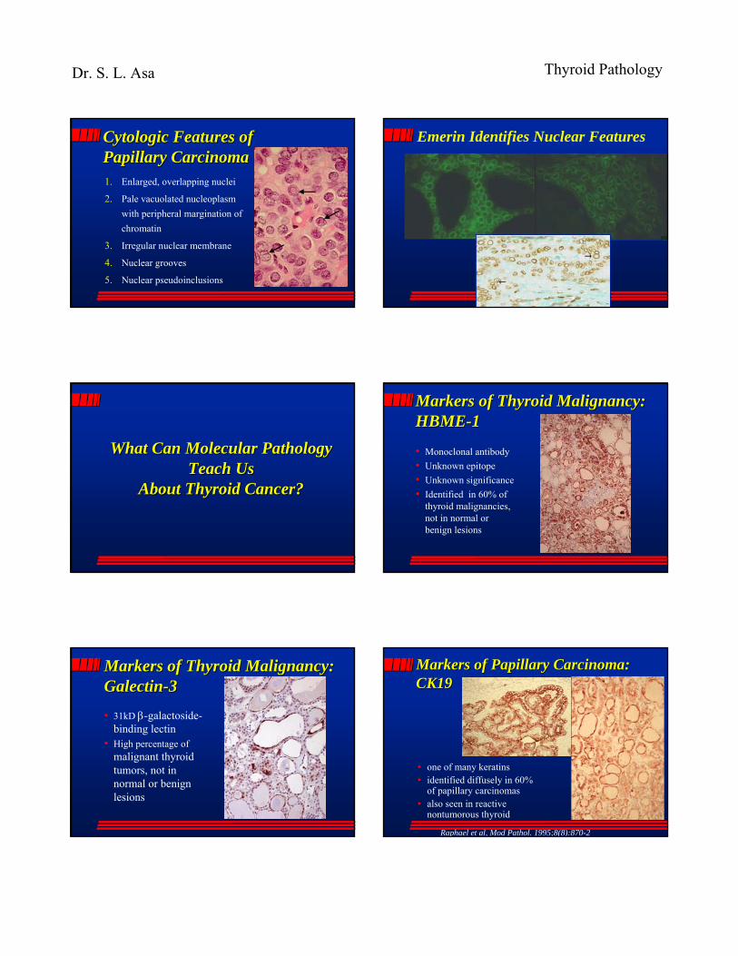
Follicular Adenomas with Follicular Adenomas with Papillary ArchitecturePapillary Architecture
• “Papillary adenomas”• Monoclonal benign neoplasms• Activating mutations of
TSH-receptor or Gsα• Plummer’s disease
(Lyons et al, Science 249:635, 1990;van Sande et al,
J Clin Endocrinol Metab 80:2577, 1995)
Follicular Adenoma & Carcinoma
• Encapsulated expansile growth
• Malignant by capsular or vascular invasion
• Hematogenous spread
Definitions: Capsular InvasionDefinitions: Capsular Invasion1. Nests, cords or cells in
capsule 2. Islands in capsule
associated with perpendicular rupture of collagen
3. In capsule beyond bulk of lesion
4. Total thickness into adjacent parenchyma
?? Artefactual trapping?? postFNA
What If There Is NO What If There Is NO Tumor Capsule?Tumor Capsule?• Capsular invasion
cannot be evaluated• Invasion must be
assessed as infiltration into
surrounding parenchyma, perineural or vascular involvement

Dr. S. L. Asa Thyroid Pathology
Classification ofClassification ofFollicular CarcinomaFollicular Carcinoma• Minimally invasive carcinoma
up to 100% 10 year survival
• Widely invasive carcinoma25-45% 10 year survival
• Angioinvasive carcinomacontroversial
Vascular Invasion by Vascular Invasion by Endocrine NeoplasmsEndocrine Neoplasms
1. Tumor cells bulging into an endothelial-lined lumen
2. Intravascular tumor nests covered with endothelium
3. Tumor casts within vessel lumen
4. Thrombus adherent to invasive tumor
? artificial implantation X
Identification of Vascular Invasionby Follicular Neoplasms• Rigid criteria
predict high likelihood of metastasis EVEN in differentiated thyroid carcinomaMete and Asa, submitted
Papillary Carcinoma:Papillary Carcinoma:A Cytologic DiagnosisA Cytologic Diagnosis
• Architecture irrelevant» Papillary, Follicular, Mixed, Solid , Cystic» Diffuse sclerosis variant is hard to recognize
• Invasion not a criterion» Encapsulated variant
• Nuclear features predict behavior
Papillary Carcinoma
• Often multifocal• Locally infiltrative• Lymphatic spread
Follicular Variant of Papillary Ca
• Encapsulatedexpansile growth
• Malignant bynuclear features
• Oftenmultifocal
• Lymphaticspread
Dr. S. L. Asa Thyroid Pathology
Cytologic Features ofCytologic Features ofPapillary CarcinomaPapillary Carcinoma1. Enlarged, overlapping nuclei
2. Pale vacuolated nucleoplasm with peripheral margination of chromatin
3. Irregular nuclear membrane
4. Nuclear grooves
5. Nuclear pseudoinclusions
Emerin Identifies Nuclear Features
What Can Molecular Pathology What Can Molecular Pathology Teach Us Teach Us
About Thyroid Cancer?About Thyroid Cancer?
Markers of Thyroid Malignancy:Markers of Thyroid Malignancy:HBMEHBME--11
• Monoclonal antibody• Unknown epitope• Unknown significance• Identified in 60% of
thyroid malignancies, not in normal or benign lesions
Markers of Thyroid Malignancy:Markers of Thyroid Malignancy:GalectinGalectin--33• 31kD β-galactoside-
binding lectin• High percentage of
malignant thyroid tumors, not in normal or benign lesions
Markers of Papillary Carcinoma: Markers of Papillary Carcinoma: CK19CK19
• one of many keratins• identified diffusely in 60%
of papillary carcinomas• also seen in reactive
nontumorous thyroid
Raphael et al, Mod Pathol. 1995;8(8):870-2
Dr. S. L. Asa Thyroid Pathology
TSH signaling MAPK signaling
ERK
MEK
BRAF
Gsa
cAMP
RAS
TSH receptor
Transcription
ERKP
PKA
CREB
CREBP
Adenylylcyclase RTK
TSH GF
P
P
Cell differentiation
Cell proliferation
P
Point mutations or rearrangement in thyroid cancers
Point mutation in hyperfunctioning
adenomas
GTP
P
Follicular cell
Mechanisms of Thyroid TumorigenesisMechanisms of Thyroid Tumorigenesis
Kondo, Ezzat and Asa, Nature Reviews Cancer 2006
Follicular Adenomas with Follicular Adenomas with Papillary ArchitecturePapillary Architecture
• “Papillary adenomas”• Monoclonal benign neoplasms• Activating mutations of
TSH-receptor or Gsα• Plummer’s disease
Lyons et al, Science 249:635, 1990;van Sande et al,
J Clin Endocrinol Metab 80:2577, 1995
BRAF MutationsBRAF Mutations
• Most common genetic event in thyroid cancer
• Diagnostic marker of PTC• Genotype-phenotype correlations
» BRAFV600E in classical variant PTC (common)» BRAFK601E in FVPTC (rare)» VK600-1E deletion (BRAFVK600-1E) in solid variant
(single case)• Prognostic significance controversial
RetRet/PTC Rearrangements/PTC Rearrangements• Chromosomal rearrangement
involving chromosome 10 ret• Fusion of the ret tyrosine kinase
to :CCDC6 (H4) = ret/PTC1*R1α = ret/PTC2NcoA4 (ele) = ret/PTC3*» Chromosome 10 inversions
most commonAt least 15 identified to date
RetRet/PTC Rearrangements/PTC RearrangementsEC TKTM
CCDC6 (H4)
R1α
NcoA4 (ele 1)
These rearrangements result in cytoplasmic protein;antibodies against ret identify the C terminus that is conserved
ret/PTC-1
ret/PTC-2
ret/PTC-3
ret
Richardson et al: Cancer Res 2009;69:4861-4869.
Different promoters drive transcript levels that modulate oncogenicity of RET/PTC oncoproteins.
Methods of Methods of RetRet/PTC Analysis/PTC Analysis• DNA
» PCR analysis difficult due to variable break-point sites leading to heterogeneous tumor profiles
• RNA» RT-PCR for ret/PTC mRNA is the “gold standard”» Variability of expression; not “all or none”
• Protein» Immunohistochemistry using antisera to C terminus
• FISH» Not widely available but promising
Rhoden et al, JCEM 2006

Dr. S. L. Asa Thyroid Pathology
RAS Mutations Characterize RAS Mutations Characterize Follicular LesionsFollicular Lesions
• Follicular Variant PTC• Follicular Adenoma• Follicular Carcinoma• Poorly Differentiated Carcinoma
Pax 8Pax 8--PPARPPARγγ 1 Fusion Oncogene1 Fusion Oncogene
• Identified in angioinvasive follicular caKroll TG et al, Science, 2000; 289:135
• Diagnostically applicable by FISH and IHC for PPARγ• Also found in PTC Nikiforova et al: AJSP2002;26(8):1016-23
• Reduced membrane stain for β-Catenin correlates with dedifferentiation
• Nuclear translocation due to exon 3 mutation in 25% of insular carcinomas and 65% of anaplastic carcinomas
Garcia-Rostan et al, Am J Pathol 2001;158:987
CTNNB1 Mutations are Found in CTNNB1 Mutations are Found in Poorly Differentiated (Insular) Poorly Differentiated (Insular) Thyroid CarcinomaThyroid Carcinoma
PIK3CA Mutations Predict PIK3CA Mutations Predict Aggressive BehaviorAggressive Behavior
• Identified in anaplastic carcinoma» Garcia-Rostan et al, Cancer Res 2005;65:10199-207» Wang et al, JCEM 2007;92:2387-90
• Accompanies other mutations in aggressive papillary carcinoma and metastases» Costa et al, Clin Endocrinol 2008;68:618-34» Ricate-Filho et al, Cancer Res 2009;69:4885-93
p53 Alterations in Thyroid Carcinomap53 Alterations in Thyroid Carcinoma
Mutations are common inAnaplastic carcinoma ↓
Immunolocalization correlates with extent of disease, extrathyroidal involvement, recurrence and poor outcome in differentiated carcinoma
Hosal et al, Endocr Pathol 1997, 8:21-28
Molecular Studies:Molecular Studies:Progression in Thyroid CancerProgression in Thyroid Cancer
ThyroidFollicular
Cell
FollicularAdenoma
FunctioningFollicularAdenoma
FollicularCarcinoma
PapillaryCarcinoma
Tall Cell Papillary
Carcinoma
Insular Carcinoma
GsTSH-R
BRAFRET/PTCTRK
p53
β-cateninPIK3CA
PPARγ
MetastaticPapillary
Carcinoma
HyperplasiaAnaplasticCarcinoma
β-cateninPIK3CARas

Dr. S. L. Asa Thyroid Pathology
What is the Clinical Significance of What is the Clinical Significance of Papillary Microcarcinoma?Papillary Microcarcinoma?
1. Potentially metastasizing
2. Metastatic focus of papillary carcinoma
3. Clinically insignificant
retret/PTC in /PTC in Multifocal Papillary CarcinomaMultifocal Papillary Carcinoma
• ret/PTC expression is highly prevalent in multifocal micropapillary thyroid cancer
• Identical ret/PTC rearrangements are found in 32% of patients» possible spread of a single tumor
• Discordant ret/PTC patterns in 68%» discrete primary tumors
Sugg et al, J Clin Endocrinol Metab 83:4116-4122, 1998
• Identical data using X-chromosome inactivationShattuck et al, N Engl J Med. 2005 Jun 9;352(23):2406-12
Implications of Implications of retret/PTC Data in /PTC Data in Multifocal Papillary CarcinomaMultifocal Papillary Carcinoma
• One major rationale for completion thyroidectomy in patients with “low risk”papillary carcinoma is unjustified
Fink et al, Modern Pathol 1996; 9: 816-820
HHüürthle Cell Tumorsrthle Cell Tumors
• Hürthle cell adenoma, Hürthle cell carcinoma» distinguished by
invasive behavior» controversial because of
unpredictable behavior
• Hürthle cell PTC» defined by papillary
architecture
Molecular Basis of HMolecular Basis of Hüürthle Cell rthle Cell Papillary CarcinomaPapillary Carcinoma
• ret/PTC identifies Hürthle cell tumors with lymph node mets» allows distinction from Hürthle cell adenoma» better prognosis than Hürthle cell carcinoma
Cheung et al, J Clin Endocrinol Metab 85: 878-882, 2000
mtDNAmtDNA, GRIM19, GRIM19• Altered ATP synthesis
Savagner et al, JCEM 2001;86:4920–4925
• mtDNA somatic eventsBonore et al, Cancer Res 2006; 66:6087–6096 Gasparre et al, PNAS 2007: 104, 9001–9006
• Mutations in non-neoplastic and neoplastic oncocytic cells » Not specific to neoplastic transformation » Associated with BRAF, ret/PTC etc
• GRIM19 (19p13.2) somatic and germline eventsMaximo et al, Virchows Arch 2000 Sobrinho-Simoes et al, Int J Surg Pathol 2005

Dr. S. L. Asa Thyroid Pathology
Molecular Diagnosis in Thyroid Molecular Diagnosis in Thyroid AspiratesAspirates-- Papillary CarcinomaPapillary Carcinoma
• ret/PTCCheung et al, J Clin Endocrinol Metab 2001
• BRAFSalvatore et al, J Clin Endocrinol Metab 2004
Improved diagnosis with combined morphology and molecular testing
After “The Anatomy Lecture of Dr. Nicolaes Tulp” – Rembrandt, 1632(Courtesy of Dr. Carlos Cordón, New York, USA)
BRAF Kinase Inhibition Arrests BRAF Kinase Inhibition Arrests Thyroid Cancer Growth In VivoThyroid Cancer Growth In Vivo
Salvatore G et al, Clin Cancer Res 2006;12:1623-9
However ………
Clinical trials have failed to show
effectiveness of BRAF inhibitors
Molecular Studies:Molecular Studies:Progression in Thyroid CancerProgression in Thyroid Cancer
ThyroidFollicular
Cell
FollicularAdenoma
FunctioningFollicularAdenoma
FollicularCarcinoma
PapillaryCarcinoma
Tall Cell Papillary
Carcinoma
Insular Carcinoma
GsTSH-R
BRAFRET/PTCTRK
p53
β-cateninPIK3CA
PPARγ
???????
MetastaticPapillary
Carcinoma
HyperplasiaAnaplasticCarcinoma
β-cateninPIK3CARas
Epigenetic Control: DNA MethylationEpigenetic Control: DNA Methylation
N Engl J Med. 2007 Feb 15;356(7):731-3
Molecular Studies:Molecular Studies:Progression in Thyroid CancerProgression in Thyroid Cancer
ThyroidFollicular
Cell
FollicularAdenoma
FunctioningFollicularAdenoma
FollicularCarcinoma
PapillaryCarcinoma
Tall Cell Papillary
Carcinoma
Insular Carcinoma
GsTSH-R
RET/PTCBRAFTRK
p53
rasβ-catenin
PPARγ
EpigeneticDysregulation
MetastaticPapillary
Carcinoma
Hyperplasia
rasβ-catenin
AnaplasticCarcinoma

Dr. S. L. Asa Thyroid Pathology
Cyclin D1 and p27 Predict Metastasis Cyclin D1 and p27 Predict Metastasis in Papillary Carcinomain Papillary Carcinoma
Khoo et al, J Clin Endocrinol Metab 2002, 87:1814-8
Vitamin D Targets p27 Degradation Vitamin D Targets p27 Degradation in Thyroid Cancerin Thyroid Cancer• VD/EB1089 induce intranuclear p27 accumulation by
diminished degradation• VD/EB1089 hypophosphorylate p27 in a phosphatase
dependent process that involves the Akt pathway but may be PTEN independent
Liu et al, Am J Pathol 2002;160:511-9• In an orthotopic model, in vivo VD administration
» decreases tumor volume» increases p27 accumulation» enhances cellular differentiation» decreases lung metastases
Dackiw et al, Endocrinology 2004;145:5840-6
• CITED-1 (L)• Galectin-3• Fibronectin ( R)• HGF, MET• TPO• COX-2• CD44V6• CD57
Prasad et al: Modern Pathology 2005;18:48-57
Are There Other Targets of VD?Are There Other Targets of VD? Fibronectin is Upregulated in Fibronectin is Upregulated in Papillary Thyroid CarcinomaPapillary Thyroid Carcinoma• Increased cDNA expression in microarray studies
of papillary carcinoma cf normal• Diminished FN immunoreactivity reported at
invading edge of aggressive thyroid cancers• Negative in poorly-differentiated and anaplastic
carcinomas• Function unclear
» Increasing invasion?» Reactive upregulation?
DownDown--regulation of FN Promotes regulation of FN Promotes Tumor Growth and MetastasisTumor Growth and Metastasis
0 5 10 15 21 26Days after cancer cell injection
Tum
or v
olum
e (m
m3 )
0
500
1000
1500
2000
2500
3000
3500
4000
4500
5000 ControlsiRNA
FN siRNA
**
*
*
Control FN-siRNA
Liu et al, Mol Endo 2005; 19(9):2349-57
Mice n # of mice with mets # of lesions/mouse pControl 9 1 single FN-siRNA 9 6 multiple <0.05
Fibronectin in Thyroid CancerFibronectin in Thyroid Cancer
• Fibronectin mediates adhesion in thyroid carcinoma and restrains tumour growth
• VD upregulates fibronectin and restores adhesiveness of thyroid carcinoma
• The PTEN/PI3 Kinase pathway is involved in FN regulation and VD action on FN and adhesion
• The mechanism underlying overexpression in papillary carcinoma is unclear, but appears to be compensatory, and is lost in aggressive and dedifferentiated thyroid cancers
Liu et al, Mol Endo 2005; 19(9):2349-57

Dr. S. L. Asa Thyroid Pathology
CEACAM1CEACAM1aka biliary glycoprotein (BGP), CD66a, Caka biliary glycoprotein (BGP), CD66a, C--CAM1 and pp120CAM1 and pp120
• A member of the CEA family (Ig superfamily)• A putative TSG
» Down-regulated in colon, prostate, liver, endometrial, bladder and breast cancer
» Reduces proliferation in human prostate cancer cell lines in vitro and in vivo
• Also implicated as an oncogene» Over-expressed in gastric cancer, non-small cell lung cancer and
malignant melanomas» Facilitates metastatic tumor spread» Shows angiogenic function as a major target of VEGF
CEACAM1 Expression Predicts Metastasis in PTC
• CEACAM1 is expressed in a small PTCswith lymph node spread
• CEACAM1 has a novel dual role in thyroid carcinoma: it has a suppressive effect on thyroid cell proliferation and increases adhesion, while promoting invasion and metastasis
Liu et al, Oncogene 2007; 26:2747-58
CEACAM1 in Thyroid CancerCEACAM1 in Thyroid Cancer• CEACAM1 is expressed in a small thyroid malignancies
with lymph node spread • CEACAM1 has a novel dual role in thyroid carcinoma:
it suppresses thyroid cell proliferation, while promoting invasion and metastasis
Liu et al, Oncogene 2007; 26:2747-58• VD inhibits CEACAM1 to promote insulin/IGF-I receptor
signaling without compromising anti-proliferative action• CEACAM1 represents a target for VD therapy which may
have potential therapeutic applicationsLiu et al, Lab Invest 2011 ;91(1):147-56
TMA Profiling Shows Divergent TMA Profiling Shows Divergent Expression of Expression of FGFRsFGFRs in the Thyroidin the Thyroid
FGFR2 is expressed exclusively in normalthyroid
FGFR1 is expressed in hyperplastic and neoplastic lesions
St Bernard et al, Endocrinology 146:1145-1153, 2005
FGFR1, PTCFGFR2, Normal thyroid
FGFR2FGFR2--IIIb Interrupts Signaling IIIb Interrupts Signaling Upstream of BRAF/MAPK Upstream of BRAF/MAPK
Kondo et al, Cancer Res 2007;67: 5461
FGFR2FGFR2--IIIb Represses MAGEIIIb Represses MAGE--A3/6A3/6
Kondo et al, Clin Cancer Res 2007;13(16):4713-20
MAGE subgroup I members , MAGE-A, B, C, are expressed in several tumors, but not in normal tissues except testis and placenta
“Cancer-testis antigens”
Dr. S. L. Asa Thyroid PathologyM
AG
E-A
3
C
ontr
ol
Time: 0 5 10 15 20 (hrs)
MAGEMAGE--A3 Promotes Migration & InvasionA3 Promotes Migration & Invasion
Liu et al, Cancer Res 2008;68:8104-8112
0%
50%
100%
150%
200%
250%
300%
Inva
sive
Cel
ls
C MAGE A3Control MAGE-A3
Tum
or v
olum
e (m
m3 )
0 5 10 15 21
0
250
500
750
1000
1250
1500
1750
2000
2250
2500Control
MAGE A3
0
0.5
1
1.5
2
2.5
Tum
or w
eigh
t (gr
am)
*
*
* *
N=20 N=20
MAGEMAGE--A3 Enhances Tumor Growth A3 Enhances Tumor Growth
Control MAGE-A3
00.5
11.5
22.5
33.5
44.5
55.5
6
Tum
or w
eigh
t (gr
am)
N=20 N=17
*
Days after injection
0
500
1000
1500
2000
2500
3000
3500
4000
4500
5000
5500
6000
6500Control
MAGE
Tum
or v
olum
e (m
m3 )
*
*
**
0 5 10 15 21 25
Control MAGE-A3
MAGEMAGE--A3 Promotes Metastasis A3 Promotes Metastasis
Control
MAGE-A3
Day 15 Day 20 Day 25
Liu et al, Cancer Res 2008;68:8104-8112
MAGE in Thyroid CancerMAGE in Thyroid Cancer• Downregulation or FN or FGFR2 increase tumor
growth and metastasis• Downregulation of FN or FGFR2 induce expression of
MAGE-A3 through histone methylation• MAGE- A3 mediates p21 down-regulation,
accelerated cell cycle progression, increased cell migration rate, invasion and metastasis
• MAGE-A3 is a functional integrator of diverse signals in mediating cancer progression
Liu et al, Cancer Res 2008;68:8104-8112
MAGE
• Normal thyroid tissue exhibits weak cytoplasmic and strong nuclear MAGE reactivity.
• Tumors exhibit an increase in cytoplasmic MAGE scores that correlates with clinical behavior» larger tumors have higher MAGE scores» correlation between MAGE cytoplasmic score and
number of lymph node metastases
Cheng et al, Endocrine Related Cancer 2009;16:455-466
Proteomic Biomarkers in PTC• 410 PTCs with morphologic and clinical data• BRAF status known• TMA analysis of:
• Histopathologic biomarkers of malignancy: Galectin-3, CK 19, HBME-1
• Cell differentiation factors: NIS, CITED-1 • Nuclear receptors: ERα, ERβ, and PPAR-γ• Adhesion molecules: CEACAM-1, Osteopontin,
Fibronectin, E-Cadherin• Cell cycle regulators: Cyclin-D1, p53, p27, p21

Dr. S. L. Asa Thyroid Pathology
BRAF Mutations & OutcomeBRAF Mutations & Outcome
Cheng et al, Clin Cancer Res 2011:17(8):2385-94
410 PTCs
ETE: ↑membranous CK19, HBME, Gal 3,OPN↓cytoplasmic HBME , CK19 ↑ nuclear ERβ, Gal3, p53
LNM:↑ membranous HBME1, CK19, Gal3↓ cytoplasmic FBN and CK19 ↑ nuclear Gal3, Erβ
VI:↑ membranous Gal3 ↓ cytoplasmic Gal3
PTC PTC ProteomeProteome
Cheng et al, Clin Cancer Res 2011:17(8):2385-94
ProteomeProteome of of Invasive PTCInvasive PTC
Cheng et al, Clin Cancer Res 2011 in press
ETE: ↑membranous CK19, HBME, Gal 3,OPN↓cytoplasmic HBME , CK19 ↑ nuclear ERβ, Gal3, p53
LNM:↑ membranous HBME1, CK19, Gal3↓ cytoplasmic FBN and CK19 ↑ nuclear Gal3, Erβ
VI:↑ membranous Gal3 ↓ cytoplasmic Gal3
PTC PTC ProteomeProteomeBy MorphologyBy MorphologyFVPTC:
ETE: ↑ membranous CK19, HBME, Gal3, OPN↓cytoplasmic CK19↑nuclear Gal3
LNM:↑membranous HBME1, CK19 and Gal3↓membranous E-Cadherin↓cytoplasmic HBME1, CK19, FBN↑ nuclear Gal3 ↓nuclear PPARγ
VI:↓ nuclear PPARγ
Classic PTC
ETE: ↑ ERβLNM: ↑cyclin D1, ↓FBNVI: ↓ p27
• The diagnosis of thyroid cancer is evolving as
molecular data clarify the significance of
morphologic features and behaviors
• Our data predict the need for targeting
epigenetic factors along with intragenic mutations
in the control of thyroid cancer progression.
ConclusionsConclusions Thanks ToThanks To…………..
♥ Shereen Ezzat
• Robyn Apel• Lei Zheng• Sonia Sugg• Mark Khoo• Carol Cheung• Wei Liu• P. Huang• Rosanne St. Bernard• Catherine Wei• Daniel Winer• Tetsuo Kondo• Xuegong Zhu• Sonia Cheng
• Alan Dackiw• Lorne Rotstein• Ana-Maria Bamberger• Christoph Bamberger• Christoph Wagener