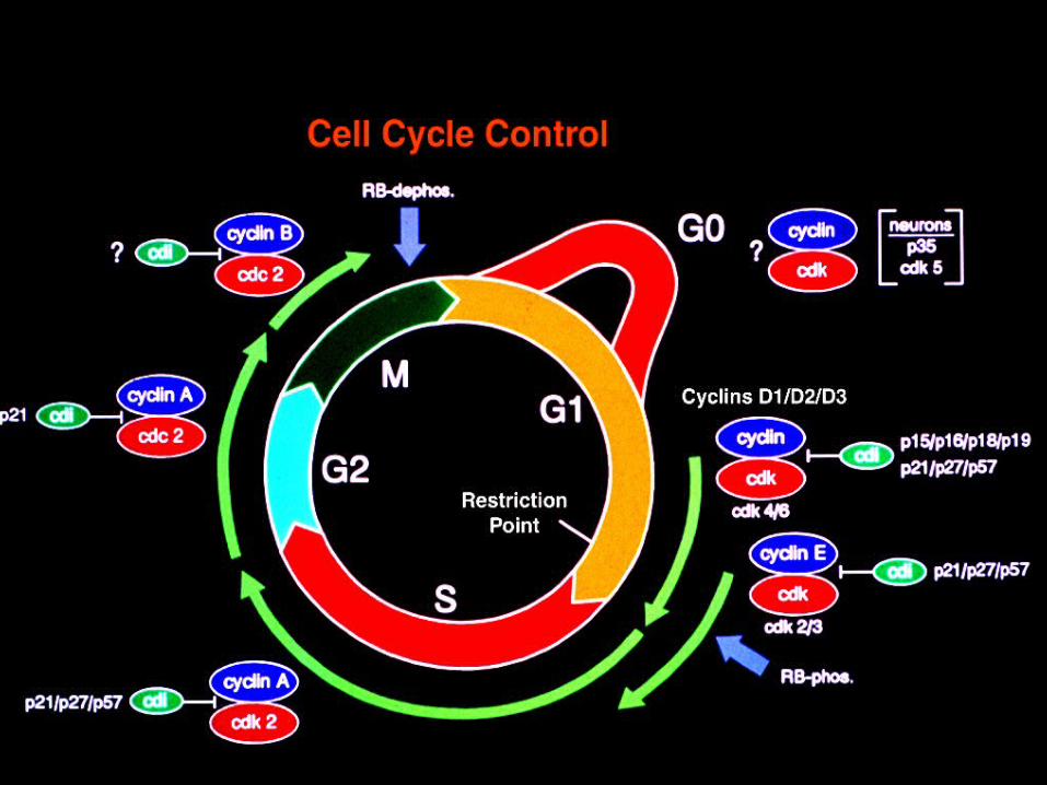
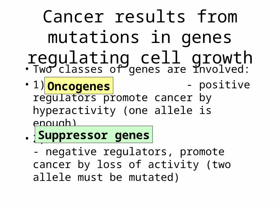
Cancer results from mutations in genes regulating cell growth
41
Cancer results from mutations in genes regulating cell growth • Two classes of genes are involved: • 1) - positive regulators promote cancer by hyperactivity (one allele is enough) • 2) - negative regulators, promote cancer by loss of activity (two allele must be mutated) Oncogenes Suppressor genes
description
Cancer results from mutations in genes regulating cell growth. Two classes of genes are involved: 1) - positive regulators promote cancer by hyperactivity (one allele is enough) - PowerPoint PPT Presentation
Transcript of Cancer results from mutations in genes regulating cell growth
Cancer results from mutations in genes regulating cell growth
• Two classes of genes are involved:
• 1) - positive regulators promote cancer by hyperactivity (one allele is enough)
• 2) - negative regulators, promote cancer by loss of activity (two allele must be mutated)
Oncogenes
Suppressor genes
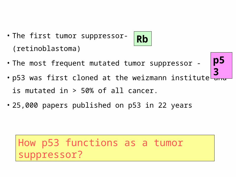
• The first tumor suppressor- (retinoblastoma)
• The most frequent mutated tumor suppressor -
• p53 was first cloned at the weizmann institute and is
mutated in > 50% of all cancer.
• 25,000 papers published on p53 in 22 years
How p53 functions as a tumor suppressor?
Rb
p53
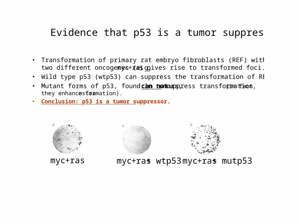
Evidence that p53 is a tumor suppressor (II)
• Transformation of primary rat embryo fibroblasts (REF) with a combination oftwo different oncogenes (e.g. myc+ras) gives rise to transformed foci.
• Wild type p53 (wtp53) can suppress the transformation of REF by oncogenes.
• Mutant forms of p53, found in tumors, can not suppress transformation (in fact,they enhance transformation).
• Conclusion: p53 is a tumor suppressor.
myc+ras myc+ras+ wtp53 myc+ras+ mutp53
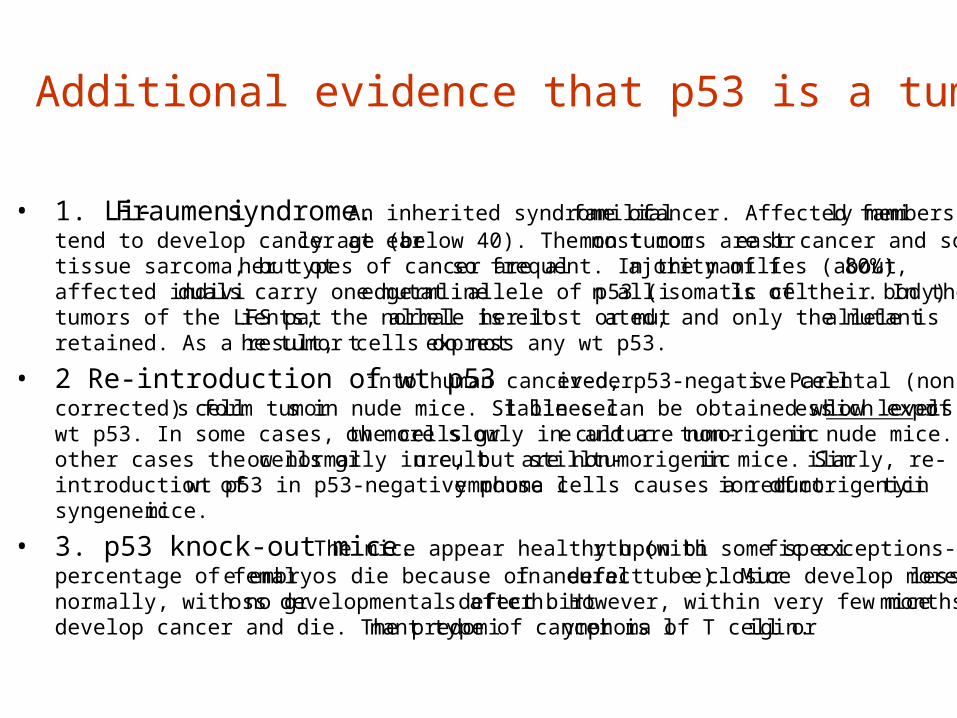
Additional evidence that p53 is a tumor suppressor
• 1. Li-Fraumeni syndrome. An inherited syndrome of familial cancer. Affected fami ly memberstend to develop cancer at early age (below 40). The most common tumors are breast cancer and softtissue sarcoma, but other types of cancer are also frequent. In the majority of families (about 80%),affected individuals carry one mutated germline allele of p53 (in all somatic cells of their body). In thetumors of the LFS patients, the normal allele is either lost or mutated, and only the mutant allele isretained. As a result, the tumor cells do not express any wt p53.
• 2 Re-introduction of wt p53 into human cancer-derived, p53-negative cells. Parental (non-corrected) cells form tumors in nude mice. Stable cell lines can be obtained which express low levels ofwt p53. In some cases, the cells grow more slowly in culture and are non-tumorigenic in nude mice. Inother cases the cells grow normally in culture, but still are non-tumorigenic in mice. Similarly, re-introduction of wt p53 in p53-negative mouse lymphoma cells causes a reduction of tumorigenicity insyngeneic mice.
• 3. p53 knock-out mice. The mice appear healthy upon birth (with some specific exceptions- apercentage of female embryos die because of a defect in neural tube closure). Mice develop more or lessnormally, with no gross developmental defects after birth. However, within very few months, all micedevelop cancer and die. The predominant type of cancer is lymphoma of T cell origin.
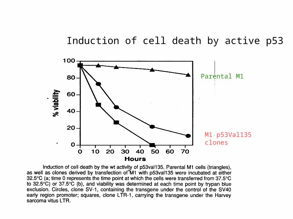
Parental M1
M1-p53Val135 clones
at 32oC
Induction of cell death by active p53
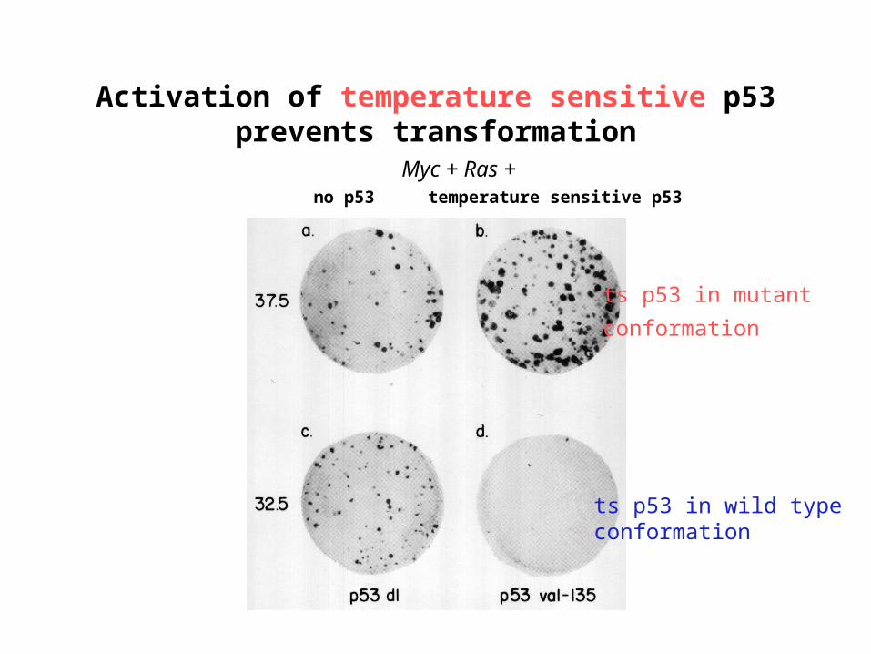
Activation of temperature sensitive p53 prevents transformation
ts p53 in mutant
conformation
ts p53 in wild type conformation
Myc + Ras +no p53 temperature sensitive p53
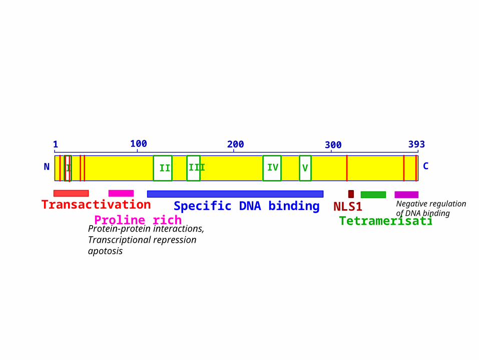
II III IV VI
Transactivation Specific DNA binding
N C
2001001 393300
NLS1Proline rich TetramerisationProtein-protein interactions,Transcriptional repressionapotosis
Negative regulation of DNA binding
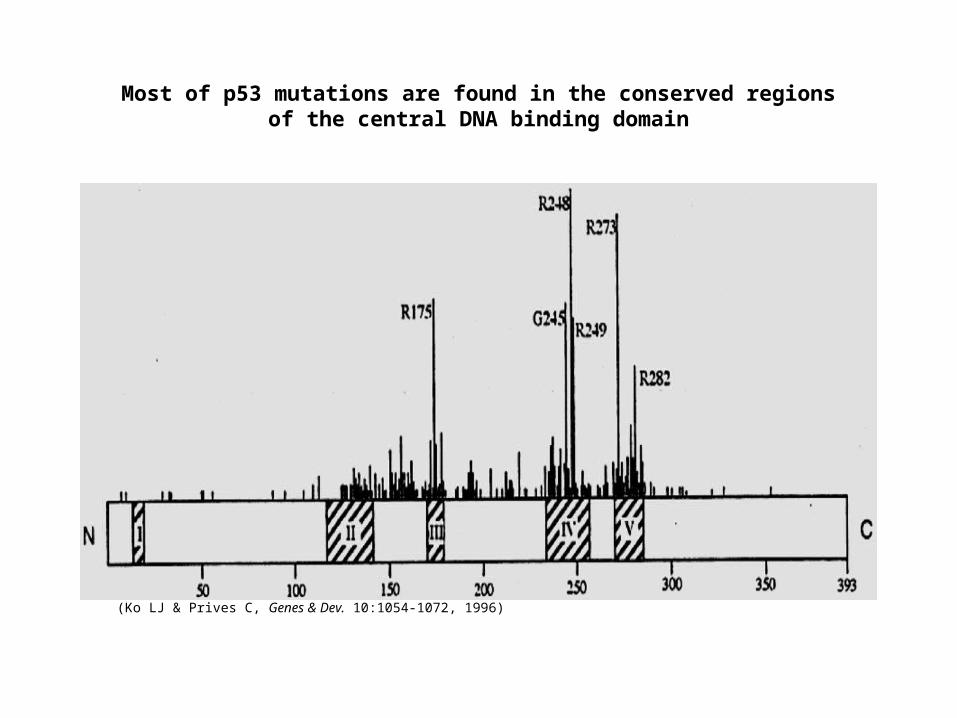
(Ko LJ & Prives C, Genes & Dev. 10:1054-1072, 1996)
Most of p53 mutations are found in the conserved regionsof the central DNA binding domain
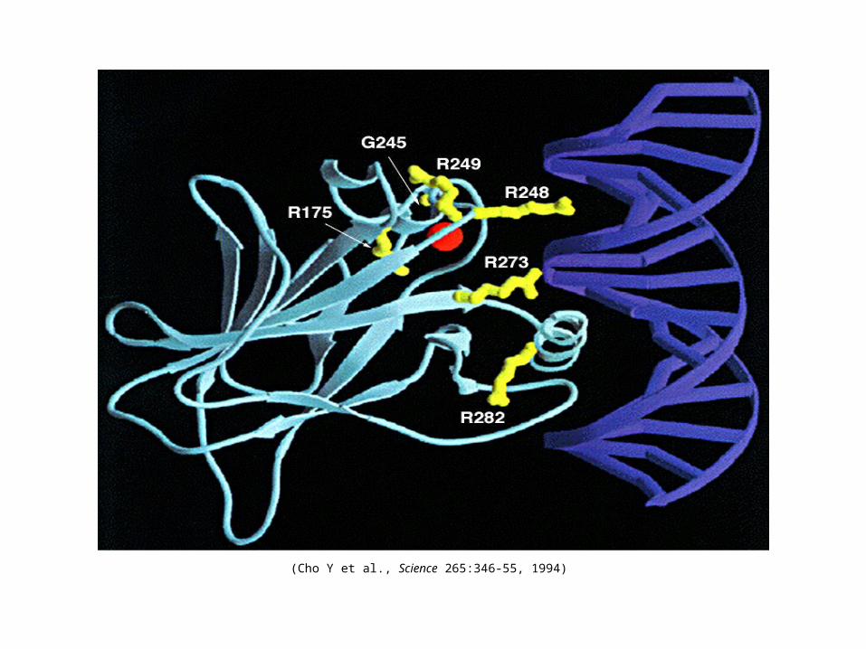
(Cho Y et al., Science 265:346-55, 1994)
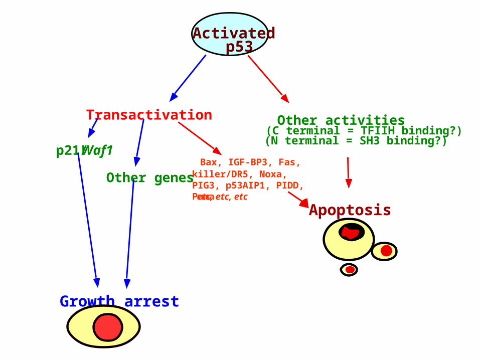
Transactivation
Growth arrest
Other activities (C terminal = TFIIH binding?)(N terminal = SH3 binding?)
Apoptosis
Bax, IGF-BP3, Fas, killer/DR5, Noxa, PIG3, p53AIP1, PIDD, Puma
p21/Waf1
Activated p53
Other genesetc, etc, etc
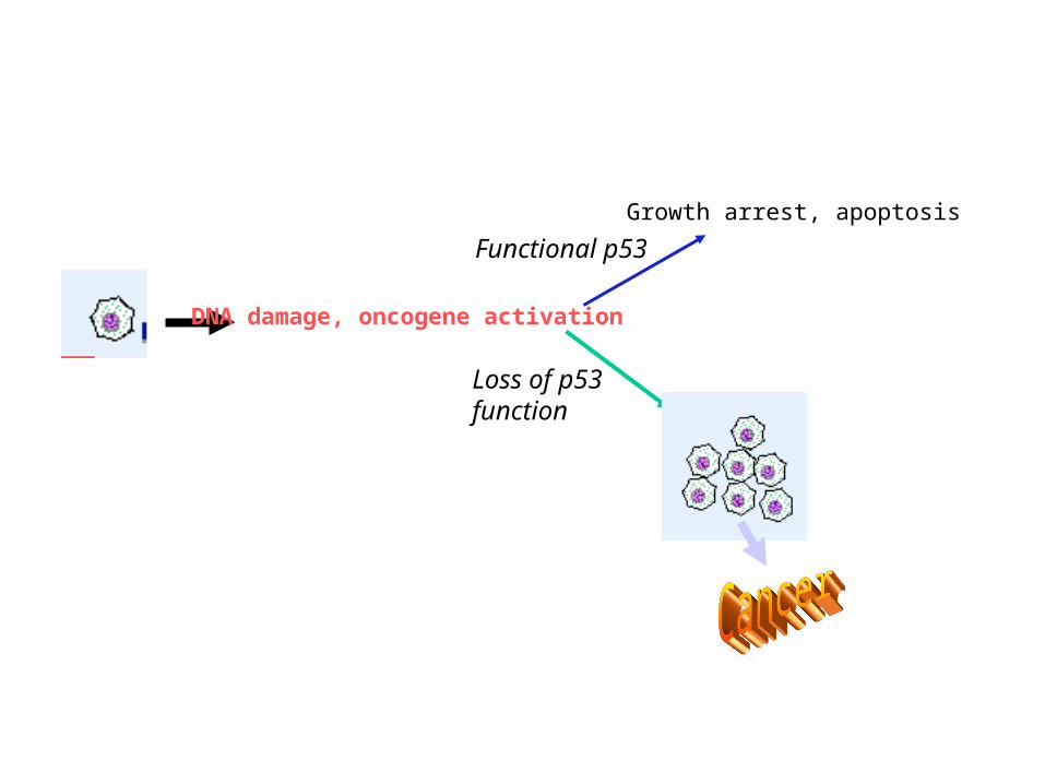
DNA damage, oncogene activation
Functional p53
Growth arrest, apoptosis
Loss of p53 function
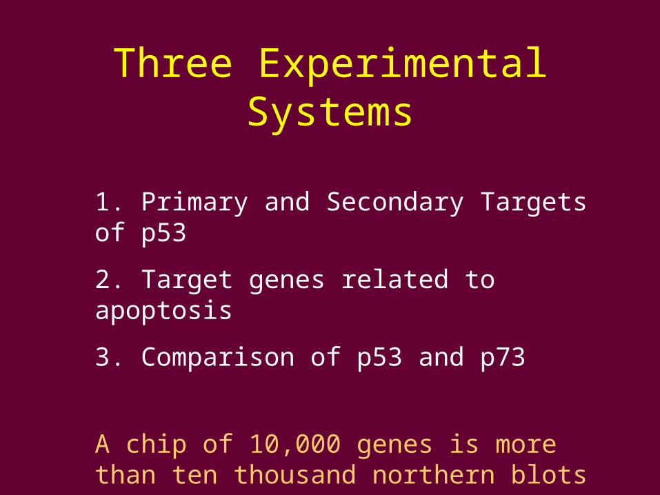
Three Experimental Systems
1. Primary and Secondary Targets of p53
2. Target genes related to apoptosis
3. Comparison of p53 and p73
A chip of 10,000 genes is more than ten thousand northern blots
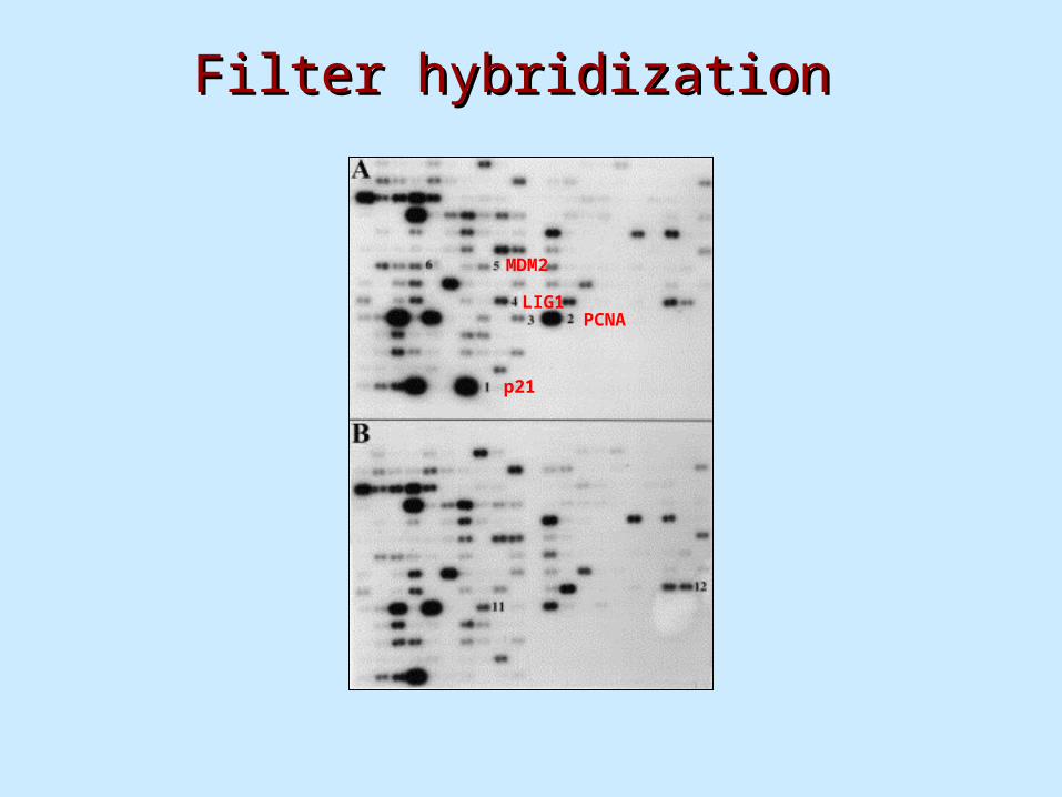
Filter hybridizationFilter hybridization
p21
PCNALIG1
MDM2
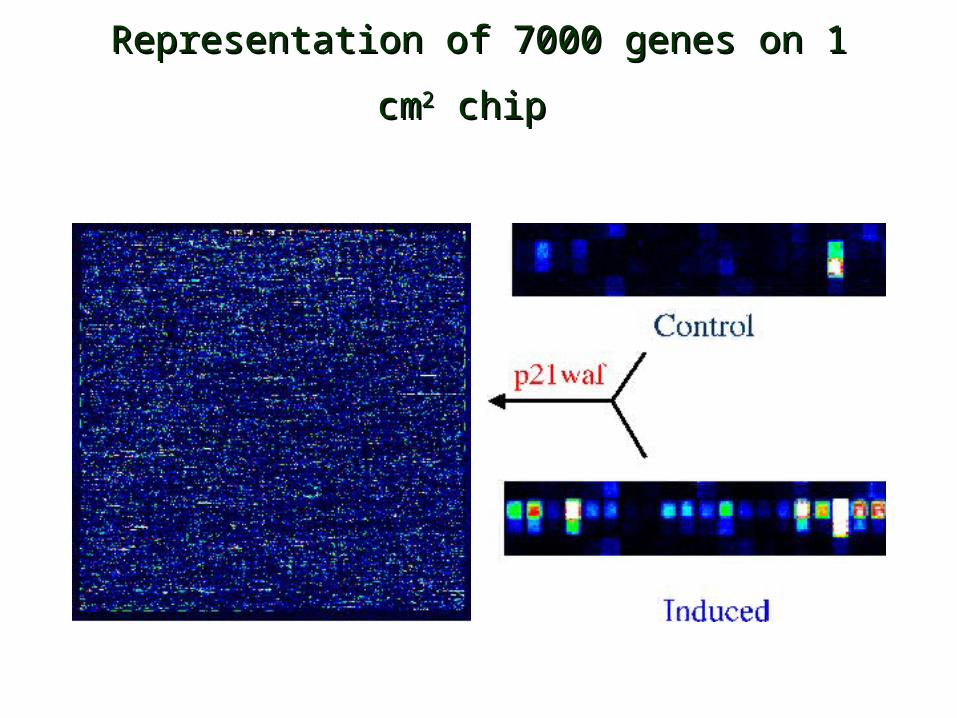
Representation of 7000 genes on 1 cmRepresentation of 7000 genes on 1 cm22 chip chip
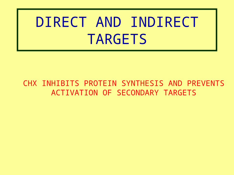
DIRECT AND INDIRECT TARGETS
CHX INHIBITS PROTEIN SYNTHESIS AND PREVENTS ACTIVATION OF SECONDARY TARGETS
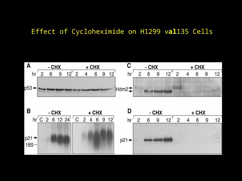
Effect of Cycloheximide on H1299 val135 Cells
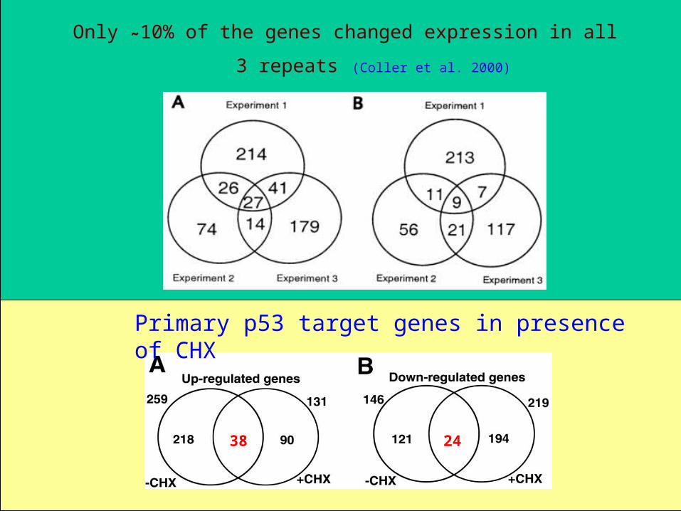
38 24
Only ˜10% of the genes changed expression in all 3
repeats (Coller et al. 2000)
Primary p53 target genes in presence of CHX
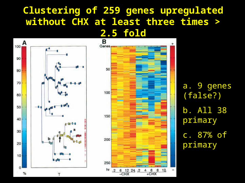
Clustering of 259 genes upregulated without CHX at least three times > 2.5 fold
a. 9 genes (false?)
b. All 38 primary
c. 87% of primary
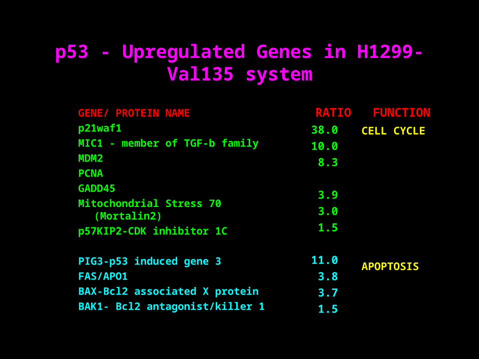
p53 - Upregulated Genes in H1299-Val135 system
GENE/ PROTEIN NAME
p21waf1
MIC1 - member of TGF-b family
MDM2
PCNA
GADD45
Mitochondrial Stress 70 (Mortalin2)
p57KIP2-CDK inhibitor 1C
PIG3-p53 induced gene 3
FAS/APO1
BAX-Bcl2 associated X protein
BAK1- Bcl2 antagonist/killer 1
38.0
10.0
8.3
3.9
3.0
1.5
11.0
3.8
3.7
1.5
RATIO FUNCTION
CELL CYCLE
APOPTOSIS
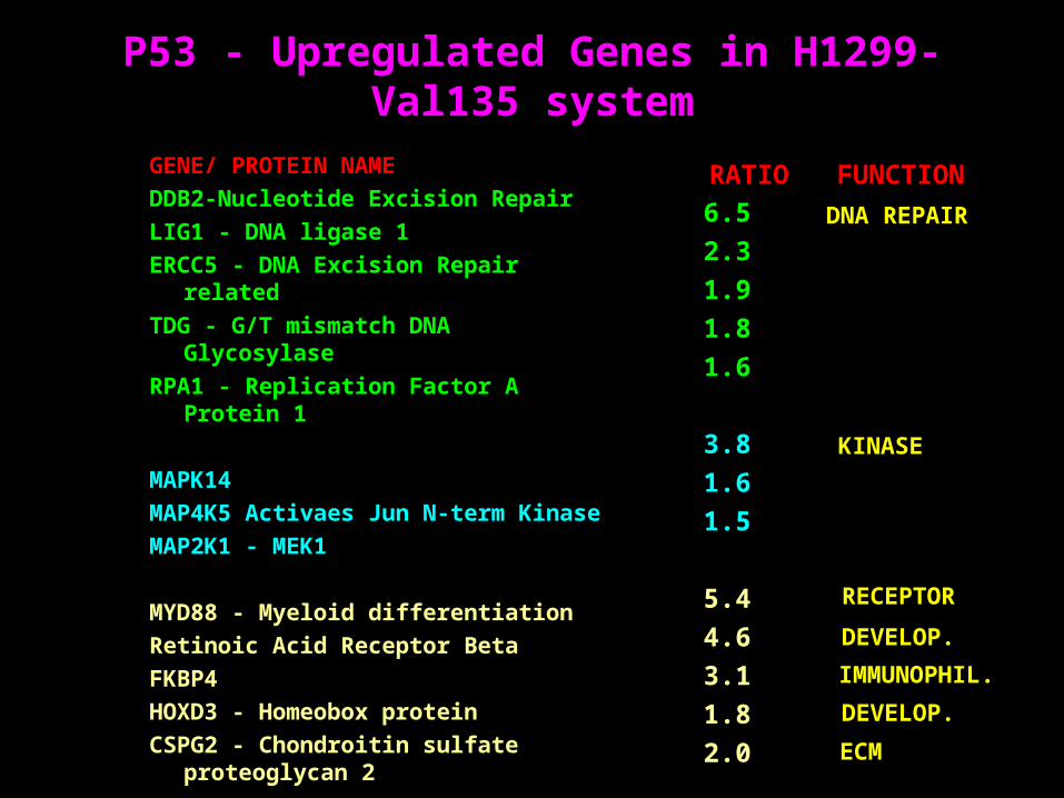
P53 - Upregulated Genes in H1299-Val135 system
GENE/ PROTEIN NAME
DDB2-Nucleotide Excision Repair
LIG1 - DNA ligase 1
ERCC5 - DNA Excision Repair related
TDG - G/T mismatch DNA Glycosylase
RPA1 - Replication Factor A Protein 1
MAPK14
MAP4K5 Activaes Jun N-term Kinase
MAP2K1 - MEK1
MYD88 - Myeloid differentiation
Retinoic Acid Receptor Beta
FKBP4
HOXD3 - Homeobox protein
CSPG2 - Chondroitin sulfate proteoglycan 2
6.5
2.3
1.9
1.8
1.6
3.8
1.6
1.5
5.4
4.6
3.1
1.8
2.0
RATIO FUNCTION
DNA REPAIR
KINASE
ECM
DEVELOP.
IMMUNOPHIL.
RECEPTOR
DEVELOP.
p53- DRIVEN APOPTOSIS
A different cell line (M1) that undergoes apoptosis by p53 at 32
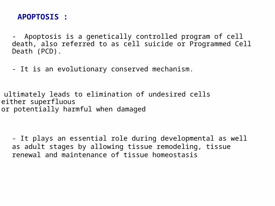
- It plays an essential role during developmental as well as adult stages by allowing tissue remodeling, tissue renewal and maintenance of tissue homeostasis
APOPTOSIS :
- Apoptosis is a genetically controlled program of cell death, also referred to as cell suicide or Programmed Cell Death (PCD).
- It is an evolutionary conserved mechanism.
- It ultimately leads to elimination of undesired cells•either superfluous•or potentially harmful when damaged
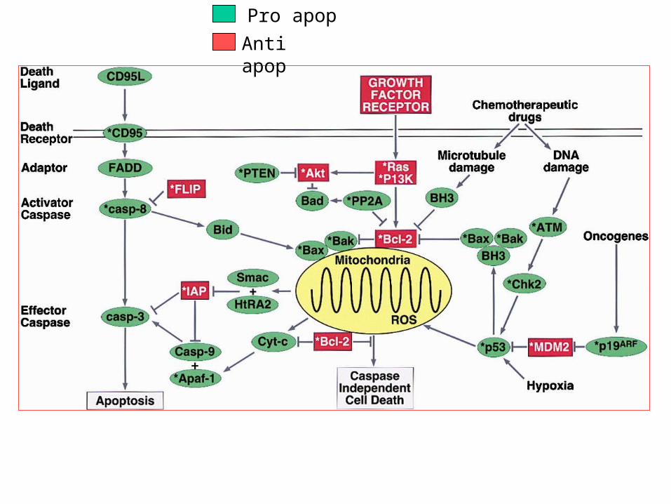
Pro apop
Anti apop
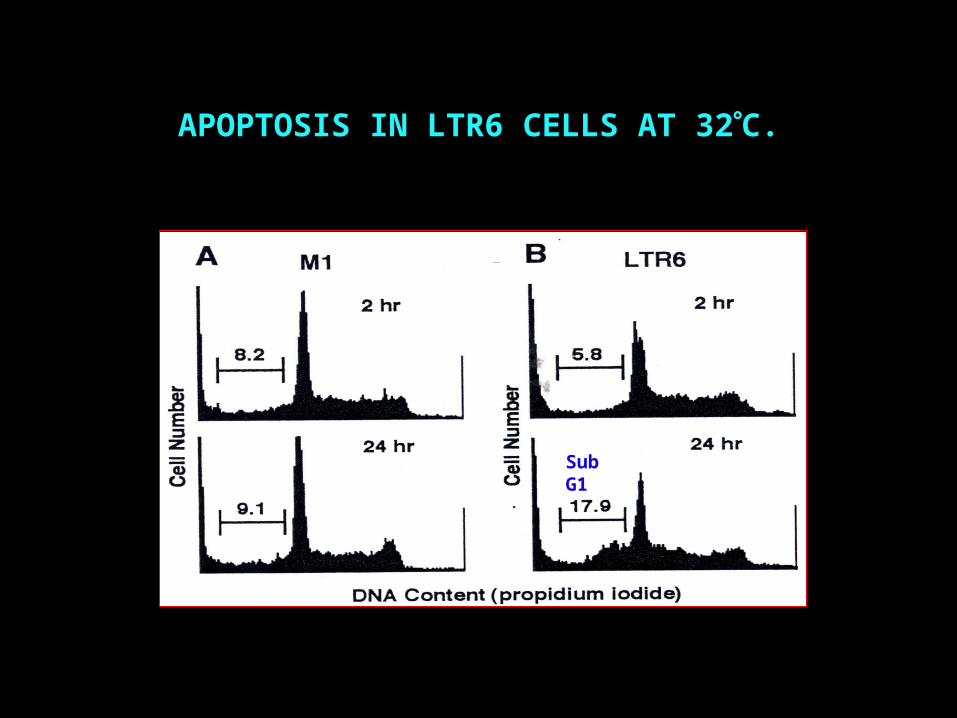
APOPTOSIS IN LTR6 CELLS AT 32C.
Sub G1
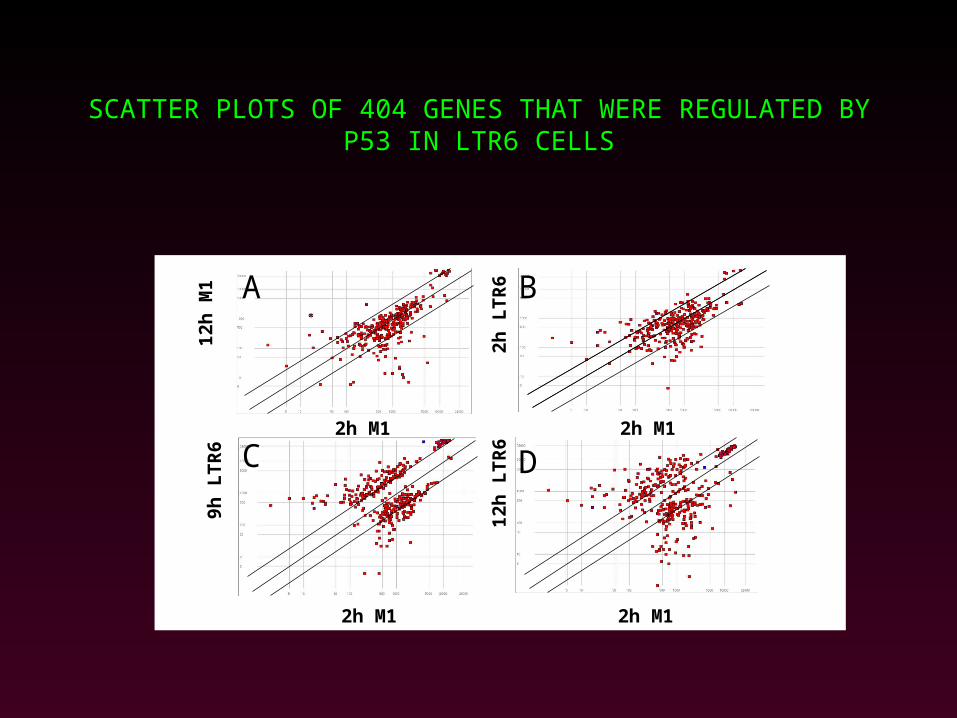
SCATTER PLOTS OF 404 GENES THAT WERE REGULATED BY P53 IN LTR6 CELLS
A
2h M1
9h L
TR
6
12h
LT
R6
2h M1
2h L
TR
6
12h
M1
2h M1
2h M1
DC
B
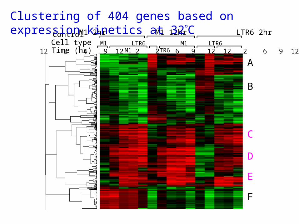
A
B
C
D
E
F
12 2 6 9 12 2 2 6 9 12 12 2 6 9 12 M1 LTR6 M1 LTR6 M1 LTR6 Time (hr)
Cell typeControl M1 2hr M1 12hr LTR6 2hr
Clustering of 404 genes based on expression kinetics at 32C
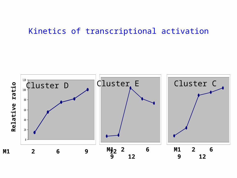
Kinetics of transcriptional activation
0
20
40
60
80
100
120
Cluster D Cluster E Cluster C
Rel
ativ
e ra
tio
M1 2 6 9 12 M1 2 6 9 12 M1 2 6 9 12
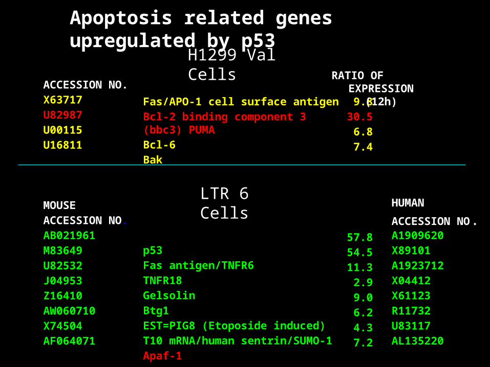
ACCESSION NO.
X63717
U82987
U00115
U16811
MOUSE
ACCESSION NO.
AB021961
M83649
U82532
J04953
Z16410
AW060710
X74504
AF064071
Fas/APO-1 cell surface antigen
Bcl-2 binding component 3 (bbc3) PUMA
Bcl-6
Bak
p53
Fas antigen/TNFR6
TNFR18
Gelsolin
Btg1
EST=PIG8 (Etoposide induced)
T10 mRNA/human sentrin/SUMO-1
Apaf-1
9.8
30.5
6.8
7.4
57.8
54.5
11.3
2.9
9.0
6.2
4.3
7.2
RATIO OF EXPRESSION (12h)
HUMAN
ACCESSION NO.A1909620
X89101
A1923712
X04412
X61123
R11732
U83117
AL135220
Apoptosis related genes upregulated by p53
H1299 Val Cells
LTR 6 Cells
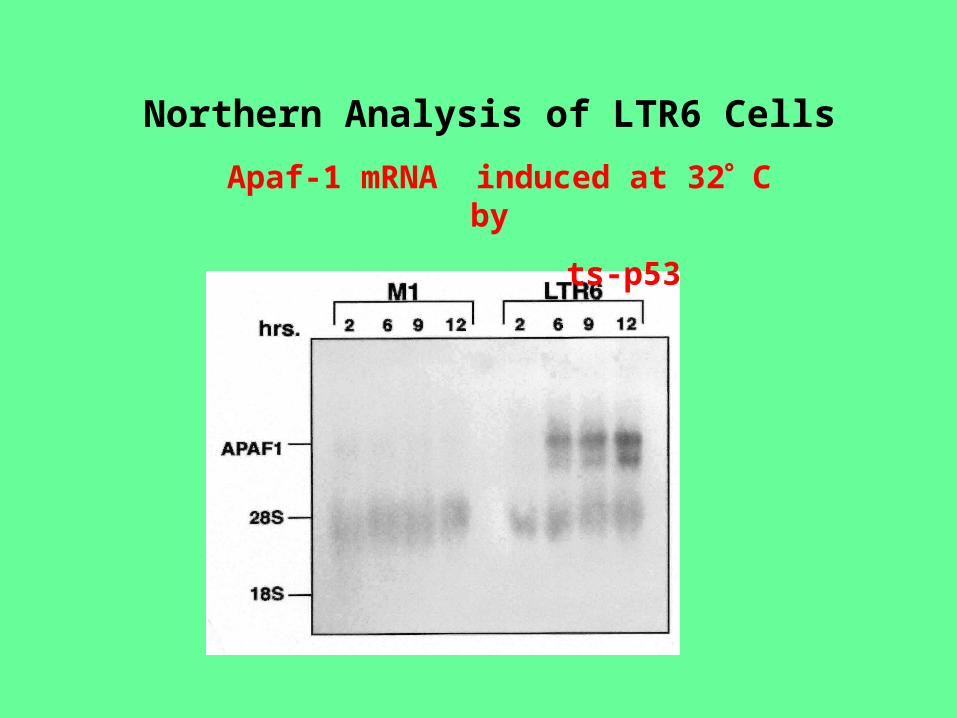
Northern Analysis of LTR6 Cells
Apaf-1 mRNA induced at 32 C by
ts-p53
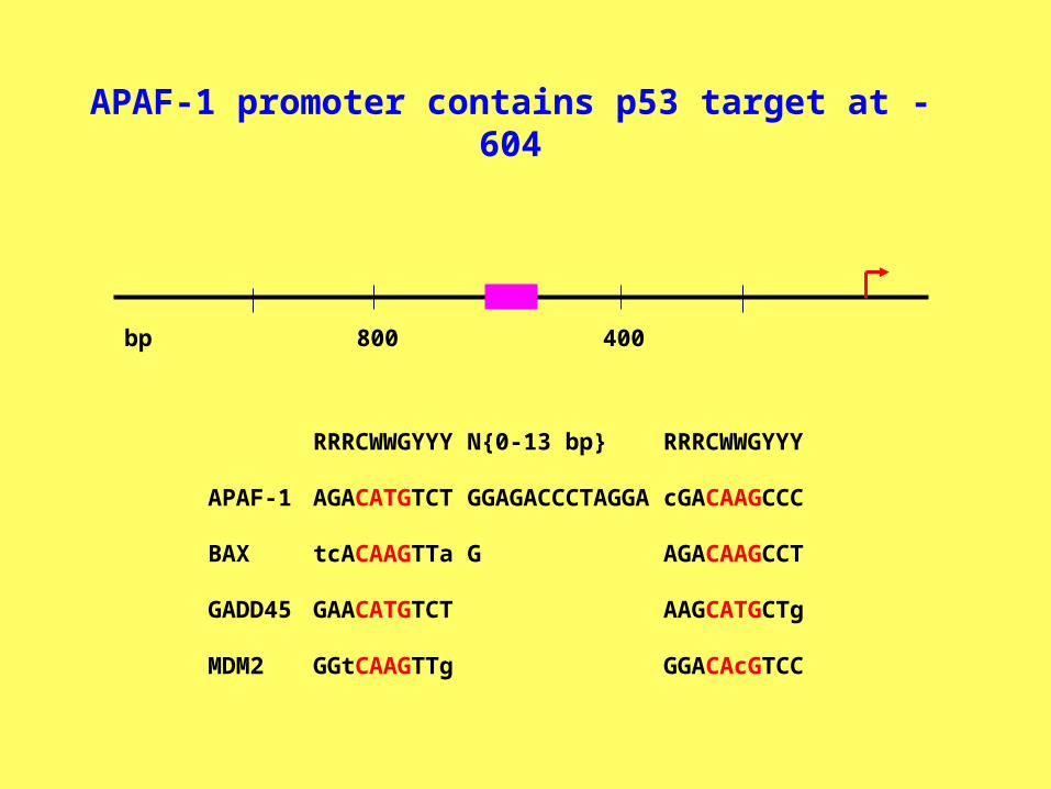
APAF-1 promoter contains p53 target at -604
RRRCWWGYYY N{0-13 bp} RRRCWWGYYY
APAF-1 AGACATGTCT GGAGACCCTAGGA cGACAAGCCC
BAX tcACAAGTTa G AGACAAGCCT
GADD45 GAACATGTCT AAGCATGCTg
MDM2 GGtCAAGTTg GGACAcGTCC
400800bp
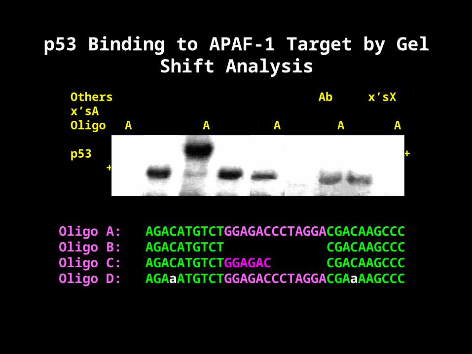
Others Ab x’sX x’sAOligo A A A A A A B C Dp53 + + + + mut + + +
Oligo A: AGACATGTCTGGAGACCCTAGGACGACAAGCCC Oligo B: AGACATGTCT CGACAAGCCCOligo C: AGACATGTCTGGAGAC CGACAAGCCCOligo D: AGAaATGTCTGGAGACCCTAGGACGAaAAGCCC
p53 Binding to APAF-1 Target by Gel Shift Analysis
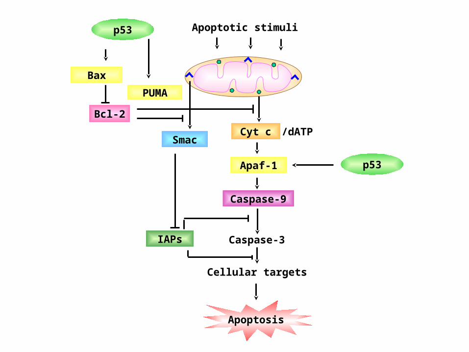
IAPs
Smac
Bcl-2
Apoptosis
Apaf-1
Caspase-9
Cyt c
Bax
Apoptotic stimuli
/dATP
Caspase-3
Cellular targets
p53
p53
PUMA
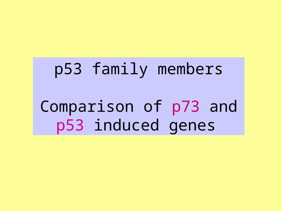
p53 family members
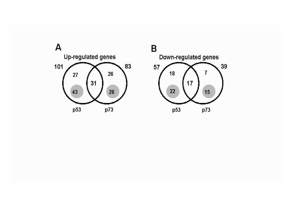
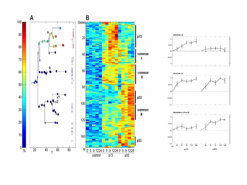
Comparison of p73 and p53 induced genes
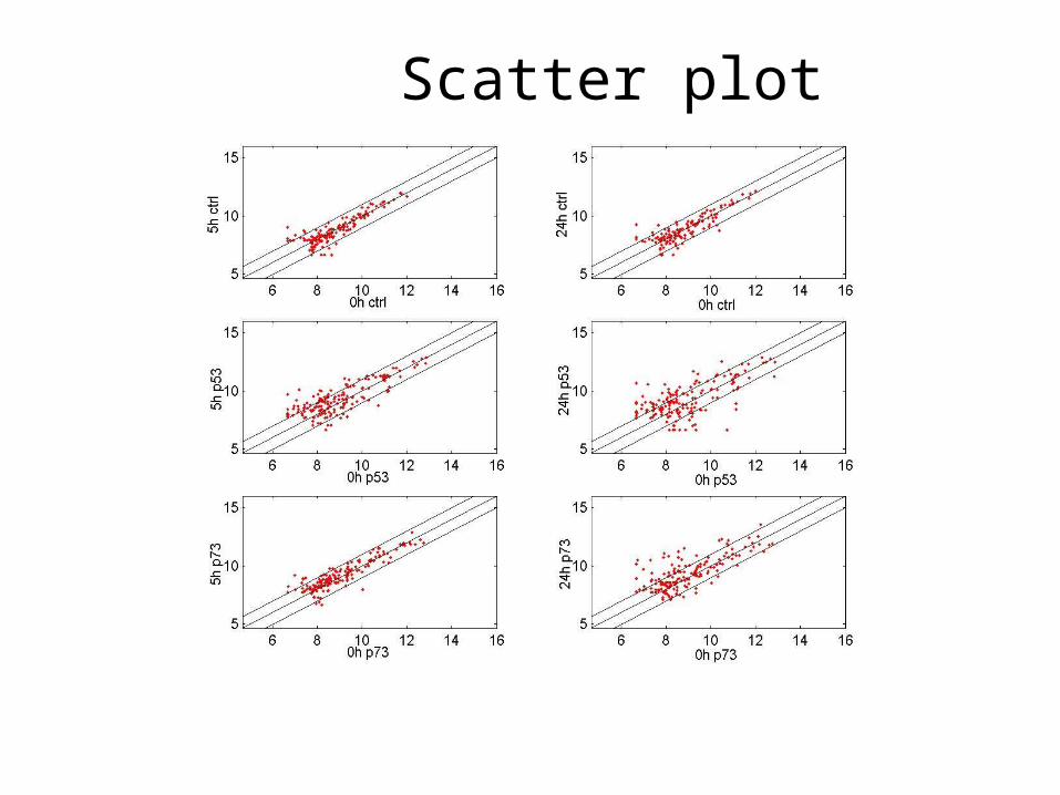
Scatter plot
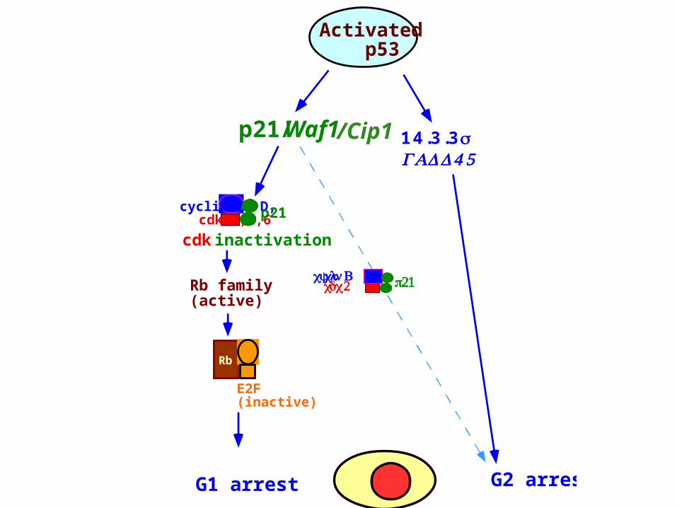
Rb family(active)
Rb
E2F(inactive)
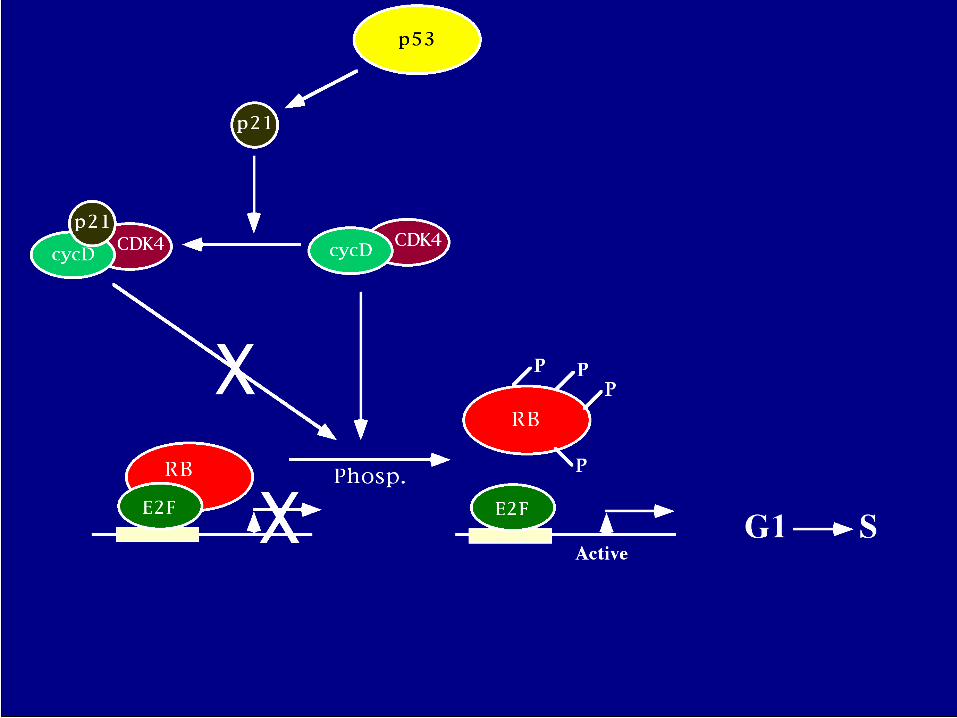
cyclin E, D, cdk 2,4,6 p21
cdk inactivation
G1 arrest
p21/Waf1
Activated p53
G2 arrest
14.3.3σGADD4 5
p21 cyclin B cdc 2
/Cip1
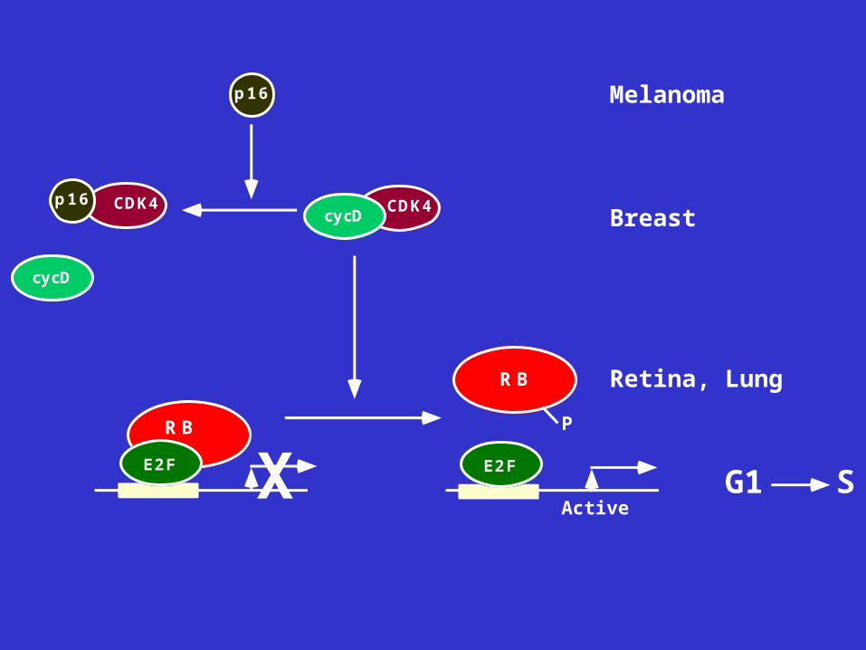
CDK4cycD
RB
E2F X
RB
P
E2F
Active
cy cD
CDK4p 1 6
p 1 6 Melanoma
Breast
Retina, Lung
G1 S