Cancer Research Immune Infiltration of Spontaneous...
12
Microenvironment and Immunology Immune Infiltration of Spontaneous Mouse Astrocytomas Is Dominated by Immunosuppressive Cells from Early Stages of Tumor Development Nhu Nam Tran Thang 1 , Madiha Derouazi 1 , Géraldine Philippin 1 , Séverine Arcidiaco 1 , Wilma Di Berardino-Besson 1 , Frédérick Masson 1 , Sabine Hoepner 1 , Cristina Riccadonna 1 , Karim Burkhardt 2 , Abhijit Guha 3 , Pierre-Yves Dietrich 1 , and Paul R. Walker 1 Abstract Immune infiltration of advanced human gliomas has been shown, but it is doubtful whether these immune cells affect tumor progression. It could be hypothesized that this infiltrate reflects recently recruited immune cells that are immediately overwhelmed by a high tumor burden. Alternatively, if there is earlier immune de- tection and infiltration of the tumor, the question arises as to when antitumor competency is lost. To address these issues, we analyzed a transgenic mouse model of spontaneous astrocytoma (GFAP-V 12 HA-ras mice), which allows the study of immune interactions with developing glioma, even at early asymptomatic stages. T cells, including a significant proportion of Tregs, are already present in the brain before symptoms develop, followed later by macrophages, natural killer cells, and dendritic cells. The effector potential of CD8 T-cells is defective, with the absence of granzyme B expression and low expression of IFN-γ, tumor necrosis factor, and interleukin 2. Overall, our results show an early defective endogenous immune response to gliomas, and local accumulation of immunosuppressive cells at the tumor site. Thus, the antiglioma response is not simply over- whelmed at advanced stages of tumor growth, but is counterbalanced by an inhibitory microenvironment from the outset. Nevertheless, we determined that effector molecule expression (granzyme B, IFN-γ) by brain-infiltrating CD8 T-cells could be enhanced, despite this unfavorable milieu, by strong immune stimuli. This potential to modulate the strong imbalance in local antiglioma immunity is encouraging for the deve- lopment and optimization of future glioma immunotherapies. Cancer Res; 70(12); 4829–39. ©2010 AACR. Introduction Interactions between cancer and the immune system have been proposed to rely on three processes: elimination (im- munosurveillance), equilibrium, and escape, leading to im- munologic sculpting of the tumor (immunoediting). Evidence for these mechanisms comes from mouse models, observation of large cohorts of immunosuppressed patients showing increased cancer incidence, and in certain tumors, correlation between immune infiltration and clinical out- come (1–3). In mice with defined immunodeficiencies or after antibody blockade/depletion of specific immunologic components, both innate and adaptive immune responses affected tumor growth (3). However, whether the theory of immunoediting is applicable to primary brain tumors has not yet been addressed. Gliomas and, in particular, malignant astrocytomas have a poor prognosis and are to date incurable. These tumors are infiltrated by macrophages, CD4 and CD8 T lymphocytes, and natural killer (NK) cells (4). However, the correlation bet- ween this immune infiltrate and clinical outcome is contro- versial, partly due to the lack of multivariate analyses taking into account tumor grade, patient treatment before and after surgery, and tumor-infiltrating immune cell phenotype. Finally, analyses of the glioma immune infiltrate are per- formed only at advanced tumor stages in patients. Thus, at least at these late stages, gliomas are not ignored by the immune system, but earlier tumor-immune interactions remain to be elucidated. Indeed, deficient immunosurveil- lance of gliomas may be explained by defective afferent immunity delaying the induction of immune effector me- chanisms, which are then overwhelmed by the tumor mass (5). Alternatively, an antiglioma immune infiltrate appea- ring earlier might shape the tumor to develop less immuno- genic variants or immune escape mechanisms. These issues must be studied in the context of the brain, as this site Authors' Affiliations: 1 Center of Oncology, 2 Neuropathology Unit, Department of Surgical Pathology, Geneva University Hospitals and University of Geneva, Geneva, Switzerland; and 3 Arthur and Sonia Labatt Brain Tumor Research Center, The Hospital for Sick Children Research Institute, and Division of Neurosurgery, Department of Surgery, Toronto Western Hospital, University Health Network, University of Toronto, Toronto, Canada Note: Supplementary data for this article are available at Cancer Research Online (http://cancerres.aacrjournals.org/). Corresponding Author: Paul R. Walker, Center of Oncology, Geneva University Hospitals, 4 rue Gabrielle-Perret-Gentil, 1211 Geneva 14, Switzerland. Phone: 41-22372-9880; Fax: 41-22372-9858; E-mail: paul. [email protected]. doi: 10.1158/0008-5472.CAN-09-3074 ©2010 American Association for Cancer Research. Cancer Research www.aacrjournals.org 4829 on May 27, 2018. © 2010 American Association for Cancer Research. cancerres.aacrjournals.org Downloaded from Published OnlineFirst May 25, 2010; DOI: 10.1158/0008-5472.CAN-09-3074
Transcript of Cancer Research Immune Infiltration of Spontaneous...

Published OnlineFirst May 25, 2010; DOI: 10.1158/0008-5472.CAN-09-3074
Microenvironment and Immunology
CancerResearch
Immune Infiltration of Spontaneous Mouse AstrocytomasIs Dominated by Immunosuppressive Cells fromEarly Stages of Tumor Development
Nhu Nam Tran Thang1, Madiha Derouazi1, Géraldine Philippin1, Séverine Arcidiaco1,Wilma Di Berardino-Besson1, Frédérick Masson1, Sabine Hoepner1, Cristina Riccadonna1,Karim Burkhardt2, Abhijit Guha3, Pierre-Yves Dietrich1, and Paul R. Walker1
Abstract
Authors' ADepartmenUniversityLabatt BraResearchSurgery, TUniversity o
Note: SupResearch O
CorresponUniversitySwitzerlandwalker@hcu
doi: 10.115
©2010 Am
www.aacr
Downlo
Immune infiltration of advanced human gliomas has been shown, but it is doubtful whether these immunecells affect tumor progression. It could be hypothesized that this infiltrate reflects recently recruited immunecells that are immediately overwhelmed by a high tumor burden. Alternatively, if there is earlier immune de-tection and infiltration of the tumor, the question arises as to when antitumor competency is lost. To addressthese issues, we analyzed a transgenic mouse model of spontaneous astrocytoma (GFAP-V12HA-ras mice),which allows the study of immune interactions with developing glioma, even at early asymptomatic stages.T cells, including a significant proportion of Tregs, are already present in the brain before symptoms develop,followed later by macrophages, natural killer cells, and dendritic cells. The effector potential of CD8 T-cells isdefective, with the absence of granzyme B expression and low expression of IFN-γ, tumor necrosis factor, andinterleukin 2. Overall, our results show an early defective endogenous immune response to gliomas, and localaccumulation of immunosuppressive cells at the tumor site. Thus, the antiglioma response is not simply over-whelmed at advanced stages of tumor growth, but is counterbalanced by an inhibitory microenvironmentfrom the outset. Nevertheless, we determined that effector molecule expression (granzyme B, IFN-γ) bybrain-infiltrating CD8 T-cells could be enhanced, despite this unfavorable milieu, by strong immune stimuli.This potential to modulate the strong imbalance in local antiglioma immunity is encouraging for the deve-lopment and optimization of future glioma immunotherapies. Cancer Res; 70(12); 4829–39. ©2010 AACR.
Introduction
Interactions between cancer and the immune system havebeen proposed to rely on three processes: elimination (im-munosurveillance), equilibrium, and escape, leading to im-munologic sculpting of the tumor (immunoediting).Evidence for these mechanisms comes from mouse models,observation of large cohorts of immunosuppressed patientsshowing increased cancer incidence, and in certain tumors,correlation between immune infiltration and clinical out-come (1–3). In mice with defined immunodeficiencies or
ffiliations: 1Center of Oncology, 2Neuropathology Unit,t of Surgical Pathology, Geneva University Hospitals andof Geneva, Geneva, Switzerland; and 3Arthur and Soniain Tumor Research Center, The Hospital for Sick ChildrenInstitute, and Division of Neurosurgery, Department oforonto Western Hospital, University Health Network,f Toronto, Toronto, Canada
plementary data for this article are available at Cancernline (http://cancerres.aacrjournals.org/).
ding Author: Paul R. Walker, Center of Oncology, GenevaHospitals, 4 rue Gabrielle-Perret-Gentil, 1211 Geneva 14,. Phone: 41-22372-9880; Fax: 41-22372-9858; E-mail: paul.ge.ch.
8/0008-5472.CAN-09-3074
erican Association for Cancer Research.
journals.org
on May 27, 201cancerres.aacrjournals.org aded from
after antibody blockade/depletion of specific immunologiccomponents, both innate and adaptive immune responsesaffected tumor growth (3). However, whether the theory ofimmunoediting is applicable to primary brain tumors hasnot yet been addressed.Gliomas and, in particular, malignant astrocytomas have a
poor prognosis and are to date incurable. These tumors areinfiltrated by macrophages, CD4 and CD8 T lymphocytes,and natural killer (NK) cells (4). However, the correlation bet-ween this immune infiltrate and clinical outcome is contro-versial, partly due to the lack of multivariate analyses takinginto account tumor grade, patient treatment before and aftersurgery, and tumor-infiltrating immune cell phenotype.Finally, analyses of the glioma immune infiltrate are per-formed only at advanced tumor stages in patients. Thus,at least at these late stages, gliomas are not ignored bythe immune system, but earlier tumor-immune interactionsremain to be elucidated. Indeed, deficient immunosurveil-lance of gliomas may be explained by defective afferentimmunity delaying the induction of immune effector me-chanisms, which are then overwhelmed by the tumor mass(5). Alternatively, an antiglioma immune infiltrate appea-ring earlier might shape the tumor to develop less immuno-genic variants or immune escape mechanisms. These issuesmust be studied in the context of the brain, as this site
4829
8. © 2010 American Association for Cancer Research.

Tran Thang et al.
4830
Published OnlineFirst May 25, 2010; DOI: 10.1158/0008-5472.CAN-09-3074
imposes such a specialized interface between cancer andthe immune system.Immune privilege of the brain, originally defined by pro-
longed allograft survival (6), has been reconsidered as a pro-cess concerning the brain parenchyma only (7). Moreover,this status is perturbed during brain inflammation. It relieson unique features such as tight regulation of traffickingthrough the blood-brain barrier, the absence of conventionallymphatic drainage, enrichment in local potent immunosup-pressive factors such as transforming growth factor-β (8),and the absence of resident parenchymal dendritic cells (DC).Endogenous brain cells such as microglial cells and astrocytescould participate in local immune responses and promote therecruitment of other cell types (9, 10). However, afferent immu-nity and cell-transported antigen egress from the brain mightstill be impaired (11). Nevertheless, in inflammatory or tumoralsituations, T cells are eventually activated, induced to prolife-rate, and imprinted with the necessary adhesionmolecules thatfacilitate their migration to the brain (12–16).Glioma cells might escape T-cell recognition by downregu-
lation of MHC molecules, and local hypoxia might impedeT-cell function (17). Active immunosuppressive mechanismsin human gliomas include soluble factors such as transfor-ming growth factor-β (18, 19) and cell surface molecules(20, 21). Several recent studies have focused on glioma-infiltrating immunosuppressive cells, of which Tregs are thebest defined (22, 23). However, the most abundant glioma-infiltrating cells are macrophages/activated microglia. Theyrepresent either an influx of peripheral monocytes thatdifferentiate into macrophages, or the in situ proliferationand activation of resident microglia. Whether they promoteor antagonize tumor growth is unclear (24).To study tumor-immune system interactions, murine
models of intracranially implanted tumors have been inva-luable, facilitating analysis of effector T-cell entry and func-tionality in the brain (14, 16, 25, 26), and of how tumors canescape immune effector mechanisms (17). However, in thesemodels, the blood-brain barrier is inevitably breached by tu-mor implantation, and transformed cells rapidly establish atumor mass, leading to an acute response, with early influx ofimmune cells into the brain. To better model slowly progres-sive glioma growth, genetic models of spontaneous cancerare now emerging (27).Here, we used a transgenic model of spontaneous astrocy-
toma (GFAP-V12HA-ras B8, referred to hereafter as RasB8mice) to study immune responses at occult stages of gliomadevelopment. In this model, activated ras is selectively ex-pressed in astrocytes under the control of the glial fibrillaryacidic protein (GFAP) promoter. Hemizygous RasB8 micegradually develop astrocytic hyperplasia and more aggressivesolitary or multifocal grade II/III astrocytoma-like lesions by12 weeks. Additional mutations occur, such as loss of PTENand p16 expression, and most mice develop symptoms anddie between 2 and 6 months of age (28, 29). At early asymp-tomatic stages, we already detected immune infiltrationof RasB8 brains, including infiltrating Tregs and granzymeB–negative CD8 T-cells, thereby unfavorable for antitumorimmunity. However, we showed that partial local immune
Cancer Res; 70(12) June 15, 2010
on May 27, 201cancerres.aacrjournals.org Downloaded from
competency could be achieved with peripheral vaccinationwith virus, or an intracranial immunogenic stimulus.
Materials and Methods
Mice and vaccinationsHemizygous transgenic RasB8 mice, endogenously expres-
sing a single allele of oncogenic V12HA-ras, have been pre-viously described (28). RasB8 mice were maintained on aCD1 background, or backcrossed at least seven generationsto the 129SvPas (H-2b) background (RasB8×129SvPas) whereindicated, and were genotyped as described. Mice exhibitingone/several of the following signs were considered sympto-matic: weight loss, ruffled fur, hunched back, decreased acti-vity/prostration, paresis/paralysis, convulsions, or irregularbreathing. Mice were sacrificed by CO2 asphyxia and all proce-dures were approved by the Institutional Ethical Committeeand the Cantonal Veterinary Office. Vaccination ofRasB8×129SvPas mice was performed using a recombinantvaccinia virus encoding β-galactosidase (VV-LacZ), whichwas kindly provided by D. Pinschewer (University of Geneva,Geneva, Switzerland). Brains were analyzed 7 days after intra-peritoneal infection with 2 × 10⁶ plaque-forming units ofVV-LacZ. The RasB8×129SvPas background was chosen tofacilitate the detection of CD8 T-cell responses to definedH-2b–restricted epitopes of VV and β-galactosidase (30).
Cell preparationsAfter transcardial perfusion with isotonic Ringer's solution,
brains were dissociated by mechanoenzymatic digestion asdescribed (14), and brain-infiltrating leukocytes (BIL) werepartially purified by ultracentrifugation (30,000 × g) on a con-tinuous 30% Percoll gradient (Fluka).
ImmunohistochemistryRinger's-perfused brains were frozen in 2-methylbutane.
Cryosections (7 μm) were fixed in 3.7% formaldehyde. Cryo-sections were microwaved before staining for Ki-67 or iso-type control (BD Biosciences). Alternatively, cryosectionswere directly stained for GFAP or with isotype control(Dako), and CD45 or with isotype control (BD Biosciences).Secondary antibodies included Envision goat anti-mousehorseradish peroxidase (Dako), donkey anti-rabbit horse-radish peroxidase (Immunoresearch), revealed with DABchromogen (Dako), and goat anti-rat biotin (MolecularProbes) used with Vectastain A+B kit (Vector Laboratories)and revealed with AEC (Sigma). Cryosections were counter-stained with hematoxylin. Images were recorded on an Axio-cam CCD camera–equipped Axiophot microscope, andanalyzed with Axiovision software (Zeiss).
Flow cytometryAfter FcR blocking, the following antibodies were used:
CD3 (145-2C11), CD4 (GK1.5, H129.19, RM4-5), CD8 (53-6.7),CD11b (M1-70), CD11c (HL3), CD25 (7D4, PC61), CD44(IM7), CD45 (30-F11), CD45R/B220 (RA3-6B2), CD49b (DX5),CD49d/anti-α4 integrin (9C10), CD69 (H1.2F3), CD103(M290), anti-α4β7 integrin/LPAM (DATK32), GR1/Ly6G
Cancer Research
8. © 2010 American Association for Cancer Research.
Immune Responses in Spontaneous Mouse Astrocytomas
Published OnlineFirst May 25, 2010; DOI: 10.1158/0008-5472.CAN-09-3074
(RB6-8C5), IFN-γ (XMG1.2), interleukin 2 (JES6-5H4), tumornecrosis factor (MP6-XT22), and appropriate control isotypesfrom BD Biosciences or BioLegend; anti-FoxP3 (FJK-16S) andcontrol isotype from eBiosciences; anti-granzyme B (GB11)and control isotype from Caltag; and CD62L from Immuno-Kontact. eBiosciences (FoxP3 staining) or BD Biosciences(IFN-γ, tumor necrosis factor, interleukin 2, granzyme B) per-meabilization and fixation kits were used according to theinstructions of the manufacturer. Where indicated, cells wererestimulated with phorbol 12-myristate 13-acetate and iono-mycin in a monensin solution for 6 hours or with a VV peptide(B8R20–27, TSYKFESV). Live leukocytes were gated on scatterand analyzed using BD Bioscience or Beckman Coulter cyto-meters, and results processed with FlowJo software (TreeStar, Inc.).
Statistical analysisStatistical analyses were performed using Prism 4.0 software
(GraphPad), and considered statistically significant if P < 0.05.
Results
Early and late brain infiltration by leukocytes inRasB8 miceAs previously shown in the initial characterization of this
model, widespread astrocytic hyperplasia leads to a gradualincrease in the number of GFAP+ astrocytes between 3 and12 weeks. Moreover, astrocytes in the neoplastic lesions exhibitnuclear atypia and higher bromodeoxyuridine labeling (29).
www.aacrjournals.org
on May 27, 201cancerres.aacrjournals.org Downloaded from
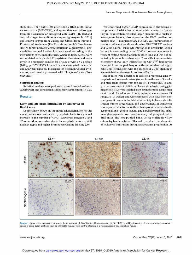
We confirmed higher GFAP expression in the brains ofsymptomatic RasB8 mice by immunohistochemistry. Hema-toxylin counterstain revealed larger pleiomorphic nuclei inastrocytoma lesions, also expressing the Ki-67 proliferationmarker (Fig. 1; Supplementary Fig. S1). We immunostainedsections adjacent to those showing Ki-67+GFAP+ regionsand found a CD45+ leukocyte infiltration in neoplastic lesions,but not in surrounding tissue. CD45 expression was lower inresident resting microglia than in other BILs and was not de-tected by immunohistochemistry. Thus, CD45 immunohisto-chemistry shows only infiltration by CD45high leukocytesrecruited from the periphery or activated resident microglialcells. This is consistent with the absence of CD45+ staining inage-matched nontransgenic controls (Fig. 1).RasB8 mice were described to develop progressive glial hy-
perplasia and low-grade astrocytomas from the age of 4 weeks,and high-grade lesions from the age of 12 weeks (29). To ana-lyze the involvement of different leukocyte subsets during glio-magenesis, BILs were isolated from asymptomatic RasB8mice(at 4, 8, and 12 weeks), and from symptomatic mice (mean, 13;range, 10–14 weeks), and were compared with BILs from non-transgenic littermates. Individual variability in leukocyte infil-tration, tumor progression, and development of symptomswas expected due to the outbred background and stochasticaccumulation of genetic lesions, and parallels variability in hu-man gliomagenesis. We therefore analyzed groups of indivi-dual mice and not pooled BILs, using multicolor flowcytometry to characterize BILs and to evaluate the dynamicsof leukocyte infiltration during astrocytoma progression. As
Figure 1. Leukocytes colocalize with pathologic lesions in ill RasB8 mice. Representative Ki-67, GFAP, and CD45 staining of corresponding neoplasticzones in serial brain sections from an ill RasB8 mouse, with control staining in a nontransgenic age-matched mouse.
Cancer Res; 70(12) June 15, 2010 4831
8. © 2010 American Association for Cancer Research.

Tran Thang et al.
4832
Published OnlineFirst May 25, 2010; DOI: 10.1158/0008-5472.CAN-09-3074
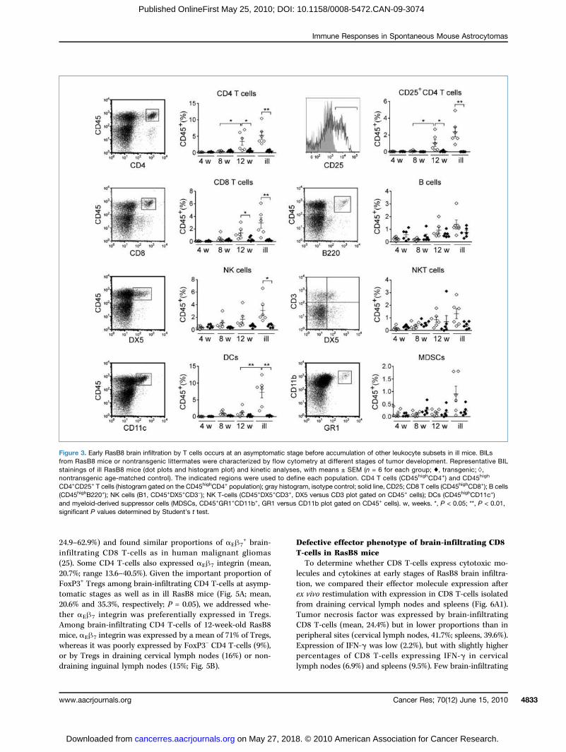
expected, statistical analysis of each group of mice was not al-ways representative of immune infiltration of individual mice,but is provided as an aid to understand the overall kinetics.The contribution of each leukocyte subset was expressed asa proportion of the total CD45+ leukocyte population, i.e., resi-dent microglia as well as infiltrating leukocytes (Figs. 2 and 3).In contrast to immunohistochemistry, resting CD45mid micro-glial cells in addition to CD45high cells are detected by flowcytometry, and constitute the major leukocyte population inBILs of nontransgenic and transgenic asymptomatic mice.However, in ill RasB8 mice, the balance switches towards pre-dominantly CD45high cells (Fig. 2A and B). At the early ages of 4and 8 weeks, there was no significant immune infiltration ofRasB8 brains. However, CD4 and CD8 T-cells (Fig. 3), but noother leukocyte subset, infiltrated the brains of still asymp-tomatic RasB8 mice by 12 weeks. A significant proportion ofCD4 T cells was CD4+CD25+, which includes Tregs. In termi-nally ill mice, infiltration by macrophages/activated microglia(Fig. 2B), CD4 and CD8 T-cells, NK cells, and DCs (Fig. 3), wasstatistically significant. Moreover, we found an even higherproportion of CD4+CD25+ Tregs. CD45highCD11c+ DCs werepredominantly CD11b+CD8−, with a low proportion (mean,1.8%) of CD11b+Gr1+ inflammatory DCs; they expressed highlevels ofMHC class II andwere CD86+ (Supplementary Fig. S2).Despite a trend for infiltration by B cells, NK T-cells, andmyeloid-derived suppressor cells (Fig. 3), their contributionto the infiltrate was variable and did not differ significantlyfrom nontransgenic littermates. Thus, at 12 weeks, whenpathologic lesions progress to high-grade astrocytomas but
Cancer Res; 70(12) June 15, 2010
on May 27, 201cancerres.aacrjournals.org Downloaded from
mice are still asymptomatic, influx of leukocytes begins witha T-cell infiltrate, including Tregs. Infiltration by macro-phages, NK cells, and DCs becomes detectable only in termi-nally ill mice. To understand the role of CD4 and CD8 T-cells inearly immune responses to astrocytomas, we consequentlyanalyzed them in 12-week-old, asymptomatic RasB8 mice.
Expression of α4β1 and αEβ7 integrins bybrain-infiltrating CD8 and CD4 T-cells at earlystages of astrocytoma progressionIn autoimmune diseases and in mice implanted intracra-
nially with fibrosarcomas, α4β1 integrin plays a key role infacilitating T-cell entry into the brain (14, 31). However, itsimportance was never addressed in human gliomas or mu-rine spontaneous astrocytomas. Because α4 integrin asso-ciates either with β1 or β7 integrins as a heterodimer atthe cell surface, upregulation of α4 and β1 in the absenceof β7 corresponds to preferential α4β1 upregulation. Inbrain-infiltrating CD8 and CD4 T-cells of 12-week-old RasB8mice, we indeed found such integrin expression (Fig. 4A) thatwas at a higher level than in T cells isolated from cervical oringuinal lymph nodes. Upregulation of β1 integrin in brain-infiltrating CD4 and CD8 T-cells confirmed its availability forpairing with α4 integrin (data not shown).In intracranially implanted tumors, the induction of αEβ7
integrin expression in CD8 T-cells correlated with theirretention in the tumor site and with an activated effectorphenotype (25). We analyzed αEβ7 integrin expression inBILs of 12-week-old RasB8 mice (Fig. 4B; mean, 42.1%; range
Figure 2. Brain infiltration is composed mainly of CD45mid resting microglia in asymptomatic RasB8 and nontransgenic mice, but of CD45high cells in ill mice.BILs from individual RasB8 mice or nontransgenic littermates were characterized by flow cytometry at 4, 8, and 12 wk (w), and in terminally ill mice, aswell as in age-matched nontransgenic (ntg) controls (n = 6 for each group; ♦, RasB8; ◊, ntg). A, representative CD45 histograms in RasB8 mice andin a nontransgenic control, age-matched to the ill mouse (solid lines, CD45 staining; gray histograms, isotype control). B, scatterplot summarizes theCD45high population at indicated times. C, CD11b versus CD45 dot plot in an ill RasB8 mouse, showing CD11b+CD45high macrophages (MAC)/activatedmicroglia (MG), and CD11b+CD45mid resting microglia. Scatterplots (with means ± SEM) summarize the MAC/MG populations at indicated times.**, P < 0.01; ***, P < 0.001, significant P values determined by Student's t test.
Cancer Research
8. © 2010 American Association for Cancer Research.
Immune Responses in Spontaneous Mouse Astrocytomas
Published OnlineFirst May 25, 2010; DOI: 10.1158/0008-5472.CAN-09-3074
24.9–62.9%) and found similar proportions of αEβ7+ brain-
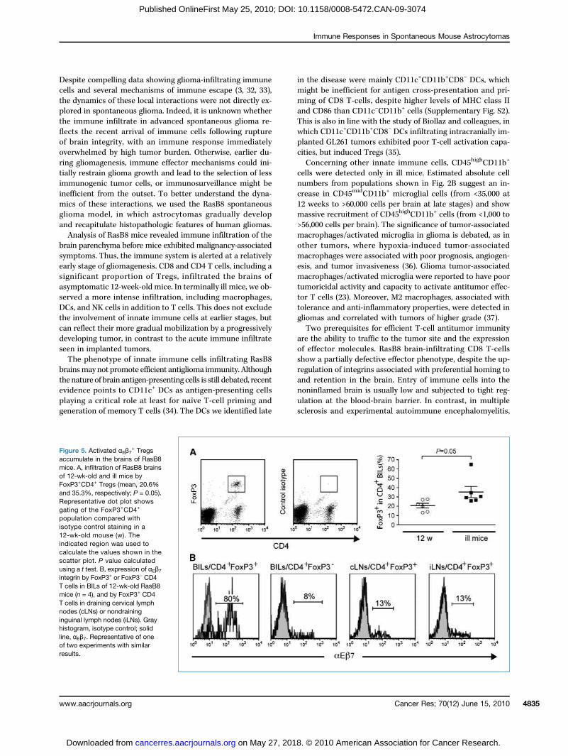
infiltrating CD8 T-cells as in human malignant gliomas(25). Some CD4 T-cells also expressed αEβ7 integrin (mean,20.7%; range 13.6–40.5%). Given the important proportion ofFoxP3+ Tregs among brain-infiltrating CD4 T-cells at asymp-tomatic stages as well as in ill RasB8 mice (Fig. 5A; mean,20.6% and 35.3%, respectively; P = 0.05), we addressed whe-ther αEβ7 integrin was preferentially expressed in Tregs.Among brain-infiltrating CD4 T-cells of 12-week-old RasB8mice, αEβ7 integrin was expressed by a mean of 71% of Tregs,whereas it was poorly expressed by FoxP3− CD4 T-cells (9%),or by Tregs in draining cervical lymph nodes (16%) or non-draining inguinal lymph nodes (15%; Fig. 5B).
www.aacrjournals.org
on May 27, 201cancerres.aacrjournals.org Downloaded from
Defective effector phenotype of brain-infiltrating CD8T-cells in RasB8 miceTo determine whether CD8 T-cells express cytotoxic mo-
lecules and cytokines at early stages of RasB8 brain infiltra-tion, we compared their effector molecule expression afterex vivo restimulation with expression in CD8 T-cells isolatedfrom draining cervical lymph nodes and spleens (Fig. 6A1).Tumor necrosis factor was expressed by brain-infiltratingCD8 T-cells (mean, 24.4%) but in lower proportions than inperipheral sites (cervical lymph nodes, 41.7%; spleens, 39.6%).Expression of IFN-γ was low (2.2%), but with slightly higherpercentages of CD8 T-cells expressing IFN-γ in cervicallymph nodes (6.9%) and spleens (9.5%). Few brain-infiltrating
Figure 3. Early RasB8 brain infiltration by T cells occurs at an asymptomatic stage before accumulation of other leukocyte subsets in ill mice. BILsfrom RasB8 mice or nontransgenic littermates were characterized by flow cytometry at different stages of tumor development. Representative BILstainings of ill RasB8 mice (dot plots and histogram plot) and kinetic analyses, with means ± SEM (n = 6 for each group; ♦, transgenic;◊,nontransgenic age-matched control). The indicated regions were used to define each population. CD4 T cells (CD45highCD4+) and CD45high
CD4+CD25+ T cells (histogram gated on the CD45highCD4+ population); gray histogram, isotype control; solid line, CD25; CD8 T cells (CD45highCD8+); B cells(CD45highB220+); NK cells (B1, CD45+DX5+CD3−); NK T-cells (CD45+DX5+CD3+, DX5 versus CD3 plot gated on CD45+ cells); DCs (CD45highCD11c+)and myeloid-derived suppressor cells (MDSCs, CD45+GR1+CD11b+, GR1 versus CD11b plot gated on CD45+ cells). w, weeks. *, P < 0.05; **, P < 0.01,significant P values determined by Student's t test.
Cancer Res; 70(12) June 15, 2010 4833
8. © 2010 American Association for Cancer Research.

Tran Thang et al.
4834
Published OnlineFirst May 25, 2010; DOI: 10.1158/0008-5472.CAN-09-3074
CD8 T-cells expressed interleukin 2 (0.2%) and granzyme B(1%). Overall brain-infiltrating CD8 T-cells in asymptomaticRasB8 mice exhibited a phenotype consistent with partiallydefective effector function, although they had a non-naïveCD62L−CD44high phenotype (data not shown). Granzyme Bexpression was absent, whether or not they were restimu-lated with phorbol 12-myristate 13-acetate/ionomycin ex vivo(1.0% and 1.3%, respectively; data not shown), and did notincrease at later stages in ill RasB8 mice (Fig. 6A3).
Induction of enhanced effector functions in T cellsinfiltrating RasB8 brainsTo determine whether the RasB8 brain microenvironment
inevitably overwhelms the effector function of CD8 T cells,we induced a more substantial immune infiltrate in responseto peripheral vaccination with VV-LacZ recombinant virus.This approach was chosen to stimulate T cells specific forthe β-galactosidase antigen encoded in the transgene (thusexpressed as an astrocytoma-associated antigen). Althoughβ-galactosidase–specific T cells were at/below the limit ofex vivo detection in VV-LacZ vaccinated mice, vaccinationdid induce T cells that infiltrated the brains of transgenicmice, and which had enhanced capacity to express IFN-γ.These T cells included viral antigen–specific CD8 T cells
Cancer Res; 70(12) June 15, 2010
on May 27, 201cancerres.aacrjournals.org Downloaded from
(Fig. 6B1), as well as CD4 T cells (Fig. 6B3). Moreover, theCD4/CD8 ratio in the BILs was significantly and consistentlydecreased after viral vaccination (Fig. 6B2). Viral vaccinationinduced no detectable T-cell infiltration of brains of non-transgenic littermate control mice (data not shown). We alsotested brain-infiltrating CD8 T-cells for granzyme B expres-sion after viral vaccination, but this remained negative(data not shown). Indeed, the only circumstances in whichwe could detect an infiltrate of granzyme B+ CD8 T-cells,was after intracranial implantation of immunogenic P815mastocytoma cells directly into the brain of RasB8×BALB/ctransgenic mice (Supplementary Fig. S3). Treg proportions inBILs were unchanged by the implantation (SupplementaryFig. S3C), indicating that the local immunosuppressive envi-ronment was permissive for CD8 T-cell expression of gran-zyme B when a sufficiently strong immune stimulus wasapplied locally.
Discussion
Analyses of human malignant astrocytoma and intracra-nially implanted mouse models highlight the complexity ofinteractions between the immune system and gliomas.
Figure 4. Brain-infiltrating CD4 and CD8 T cells in 12-wk-old RasB8 mice express integrins involved in tropism to and retention in the brain. Representativedot plots and histogram plots are shown, and results are summarized in graphs in which columns represent expression of integrins in individual mice.A, given expression of α4 integrin either as α4β7 or α4β1 heterodimer at the cell surface, expression of α4β1 integrin in RasB8 brain-infiltrating CD4 andCD8 T cells is inferred by positive α4 and negative α4β7 stainings. B, expression of αEβ7 integrin by CD4 and CD8 T cells in BILs of 12-wk-old RasB8 mice.Gray histogram, isotype control; solid line, αEβ7. The percentages noted in the histograms are from individual representative mice.
Cancer Research
8. © 2010 American Association for Cancer Research.
Immune Responses in Spontaneous Mouse Astrocytomas
Published OnlineFirst May 25, 2010; DOI: 10.1158/0008-5472.CAN-09-3074
Despite compelling data showing glioma-infiltrating immunecells and several mechanisms of immune escape (3, 32, 33),the dynamics of these local interactions were not directly ex-plored in spontaneous glioma. Indeed, it is unknown whetherthe immune infiltrate in advanced spontaneous glioma re-flects the recent arrival of immune cells following ruptureof brain integrity, with an immune response immediatelyoverwhelmed by high tumor burden. Otherwise, earlier du-ring gliomagenesis, immune effector mechanisms could ini-tially restrain glioma growth and lead to the selection of lessimmunogenic tumor cells, or immunosurveillance might beinefficient from the outset. To better understand the dyna-mics of these interactions, we used the RasB8 spontaneousglioma model, in which astrocytomas gradually developand recapitulate histopathologic features of human gliomas.Analysis of RasB8 mice revealed immune infiltration of the
brain parenchyma before mice exhibited malignancy-associatedsymptoms. Thus, the immune system is alerted at a relativelyearly stage of gliomagenesis. CD8 and CD4 T cells, including asignificant proportion of Tregs, infiltrated the brains ofasymptomatic 12-week-old mice. In terminally ill mice, we ob-served a more intense infiltration, including macrophages,DCs, and NK cells in addition to T cells. This does not excludethe involvement of innate immune cells at earlier stages, butcan reflect their more gradual mobilization by a progressivelydeveloping tumor, in contrast to the acute immune infiltrateseen in implanted tumors.The phenotype of innate immune cells infiltrating RasB8
brainsmay not promote efficient antiglioma immunity. Althoughthe nature of brain antigen-presenting cells is still debated, recentevidence points to CD11c+ DCs as antigen-presenting cellsplaying a critical role at least for naïve T-cell priming andgeneration of memory T cells (34). The DCs we identified late
www.aacrjournals.org
on May 27, 201cancerres.aacrjournals.org Downloaded from
in the disease were mainly CD11c+CD11b+CD8− DCs, whichmight be inefficient for antigen cross-presentation and pri-ming of CD8 T-cells, despite higher levels of MHC class IIand CD86 than CD11c−CD11b+ cells (Supplementary Fig. S2).This is also in line with the study of Biollaz and colleagues, inwhich CD11c+CD11b+CD8− DCs infiltrating intracranially im-planted GL261 tumors exhibited poor T-cell activation capa-cities, but induced Tregs (35).Concerning other innate immune cells, CD45highCD11b+
cells were detected only in ill mice. Estimated absolute cellnumbers from populations shown in Fig. 2B suggest an in-crease in CD45midCD11b+ microglial cells (from <35,000 at12 weeks to >60,000 cells per brain at late stages) and showmassive recruitment of CD45highCD11b+ cells (from <1,000 to>56,000 cells per brain). The significance of tumor-associatedmacrophages/activated microglia in glioma is debated, as inother tumors, where hypoxia-induced tumor-associatedmacrophages were associated with poor prognosis, angiogen-esis, and tumor invasiveness (36). Glioma tumor-associatedmacrophages/activated microglia were reported to have poortumoricidal activity and capacity to activate antitumor effec-tor T cells (23). Moreover, M2 macrophages, associated withtolerance and anti-inflammatory properties, were detected ingliomas and correlated with tumors of higher grade (37).Two prerequisites for efficient T-cell antitumor immunity
are the ability to traffic to the tumor site and the expressionof effector molecules. RasB8 brain-infiltrating CD8 T-cellsshow a partially defective effector phenotype, despite the up-regulation of integrins associated with preferential homing toand retention in the brain. Entry of immune cells into thenoninflamed brain is usually low and subjected to tight reg-ulation at the blood-brain barrier. In contrast, in multiplesclerosis and experimental autoimmune encephalomyelitis,
Figure 5. Activated αEβ7+ Tregs
accumulate in the brains of RasB8mice. A, infiltration of RasB8 brainsof 12-wk-old and ill mice byFoxP3+CD4+ Tregs (mean, 20.6%and 35.3%, respectively; P = 0.05).Representative dot plot showsgating of the FoxP3+CD4+
population compared withisotype control staining in a12-wk-old mouse (w). Theindicated region was used tocalculate the values shown in thescatter plot. P value calculatedusing a t test. B, expression of αEβ7integrin by FoxP3+ or FoxP3− CD4T cells in BILs of 12-wk-old RasB8mice (n = 4), and by FoxP3+ CD4T cells in draining cervical lymphnodes (cLNs) or nondraininginguinal lymph nodes (iLNs). Grayhistogram, isotype control; solidline, αEβ7. Representative of oneof two experiments with similarresults.
Cancer Res; 70(12) June 15, 2010 4835
8. © 2010 American Association for Cancer Research.

Tran Thang et al.
4836
Published OnlineFirst May 25, 2010; DOI: 10.1158/0008-5472.CAN-09-3074
in vitro and in vivo experiments with anti-α4 blocking anti-bodies showed a key functional role of α4β1 in controllingthe entry of T cells into the brain (31, 38, 39). Less is knownin tumoral situations, although α4β1 integrin facilitates T-cell
Cancer Res; 70(12) June 15, 2010
on May 27, 201cancerres.aacrjournals.org Downloaded from
access to the brain in acute responses to intracranially im-planted fibrosarcomas and melanomas (14, 16). We showfor the first time that this integrin is also upregulated inT cells infiltrating spontaneous gliomas. Furthermore, we
Figure 6. CD8 T cells infiltrating brains of RasB8 mice exhibit a partially defective effector phenotype but expression of IFN-γ by brain-infiltrating CD4and CD8 T cells could be stimulated by vaccination. A, defective effector phenotype of brain-infiltrating CD8 T cells. Representative stainings of tumornecrosis factor (TNF), IFN-γ, interleukin 2 (IL2), and granzyme B (GrzB) expression gated on CD8 T cells from BILs, draining cervical lymph nodes, or spleensof 12-wk-old RasB8 mice (A1). Quadrants were defined according to isotype controls and used to calculate the values shown in the histogram withmeans and SEM (A2, n = 6). A representative dot plot for granzyme B expression by brain-infiltrating CD8 T cells in an ill RasB8 mouse is shown andcompared with isotype control (A3, n = 4, mean expression granzyme B+ CD8 T cells, 1.96 ± 0.79%). Numbers above the quadrants correspond topercentages of CD8 T cells. B, peripheral vaccination with β-galactosidase recombinant vaccinia virus (VV-LacZ). BILs isolated from infected (Vacc) andnoninfected (Non-Vacc) RasB8×129SvPas mice were analyzed 7 days after infection. IFN-γ expression gated on CD8 T cells is shown after ex vivorestimulation with phorbol 12-myristate 13-acetate/ionomycin (PMA-iono) or with a VV peptide (B8R20–27, TSYKFESV; B1). Quadrants in representative dot plotswere defined according to isotype control staining. Values in the histogram represent means from all mice, ± SEM (n = 4). Ratio of CD4 to CD8 T cells in BILsfrom VV-LacZ noninfected and infected mice is also represented with means and SEM (B2). Dot plots of IFN-γ expression gated on CD4 T cells from BILs ofVV-LacZ noninfected and infectedmice are shown (n = 2), with quadrants defined according to isotype control staining (B3). P value was determined with ANOVA(A2) or Student's t test (B1 and B2; **, P < 0.01; ***, P < 0.001). The numbers above the quadrants correspond to percentages of CD8 or CD4 T cells as indicated.
Cancer Research
8. © 2010 American Association for Cancer Research.

Immune Responses in Spontaneous Mouse Astrocytomas
Published OnlineFirst May 25, 2010; DOI: 10.1158/0008-5472.CAN-09-3074
noted the expression of αEβ7 integrin by CD8 T-cells, whichwas associated with their retention in the brain (25), andwith granule exocytosis and antitumor activity in E-cadherin–expressing lung cancers (40). E-cadherin is not detected ingliomas, but it is assumed that other αEβ7 ligands exist be-cause E-cadherin–independent adhesion of αEβ7
+ lympho-cytes to endothelial and epithelial cells was described (41).It is likely that the transforming growth factor-β–rich gliomamicroenvironment plays a role in the induction of αEβ7
integrin (42). Indeed, in RasB8 brains, we confirmed trans-forming growth factor-β expression by quantitative reversetranscription-PCR (Supplementary Fig. S4).However, CD8 T-cells infiltrating RasB8 brains strikingly
differed from those observed in implanted models. In amodel using intracranial implantation of fibrosarcomas,antigen-experienced CD8+ T cells proliferated in the brainand upregulated their IFN-γ and granzyme B expression(25). CD8 T-cells infiltrating intracranially implanted GL261glioma similarly expressed effector molecules (43),4 althoughthese cells did not enable GL261 tumor rejection withoutTLR stimulation (44) or depletion of Tregs (43, 45). In RasB8mice, inefficient priming is one possible explanation for pooreffector molecule expression by brain-infiltrating CD8 T-cells.Indeed, Thomas and colleagues showed that brain tumorantigens draining to cervical lymph nodes generated a low-magnitude local CD8 T-cell response (46). Although in RasB8mice, elevated numbers of activated T cells were not de-tected in cervical lymph nodes (data not shown), this is likelydue to the gradual kinetics of tumorigenesis. Moreover, cen-tral tolerance to the β-galactosidase component of the trans-gene was absent because CD4 and CD8 T-cells proliferated inRasB8 mice immunized with β-galactosidase protein (datanot shown). Another hypothesis for the lack of effector mole-cule expression implicates local immunosuppressive factorsand cells. These could progressively inhibit proliferation andfull activation of CD8 T-cells early after infiltration. The im-portance of Tregs was addressed in acute immune responsesto intracranially implanted gliomas (33, 45); Treg depletionalone or in combination with vaccination had an effect onsurvival and antitumor immunity (43, 45, 47, 48). Tregs accu-mulate simultaneously with CD8 T-cells in the brains ofasymptomatic RasB8 mice and express αEβ7
+ integrin, whichcorrelates with an effector/memory phenotype, and their re-direction to inflammatory sites (49). Moreover αE
+ Tregswere recently shown to accumulate in tumors and to potentlysuppress antitumor CD8 T-cells (50). Tumor-associatedmacrophages/activated microglia observed in ill mice mayalso further perturb the local response and favor tumor pro-gression. These late effects are consistent with observations inhuman glioma (23, 37). However, importantly, the possibilityto study early phases of the immune response in the RasB8model has revealed Tregs as the main early immunosuppres-sive cells, suggesting that they tilt the balance towards tumorescape.
4 Our unpublished data.
www.aacrjournals.org
on May 27, 201cancerres.aacrjournals.org Downloaded from
Our peripheral vaccination experiments (Fig. 6B) indicatedthat a potentially clinically relevant approach could tilt the in-tracranial immune balance towards Tc1/Th1 (IFN-γ secre-ting) effector functions. Moreover, even symptomatic RasB8mice show unimpaired in vitro T-cell mitogen responses,and only minor changes in CD4/CD8 T-cell ratios in lymphnodes (Supplementary Fig. S5), suggesting that peripheralstimulation of antitumor immunity might be feasible. Howev-er, the absence of granzyme B expression by brain-infiltrating Tcells after vaccination could indicate that a stronger local im-mune stimulus (as illustrated in Supplementary Fig. S3), or in-hibition of Tregs or factors such as STAT-3, will be necessaryfor the induction of polyfunctional antitumor T cells expres-sing a range of effector molecules.These encouraging results suggest that future immu-
notherapies might function in the context of local Tregs, pro-vided the immune stimulation and brain tumor infiltration byeffector T cells are sufficient. Moreover, observations made inRasB8 mice at early stages might be relevant for the design ofcomplementary immunotherapies for unresectable lowergrade gliomas. Other approaches to generate mouse modelsrecapitulating progressive genetic alterations and histopatho-logic features of human glioma have been or are being devel-oped. These include models allowing retroviral gene transferto selected precursor cells, or models using transgenesis totarget individual or multiple genetic pathways (27). However,to our knowledge, these models have not been used to exam-ine the issue of host immunity or immunosuppression.Our study has taken a first step in addressing early
phases and dynamics of the immune response in spontaneousglioma. Elimination or equilibrium phases are unlikely to takeplace in these tumors in view of the early unbalanced responsetowards immunosuppression. The lack of an effective pre-existing CD8 T-cell response, the abundant Treg infiltrate,and the potential role of tumor-associated macrophages atlate stages of disease provide specific targets for future multi-modal glioma therapies.
Disclosure of Potential Conflicts of Interest
No potential conflicts of interest were disclosed.
Acknowledgments
We acknowledge the Geneva University Genomics and Flow CytometryCore Facilities, and laboratory members for support and advice. We aregrateful to Daniel Pinschewer, Burkhard Ludewig, Immanuel Luescher, andPhilippe Guillaume for reagents and advice for the viral vaccinationexperiments. We thank Arun Kamath and Valérie Dutoit for helpful discussions.
Grant Support
Association for International Cancer Research (05/465), Oncosuisse (OCS/01754/08/2005), Ligue Genevoise contre le Cancer (0514), Kisane Foundation(P.R. Walker), and the Cancer and Solidarity Foundation (N.N. Tran Thang).
The costs of publication of this article were defrayed in part by the paymentof page charges. This article must therefore be hereby marked advertisementin accordance with 18 U.S.C. Section 1734 solely to indicate this fact.
Received 08/17/2009; revised 03/29/2010; accepted 04/21/2010; publishedOnlineFirst 05/25/2010.
Cancer Res; 70(12) June 15, 2010 4837
8. © 2010 American Association for Cancer Research.
Tran Thang et al.
4838
Published OnlineFirst May 25, 2010; DOI: 10.1158/0008-5472.CAN-09-3074
References
1. Galon J, Costes A, Sanchez-Cabo F, et al. Type, density, and loca-tion of immune cells within human colorectal tumors predict clinicaloutcome. Science 2006;313:1960–4.
2. Leffers N, Gooden MJ, de Jong RA, et al. Prognostic significance oftumor-infiltrating T-lymphocytes in primary and metastatic lesions ofadvanced stage ovarian cancer. Cancer Immunol Immunother 2009;58:449–59.
3. Dunn GP, Koebel CM, Schreiber RD. Interferons, immunity and can-cer immunoediting. Nat Rev Immunol 2006;6:836–48.
4. Dunn GP, Dunn IF, Curry WT. Focus on TILs: prognostic significanceof tumor infiltrating lymphocytes in human glioma. Cancer Immun2007;7:12.
5. Walker PR, Calzascia T, de Tribolet N, Dietrich PY. T-cell immuneresponses in the brain and their relevance for cerebral malignancies.Brain Res Brain Res Rev 2003;42:97–122.
6. Medawar PB. Immunity to homologous grafted skin; the fate ofskin homografts transplanted to the brain, to subcutaneous tis-sue, and to the anterior chamber of the eye. Br J Exp Pathol1948;29:58–69.
7. Galea I, Bechmann I, Perry VH. What is immune privilege (not)?Trends Immunol 2007;28:12–8.
8. Carson MJ, Doose JM, Melchior B, Schmid CD, Ploix CC. CNSimmune privilege: hiding in plain sight. Immunol Rev 2006;213:48–65.
9. Hanisch UK, Kettenmann H. Microglia: active sensor and versatileeffector cells in the normal and pathologic brain. Nat Neurosci2007;10:1387–94.
10. Farina C, Aloisi F, Meinl E. Astrocytes are active players in cerebralinnate immunity. Trends Immunol 2007;28:138–45.
11. Bechmann I, Galea I, Perry VH. What is the blood-brain barrier (not)?Trends Immunol 2007;28:5–11.
12. Cayrol R, Wosik K, Berard JL, et al. Activated leukocyte cell adhesionmolecule promotes leukocyte trafficking into the central nervous sys-tem. Nat Immunol 2008;9:137–45.
13. Walter L, Albert ML. Cutting edge: cross-presented intracranial anti-gen primes CD8+ T cells. J Immunol 2007;178:6038–42.
14. Calzascia T, Masson F, Di Berardino-Besson W, et al. Homing phe-notypes of tumor-specific CD8 T cells are predetermined at the tu-mor site by crosspresenting APCs. Immunity 2005;22:175–84.
15. Prins RM, Shu CJ, Radu CG, et al. Anti-tumor activity and traffickingof self, tumor-specific T cells against tumors located in the brain.Cancer Immunol Immunother 2008;57:1279–89.
16. Sasaki K, Zhu X, Vasquez C, et al. Preferential expression of verylate antigen-4 on type 1 CTL cells plays a critical role in traffick-ing into central nervous system tumors. Cancer Res 2007;67:6451–8.
17. Walker PR, Prins RM, Dietrich P-Y, Liau LM. Harnessing T-cell immu-nity to target brain tumors. In: Van Meir EG, editor. CNS cancer:models, markers, prognostic factors, targets and therapeutic ap-proaches. New York: Humana Press; 2009, p. 1165–218.
18. Liau LM, Prins RM, Kiertscher SM, et al. Dendritic cell vaccination inglioblastoma patients induces systemic and intracranial T-cellresponses modulated by the local central nervous system tumormicroenvironment. Clin Cancer Res 2005;11:5515–25.
19. Weller M, Fontana A. The failure of current immunotherapy formalignant glioma. Tumor-derived TGF-β, T-cell apoptosis, and theimmune privilege of the brain. Brain Res Brain Res Rev 1995;21:128–51.
20. Saas P, Walker PR, Hahne M, et al. Fas ligand expression by astro-cytoma in vivo: maintaining immune privilege in the brain? J ClinInvest 1997;99:1173–8.
21. Wintterle S, Schreiner B, Mitsdoerffer M, et al. Expression of theB7-related molecule B7-1 by glioma cells: a potential mechanismof immune paralysis. Cancer Res 2003;63:7462–7.
22. El Andaloussi A, Lesniak MS. CD4+ CD25+ FoxP3+ T-cell infiltrationand heme oxygenase-1 expression correlate with tumor grade inhuman gliomas. J Neurooncol 2007;83:145–52.
23. Hussain SF, Yang D, Suki D, Aldape K, Grimm E, Heimberger AB.The role of human glioma-infiltrating microglia/macrophages in
Cancer Res; 70(12) June 15, 2010
on May 27, 201cancerres.aacrjournals.org Downloaded from
mediating antitumor immune responses. Neuro-oncol 2006;8:261–79.
24. Graeber MB, Scheithauer BW, Kreutzberg GW. Microglia in braintumors. Glia 2002;40:252–9.
25. Masson F, Calzascia T, Di Berardino-Besson W, de Tribolet N,Dietrich PY, Walker PR. Brain microenvironment promotes the finalfunctional maturation of tumor-specific effector CD8+ T cells.J Immunol 2007;179:845–53.
26. Plautz GE, Mukai S, Cohen PA, Shu S. Cross-presentation of tumorantigens to effector T cells is sufficient to mediate effective immu-notherapy of established intracranial tumors. J Immunol 2000;165:3656–62.
27. Huse JT, Holland EC. Genetically engineered mouse models of braincancer and the promise of preclinical testing. Brain Pathol 2009;19:132–43.
28. Ding H, Roncari L, Shannon P, et al. Astrocyte-specific expression ofactivated p21-ras results in malignant astrocytoma formation in atransgenic mouse model of human gliomas. Cancer Res 2001;61:3826–36.
29. Shannon P, Sabha N, Lau N, Kamnasaran D, Gutmann DH, Guha A.Pathological and molecular progression of astrocytomas in a GFAP:12 V-Ha-Ras mouse astrocytoma model. Am J Pathol 2005;167:859–67.
30. Ludewig B, Ochsenbein AF, Odermatt B, Paulin D, Hengartner H,Zinkernagel RM. Immunotherapy with dendritic cells directed againsttumor antigens shared with normal host cells results in severe auto-immune disease. J Exp Med 2000;191:795–804.
31. Engelhardt B, Kappos L. Natalizumab: targeting α4-integrins in mul-tiple sclerosis. Neurodegener Dis 2008;5:16–22.
32. Parsa AT, Waldron JS, Panner A, et al. Loss of tumor suppressorPTEN function increases B7-1 expression and immunoresistance inglioma. Nat Med 2007;13:84–8.
33. Grauer OM, Nierkens S, Bennink E, et al. CD4+FoxP3+ regulatoryT cells gradually accumulate in gliomas during tumor growth andefficiently suppress antiglioma immune responses in vivo. Int J Can-cer 2007;121:95–105.
34. Greter M, Heppner FL, Lemos MP, et al. Dendritic cells permit im-mune invasion of the CNS in an animal model of multiple sclerosis.Nat Med 2005;11:328–34.
35. Biollaz G, Bernasconi L, Cretton C, et al. Site-specific anti-tumor im-munity: differences in DC function, TGF-β production and numbersof intratumoral Foxp3+ Treg. Eur J Immunol 2009;39:1323–33.
36. Allavena P, Sica A, Garlanda C, Mantovani A. The Yin-Yang of tumor-associated macrophages in neoplastic progression and immune sur-veillance. Immunol Rev 2008;222:155–61.
37. Komohara Y, Ohnishi K, Kuratsu J, Takeya M. Possible involvementof the M2 anti-inflammatory macrophage phenotype in growth ofhuman gliomas. J Pathol 2008;216:15–24.
38. Baron JL, Madri JA, Ruddle NH, Hashim G, Janeway CA, Jr. Surfaceexpression of α4 integrin by CD4 T cells is required for their entry intobrain parenchyma. J Exp Med 1993;177:57–68.
39. Engelhardt B, Laschinger M, Schulz M, Samulowitz U, Vestweber D,Hoch G. The development of experimental autoimmune encephalo-myelitis in the mouse requires α4-integrin but not α4β7-integrin.J Clin Invest 1998;102:2096–105.
40. Le Floc'h A, Jalil A, Vergnon I, et al. αEβ7 integrin interaction withE-cadherin promotes antitumor CTL activity by triggering lytic gran-ule polarization and exocytosis. J Exp Med 2007;204:559–70.
41. Strauch UG, Mueller RC, Li XY, et al. Integrin αE(CD103)β7 med-iates adhesion to intestinal microvascular endothelial cell lines viaan E-cadherin-independent interaction. J Immunol 2001;166:3506–14.
42. Ling KL, Dulphy N, Bahl P, et al. Modulation of CD103 expressionon human colon carcinoma-specific CTL. J Immunol 2007;178:2908–15.
43. Grauer OM, Sutmuller RP, van Maren W, et al. Elimination of regula-tory T cells is essential for an effective vaccination with tumor lysate-pulsed dendritic cells in a murine glioma model. Int J Cancer 2008;122:1794–802.
Cancer Research
8. © 2010 American Association for Cancer Research.
Immune Responses in Spontaneous Mouse Astrocytomas
Published OnlineFirst May 25, 2010; DOI: 10.1158/0008-5472.CAN-09-3074
44. Grauer OM, Molling JW, Bennink E, et al. TLR ligands in the localtreatment of established intracerebral murine gliomas. J Immunol2008;181:6720–9.
45. El Andaloussi A, Han Y, Lesniak MS. Prolongation of survival follo-wing depletion of CD4+CD25+ regulatory T cells in mice with exper-imental brain tumors. J Neurosurg 2006;105:430–7.
46. Thomas DL, Kranz DM, Roy EJ. Experimental manipulations of affer-ent immune responses influence efferent immune responses to braintumors. Cancer Immunol Immunother 2008;57:1323–33.
47. Fecci PE, Sweeney AE, Grossi PM, et al. Systemic anti-CD25 mono-clonal antibody administration safely enhances immunity in murine
www.aacrjournals.org
on May 27, 201cancerres.aacrjournals.org Downloaded from
glioma without eliminating regulatory T cells. Clin Cancer Res2006;12:4294–305.
48. Maes W, Rosas GG, Verbinnen B, et al. DC vaccination with anti-CD25 treatment leads to long-term immunity against experimentalglioma. Neuro-oncol 2009;11:529–42.
49. Huehn J, Siegmund K, Lehmann JC, et al. Developmental stage,phenotype, and migration distinguish naive- and effector/memory-like CD4+ regulatory T cells. J Exp Med 2004;199:303–13.
50. Lin YC, Chang LY, Huang CT, et al. Effector/memory but not naiveregulatory T cells are responsible for the loss of concomitant tumorimmunity. J Immunol 2009;182:6095–104.
Cancer Res; 70(12) June 15, 2010 4839
8. © 2010 American Association for Cancer Research.

2010;70:4829-4839. Published OnlineFirst May 25, 2010.Cancer Res Nhu Nam Tran Thang, Madiha Derouazi, Géraldine Philippin, et al. Tumor DevelopmentDominated by Immunosuppressive Cells from Early Stages of Immune Infiltration of Spontaneous Mouse Astrocytomas Is
Updated version
10.1158/0008-5472.CAN-09-3074doi:
Access the most recent version of this article at:
Material
Supplementary
http://cancerres.aacrjournals.org/content/suppl/2010/05/25/0008-5472.CAN-09-3074.DC1
Access the most recent supplemental material at:
Cited articles
http://cancerres.aacrjournals.org/content/70/12/4829.full#ref-list-1
This article cites 49 articles, 17 of which you can access for free at:
Citing articles
http://cancerres.aacrjournals.org/content/70/12/4829.full#related-urls
This article has been cited by 9 HighWire-hosted articles. Access the articles at:
E-mail alerts related to this article or journal.Sign up to receive free email-alerts
Subscriptions
Reprints and
To order reprints of this article or to subscribe to the journal, contact the AACR Publications
Permissions
Rightslink site. Click on "Request Permissions" which will take you to the Copyright Clearance Center's (CCC)
.http://cancerres.aacrjournals.org/content/70/12/4829To request permission to re-use all or part of this article, use this link
on May 27, 2018. © 2010 American Association for Cancer Research.cancerres.aacrjournals.org Downloaded from
Published OnlineFirst May 25, 2010; DOI: 10.1158/0008-5472.CAN-09-3074