cancer MDA-MB-231 cells demolition increases the ...
21
Page 1/21 Fine particulate matter PM2.5 generated by building demolition increases the malignancy of breast cancer MDA-MB-231 cells Chun-Wen Cheng Chung Shan Medical University Gwo-Tarng Sheu Chung Shan Medical University Jing-Shiuan Chou Chung Shan Medical University Pei-Han Wang Chung Shan Medical University Yu-Chun Cheng Fu Jen Catholic University Chane-Yu Lai ( [email protected] ) Chung Shan Medical University https://orcid.org/0000-0003-4365-8673 Research Keywords: Demolition, PM2.5, heavy metals, breast cancer, malignancy Posted Date: February 10th, 2020 DOI: https://doi.org/10.21203/rs.2.23043/v1 License: This work is licensed under a Creative Commons Attribution 4.0 International License. Read Full License Version of Record: A version of this preprint was published at Chemosphere on February 1st, 2021. See the published version at https://doi.org/10.1016/j.chemosphere.2020.129028.
Transcript of cancer MDA-MB-231 cells demolition increases the ...

Page 1/21
Fine particulate matter PM2.5 generated by buildingdemolition increases the malignancy of breastcancer MDA-MB-231 cellsChun-Wen Cheng
Chung Shan Medical UniversityGwo-Tarng Sheu
Chung Shan Medical UniversityJing-Shiuan Chou
Chung Shan Medical UniversityPei-Han Wang
Chung Shan Medical UniversityYu-Chun Cheng
Fu Jen Catholic UniversityChane-Yu Lai ( [email protected] )
Chung Shan Medical University https://orcid.org/0000-0003-4365-8673
Research
Keywords: Demolition, PM2.5, heavy metals, breast cancer, malignancy
Posted Date: February 10th, 2020
DOI: https://doi.org/10.21203/rs.2.23043/v1
License: This work is licensed under a Creative Commons Attribution 4.0 International License. ReadFull License
Version of Record: A version of this preprint was published at Chemosphere on February 1st, 2021. See thepublished version at https://doi.org/10.1016/j.chemosphere.2020.129028.
Page 2/21
AbstractBackground
PM2.5 is associated with increased risk of mortality for a variety of cancers and all subjects, includingbreast cancer in females, and lung cancer in males. This study investigates the effects of water-extractedPM2.5 on a triple-negative breast cancer (TNBC) cell line, MDA-MB-231, by sampling suspendedparticulates around a building demolition site.
Methods
PM2.5 particles were obtained using a high-�ow TISCH sampler. Being water-soluble, they were extractedfrom sampled �lters using an ultrasonic oscillator and then freeze-dried. The heavy metal components ofsoluble PM2.5 particle was analyzed by ICP-MS. Cell viability was evaluated by MTT assay for cells thatwere exposed to PM2.5. Wound healing and transwell cell migration and invasion assays were used tomeasure cell motility and the invasiveness of cancer cells that had been exposed to PM2.5 into a chemo-attractant substance. Possible mechanisms of cancer malignancy were analyzed by Western blot analysis.
Results
The results revealed that nearby PM2.5 concentrations increased signi�cantly during the deconstruction ofbuildings, and the Cd, Cu, Pb, Zn and Cr contents of soluble PM2.5 also signi�cantly increased. Followingexposure to PM2.5, the survival rate of breast cancer cells was signi�cantly higher than that of the controlgroup. Soluble PM2.5-treated cells also had a higher migration capacity, as determined by wound healingand transwell migration assays. The signaling pathway of FAK/PI3K/AKT proteins was more activated inPM2.5-treated cells than the control group. The data show that increased levels of Aurora B and Bcl-2 wereassociated with cell proliferation. Elevated levels of cathepsins D, β-catenin, N-cadherin, vimentin andMMP-9 were associated with breast cancer cell metastasis
Conclusion
Soluble PM2.5 that is generated in building demolition may have a role in the promotion/progression ofsurviving in TNBC cells, increasing the malignancy of breast cancer. The prevention of environmentalPM2.5 from deconstruction is strongly recommended.
IntroductionThe demolition of a building can produce large amounts of particulate matter (PM), usually seriouslydegrading ambient air quality in implosion areas (Beck et al. 2003). An extremely high concentration of PMthat is generated during the demolition of buildings may be inhaled by �eld workers and people who livenearby (Farhad Azarmi 2016). The assessment of possible pathogenicity under such environmental
Page 3/21
exposure to speci�c substances with aerodynamic diameters of less than 2.5 µm (PM2.5) remains animportant issue. Studies of the impact of PM2.5 at real-world demolition sites, especially on breast cancer,are still very limited.
The collapse of the New York World Trade Center (WTC) Twin Towers on September 11, 2001, led to anestimated release of 10 million tons of material, exposing more than 300,000 rescue workers and New YorkCity (NYC) residents and local workers to WTC particulate matter (Aldrich et al., 2010; Claudio, 2001;Landrigan, 2001). After the collapse of the WTC building, many neighboring places were stuck in the initialdust/smoke cloud (4 to 8 h) and Lower Manhattan was brie�y exposed to PM2.5 levels in air in the mg/m3
(thousands of µg/m3) range (Lippmann et al., 2015). The toxicological and physical properties of WTC-PMhave been described elsewhere (Lioy et al., 2002; McGee et al., 2003). WTC-PM comprised mostly poweredconcrete, plastics and other hydrocarbons. WTC-PM was found to be highly alkaline, with pH 9–11 (Lioy etal., 2006; McGee et al., 2003). Exposure to �ne (PM2.5) and coarse (PM53, > 53 microns) PM has beenassociated with the development of lung injuries and high sensitive immune responses (Rom et al., 2002;Weiden et al. 2015).
Evidence that causally links air pollution to breast cancer risk remains controversial. A recent studyaddressed increased breast cancer risk that is associated with environmental air pollutants, includingnitrogen dioxide (NO2), polycyclic aromatic hydrocarbons (PAHs), carbon monoxide, sulfur dioxide, volatileorganic compounds and PM2.5 (Crouse et al., 2010; Hystad et al., 2015; Parikh and Wei, 2016; Wei et al.,2012). A positive relationship between PM2.5 and the risk of death from breast cancer has been mentioned(Tagliabue et al., 2016). Mammographic breast density is a well-established strong risk indicator for breastcancer and women are at higher risk of developing breast cancer because they are exposed to a highermean of PM2.5 concentration (Yaghjyan et al., 2017). However, some works have found no signi�cantcorrelation between breast cancer and PM2.5 (Andersen et al., 2017; Hart et al., 2016; Reding et al., 2015).Interestingly, women who are estrogen receptor-positive (ER+) may develop breast cancer upon prolongedexposure to a xenoestrogenic compound, leading to the tumorigenesis of mammary epithelial cells (Huo etal., 2013). Positive correlations exist between exposure to environmental estrogen-expelling agents andhormone receptor-positive breast cancer risk, and between levels of cadmium compounds to which aperson is exposed and risk of hormone receptor-negative tumors (Liu et al., 2015). Approximately 15% ofall invasive breast cancers are triple-negative breast cancers (TNBC) that lack estrogen receptor (ER),progesterone receptor (PR), and HER2 (human epidermal growth factor receptor 2) expression, and exhibita distinct pattern of recurrence with unfavorable outcomes (Dent et al., 2007). To identify the underlyingmolecular mechanism by which PM2.5 acts on TNBC tumor cell malignancies, concentrations of PM2.5
were measured during building demolition following the collection of smaller particles than PM2.5 asexposure sources. In this investigation, invasive MDA-MB-231 cells were treated with water-solubleextracted PM2.5. PM2.5-induced cancer characteristics were studied by cell viability and migration assays.The results demonstrated the carcinogenic potential of PM2.5 particles in building demolition environmentsto exacerbate the progression of tumor cells. These �ndings can improve our understanding of the needfor optimal air quality management during building demolition to prevent cancer cell malignancy.
Page 4/21
Materials And Methods
Collection of the PM2.5 that is generated by buildingdemolitionAirborne particles were obtained using a TISCH high-�ow sampler (TE-6070) and a high-volume cascadeimpactor (TE-231, Tisch Environmental, Cleves, Ohio. USA). Suspended particulates enter the cascadeimpactor through the �rst set of parallel slots in the �rst stage. Particulates with high inertial force that aretoo large to pass to the next stage are impacted on the quartz �ber �lter (Pall, USA) and the smallerparticles remain in the air stream and travel to the next stage. The slots become successively smaller andmost of the particulates eventually become impacted on one of the collection stages in the �lter. Beyondthe last stage, the smallest particles will be collected on the backup �lter, which will be weighed todetermine PM content. The �lter was dried at 50 oC for 24 h and then incubated in a humidi�er for 24 h.PM10 − 2.5 (< 10 − 2.5 µm) and PM2.5 (< 2.5 µm) were collected using 5.625”x 5.375” and 8”x10” �lters,respectively. To evaluate concentrations of PM, collection was carried out in an open area next to ademolition site at a constant �ow rate of 1.3 m3/min for 24 h. PM was collected during demolition fromDecember 23, 2016 to January 13, 2017. For comparison with the demolition-generated PM, airborne PMwas collected at the same collection site 14 months later (2018-03-20 to 2018-03-30).
Preparation of water-soluble PM2.5 extractsThe sampled PM2.5 �lter was weighed; cut into small pieces, and then transferred into a 50 ml tube thatcontained enough double-distilled water for a 30 min sonication. The PM2.5 suspension was centrifuged at
13,000 x g for 10 min at 4 oC and �ltered using a 0.22 µm syringe �lter. To obtain concentrated PM, the�ltered suspension was dried in a vacuum dryer (VIRTIS) at 50 oC until completely dry. A total of101.556 mg of PM2.5 was estimated for initial sonication and 27.368 mg of PM2.5 was recovered anddissolved in 100 mL double-distilled water to perform an in vitro assay. The control for the assay wasprepared from a blank quartz �ber �lter that was went through all the steps of extraction except forexposure to PM.
Cell cultureThe triple-negative breast cancer cell line MDA-MB-231 was purchased from the Bioresources Collectionand Research Center (Hsinchu, Taiwan). Cells were maintained in Dulbecco’s modi�ed Eagle’s medium(DMEM; Gibco, Carlsbad, CA) with 10% fetal bovine serum (FBS, Gibco), 100 IU/mL penicillin G, and100 g/mL streptomycin in a humidi�ed 37 oC environment with 5% CO2.
MTT assayHuman MDA-MB-231 cells were seeded in 24-well plates (3 × 10⁴ cells/well) for 20 h and then treated with0.5 mL of PM2.5 (200, 400 and 600 ∝g/mL) in fresh medium. The MTT assay (Sigma Aldrich, USA) wasused to determine relative cell growth after 48 h. Following removal of the culture medium, 200 ∝L of

Page 5/21
0.5 mg/mL MTT in fresh medium was added and then incubation was performed for 3 h at 37 oC. Anyremaining crystals were dissolved in 500 µL isopropanol. The absorbance (A) was measured in amicroplate reader (Biotek, Winooski, VT, USA) at a wavelength of 570 nm.
Colony formation assayMDA-MB-231 cells were seeded in a 60 mm dish (5 × 10⁵ cells/well) and treated with 600 µg/mL of PM2.5
or a control for 24 h at 37 oC of incubation. The cells were trypsinized and collected. A total of 1000 cellswere seeded in a 60 mm dish and eventually cultured for seven days. The colonies thus formed were �xedwith ice-cold methanol for 15 min and then stained with 0.4% crystal violet for 15 min, before beingwashed in phosphate buffered saline (PBS) solution. Survival fractions were calculated by normalizationto the appropriate control group.
Wound healing assayMDA-MB-231 cells were treated with 600 µg/mL of PM2.5 or a control for 24 h and collected as described
above. Cells were counted and adjusted to a concentration of 3 × 10⁵ cells/mL. The culture-insert (ibidi80206, Martinsried, Germany) was loaded into a 60 mm dish; then 100 µL of the prepared cells was addedto both chambers to yield a total of 3 × 10⁴ cells. The dish was maintained at 37 °C, 5% CO2 for 24 h andthen the culture-insert was carefully removed. The chambers were then rinsed using PBS solution. The �rstphotograph was taken as 0 h and 1% FBS in fresh medium (4 mL) was added to induce would healing.After 16 h of incubation, the medium was removed and rinsed in PBS, and then the second photographwas taken. Microscopic images of a representative �eld of the cell-free space were obtained at 0 and 16 h,and the numbers of cells were calculated in ImageJ software (Java 1.8.0_112, imagej.nih.gov).
Transwell migration assayMDA-MB-231 cells migration was characterized using a transwell migration assay with 24-well hanging-inserts that were �tted with an 8 µm-pore-size membrane (Millicell Cell Culture Inserts Category No.MCEP24H48). A total of 5 × 10⁴ serum free cells (200 µL) were seeded in triplicate in culture medium ontothe apical surface of each hanging-insert and placed into wells that contained 10% FBS in culture medium(500 µL). The plate was incubated for 16 h and the lower surface of the insert was �xed with 100%methanol and stained with 0.4% crystal violet for 15 min. Non-migrating cells were removed from the uppersurface using a cotton stick and the migrated cells were counted.
Western blot analysisProteins (20 ∝g) that had been separated by SDS-PAGE were transferred onto an Immobilon-P membranethat was then subjected to western blotting using a suitable primary antibody against human FAK (GeneTex), p-FAK(Y925), p38, p-p38, ERK1/2, pERK1/2, JNK, pJNK and vimentin (Cell Signaling Technology,Danvers, MA), PI3K p110, PI3K p85α, Akt1/2/3, N-cadherin, BCL2 and β-catenin (Santa Cruz Biotechnology,Dallas, TX). Anti-Aurora B, -cathepsin D, -MMP9 were purchased from Abcam. The antibody againstGAPDH (Cell Signaling Technology) was the endogenous control to which the expression of the proteins ofinterest was normalized. An appropriate horseradish peroxidase–conjugated secondary antibody was

Page 6/21
used to detect each immunoreactive protein and was visualized with an enhanced chemiluminescenceassay (Western Blotting Luminol Reagent; Santa Cruz Biotechnology). Band intensity was quanti�ed bydensitometry (Digital Protein DNA Imagineware, Huntington Station, NY).
Analysis of metal components of water-extracted PM2.5
The vacuum-dried pellet of PM2.5 was resuspended with double-distilled water and analyzed by ICP-MS(PerkinElmer/NexION 300X).
Statistical analysisData are presented as mean ± standard deviation (S.D.). All cell-based experiments were performed intriplicate for each group of assay test. The statistical signi�cance of difference in results among testgroups was determined using Student’s t-test. The p-values are represented as *, < 0.05; **, < 0.01; and ***,< 0.001.
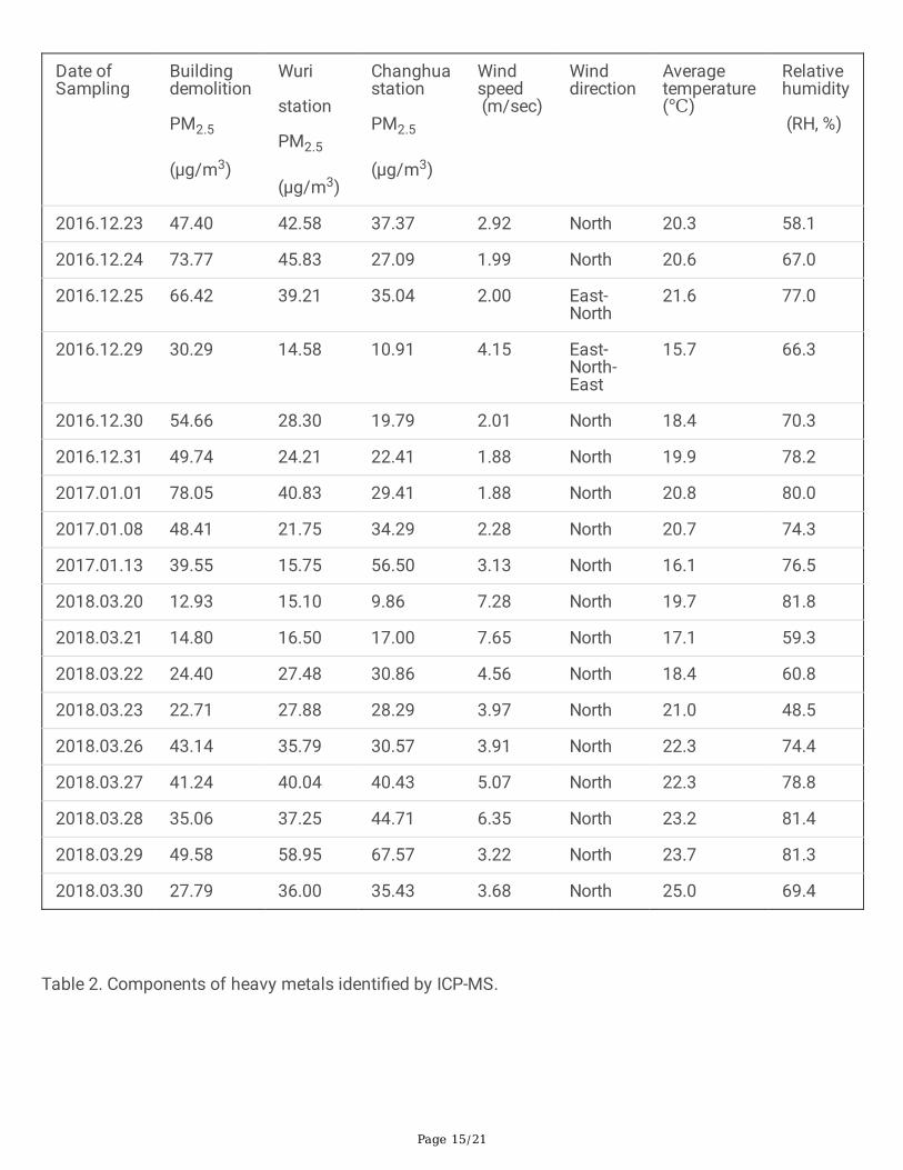
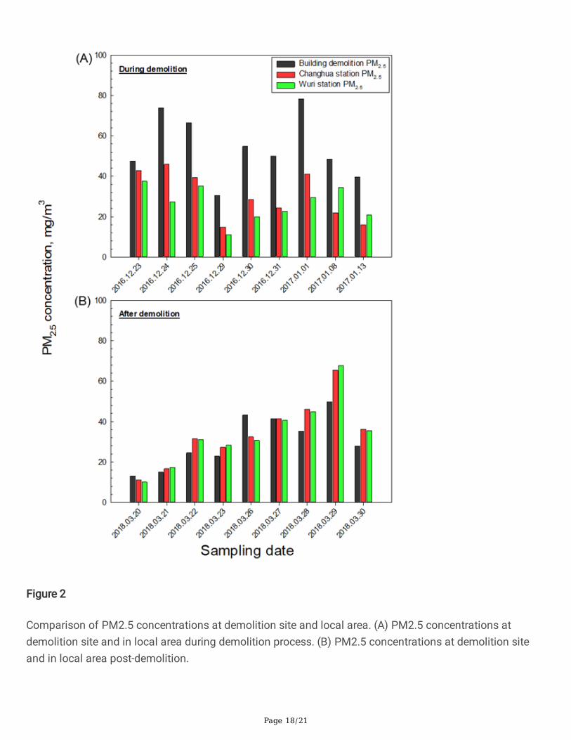
ResultsFigure 1 Schematic diagram of demolition location and regional monitoring stations around sampling site.A four-�oor building was demolished from 2016-12-29 to 2017-01-13. The PM concentration at ademolition site depends strongly on numerous factors, including local and regional PM sources,meteorological conditions and geography. To con�rm the concentration of PM in air that was generated byvarious sources, measurements were made at the building demolition site and the local monitoringstations (station A and station B) before and after the deconstruction process, as indicated in Table 1. Thefactors were wind speed, temperature and relative humidity (RH). Based on meteorological data from2016-12-29, 2018-03-20 and 2018-03-21, high-velocity wind might have reduced PM2.5 levels in the localair, this �nding is consistent with the low PM2.5 concentration that was derived from a correlation analysisthat was based on 22 months of observation at 68 major cities in seven geographical regions in China(Yang et al., 2017). Wind speed has similarly been negatively correlated with PM2.5 level. During thedemolition process, the concentration of PM2.5 at the collection point signi�cantly exceeded that in thesurrounding area (Fig. 2A). Fourteen months later, PM2.5 concentrations in the air at the collection pointwere normally lower than in the local surrounding area (Fig. 2B).
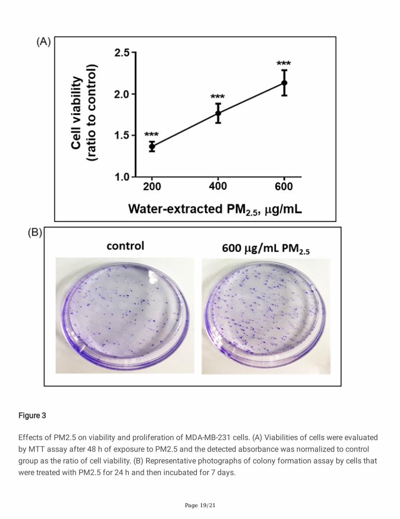
To assess the effect of PM2.5 on the progression of breast cancer cells, MDA-MB-231 breast cancer cellswere treated with water-extracted PM2.5 for 48 h and analyzed using an MTT survival assay (Fig. 3A). Thehigher PM2.5 concentrations resulted in a tumor cell viability of double that of the control group. Theproliferation capacity of breast cancer cells was examined using a colony formation assay. After 24 h ofexposure to 600 µg/mL PM2.5, the number of colonies of the PM2.5-treated breast cancer cells signi�cantlyexceeded that in the control group after seven days of incubation (Fig. 3B). These data provide strongevidence that water-extracted PM2.5 enhances the survival of MDA-MB-231 breast cancer cells.
Page 7/21
The metastatic potential of breast cancer is associated with poor prognosis in patients with a shortsurvival time and high recurrence rate (Stovgaard et al., 2018). Therefore, the effect of PM2.5 on themotility of MDA-MB-231 cells was determined using a wound healing assay (Figs. 4A and B). Similarly,treatment with 600 µg/mL PM2.5 signi�cantly enhanced the motility of MDA-MB-231 cells followingincubation for 16 h. The results of transwell migration assays showed that PM2.5 promotes the verticalmigration capacity of the cells, verifying that water-extracted PM2.5 increases the invasiveness of breastcancer cells (Fig. 4C).
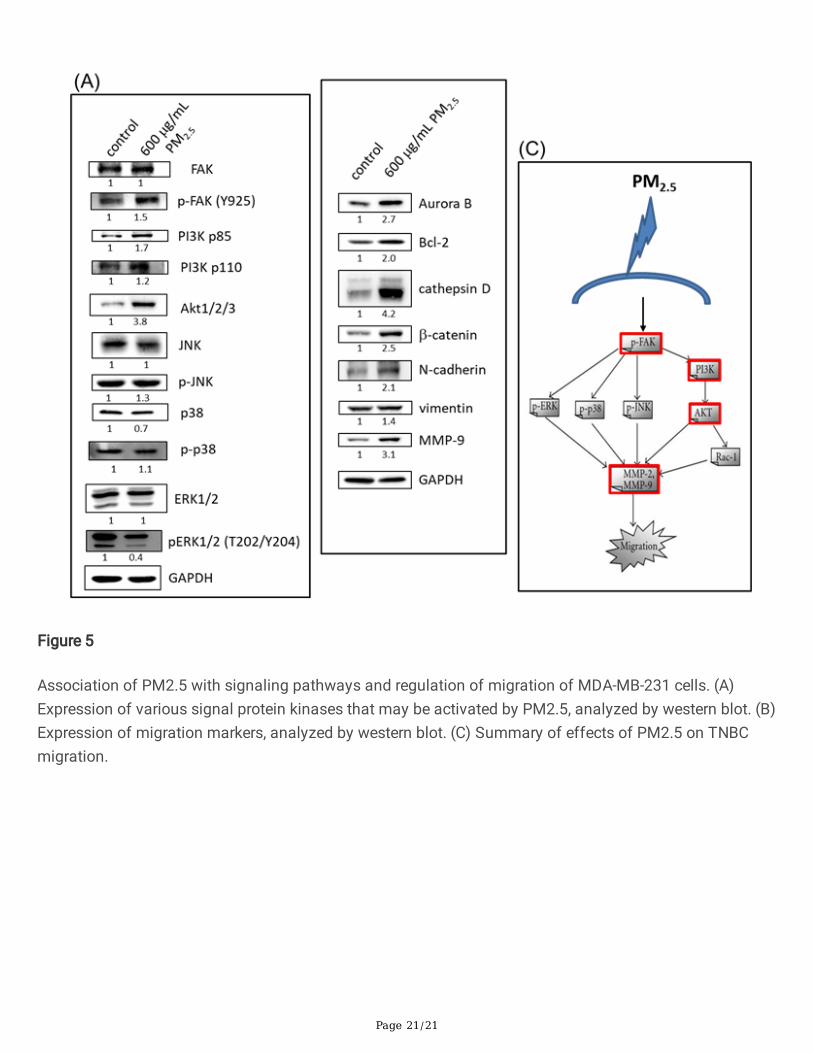
During tumor metastasis, cancer cells undergo an epithelial-mesenchymal transition (EMT) in whichepithelial tumor parts are converted into aggressive and metastatic ones, which are known as amesenchymal tumor phenotype with the loss of cell-cell adhesive function (Liao and Yang, 2017; Thiery etal., 2009). Focal adhesion kinase (FAK) is a cytoplasmic non-receptor tyrosine kinase whosephosphorylation at the Tyr397 residue by integrins results in the activation of the phosphoinositide 3-kinase (PI3K)/AKT signaling pathway that promotes tumor cell adhesion and invasion during EMT. Theconsequence is cancer cell metastasis (Behmoaram et al. 2008) (LoRusso 2016). As expected, after 48 hof treatment with water-extracted PM2.5 increased phosphorylation of FAK at Tyr925 (p-FAK-Y925) iscorrelated with high levels of both PI3K p85α and p110 subunits as well as the overexpression ofphosphorylated-AKT (Fig. 5A). In contrast, water-extracted PM2.5 that is produced by building demolitionhas no signi�cant effect on ERK, JNK or p38 MMPK expressions (Fig. 5A).
The characteristics of EMT include reduced intercellular adhesion, acquisition of mesenchymal markers(including vimentin and N-cadherin) and loss of epithelial markers (such as E-cadherin) (Lamouille et al.,2014). Aurora B kinase is one of the major kinases that is responsible for the �delity of mitosis. Anelevated level of Aurora B kinase contributes to chemoresistance and predicts poor prognosis of breastcancer (Zhang et al. 2015). As presented in Fig. 5B, the upregulation of Aurora B kinase and Bcl-2 supportshigher viability and proliferation (Fig. 3). PM2.5 treatment signi�cantly increased the level of β-catenin, asresulted in the overexpression of N-cadherin, vimentin and MMP-9 proteins. Therefore, Fig. 5C displays ahypothetical model of the induction of TNBC migration by PM2.5, which demonstrates that exposure toPM2.5 activates the FAK/PI3K/AKT signaling pathway to regulate the process of EMT with the help ofcathepsin D, β-catenin, N-cadherin, vimentin and MMP-9, increasing migration.
Growing evidence shows that prolonged exposure to heavy metals is associated with a poor prognosis ofcancer. The effects of MAPK and PI3K/AKT signaling pathways on heavy metal-induced carcinogenesis intumors in the lung have been reported (Ohgami et al., 2015). Accordingly, metal species in water-solublePM2.5 that promote the development of cancer cells by activating the FAK/PI3K/AKT pathway, and thereby,enhance cell proliferation, migration, and metastasis. Inductively coupled plasma mass spectrometry (ICP-MS) analysis revealed that the amounts of the �ve heavy metals, cadmium (Cd), copper (Cu), lead (Pb),zinc (Zn) and chromium (Cr) were higher in the water-soluble PM2.5 extracts that were generated bybuilding demolition than in the post-control sample from the surrounding area (Table 2). The data on theheavy metals were normalized using the environmental factors that are listed in Table 3. Interestingly, our
Page 8/21
hypothesis was supported by observations that increased concentrations of Cd and Cu in the water-solublePM2.5 extracts that were obtained from a building demolition site adversely affected colony formation by,and the motility of, MDA-MB-231 cells through activation of the PI3K/AKT pathway.
Discussion And ConclusionSeveral investigations have addressed the effect of water-soluble PM2.5 on the development of cancercells, considering the close relationship between human exposure to PM2.5 and the risk of circulatingPM2.5, which promotes lung tumor invasiveness, including the promotion and progression of the tumorduring EMT. The effects of building demolition PM2.5 on breast cancer cells was also examined. In this
study, 1872 m3 of air was analyzed over nine days of demolition and 101.556 mg of PM2.5 was obtained.A total of 27.368 mg PM2.5 was obtained after extraction with water, freeze-drying and resuspension.Although some meteorological factors affect PM2.5 concentration, building demolition clearly generates ahigh level of ambient PM2.5 in the surroundings. The fact that the level of PM2.5 in the cellular model hereinexceeds the ambient PM2.5 level is a serious unavoidable shortcoming of the model. For example, a
200 µg/mL water-extracted PM2.5 is equivalent to 53.88 g/m3 ambient PM2.5, which represents more thanten years of exposure to ambient PM2.5 levels following the collapse of the WTC buildings during the 911event. Notably, although this extraordinary high PM2.5 exposure does not occur in the real world, molecularevidence reveals a potentially high risk of cancer malignancy, especially for women with breast cancer,upon exposure to relatively high ambient PM2.5 levels.
The underlying cause of death in the majority of breast cancer patients is metastasis (Narod et al., 2015),of which the main characteristic is EMT. The MDA-MB-231 cell line has a high capacity for distantmetastasis, and manifestation with malignant mesenchymal features arises from the loss of E-cadherin(Luo et al., 2018). The biochemical data herein reveal that E-cadherin loss the overexpression of N-cadherinand β-catenin screening in cells that had been treated with PM2.5. PM2.5-treated MDA-MB-231 cellsexhibited an increased level of cathepsin D with overexpression of Aurora kinase B, vimentin and MMP-9,which promotes metastasis. The overexpression of cathepsin D enhanced breast cancer cell metastasis byinducing the expression of intercellular cell adhesion molecule-1 (ICAM-1) (Zhang et al. 2018) and hasbeen used as an independent marker of a poor prognosis of breast cancer (Dey et al., 2013; Foekens et al.1999). Many studies have shown that exposure to PM2.5 stimulates lung cancer and epithelium cell
motility. Exposure to PM2.5 (50 µg/cm2 concentrations of PM2.5 for 72 h) induces the proliferation andmotility of cells of various lung cancer cell lines (Yang et al., 2016). At PM2.5 dose ≥ 60 µg/mL, cellapoptosis is evaded, promoting cell proliferation and sustained angiogenesis through the activation of thePI3K-AKT signaling pathway (Zheng et al., 2017). Five components (organic carbon, PAH, Zn, As, V) ofPM2.5 mostly from tra�c emissions were strongly associated with cancer progression, and Zn has acritical role in the activation of PI3K-AKT signal transduction (Zheng et al., 2017; Chen et al., 2013).According to our data and above reports, Zn in water-extracted PM2.5 strongly promotes TNBC malignancy.The particles and their solvent extracts had a range of effects on the cell lines, such as the generation of

Page 9/21
reactive oxygen species (ROS) and an increase in DNA strand breakage (which is concentration-dependent). Additionally, PM pollution as a result of demolition activity has an adverse effect on the healthof people who live close to the demolition sites, especially since exposure to a high dose of PM2.5 isassociated with FAK/PI3K/AKT signaling activation and enhanced EMT in breast cancer cells. These�ndings are supported by health assessments that demonstrate that exposure to PM2.5 becomes moreimportant when the surroundings are densely built residential areas or sensitive areas, such as schoolsand hospitals (Farhad &Azarmi, 2016; Azarmi et al., 2016).
Scienti�c research into the effects of airborne particulates that are generated by building demolition sincethe September 11th event in New York city have shown that the demolition of buildings generates largequantities of PM, which can be inhaled by on-site workers and people who live in the neighborhood.Evidence suggests that long-term exposure to airborne particles triggers cell mutations and increase therisk of breast cancer, but the toxicological mechanisms are unclear. The discovery of estrogen-mimickingcompounds in the environment and the synergistic activity of many of these on estrogen receptors haveled researchers to hypothesize the role of xenoestrogenic substances in the environment that mimicestrogen in increasing breast cancer risk (Arnold et al., 1996). However, not all breast cancers areresponsive to this hormone and its analogs in environmental pollutants. Based on the breast cancersubtypes estrogen receptor (ER) and progesterone receptor (PR) status, ER-positive (ER(+)) and ER-negative (ER(-)) breast cancers have distinctly different risk factors and possibly, therefore, differentetiologies (Althuis et al., 2004). Depending on the etiological differences between breast cancer subtypes,ER(+) breast cancers are associated with estrogen-related factors, including early menarche, number ofpregnancies, and late-age childbearing (Chen et al., 2013). However, the incidence rate of ER(-) breastcancers in Western populations has been shown to be related to county-level environmental factors, suchas pesticide use, toxic air emissions, and pollution from urban activities. In this investigation, MDA-MB-231invasive, rather than ER(+),breast cancer cells were used to study the effect of water-extracted buildingdemolition PM2.5 on ER(-) breast cancer cell progression. Exposure to water-extracted PM2.5 enhanced thePI3K/Akt signal transduction pathway in MDA-MB-231 cells, favoring breast cancer cell invasiveness.Moreover, the signi�cantly elevated concentrations of the heavy metals cadmium, copper, lead, zinc andchromium were found in the water-extracted PM2.5 during building demolition. Long-term exposure toPM2.5 in Italy has been associated with increased risk of death due to breast cancer (Tagliabue et al.,2016). Interestingly, the �ndings herein are consistent with the previous epidemiological observations ofItalian women who live near incinerators, who have been found to have an elevated risk of breast cancermortality. These women live in areas with a very high total concentration (> 2 ng/m3) of heavy metals,including lead, cadmium, chromium, cobalt, and copper. The lowest measured ambient concentrations ofthese heavy metals are of the order of < 0.5 ng/m3 (Ranzi et al. 2011). Beyond this investigation of theeffects of ambient exposure to coarse and �ne particle emissions from building demolition sites on breastcancer, identifying genetic factors that respond to ambient air pollution and breast cancer would be veryinformative (Brody et al. 2007). Polycyclic aromatic hydrocarbons (PAHs), which can cause oxidativestress (Mordukhovich et al. 2010), stimulate cell proliferation and mammary tumors in laboratory animals,leading to neoplasia in breast cancer, warrant particular attention. To the best of our knowledge, this
Page 10/21
investigation is the �rst to highlight the promotion by building demolition PM2.5 of breast tumorprogression, mediated by PI3K signaling, enhancing the invasion and migration of ER(-) breast cancercells. This information may contribute to better estimates of ambient air pollution to identify areas thatexperience disproportionate effects of outdoor air pollution.
DeclarationsEthics approval and consent to participate
Not applicable.
Consent for publication
Not applicable.
Availability of data and materials
All data generated or analyzed during this study are included in this published article.
Competing interests
The authors declare that they have no competing interests.
Funding
No funding was received.
Authors' contributions
C.-Y.L., C.-W.C., G.-T.S. and Y.-C. C. planned work and designed experiments. G.-T.S., C.-Y.L. and C.-W.C.wrote manuscript. J.-S.C. and P.-H.W. collected PM and performed experimental sampling and testing. C.-W.C., G.-T.S. and C.-Y.L. performed statistical analysis. All authors analyzed and discussed the results andcommented on the manuscript.
Acknowledgements
We thank the SDI Group, Taiwan, for their assistance with PM sampling.
Authors' information
Chun-Wen Cheng, email: [email protected];
Gwo-Tarng Sheu, email: [email protected];
Jing-Shiuan Chou, email: [email protected];
Pei-Han Wang, email: [email protected];
Page 11/21
Yu-Chun Cheng, email: [email protected];
Chane-Yu Lai, email: [email protected] . Rm. 1215A, No.110, Sec.1, Chien-Kuo N. Rd. Taichung, Taiwan.
References1. Aldrich TK, Gustave J, Hall CB, Cohen HW, Webber MP, Zeig-Owens R, Cosenza K, Christodoulou V,
Glass L, Al-Othman F and others. 2010. Lung function in rescue workers at the World Trade Centerafter 7 years. N Engl J Med 362(14):1263-72.
2. Althuis MD, Fergenbaum JH, Garcia-Closas M, Brinton LA, Madigan MP, Sherman ME. 2004. Etiologyof hormone receptor-de�ned breast cancer: a systematic review of the literature. Cancer EpidemiolBiomarkers Prev 13(10):1558-68.
3. Andersen ZJ, Ravnskjaer L, Andersen KK, Loft S, Brandt J, Becker T, Ketzel M, Hertel O, Lynge E,Brauner EV. 2017. Long-term Exposure to Fine Particulate Matter and Breast Cancer Incidence in theDanish Nurse Cohort Study. Cancer Epidemiol Biomarkers Prev 26(3):428-430.
4. Arnold SF, Klotz DM, Collins BM, Vonier PM, Guillette LJ, Jr., McLachlan JA. 1996. Synergisticactivation of estrogen receptor with combinations of environmental chemicals. Science272(5267):1489-92.
5. Azarmi F, Kumar P, Marsh D, Fuller G. 2016. Assessment of the long-term impacts of PM10 and PM2.5particles from construction works on surrounding areas. Environ Sci Process Impacts 18(2):208-21.
�. Beck CM, Geyh A, Srinivasan A, Breysse PN, Eggleston PA, Buckley TJ. 2003. The impact of a buildingimplosion on airborne particulate matter in an urban community. J Air Waste Manag Assoc53(10):1256-64.
7. Behmoaram E, Bijian K, Jie S, Xu Y, Darnel A, Bismar TA, Alaoui-Jamali MA. 2008. Focal adhesionkinase-related proline-rich tyrosine kinase 2 and focal adhesion kinase are co-overexpressed in early-stage and invasive ErbB-2-positive breast cancer and cooperate for breast cancer cell tumorigenesisand invasiveness. Am J Pathol 173(5):1540-50.
�. Brody JG, Rudel RA, Michels KB, Moysich KB, Bernstein L, Att�eld KR, Gray S. 2007. Environmentalpollutants, diet, physical activity, body size, and breast cancer: where do we stand in research toidentify opportunities for prevention? Cancer 109(12 Suppl):2627-34.
9. Chen ST, Lin CC, Liu YS, Lin C, Hung PT, Jao CW, Lin PH. 2013. Airborne particulate collected fromcentral Taiwan induces DNA strand breaks, Poly(ADP-ribose) polymerase-1 activation, and estrogen-disrupting activity in human breast carcinoma cell lines. J Environ Sci Health A Tox Hazard SubstEnviron Eng 48(2):173-81.
10. Claudio L. 2001. Environmental aftermath. Environ Health Perspect 109(11):A528-36.
11. Crouse DL, Goldberg MS, Ross NA, Chen H, Labreche F. 2010. Postmenopausal breast cancer isassociated with exposure to tra�c-related air pollution in Montreal, Canada: a case-control study.Environ Health Perspect 118(11):1578-83.
Page 12/21
12. Dent R, Trudeau M, Pritchard KI, Hanna WM, Kahn HK, Sawka CA, Lickley LA, Rawlinson E, Sun P,Narod SA. 2007. Triple-negative breast cancer: clinical features and patterns of recurrence. Clin CancerRes 13(15 Pt 1):4429-34.
13. Dey N, Barwick BG, Moreno CS, Ordanic-Kodani M, Chen Z, Oprea-Ilies G, Tang W, Catzavelos C,Kerstann KF, Sledge GW, Jr. and others. 2013. Wnt signaling in triple negative breast cancer isassociated with metastasis. BMC Cancer 13:537.
14. Farhad Azarmi PK. 2016. Ambient exposure to coarse and �ne particle emissions from buildingdemolition. Atmospheric Environment 137:62-79.
15. Foekens JA, Look MP, Bolt-de Vries J, Meijer-van Gelder ME, van Putten WL, Klijn JG. 1999. Cathepsin-D in primary breast cancer: prognostic evaluation involving 2810 patients. Br J Cancer 79(2):300-7.
1�. Hart JE, Bertrand KA, DuPre N, James P, Vieira VM, Tamimi RM, Laden F. 2016. Long-term ParticulateMatter Exposures during Adulthood and Risk of Breast Cancer Incidence in the Nurses' Health Study IIProspective Cohort. Cancer Epidemiol Biomarkers Prev 25(8):1274-6.
17. Huo Q, Zhang N, Wang X, Jiang L, Ma T, Yang Q. 2013. Effects of ambient particulate matter onhuman breast cancer: is xenogenesis responsible? PLoS One 8(10):e76609.
1�. Hystad P, Villeneuve PJ, Goldberg MS, Crouse DL, Johnson K, Canadian Cancer RegistriesEpidemiology Research G. 2015. Exposure to tra�c-related air pollution and the risk of developingbreast cancer among women in eight Canadian provinces: a case-control study. Environ Int 74:240-8.
19. Lamouille S, Xu J, Derynck R. 2014. Molecular mechanisms of epithelial-mesenchymal transition. NatRev Mol Cell Biol 15(3):178-96.
20. Landrigan PJ. 2001. Health consequences of the 11 September 2001 attacks. Environ Health Perspect109(11):A514-5.
21. Liao TT, Yang MH. 2017. Revisiting epithelial-mesenchymal transition in cancer metastasis: theconnection between epithelial plasticity and stemness. Mol Oncol 11(7):792-804.
22. Lioy PJ, Pellizzari E, Prezant D. 2006. The World Trade Center aftermath and its effects on health:understanding and learning through human-exposure science. Environ Sci Technol 40(22):6876-85.
23. Lioy PJ, Weisel CP, Millette JR, Eisenreich S, Vallero D, Offenberg J, Buckley B, Turpin B, Zhong M,Cohen MD and others. 2002. Characterization of the dust/smoke aerosol that settled east of the WorldTrade Center (WTC) in lower Manhattan after the collapse of the WTC 11 September 2001. EnvironHealth Perspect 110(7):703-14.
24. Lippmann M, Cohen MD, Chen LC. 2015. Health effects of World Trade Center (WTC) Dust: Anunprecedented disaster's inadequate risk management. Crit Rev Toxicol 45(6):492-530.
25. Liu R, Nelson DO, Hurley S, Hertz A, Reynolds P. 2015. Residential exposure to estrogen disruptinghazardous air pollutants and breast cancer risk: the California Teachers Study. Epidemiology26(3):365-73.
2�. LoRusso PM. 2016. Inhibition of the PI3K/AKT/mTOR Pathway in Solid Tumors. J Clin Oncol34(31):3803-3815.
Page 13/21
27. Luo CW, Wu CC, Chang SJ, Chang TM, Chen TY, Chai CY, Chang CL, Hou MF, Pan MR. 2018. CHD4-mediated loss of E-cadherin determines metastatic ability in triple-negative breast cancer cells. ExpCell Res 363(1):65-72.
2�. McGee JK, Chen LC, Cohen MD, Chee GR, Prophete CM, Haykal-Coates N, Wasson SJ, Conner TL,Costa DL, Gavett SH. 2003. Chemical analysis of World Trade Center �ne particulate matter for use intoxicologic assessment. Environ Health Perspect 111(7):972-80.
29. Mordukhovich I, Rossner P, Jr., Terry MB, Santella R, Zhang YJ, Hibshoosh H, Memeo L, Mansukhani M,Long CM, Garbowski G and others. 2010. Associations between polycyclic aromatic hydrocarbon-related exposures and p53 mutations in breast tumors. Environ Health Perspect 118(4):511-8.
30. Narod SA, Iqbal J, Giannakeas V, Sopik V, Sun P. 2015. Breast Cancer Mortality After a Diagnosis ofDuctal Carcinoma In Situ. JAMA Oncol 1(7):888-96.
31. Ohgami N, Yamanoshita O, Thang ND, Yajima I, Nakano C, Wenting W, Ohnuma S, Kato M. 2015.Carcinogenic risk of chromium, copper and arsenic in CCA-treated wood. Environ Pollut 206:456-60.
32. Parikh PV, Wei Y. 2016. PAHs and PM2.5 emissions and female breast cancer incidence in metroAtlanta and rural Georgia. Int J Environ Health Res 26(4):458-66.
33. Ranzi A, Fano V, Erspamer L, Lauriola P, Perucci CA, Forastiere F. 2011. Mortality and morbidity amongpeople living close to incinerators: a cohort study based on dispersion modeling for exposureassessment. Environ Health 10:22.
34. Reding KW, Young MT, Szpiro AA, Han CJ, DeRoo LA, Weinberg C, Kaufman JD, Sandler DP. 2015.Breast Cancer Risk in Relation to Ambient Air Pollution Exposure at Residences in the Sister StudyCohort. Cancer Epidemiol Biomarkers Prev 24(12):1907-9.
35. Rom WN, Weiden M, Garcia R, Yie TA, Vathesatogkit P, Tse DB, McGuinness G, Roggli V, Prezant D.2002. Acute eosinophilic pneumonia in a New York City �re�ghter exposed to World Trade Center dust.Am J Respir Crit Care Med 166(6):797-800.
3�. Stovgaard ES, Nielsen D, Hogdall E, Balslev E. 2018. Triple negative breast cancer - prognostic role ofimmune-related factors: a systematic review. Acta Oncol 57(1):74-82.
37. Tagliabue G, Borgini A, Tittarelli A, van Donkelaar A, Martin RV, Bertoldi M, Fabiano S, Maghini A,Codazzi T, Scaburri A and others. 2016. Atmospheric �ne particulate matter and breast cancermortality: a population-based cohort study. BMJ Open 6(11):e012580.
3�. Thiery JP, Acloque H, Huang RY, Nieto MA. 2009. Epithelial-mesenchymal transitions in developmentand disease. Cell 139(5):871-90.
39. Wei Y, Davis J, Bina WF. 2012. Ambient air pollution is associated with the increased incidence ofbreast cancer in US. Int J Environ Health Res 22(1):12-21.
40. Weiden MD, Kwon S, Caraher E, Berger KI, Reibman J, Rom WN, Prezant DJ, Nolan A. 2015. Biomarkersof World Trade Center Particulate Matter Exposure: Physiology of Distal Airway and Blood Biomarkersthat Predict FEV(1) Decline. Semin Respir Crit Care Med 36(3):323-33.
41. Yaghjyan L, Arao R, Brokamp C, O'Meara ES, Sprague BL, Ghita G, Ryan P. 2017. Association betweenair pollution and mammographic breast density in the Breast Cancer Surveilance Consortium. Breast
Page 14/21
Cancer Res 19(1):36.
42. Yang B, Chen D, Zhao H, Xiao C. 2016. The effects for PM2.5 exposure on non-small-cell lung cancerinduced motility and proliferation. Springerplus 5(1):2059.
43. Yang Q, Yuan Q, Li T, Shen H, Zhang L. 2017. The Relationships between PM2.5 and MeteorologicalFactors in China: Seasonal and Regional Variations. Int J Environ Res Public Health 14(12).
44. Zhang C, Zhang M, Song S. 2018. Cathepsin D enhances breast cancer invasion and metastasisthrough promoting hepsin ubiquitin-proteasome degradation. Cancer Lett 438:105-115.
45. Zhang Y, Jiang C, Li H, Lv F, Li X, Qian X, Fu L, Xu B, Guo X. 2015. Elevated Aurora B expressioncontributes to chemoresistance and poor prognosis in breast cancer. Int J Clin Exp Pathol 8(1):751-7.
4�. Zheng L, Liu S, Zhuang G, Xu J, Liu Q, Zhang X, Deng C, Guo Z, Zhao W, Liu T and others. 2017. SignalTransductions of BEAS-2B Cells in Response to Carcinogenic PM2.5 Exposure Based on a Micro�uidicSystem. Anal Chem 89(10):5413-5421.
TablesTable 1. Sampling duration and monitoring sites with PM2.5 concentrations and meteorological data.
Page 15/21
Date ofSampling
Buildingdemolition
PM2.5
(μg/m3)
Wuri
station
PM2.5
(μg/m3)
Changhuastation
PM2.5
(μg/m3)
Windspeed (m/sec)
Winddirection
Averagetemperature(℃)
Relativehumidity
(RH, %)
2016.12.23 47.40 42.58 37.37 2.92 North 20.3 58.1
2016.12.24 73.77 45.83 27.09 1.99 North 20.6 67.0
2016.12.25 66.42 39.21 35.04 2.00 East-North
21.6 77.0
2016.12.29 30.29 14.58 10.91 4.15 East-North-East
15.7 66.3
2016.12.30 54.66 28.30 19.79 2.01 North 18.4 70.3
2016.12.31 49.74 24.21 22.41 1.88 North 19.9 78.2
2017.01.01 78.05 40.83 29.41 1.88 North 20.8 80.0
2017.01.08 48.41 21.75 34.29 2.28 North 20.7 74.3
2017.01.13 39.55 15.75 56.50 3.13 North 16.1 76.5
2018.03.20 12.93 15.10 9.86 7.28 North 19.7 81.8
2018.03.21 14.80 16.50 17.00 7.65 North 17.1 59.3
2018.03.22 24.40 27.48 30.86 4.56 North 18.4 60.8
2018.03.23 22.71 27.88 28.29 3.97 North 21.0 48.5
2018.03.26 43.14 35.79 30.57 3.91 North 22.3 74.4
2018.03.27 41.24 40.04 40.43 5.07 North 22.3 78.8
2018.03.28 35.06 37.25 44.71 6.35 North 23.2 81.4
2018.03.29 49.58 58.95 67.57 3.22 North 23.7 81.3
2018.03.30 27.79 36.00 35.43 3.68 North 25.0 69.4
Table 2. Components of heavy metals identi�ed by ICP-MS.

Page 16/21
Cd Cu Hg Pb Ni As Zn Cr V
Control mg/L N.D. 2.38 N.D. 1.2 N.D. N.D. 49.0 4.30 N.D.
PM2.5 mg/L 61.9 1780 N.D. 927 160 111 10800 307 110
Post-Ctl mg/L 32.9 1400 N.D. 655 189 112 7040 133 445
Table 3. Concentrations of heavy metals in PM2.5 collected at building demolition site.
Cd Cu Hg Pb Ni As Zn Cr V
PM2.5 ng/L 0.367 10.565 N.D. 5.502 0.95 0.659 64.103 1.822 0.65
Post-Ctl ng/L 0.195 8.31 N.D. 3.888 1.122 0.665 41.785 0.789 2.641
Figures
Page 17/21
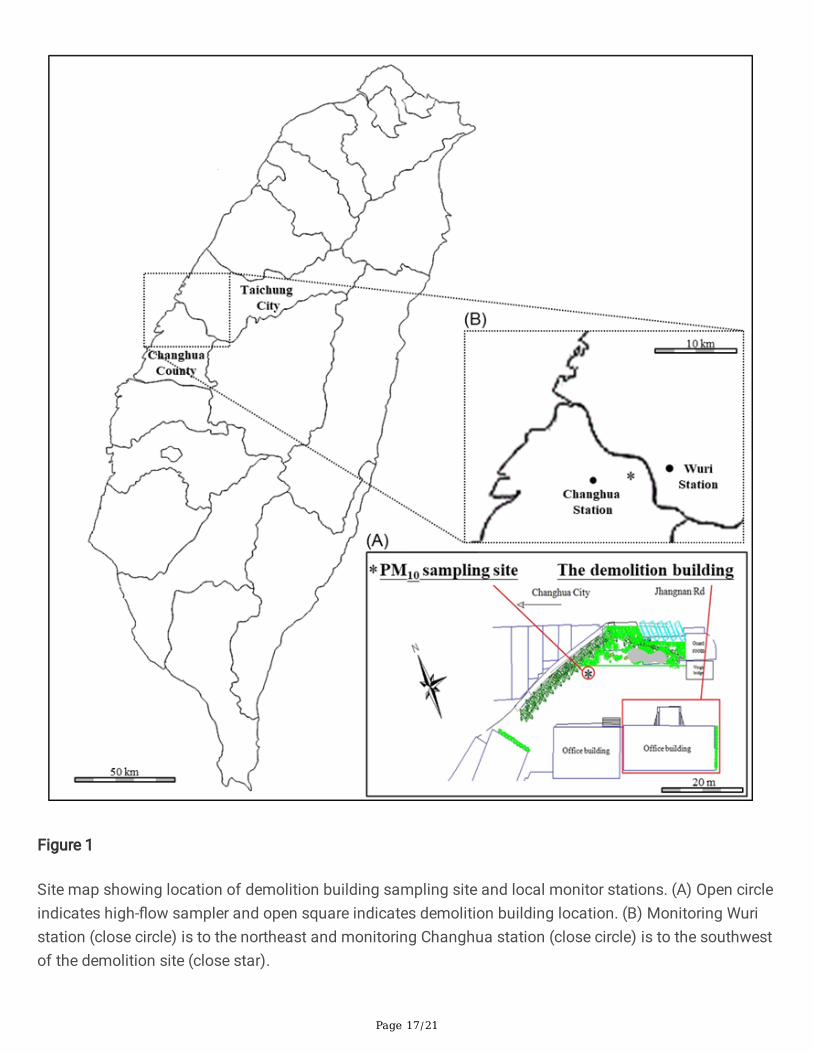
Figure 1
Site map showing location of demolition building sampling site and local monitor stations. (A) Open circleindicates high-�ow sampler and open square indicates demolition building location. (B) Monitoring Wuristation (close circle) is to the northeast and monitoring Changhua station (close circle) is to the southwestof the demolition site (close star).
Page 18/21
Figure 2
Comparison of PM2.5 concentrations at demolition site and local area. (A) PM2.5 concentrations atdemolition site and in local area during demolition process. (B) PM2.5 concentrations at demolition siteand in local area post-demolition.
Page 19/21
Figure 3
Effects of PM2.5 on viability and proliferation of MDA-MB-231 cells. (A) Viabilities of cells were evaluatedby MTT assay after 48 h of exposure to PM2.5 and the detected absorbance was normalized to controlgroup as the ratio of cell viability. (B) Representative photographs of colony formation assay by cells thatwere treated with PM2.5 for 24 h and then incubated for 7 days.

Page 20/21
Figure 4
Effects of PM2.5 on migration of MDA-MB-231 cells. (A) PM2.5-treated cells were analyzed using woundhealing assay and the representative photographs are displayed. (B) Cells that migrated across the solidline were counted. (C) Representative photographs of transwell migration assay are shown. Cells weretreated with PM2.5 for 24 h and allowed to migrate for 16 h before staining.
Page 21/21
Figure 5
Association of PM2.5 with signaling pathways and regulation of migration of MDA-MB-231 cells. (A)Expression of various signal protein kinases that may be activated by PM2.5, analyzed by western blot. (B)Expression of migration markers, analyzed by western blot. (C) Summary of effects of PM2.5 on TNBCmigration.