Cancer Classification Using Gene Expression Datahanj.cs.illinois.edu/pdf/is03_cancer.pdf · Cancer...
35
Cancer Classification Using Gene Expression Data Ying Lu Jiawei Han Department of Computer Science University of Illinois at Urbana-Champaign Urbana, IL 61801, USA Email: {yinglu, hanj}@uiuc.edu Corresponding author: Ying Lu ([email protected]), DCL, Department of Computer Science, 1304 Springfield Ave., University of Illinois at Urbana-Champaign, Urbana, IL 61801, USA. Phone: (217) 244-3570 Fax: (217) 244-6500 Abstract The classification of different tumor types is of great importance in cancer diagnosis and drug discovery. However, most previous cancer classification studies are clinical-based and have limited diagnostic ability. Cancer classification using gene expression data is known to contain the keys for addressing the fundamental problems relating to cancer diagnosis and drug discovery. The recent advent of DNA microarray technique has made simultaneous monitoring of thousands of gene expressions possible. With this abundance of gene expression data, researchers have started to explore the possibilities of cancer classification using gene expression data. Quite a number of methods have been proposed in recent years with promising results. But there are still a lot of issues which need to be addressed and understood. In order to gain deep insight into the cancer classification problem, it is necessary to take a closer look at the problem, the proposed solutions and the related issues all together. In this survey paper, we present a comprehensive overview of various proposed cancer classification methods and evaluate them based on their computation time, classification accuracy and ability to reveal biologically meaningful gene information. We also introduce and evaluate various proposed gene selection methods which we believe should be an integral preprocessing step for cancer classification. In order to obtain a full picture of cancer classification, we also discuss several issues related to cancer classification, including the biological significance vs. statistical significance of a cancer classifier, the asymmetrical classification errors for cancer classifiers, and the gene contamination problem. Keywords: cancer classification, gene expression data. 1 Introduction Cancer research is one of the major research areas in the medical field. Accurate prediction of different tumor types has great value in providing better treatment and toxicity minimization on the patients. 1
Transcript of Cancer Classification Using Gene Expression Datahanj.cs.illinois.edu/pdf/is03_cancer.pdf · Cancer...

Cancer Classification Using Gene Expression Data
Ying Lu Jiawei Han
Department of Computer Science
University of Illinois at Urbana-Champaign
Urbana, IL 61801, USA
Email: {yinglu, hanj}@uiuc.edu
Corresponding author: Ying Lu ([email protected]), DCL, Department of Computer Science, 1304 Springfield
Ave., University of Illinois at Urbana-Champaign, Urbana, IL 61801, USA. Phone: (217) 244-3570 Fax: (217)
244-6500
Abstract
The classification of different tumor types is of great importance in cancer diagnosis and drug
discovery. However, most previous cancer classification studies are clinical-based and have limited
diagnostic ability. Cancer classification using gene expression data is known to contain the keys
for addressing the fundamental problems relating to cancer diagnosis and drug discovery. The
recent advent of DNA microarray technique has made simultaneous monitoring of thousands of
gene expressions possible. With this abundance of gene expression data, researchers have started
to explore the possibilities of cancer classification using gene expression data. Quite a number of
methods have been proposed in recent years with promising results. But there are still a lot of issues
which need to be addressed and understood.
In order to gain deep insight into the cancer classification problem, it is necessary to take a
closer look at the problem, the proposed solutions and the related issues all together. In this
survey paper, we present a comprehensive overview of various proposed cancer classification methods
and evaluate them based on their computation time, classification accuracy and ability to reveal
biologically meaningful gene information. We also introduce and evaluate various proposed gene
selection methods which we believe should be an integral preprocessing step for cancer classification.
In order to obtain a full picture of cancer classification, we also discuss several issues related to cancer
classification, including the biological significance vs. statistical significance of a cancer classifier, the
asymmetrical classification errors for cancer classifiers, and the gene contamination problem.
Keywords: cancer classification, gene expression data.
1 Introduction
Cancer research is one of the major research areas in the medical field. Accurate prediction of different
tumor types has great value in providing better treatment and toxicity minimization on the patients.
1

Previously, cancer classification has always been morphological, and clinical based. These conventional
cancer classification methods are reported to have several limitations [Azu00] in their diagnostic abil-
ity. It has been suggested that specifications of therapies according to tumor types differentiated by
pathogenetic patterns may maximize the efficacy of the patients [Aea00, GST+99, VDBea02, PTGea02,
Zea01, Sea01, DGB02, VJ02, Aea00, DPBea96]. Also, the existing tumor classes has been found to
be heterogeneous and comprises of diseases that are molecularly distinct and follow different clinical
courses.
In order to gain a better insight into the problem of cancer classification, systematic approaches
based on global gene expression analysis have been proposed. The expression level of genes are known
to contain the keys to address fundamental problems relating to the prevention and cure of diseases,
biological evolution mechanisms and drug discovery. The recent advent of microarray technology has
allowed the simultaneous monitoring of thousands of genes, which motivated the development in cancer
classification using gene expression data [GST+99, STM+00, LA01, NR02, Ber00]. Though still in its
early stages of development, results obtained so far seemed promising .
Different classification methods from statistical and machine learning area have been applied to
cancer classification, but there are some issues that make it a nontrivial task. The gene expression
data is very different from any of the data these methods had previously dealt with. First, it has
very high dimensionality, usually contains thousands to tens of thousands of genes. Second, publicly
available data size is very small, all below 100. Third, most genes are irrelevant to cancer distinction.
It is obvious that those existing classification methods were not designed to handle this kind of data
efficiently and effectively. Some researchers proposed to do gene selection prior to cancer classification.
Performing gene selection helps to reduce data size thus improving the running time. More importantly,
gene selection removes a large number of irrelevant genes which improves the classification accuracy
[GWB+00]. Due to the important role it plays in cancer classification, we also study the various
proposed gene selection methods in this paper.
Besides gene selection, there are several issues related to cancer classification that are of great
concern to researchers. These issues are derived from the biological context of the problem, and the
medical importance of the result. These issues include statistical relevance vs. biological relevance of
cancer classifiers, asymmetrical classification errors and the gene contamination problem. We believe
that in order to have an in-depth understanding of the problem, it is necessary to study both the
problem and its related issues and look at them all together.
The paper is organized as follows: In Section 2, we give a biological background information and
problem definition. In Section 3, we first give a detailed description of the proposed gene classification
methods, followed by an evaluation of the methods and end with a unified view of the methods. We
present the description and evaluation of the various kinds of gene selection methods in Section 4. The
related issues of cancer classification are discussed in Section 5. We conclude the paper in Section 6.
2

2 Background Information and Problem Statement
In this section, we first provide some basic biological background knowledge . Readers familiar with
the background can skip this part. Then we introduce some terminologies and define the problem of
cancer classification using gene expression data. Some classification challenges that are unique to the
gene expression data are stated at the end.
2.1 Biological Background Information
We first give some fundamental knowledge in molecular biology. Cells are the fundamental working
units of every living system. All the instructions needed to direct their activities are contained within
the chemical deoxyribonucleic acid or DNA. A DNA molecule is a double-stranded polymer composed
of four basic molecular units called nucleotides. DNA from all organisms is made up of the same
chemical and physical components. Each nucleotide comprises a phosphate group, a deoxyribose sugar
and one of the four nitrogen bases. The nitrogen bases are adenine(A), guanine(G), cytosine(C) and
thymine(T). The halves of the double helix structures are held together by the hydrogen bonds between
the nitrogen bases through the base pairs : A with T, C with G. Each strand in the DNA double helix
can be seen as a chemical “mirror image” of the other. If there is an A on one strand, there will always
be a T opposite it on the other, if there is a C on one strand, then its partner will always be G.
DNA sequence is a particular arrangement of the base pairs in the DNA strand, e.g., CTTGAATC-
CCG. The arrangement spells out the exact instructions required to create a particular organism with
its own unique characteristics. DNA is called the blueprints of all living organisms, since the compo-
nents of the strand encode all the information necessary for building and maintaining life, from simple
bacteria to remarkably complex human beings. The unusual double helix structure of DNA molecules
gives DNA special properties. These properties allow the information stored in DNA to be preserved
and passed from one cell to another and from parents to offspring. When a cell divides to form two
new daughter cells, DNA is replicated by untwisting the two strands of the double helix and using each
strand as a template for building its chemical mirror image.
The entire DNA sequence that codes for a living thing is called its genome. The genome is an
organism’s complete set of DNA. Genomes vary widely in size: the smallest known genome for a
free-living organism (a bacterium) contains about 600,000 DNA base pairs, while human and mouse
genomes have about 3 billion DNA base pairs. Except for mature red blood cells, all human cells
contain a complete genome. The genome does not function as one long sequence, but is divided into a
set of genes.
A gene is a small, defined section of the entire genomic sequence, each has a specific and unique pur-
pose. There are three types of genes, namely the protein-coding genes, the RNA-specifying genes and
the untranscribed genes. Protein-coding genes are templates for generating molecules called proteins.
RNA-specifying genes are templates for chemical machines. The RNA-specifying genes provides the
template for the synthesis of a variety of RNA molecules. Untranscribed genes are regions of genomic
DNA that have some functional purpose but do not achieve that purpose through transcription or
3

translation for the creation of new molecules.
2.1.1 Gene Expression and DNA Microarray Technology
DNA act as a template for making copies of itself but also as a blueprint for a molecule called
RNA(ribonucleic acid). The genome provides a template for the synthesis of a variety of RNA
molecules. The main types of RNA are messenger RNA(mRNA), transfer RNA(tRNA), and ribo-
somal RNA(rRNA).
The expression of the genetic information stored in the DNA molecule occurs in two stages: (i) tran-
scription stage where the DNA molecule is transcribed into mRNA, (ii) translation stage where mRNA
is translated into the amino acid sequences of the proteins that perform various cellular functions.
The process of transcribing a gene’s DNA sequence into RNA is called gene expression. A gene’s
expression level indicates the approximate number of copies of that gene’s RNA produced in a cell
and it is correlated with the amount of the corresponding proteins made. It has been shown that
specific patterns of gene expression occur during different biological states such as embryogenesis,
cell development, and during normal physiological responses in tissues and cells [Rus00]. Thus the
expression of a gene provides a measure of activity of a gene under certain biochemical conditions.
It is known that certain diseases, such as cancer, are reflected in the change of the expression values
of certain genes. Normal cells can evolve into malignant cancer cells through a series of mutations
in genes that control the cell cycle, apoptosis and genome integrity, etc. [BDBF+00]. Studies on the
use of DNA microarrays have supported the effectiveness of gene expression patterns for identifying
different gene functions and cancer diagnosis.
Microarrays and serial analysis of gene expressions are two recent technologies for measuring the
thousands of genome-wide expression values in parallel. The former, which consists of cDNA microar-
rays [SSDB95] and high-density oligonucleotide arrays [LDB+96], measures the relative levels of mRNA
abundance between different samples, while the latter measures the absolute level.
cDNA microarray analysis is a relatively new molecular biology method that expands on classic
probe hybridization methods to provide access to thousands of genes at once. Therefore allowing the
recording of expression levels of thousands of genes simultaneously. cDNA microarrays consists of
thousands of individual DNA sequences printed in a high density array on a glass microscope. Each
data point produced by a DNA microarray hybridization experiment represents the ratio of expression
levels of a particular gene under two different experimental conditions. The result, from an experiment
with m genes on a single chip, is a series of m expression level ratios. The numerator of the ratio is the
expression level of the gene in the varying conditions of interest, and the denominator is the expression
level of the gene in some reference condition.
Serial analysis of gene expression, or SAGE, is a technique designed to take advantage of high-
throughput sequencing technology to obtain a quantitative profile of cellular gene expression. The
SAGE technique does not measure the expression level of a gene, but quantifies a ”tag” which represents
the transcription product of that gene. A tag in this case is a nucleotide sequence of defined length.
4

The original length of the tag was nine bases, current SAGE protocols produce a ten to eleven base
tag. The data product of the SAGE technique is a list of tags, with their corresponding count values,
which is a digital representation of cellular gene expression.
Most existing cancer classification methods uses DNA microarray expression data. All the proposed
methods in this paper tested their performance on such data.
2.2 The Cancer Classification Problem
Classification problem has been extensively studied by researchers in the area of statistics, machine
learning and databases. Many classification algorithms have been proposed in the past, such as the
decision tree methods, the linear discrimination analysis, the bayesian network, etc. For the last
few years, researchers have started paying attention to the cancer classification using gene expression
[GST+99, BDBF+00]. Studies have shown that gene expression changes are related with different types
of cancers.
Most proposed cancer classification methods are from the statistical and machine learning area,
ranging from the old nearest neighbor analysis, to the new support vector machines. There is no single
classifier that is superior over the rest. Some of the methods only works well on binary-class problems
and not extensible to multi-class problems, while others are more general and flexible. One thing to
note for most of those proposed algorithms on gene classification is that the authors are only concerned
with the accuracy of the classification and did not pay much attention to the running time(in fact,
most gene classifiers proposed are quite computationaly expensive).
Cancer classification using gene expression data stands out from the other previous classification
data due to its unique nature and application domain. Through this survey, we hope to gain some
insight into the problem of cancer classification in aid of further developing more effective and efficient
classification algorithms.
2.2.1 Terminologies and Problem Statement
We define and introduce some terminologies and notations that we will use throughout the section
for the problem of cancer classification using gene expression data, termed cancer classification, for
brevity.
Let X1,X2, . . . ,Xm be random variables for genes G1, G2, . . . , Gm respectively, where Xi has domain
dom(Xi) which is the range of expression values for gene Gi. Let C be the random variable for the
class labels, and dom(C) = {1, . . . ,K}, where K denotes the total number of classes.
Let t = {t.X1, t.X2, . . . , t.Xm} denotes a size m tuple of expression values for m genes. Let T =
{(t1, c1), (t2, c2), . . . , (tn, cn)} denoting a training set of n tuples, where i = {1, 2, . . . , n},ci ∈ dom(C)
is the class label of tuple ti, Let the test set be S = {t1, t2, . . . , tl} where l is the size of the test set.
A classifier is a function Class with two arguments, T and s, where T denotes the training samples
and s is a testing sample. Function Class returns a class prediction for sample s. The classification
accuracy is defined as the number of correct predictions made by the classifier on a set of testing tuples
5

using the function Class trained on the training tuples.
Cancer Classification Problem: Given a training set T = {(t1, c1), (t2, c2), . . . , (tn, cn)}, where
tis are independent m-dimensional random data tuples of gene expression values, m is the total number
of genes, ti = (ti.X1, ti.X2, . . . , ti.Xm), m ≫ n and ci ∈ dom(C) is the class label of the ith tuple. Given
a test set S = {s1, s2, . . . , sl}. Each si is a gene expression data tuple of length m. Each si is in the form
of {si.X1, si.X2, . . . , si.Xm}, where xj is the expression value of gene j. Find a classification function
Class, that gives maximal classification accuracy on S.
2.2.2 The Challenges
There have been extensive studies done in the past on the classification problem by the statistical,
machine learning and database research community. But gene classification as a new area of research
poses new challenges due to its unique problem nature. Here we elaborate on some of these challenges.
First challenge comes from the unique nature of the available gene expression data set. Though
the successful application of cDNA microarrays and the high-density oligonucleotides have made fast
simultaneous monitoring of thousands of gene expressions possible and inexpensive, the publicly avail-
able gene expression data set size still remains small. Most of these data, such as the Colon tissue
samples, the Leukemia data set, etc., has sample size below 100, On the contrary, the attribute space,
or the number of genes, of the data is enormous: there are usually thousands to hundred thousands
of genes present in each tuple. If the samples are mapped to points in the attribute space, then the
samples can be viewed as very sparse points in a very high dimensional space. Most existing classifi-
cation algorithms were not designed with this kind of data characteristics in mind. Such a situation
of sparseness and high dimensionality is a big challenge for most classification algorithms. Overfitting
is a major problem due to the high dimension, while the small data size makes it worse. Also, with so
many genes in the tuple, it will be a big challenge on the computation time. Therefore, developing an
effective and efficient classification algorithm for cancer classification is not an easy task.
Second challenge comes from the presence of noise inherent in the data set. These noise can be
categorized into biological noise and technical noise [BDBF+00]. Biological noise refers to the noise
introduced by genes that are not relevant for determination of the cancer classes. In fact, most of the
genes are not related to the cancer classes. Technical noise refers to the noises that are introduced
at the various stages of data preparation whereas biological noise are associated with the non-uniform
genetic backgrounds of the samples or the misclassification of the samples. Coupled with small sample
size, the presence of noise makes accurate classification of data difficult.
Third challenge involves dealing with a huge number of irrelevant attributes(genes). Though irrele-
vant attributes are present in almost every kind of data sets researchers have dealt with previously, but
the ratio of irrelevant attributes to the relevant attributes is not as huge as that in the gene expression
data. In most gene expression data set, the number of relevant genes only occupy a small portion of
the total number of genes. Most genes are not cancer related. The presence of these irrelevant genes
interferes with the discrimination power of those relevant attributes. This not only incurs extra compu-
tation time in both the training and testing phase of the classifier, but also increases the classification
6

difficulty. One way to handle this is to incorporate a gene selection mechanism to select a group of
relevant genes. Then cancer classifiers can be built on top of these selected genes. Another way is to
incorporate the selection of relevant genes inside the training phase of the classifier. Performing cancer
classification efficiently and effectively using either way is a nontrivial process, thus requiring further
exploration.
Fourth challenge arises from the application domain of cancer classification. Accuracy is important
in cancer classification, but it is not the only goal we want to achieve. Biological relevancy is another
important criterion, since any biological information revealed during the process can help in further
gene function discovery and other biological studies. Some useful information can be gained from
the classification process is the determination of the genes that work as a group in determining the
cancerous tissues or cells or the genes that are under-expressed or over-expressed in certain tissues or
cells. All these would help biologists in gaining more understanding about the genes and how they
work together and interact with each other. Therefore biologists are more interested in classifiers that
not only produce high classification accuracy but also reveal important biological information.
2.3 Publicly Available Cancer Data Sets from cDNA Microarray
Currently, there is no central repository for human expression data. Below are several publicly available
gene expression data from DNA microarray that are widely used by reseachers for cancer classification
experiments.
The first data set is the Colon cancer data(http://microarray.princeton.edu/oncology). This data
consists of 62 samples of colon epithelial cells from colon-cancer patients. The samples consists of
tumor bipsies collected from tumors, and normal biopsies collected from healthy part of the colons of
the same patient. The number of genes in the data set is more than 2000.
The second data set is the Ovarian cancer data. It consists of 32 samples, 15 of which are ovary
biopsies of ovarian carcinomas, 13 of which are biopsies of normal ovaries and 4 samples belong to other
tissues. 28 of the samples are labeled.
The third data set is the Leukemia data(http://www.genome.wi.mit.edu/MPR). This data consists
of 72 samples. The samples consists of two types of leukemia, 25 of AML and 47 of ALL. The samples
are taken from 63 bone marrow samples and 9 peripheral blood samples. There are 7192 genes in the
data set.
The fourth data set is the Lymphoma data set(http://genome-www.stanford.edu/lymphoma). This
data consists of 81 samples with 4,482 genes. In the 81 samples, 29 of them belong to class 1, 9 sample
belong to class 2 and 43 samples belong to class 3.
The fifth data set is the NCI data set. This data consists of 60 samples of 9,703 genes from
the National Cancer Institute(NCI)’s anti-cancer drug screen. The 60 samples belong to a variety of
classes: 7 belongs to breast class, 5 belongs to central nervous system class, 7 belongs to the colon
class, 6 belongs to leukemia class, 8 in melanoma class, 9 in non-small-cell-lung-carcinoma class, 6 in
ovarian class, 2 in prostate class, 9 in renal class and 1 of unknown class.
7

There is another data set from NCI. It consists of 218 normal tissue samples and 90 cancerous
tissue samples spanning across 14 different cancer types. There are 16,063 genes in this data set.
3 Cancer Classification Methods
In this section we first give a detailed description of some common classification methods used for
cancer classification. We then follow with a preliminary evaluation of the methods based on their
performance and biological relevance. we hope that the addressing of some of the important issues in
cancer classification would assist in developing better techniques in the future. We conclude the section
with a summarization of the methods.
3.1 Fisher’s Linear Discriminant Analysis(FLDA)
FLDA was originally proposed and applied by [Fis36]. It is a nonparametric method that finds a
projection matrix P which reshapes the data set to maximize the class separability. Class separability
is defined to be the ratio of the between-class scatter matrix to the within-class scatter matrix. This
projection defines features that are optimally discriminating.
Let xi be a set of N column vectors of dimension D. The mean of the data set is µx = 1N
∑Ni=1 xi.
Suppose there are K classes, c1, c2, . . . , cK . Then the mean of class k having Nk members is µxk =1
Nk
∑
xi∈Ckxi.
Therefore, the between-class scatter matrix can be defined as:
Se =K
∑
k=1
Nk(µxk − µk)(µxk − µx)T
And the within-class scatter matrix defined as :
Sn =K
∑
k=1
∑
xi∈Ck
(xi − µxk)(xi − µxk)T
The transformation matrix that repositions the data to be most separable is the matrix P that
maximizes det(P T SeP )det(P T SnP )
.
Let w1, w2, . . . , wD be the generalized eigenvectors of Se and Sn. Then P = [p1, p2, . . . , pD]. This
gives a projection space of dimension D. A projection space of dimension d ≤ D can be defined by using
the generalized eigenvectors with the largest d eigenvalues to give Pd = [p1, p2, . . . , pd]. The projection
of vector x into a subspace of dimension d is then y = W Td x. Thus the generalized eigenvectors are the
eigenvectors of SeS−1n .
[DFS00] applied FLDA to the cancer classification problem. Given a training set T of size n, each
tuple is in the form (ti, ci), where ti is in the form (ti.X1, ti.X2, . . . , ti.Xm) which is a vector of the
expression values of m genes in tuple i and ci is the class label associated with ti. tis can be viewed as
an n × m matrix, M , of gene expression values, where row i corresponds to the ith tuple and column
8

j corresponds to the expression values of gene j in the n samples. This method tries to find the linear
combination Ma of the columns of M that has large ratio of between-class sum of squares to within-class
sum of squares, where a is the transformation matrix.
Since Se and Sn are the between-class scatter matrix and within-class scatter matrix respectively,
Ma has the ratio of between-class sum of squares to within-class sum of squares given by a′Sea/a′Sna.
The extreme values of a′Sea/a′Sna is obtained from the eigenvalues and eigenvectors of the matrix
S−1n Se. S−1
n Se has at most h = min(K − 1,m) non-zero eigenvalues, λ1 ≥ λ2 ≥ . . . ≥ λh, and
corresponding linearly independent eigenvectors,v1 , v2, . . . , vh. Note that K denotes the number of
classes present in the data, and m is the attribute space. Then, for any sample t, the discriminant
variables are defined to be uk = tvl, where l = 1, 2, . . . , h and vl maximizes a′Sea/a′Sna.
Let s = (s.X1, s.X2, . . . , s.Xm) be a test sample, where s.Xi, i = 1, 2, . . . ,m, denotes the expression
value of gene i. Let ck be a 1 × m vector of average expression values of m genes in training tuples
belonging to class k. Let ck denotes the vector of average gene expression values for tuples in class k.
The correlation between s and each class is measured using the squared Euclidean distance of s and
ck, denoted as dk(s), where
dk(s) =h
∑
l=1
((s − ck)vl)2
Class k is assigned to s if the Euclidean distance between s and ck is minimum.
Formally, given training samples T and a test sample s, the FLDA classifies s using the following
classification function:
Class(T, s) = argminkdk(s)
3.2 Weighted Voting of Informative Genes - GS Method
The weighted voting method is proposed by Golub and Slonim et al. [GST+99, STM+00] for classifying
binary class data. The GS method is a correlation based classifier. The assignment of classes is based
on the weighted voting of the expression values of a group of “informative genes” in the test tuple.
The informative genes are genes that have high correlation with the class labels. Let the expression
values of gene g in n training samples be represented by an expression vector g = (e1, e2, . . . , en),
where ei denotes the expression value of g in tuple i. Let vector c = (c1, c2, . . . , cn) be the class vector
denoting the classes of tuple i. Let (µ1(g), σ1(g)) and (µ2(g), σ2(g)) denote the mean and the standard
deviation of the log10 of the expression values of g in class 1 and class 2 respectively. Then, the level
of correlation, P (g, c), between the expression values of gene g and the class vector c is measured using
“signal-to-noise” ratio(SNR).
P (g, c) = (µ1(g) − µ2(g))/(σ1(g) + σ2(g))
Intuitively, this metric favors genes with expression values that span a big range, has small variation
within the same class and big variation between different classes. The value of |P (g, c)| is proportional
to the correlation between the gene expression vector and the class vector. The sign of P (g, c) denotes
9

which of the two classes the gene is more correlated with and the magnitude denotes the degree of
correlation. Positive P -values denotes higher correlation with class 1 and negative P -values denotes
higher correlation with class 2. The larger the magnitude, the stronger the correlation.
The “informative genes”, IG, are selected as follows: let L be the user input parameter for the
number of informative genes to be selected. Then the GS method selects L/2 genes having the highest
positive P values and L/2 genes having the highest negative values.
For each g ∈ IG, define parameters (ag, bg), where ag = P (g, c), bg = (µ1(g) + µ2(g))/2. ag reflects
the correlation of the expression values of g in the training data with the classes. bg denotes the average
of the mean log10 expression values of g of training tuples in the two classes. Let µ and σ denote the
mean and the standard deviation of the expression values of gene g in the training tuples. Given a test
tuple s, where s = (s1, s2, . . . , sm). The class label of s is determined as follows: For each gene g ∈
IG with expression value in s denoted by sg, the normalized log10 expression value of g is defined as
Norg = log10((sg − µ)/σ). Define the vote of gene g as vg = ag(Norg − bg), where the sign of the vote
indicates the class(positive for class 1 and negative for class 2).
Intuitively, each “informative gene” casts a “weighted” vote for one class, where the magnitude
depends on the expression level of the gene in the test tuple and the degree of correlation of that gene
has over the training set.
The total vote for class 1, V1, by IG is the sum of all the positive votes, and the total vote for class
2, V2, is the sum of all the absolute values of the negative votes. Let Vwin be the total vote of the class
that has the higher total votes, and Vlose be the total vote of the class with lower total votes. Then
the prediction strength, PS, of the vote cast by IG is defined as PS = (Vwin − Vlose)/(Vwin + Vlose).
PS denotes the relative margin on victory over the vote.
A “prediction strength threshold”, pst, is used to determine if the prediction of the weighted voting
is strong enough to assign the majority class to the test tuple. If PS ≥ pst, then the winning class is
assigned to be the class label of s, otherwise, the weighted voting is considered to be too weak to assign
the test sample to the voted class, thus assigning “Uncertain” as the class label to the test tuple.
3.3 Artificial Intelligence Approaches
Below we describe the Naive Bayesian classifier and the artificial neural network method.
3.3.1 Probabilistic Induction: Naive Bayes(NB) Method
In general, Naive Bayes method uses probabilistic induction to assign class labels to test tuples, as-
suming independence among the attributes.
[KSHR00, FNP00] used the Naive Bayes algorithm for gene classification. In applying Naive Bayes
method to gene classification, the method models each class as a set of Gaussian distributions: one for
each gene from the training samples. Let K be the number of classes, and Ck denotes class k. Each
class Ck is modeled using a set of Gaussian distributions, one for each gene in the training data set:
10
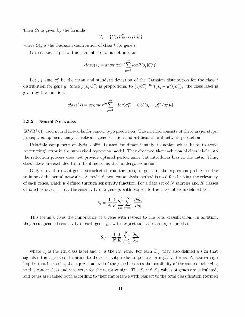
Then Ck is given by the formula:
Ck = {C1k , C2
k , . . . , Cmk }
where Cik, is the Gaussian distribution of class k for gene i.
Given a test tuple, s, the class label of s, is obtained as:
class(s) = argmaxmi (
m∑
g=1
logP (sg|Cgi ))
Let µgi amd σg
i be the mean and standard deviation of the Gaussian distribution for the class i
distribution for gene g. Since p(sg|Cgi ) is proportional to (1/σg
i )−0.5((sg − µgi )/σ
gi )2, the class label is
given by the function:
class(s) = argmaxmi
m∑
g=1
[−log(σgi ) − 0.5((sg − µg
i )/σgi )2]
3.3.2 Neural Networks
[KWR+01] used neural networks for cancer type prediction. The method consists of three major steps:
principle component analysis, relevant gene selection and artificial neural network prediction.
Principle component analysis [Jol86] is used for dimensionality reduction which helps to avoid
“overfitting” error in the supervised regression model. They observed that inclusion of class labels into
the reduction process does not provide optimal performance but introduces bias in the data. Thus,
class labels are excluded from the dimensions that undergo reduction.
Only a set of relevant genes are selected from the group of genes in the expression profiles for the
training of the neural networks. A model dependent analysis method is used for checking the relevancy
of each genes, which is defined through sensitivity function. For a data set of N samples and K classes
denoted as c1, c2, . . . , ck, the sensitivity of a gene gi with respect to the class labels is defined as
Si =1
N
1
K
N∑
j=1
K∑
m=1
∣
∣
∣
∣
∂cm
∂gi
∣
∣
∣
∣
This formula gives the importance of a gene with respect to the total classification. In addition,
they also specified sensitivity of each gene, gi, with respect to each class, cj , defined as
Sij =1
N
1
K
N∑
m=1
∣
∣
∣
∣
∂cj
∂gi
∣
∣
∣
∣
where cj is the jth class label and gi is the ith gene. For each Sij, they also defined a sign that
signals if the largest contribution to the sensitivity is due to positive or negative terms. A positive sign
implies that increasing the expression level of the gene increases the possibility of the sample belonging
to this cancer class and vice versa for the negative sign. The Si and Sij values of genes are calculated,
and genes are ranked both according to their importance with respect to the total classification (termed
11

as total rank) and to their importances with respect to each individual cancer class (termed as separate
rank). Based on the separate rank, the genes are then classified according to the cancer class in which
they are highly expressed.
While deciding the number of genes to be selected for the classification process, it was observed
that selection of 96 genes gives the best performance for the data set they used(88 samples of 6567
genes in which 63 are used in the training process and 25 used in the test).
The class prediction was done using an Artificial Neural Network(ANN) classifier[Bis95]. The ANN
classifier consists of linear perceptrons of 10 input nodes, which corresponds to 10 principle components,
and 4 output nodes, which corresponds to the 4 different class labels in the input data. In total, they
used 44 parameters. 3-fold cross validation were used for the prediction procedure. Samples were
shuffled and split into 3 equal sized groups where 2 of them were used as the training set and 1 was
used as the testing set. The experiment was repeated for 1250 times by random shuffling the samples.
The class label of each test sample was assigned using majority voting of the results obtained for each
test sample in the 1250 tests. To allow the rejection of assigning a class to a test sample, they defined
a distance function, dk, which measures the distance from a sample to the ideal vote for each cancer
class
dk = 1/2k
∑
i=1
(oi − δi,k)2
where k is the cancer class, oi is the average vote for cancer class i and δi,k is unity if i corresponds to
class k and 0 otherwise. The distance is normalized such that the distance between two ideal samples
belonging to different classes is unity. For each class, the empirical probability distribution of its
distance was generated. They defined the 95th percentile of the probability distribution as the cutoff
value for confidence. Samples outside of the 95th percentile of probability distribution of the classes
were not assigned any class.
3.4 Decision Tree - Recursive Partitioning
Decision tree, also known as classification trees, is a well know classification method[BFOS84]. It has
been widely used in classification applications and many extensions/variations have been proposed[RS98,
Utg89, SAM96, GGRL99].
A decision tree consists of a set of internal nodes and leaf nodes. The internal nodes are associated
with a splitting criterion which consists of a splitting attribute and one or more splitting predicates
defined on this attribute. The leaf nodes are labeled with a single class label. The construction of
the decision tree is usually a two-phase process. In phase 1, the growing phase, an overgrown decision
tree is built from the training data. The splitting criterion at each internal node is chosen to split the
data sets into subsets that have better class separability, thus minimizing the misclassification error.
In phase 2, the pruning phase, the tree is pruned using some heuristics to avoid overfitting of data
which tends to introduce classification error on the test data.
[ZYSX01] proposed a recursive partitioning cancer classification method based on decision tree.
This method constructs a binary decision tree. A purity based entropy function is used to determine
12

the splitting criterion at each internal node. The function is defined as
Plog(P ) + (1 − P )log(1 − P )
where P is the probability of a tuple being normal. During the process of tree construction, this
function is applied to find the best gene to split and the best splitting criterion for the chosen gene.
This is done by test each unused gene on all of its possible splitting points using the purity entropy
function and select the one that gives the best result.
For testing the classification accuracy, they used 5-way localized cross-validation to reduce the
chances of overfitting due to small data set size. The localized procedure keeps the same splitting
gene in each internal node, but does cross validation on different splitting point of that gene. The
performance test was done on the colon tissue data set from NCI and the result of the cross validation
showed a misclassification rate of 6-8%.
3.5 Similarity Based Methods - NN and CAST
Here, we describe two multiclass classification methods based on similarity measure. Compared to
the other methods, these two methods are less prone to noise and bias in the data But they have the
disadvantage of not being able to scale well.
3.5.1 Nearest Neighbor Analysis
This method is based on a distance metric between the testing tuples and the training tuples. The main
idea of the method is for each testing sample s, find one training sample t with most similar expression
value, according to a distance measure. The class label of t is then assigned to s. The distance metric
can be any similarity measure based on attribute values, for example, the Pearson correlation function,
the Euclidean distance function, etc.
[BDBF+00] used the Pearson correlation as a measure of similarity. Let s be a testing sample, and
t be a training sample. Then s and t can be viewed as vectors of m gene expression values. Let E(x)
and E(x) represent the expected value and the variance of a vector x, respectively. Then the Pearson
correlation function between two vectors, s and t, is given by:
P (s,t) =E((si − E(s))(ti − E(t)))
√
V ar(s)V ar(t)
Given a testing sample s, and a set of training tuples T containing pairs of the form (ti, ci) where tis
are the expression values of genes and ci is the class label of the tuple, the Pearson correlation function
is evaluated for every (s, ti) pair, where i ∈ {1 . . . n}. The tuple t that has most similar expression
value with s is the one that maximizes P , given by argmaxni (P (s, ti)). The class label of t is then
assigned to s.
Therefore, the nearest neighbor classifier can be formally expressed as:
Class(T, s) = class(argmaxiP (s, ti))
13

where class returns the class of the training tuple that has the highest P value.
The Nearest Neighbor(NN) method can be extended to the K-Nearest Neighbor(KNN) method, as
proposed by [EH51] and applied to gene classification by [DFS00]. KNN differs from NN that the class
label of a testing sample s is assigned using majority vote from K training tuples that are most similar
to s according to a distance measure function. In [DFS00] a correlation metric, R, is used to measure
the similarity between pairs of samples. The correlation metric R is given by
R(s, t) =
∑mj=1(sj − s)(tj − t)
√
∑mj=1(sj − s)2
√
∑mj=1(tj − t)2
The classification proceeds in two steps. In first step, for each test sample s, K training samples
with highest similarity to s are picked using the correlation metric R. In step two, for each test sample
s, its class is determined using majority voting of the classes of its K most similar neighbors.
Obviously the value of K affects the performance of the method. This is especially notable in the
case of gene classification, since the data sets is small but has enormous number of attributes. [DFS00]
determines the value of K using leave-one-out cross validation on the training set. Each training
sample, t, is treated as a test sample, the distance measure is used to check its correlation with every
other training tuples. The k most correlated training tuples are selected to vote for t’s class. Every
training tuple’s original class is compared with its assigned class to get the total number of erroneous
assignment for the current value of k.
This process is repeated for different values of k to find the one that gives least error to be used as
in the classification of testing tuples. In [DFS00], the method checked for k = 1, 2, . . . , 21.
NN is simpler than KNN since it only requires finding one most similar training tuple while the
KNN method needs to first determine the value of k. But KNN has several advantages over NN. First,
in the case of mislabeled training tuples, it will have much greater effect on the classification result of
NN since one mislabel will result in misclassifying all the test tuples that are most similar to it. Also,
KNN is less prone to bias in the data and more tolerable to noise since it makes use of several training
tuples to determine the class of a test tuple instead of one tuple.
3.5.2 Cluster-based Method: CAST
[BDBF+00] developed a cluster-based classifier, CAST. It is motivated from the observation made by
Alon et al[ABN+99] on the hierarchical clustering on the colon data (see data set description): the
topmost division divides the samples into two groups: one group contains mostly normal samples and
the other mostly tumor samples.
The main idea is to group the training tuples into different clusters based on their similarity with
the tuples already in the cluster and remove those that no longer has much similarity with the current
cluster. CAST uses a threshold parameter, p, to control the granularity of the clusters. During the
training phase, a training tuple is grouped into a cluster if it has high similarity with the tuples inside
the cluster. A tuple is said to have high similarity with a group of tuples if the similarity measure
between them is at least p (the authors used Pearson correlation between the gene expression values
14

as a measure of similarity, though any similarity measure can be used). After inclusion of the new
tuple into a cluster, the similarity score of the tuples in the cluster is again evaluated. A tuple will be
“kicked out” if its new similarity score with the updated cluster is below p.
CAST creates one cluster at a time. During the training phase, it alternates between adding tuples
with satisfying similarity scores to the cluster and removes those with unsatisfying similarity scores.
Eventually, one cluster is formed with tuples all having similarity score above p. It then goes on to
create another cluster until every tuple has been included inside a cluster.
CAST selects the best p value through a measure of cluster structure compatibility. Intuitively,
compatibility measure penalizes the cluster structures that have tuples of the same classes separated
into different clusters and tuples of different classes being grouped into the same cluster. They define
the compatibility score as the sum of the number of pairs of tuples that have different classes and
assigned to different cluster and the number of pairs of tuples that have the same class and assigned
to the same clusters.
In selecting the best p value, the algorithm iteratively considers different values for p using binary
search. For each p, it uses CAST to cluster the training tuples and then apply compatibility measure to
the clusters. The p value that gives the highest compatibility score is chosen to used in the classification
phase.
Given a test sample s, and a group of training tuples T , CAST tries different values of p to cluster
s and T to find the best cluster structure (note that the compatibility measure is only done on the
training tuples). The class label of s is determined through majority vote of the training tuples in the
cluster where s belongs to. If there is no majority class in the cluster, or the majority is too small to
be confident, then s is assigned to the “Uncertain” class.
3.6 Max-Margin Classifiers
The training process of a classifier can be viewed as a process of finding an hyperplane that separates
the training tuples into different groups according to their classes. The margin of the hyperplane is
defined as the distance from the hyperplane to the sets of points that are closest to it. A hyperplane
with small margin is not as confident as the one with large margin. Given a slightly different training
data, the hyperplane would change, thus changing the classification of points that lie close to the
hyperplane. The definition of margin suggests that if a classifier is able to separate the points in a way
that maximizes the margin, then it will be less subjective to overfitting and have better classification
performance.
For the expression data used in cancer classification, they can be viewed as very sparse points
in very high dimensional space. For such kind of data, it is very easy to find several hyperplanes
that linearly separate the training tuples, yet easily subject to overfitting. Therefore, max-margin
classifiers [FS98, SBS00] are good choices for dealing with this kind of data. Here we introduce two
max-margin classification algorithms, Support Vector Machine and Boosting that has been applied on
cancer classification.
15
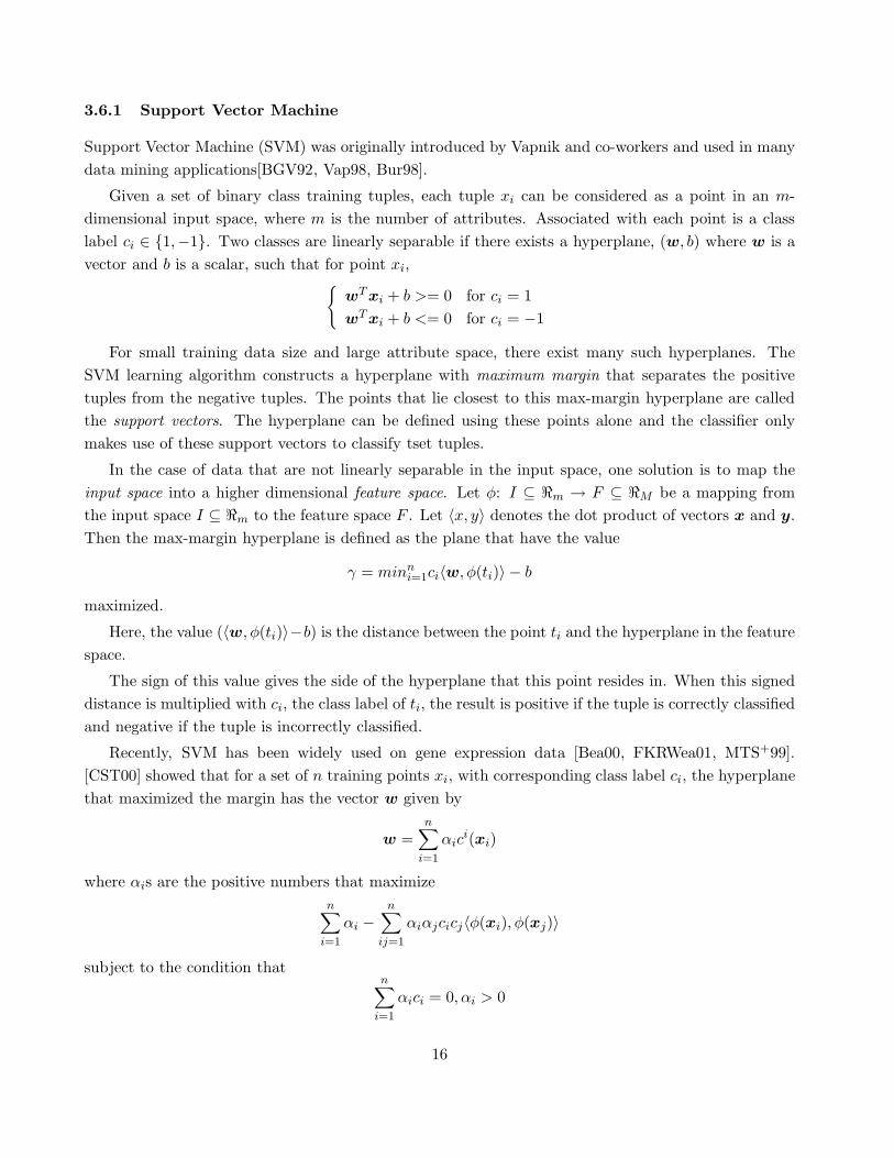
3.6.1 Support Vector Machine
Support Vector Machine (SVM) was originally introduced by Vapnik and co-workers and used in many
data mining applications[BGV92, Vap98, Bur98].
Given a set of binary class training tuples, each tuple xi can be considered as a point in an m-
dimensional input space, where m is the number of attributes. Associated with each point is a class
label ci ∈ {1,−1}. Two classes are linearly separable if there exists a hyperplane, (w, b) where w is a
vector and b is a scalar, such that for point xi,{
wT xi + b >= 0 for ci = 1
wT xi + b <= 0 for ci = −1
For small training data size and large attribute space, there exist many such hyperplanes. The
SVM learning algorithm constructs a hyperplane with maximum margin that separates the positive
tuples from the negative tuples. The points that lie closest to this max-margin hyperplane are called
the support vectors. The hyperplane can be defined using these points alone and the classifier only
makes use of these support vectors to classify tset tuples.
In the case of data that are not linearly separable in the input space, one solution is to map the
input space into a higher dimensional feature space. Let φ: I ⊆ ℜm → F ⊆ ℜM be a mapping from
the input space I ⊆ ℜm to the feature space F . Let 〈x, y〉 denotes the dot product of vectors x and y.
Then the max-margin hyperplane is defined as the plane that have the value
γ = minni=1ci〈w, φ(ti)〉 − b
maximized.
Here, the value (〈w, φ(ti)〉−b) is the distance between the point ti and the hyperplane in the feature
space.
The sign of this value gives the side of the hyperplane that this point resides in. When this signed
distance is multiplied with ci, the class label of ti, the result is positive if the tuple is correctly classified
and negative if the tuple is incorrectly classified.
Recently, SVM has been widely used on gene expression data [Bea00, FKRWea01, MTS+99].
[CST00] showed that for a set of n training points xi, with corresponding class label ci, the hyperplane
that maximized the margin has the vector w given by
w =n
∑
i=1
αici(xi)
where αis are the positive numbers that maximize
n∑
i=1
αi −n
∑
ij=1
αiαjcicj〈φ(xi), φ(xj)〉
subject to the condition thatn
∑
i=1
αici = 0, αi > 0
16

It turns out that only for the support vectors, their αi values are non-zero. Therefore, only the
support vectors are required to define the max-margin hyperplane. This implies that only the training
tuples with non-zero αi values have to be stored in memory for the classification process.
[FCD+01, BDBF+00, RTRea01] applied SVM technique to the cancer classification problem. Given
a set of gene expression training tuples of size n, each tuple is a pair in the form of (ti, ci), where tis
are the vectors of the expression values of m genes, and cis are the corresponding class labels and ci
∈ {1,−1}. Let the max-margin hyperplane be denoted by the vector w0 and b0. Given a test tuple s
with class label c, the classification result produced by the SVM on s is given by its relationship with
respect to the hyperplane generated, The result is given by:
Class(s) = sign(c(〈w0, φ(s)〉 − b0)
If the returned sign is positive, it means s is correctly classified, otherwise s is incorrectly classified.
Sometimes when the training samples have mislabeled samples, the tuples may not be linearly sep-
arable in the feature space. In other times, for the sake of error tolerance and overfitting avoidance,
perfect linear separation may not be desirable. For these cases, it is better to allow some training sam-
ples to fall to the wrong side of the hyperplane [Bea00, FCD+01]. Soft margin and margin-distribution
[JTC99] SVM are developed for this purpose. Through some parameter tuning, which controls the
overall training error or a specific false positive or false negative error, desired training error can be
achieved.
The ability of SVM for producing hyperplane with maximized margin and for tuning the amount
of training errors allowed has made SVM especially suitable for the gene expression data classification.
3.6.2 Aggregated Classifiers: Boosting
Aggregated classifiers are built from the aggregation of multiple versions of the class predictors using
majority voting [Bre96, Bre98]. It is useful for improving the accuracy of the classifiers that are unstable
to small changes of the learning set, for reducing the overfitting problem due to small training data set
and for improving the accuracy of those “weak learner” classifiers.
The idea is to generate multiple versions of classifiers over the training data in iterations. In each
iteration, the training data are sampled with replacement by putting more emphasis on the training
tuples that were wrongly classified in the previous iteration. The final aggregated classifier is a weighted
voting of the classifiers built during each iteration. The weight is dependent on the accuracy of the
classifier built from the data it was being trained.
Here we describe AdaBoost algorithm proposed by [FS97] for binary-class problems. Given a
training set T = {(t1, c1), (t2, c2), . . . , (tn, cn)}, and a classification algorithm L. Since there are only
two class labels, we could map one class label to -1 and the other to 1 for easier classification using
boosting.
The initial distribution of weights of each tuple is the same, given by W1(ti) = 1/n, where i =
1, . . . , n. In iteration i, call to L with weights distribution Wi will return a classifier Li. The error, ǫi,
17

of Li is given by∑n
j=1 Wi(tj){ci 6= Li(tj)}. Here, {ci 6= Li(tj)} checks if the classifier classifies tuple tj
correctly, it returns 1 if the statement is true (false classification) and 0 otherwise.
The weight of Li is then wi = 12 log 1−ǫi
ǫi. The new weight distribution of T is then given by
Wi+1(ti) ∝ Wi(ti)e−wiciLi(ti)
such that∑n
i Wi+1(ti) = 1. Suppose there are x number of classifiers trained. Then given a test sample
s, the class of s is given by
Class(s) = sign(x
∑
i=1
wiLi(s))
.
The margin mi of a tuple ti with class label ci can be defined as:
mi = ci
K∑
j=1
wjClassi(ti)
Positive mi means ti is correctly classified and otherwise ti is incorrectly classified. The magnitude
of mi is proportional to the confidence of the classification. The smaller the magnitude, the less
confident.
During the iterations of the classifier construction, if a tuple is misclassified in the current iteration,
it will be assigned a greater weight in the next round. Thus, the goal of the algorithm is to “concentrate”
on the hard samples that always get misclassified to achieve optimal classification. This actually
corresponds to the effort of trying to enlarge the margin of the training tuples.
Theoretically, boosting can be thought of as a max-margin classification technique similar to SVM
[BDBF+00]. [SFBL98, MBB98] also showed that the generalization error of boosting depends on the
distribution of margins of the training tuples.
[BDBF+00, DFS00] applied boosting for cancer classification. [BDBF+00] used a very naive decision
function to act as a weak classifier. The decision function f is given as:
f(g : i, t, d) =
{
d g[i] > t
−d g[i] < t
f takes in three parameters, g : i, t, and d, where g denotes the vector of gene expression values,
thus g : i denotes the value of ith gene in the vector, t denotes the split point for gene i, and d is the
class of g, d ∈ {−1, 1}. This decision function is applied to every gene and checks through every possible
split point to find the one that gives the best class separation. The boosting process is thus iterations
of split point evaluations for every gene in the tuples. The test samples are then classified according
to the weighted voting of the classes given by the split point decision functions in each iteration.
[DFS00] applied boosting technique using the CART decision tree as the classifier. Both method
achieved satisfactory results even though neither classification algorithm is well suited for the cancer
data set. The credit should go to the max-margin idea in the boosting technique that concentrates on
tuples that can easily get misclassified and try to find the separation that maximizes the margin.
18
3.7 Evaluation
We believe that for cancer classification, besides computation time and classification accuracy, biological
relevance such as information about gene interaction, marker genes and etc(we will elaborate it in the
next section of related issues) should also be one of the evaluation criteria.
This is due to the fact that biologists not only expect cancer classification gives correction classifi-
cation but also want to generate more information about the genes for cancer-related studies such as
drug discover and have a better understanding of tumor development.
Below, we give a rough evaluation of the cancer classifiers based on their classification performance
and biological relevance.
3.8 Weighted Voting: GS Method
The GS method is simple and works well in some data such as the Leukemia data set. But its simplicity
also results in some limitations. First, it is only applicable for binary class problem. Typical cancer
classification involves identifying of more than two classes of cancer. In this case, GS algorithm will
not be effective. Second, it chooses an equal number of genes that have high correlation with the two
classes. This means that it is only effective in classifying data sets that are unbiased. Biased in this
sense means that there are unequal number of genes favoring the two classes with the same correlation
strength. If the majority genes of a data set are highly correlated with one class, then this method will
not produce satisfactory classification result since it always choose an equal number of genes in both
classes to vote for the majority.
3.9 Similarity-based Classifiers: KNN and CAST
The similarity methods(KNN and CAST) base their classification on the similarity of the expression
values of each gene, this makes them less prone to noise and bias in the data. Both methods tries to
find for a test tuple s, a set of training tuples that it has high similarity with and use majority voting
of these training tuples to determine the class of s. The disadvantage of these methods is due to their
non-scalability. Every testing tuple needs to be checked against every training tuple in order to find
the most similar ones. These method works fine when the data size is small, but they suffer when the
data size is large.
Comparatively, KNN is less computationally expensive than CAST since the similarity score is
only evaluated once for every test and training pair. Whereas in CAST, it has to repeatedly evaluate
the similarity score of each tuple in the clusters against that of the rest during the formation of each
cluster. This is repeated many times in order to find the best granularity control parameter. We feel
that it might not be practical to apply this method to cancer classification since it requires too much
computational time. As the availability of gene expression data increases, this method tends to loose
its advantage over other more scalable methods.
19
3.10 Max-Margin Classifiers: SVM and Boosting
Support Vector Machine has been well exploited by researchers in various fields from text categorization,
pattern recognition to protein function prediction. Recently, SVM has been used in cancer classification.
ability to deal with high dimensional data, avoidance of overfitting, and robustness with noise and
sparseness of solution all make it well suited for application to cancer classification. Another benefit
offered by SVM is scalability [Tre01]. The number of support vectors selected by the learning algorithm
is usually small, even with large training set [BGL+99, BDBF+00, JTC99]. This is essential since the
amount of available gene expression data will soon increase dramatically. One drawback that limits
its original application is only for binary class problems. Some solutions were suggested to overcome
this limitation, such as redefining the multiclass problem into binary, or iteratively performing binary
classification until all classes has been separated, but none was very effective. Recently, extensions to
multiclass SVM were being studied[CS00, LLW01, HL01], its effectiveness and performance is still an
on-going research problem.
For the current available data characteristics, boosting achieves comparable classification perfor-
mance with respect to other methods. Since boosting is usually applied to weak learners to improve
classification accuracy through repeated classification of the weighted training tuples, it is quite time
consuming. In addition, existing classification methods do produce comparable classification accuracy,
thus we feel that the boosting method does not have any superiority over other methods.
3.11 Bayesian Network and Neural Networks
Bayesian network method is simple and can be applied to multiclass classification problem. But there
are two issues that restricts its performance in cancer classification. First issue is its assumption
of orthorgonality among genes. It is known that in most cases, the expression values of genes are
correlated. Also, gene interaction is of important interest for the biologists. Assuming independence
not only ignores important classification information embedded in the data set which might result in
inaccurate classification, but it is also incapable of revealing any biological information in the data
since the whole process is a black box. Another issue is that it assumes a Gaussian distribution of
the data. And in the cases of data set that does not follow such a distribution, it proposed to do
some transformation such as taking the logarithmic or square root of the gene expression values. But
still, the assumption that the classes must follow a certain distribution is too restrictive, especially in
the case of gene expression data which are hard to say what kind of distribution the data follow. We
feel that though this method might be useful in classifying certain types of expression data, but its
performance is limited due to its assumptions.
Neural network method has comparable result with the other methods. But similar to the naive
bayes method, it does the classification in a black box manner. The user are not given any information
on how the genes are correlated, which set of genes is more effective for classification, etc..
20
3.12 Decision Trees
Decision trees have been attractive in data mining environment for several reasons. First, their results
are interpretable. Second, they do not require any parameter input. Third, the construction process is
relatively fast. We believe that these advantages are also applicable to cancer classification using gene
expression data. An additional advantage is the scalability of decision trees. There are several scalable
decision tree algorithms [GGRL99, SAM96] available, as the date size increases, these algorithms might
be useful for providing satisfying performance over large gene expression data.
3.12.1 Classification Accuracy
From the experiments conducted in[BDBF+00], it found that SVM has the highest accuracy on the
leukemia and ovarian data set, while CAST performs better for the Colon data set and Boosting using
simple decision stump outperforms NN in all three data sets.
[KSHR00] compared NB method with GS method and found that NB outperforms GS method for
the leukemia and ovarian data sets while GS outperformed NB in the colon data set. The NB classifier
achieved 100% accuracy on the leukemia data set and 84%accuracy in the ovarian and colon data set.
[DFS00] observed that the classification accuracy for the NCI data set is much lower than that of
leukemia and colon data set. They contributed it to the small data size (60) and wide range of classes
(10). We believe that for such a small data size and large number of classes, overfitting is the main
problem causing the degrading of performance.
Some of these approaches perform classification after gene selection. Experiments show that the
quality and quantity of genes selected has quite big effect on the classification results, as expected.
Overall, the proposed methods all showed good performance on some data sets and there is no one
that is superior than the rest on all data sets. Since the amount of gene expression data available is
small, the classification accuracy of various algorithms cannot be compared extensively. It would be
necessary to perform an extensive comparison of all the proposed methods on the available data sets in
order to really judge which method gives the best classification accuracy. This could be one direction
for future work.
3.12.2 Biological Relevance
One important difference between cancer classification and other previous classification applications
is the amount of useful information revealed during the classification process. Obviously, accuracy is
very important in cancer classification since it helps in accurate diagnosis of cancer patients. On the
other hand, we still have limited knowledge about the cancer disease, biologists wish to gain more
understanding of gene interaction and other biological information related to cancer development.
Thus, cancer classification is not merely about classification accuracy, but also about identifying useful
information for studying gene interactions. A cancer classifier that does not explore the biological
information within the gene expression has only achieved part of the classification goal. Here, we give
21

a categorization of the methods described in this section to reflect their biological relevance in using and
revealing the gene interaction information. The methods can be grouped roughly into three categories.
The first category corresponds to methods that totally ignore the context of the data, the represen-
tatives in this category are FLDA (or any statistical based methods), the naive bayesian method and
the neural network method. These methods only looks at the data as a set of distributions and make
classification decision based on the distribution of data values regardless of the context meaning of the
data.
The second category corresponds to methods that makes use of the correlation among genes in the
data but do so in a “black box” fashion. NN, KNN, perceptrons, artificial neural networks, CAST and
SVM belongs to this category. The classifiers in this category makes use of the correlation between the
expression values of genes. But they do not give any insight into the structure of the data.
The third category corresponds to the methods that are capable of exploring and revealing corre-
lations between the genes. The representative in this category is the classification tree based method-
recursive partitioning. Classification trees reveal the relationships among the genes and do so through a
stepwise predictor selection. This process of selection and splitting provides the biologists some insight
into the structure of the data and the correlation information among the genes. The gene expression
data have high dimensionality and small training data size, there might be times when decision tree
method fail to give good classification accuracy due to noise and overfitting. Applying boosting with
classification tree might be a good solution to improve accuracy in this type of situations though the
information about gene correlations are less visible.
From this categorization, it can be seen that most cancer classifiers are still lacking in the biological
relevance aspect. Further development in this direction is necessary.
3.13 A Unified View
Table 1 is a summarization of the various methods introduced in this section.
Multi- Strategy Biologically Scalability
class meaningful
SVM No Max-Margin No Good
Boosting(Decision-tree) Yes Max-Margin Yes Classifier Dependent
Decision tree Yes Entropy Function Yes Good
KNN(K ≤ 1) Yes Similarity No Not Scalable
CAST Yes Similarity No Not Scalable
GS No Weighted Voting Yes (gene selection) Fair
FLDA Yes Discriminant Analysis No Fair
Neural Network Yes Perceptrons No Fair
Naive Bayes Yes Distribution modelling No Fair
Table 1: Summarization of cancer classifiers
22

It can be seen that some of the classification methods used for cancer classification do not pro-
vide much biologically relevant information to the user. In order to fully achieve the goals of cancer
classification which are accuracy and bio-relevancy, it is necessary to either develop new classification
algorithms aimed at providing good cancer classification accuracy while at the same time revealing
more information for gene interaction, or modify the existing algorithms to take care of the bio-related
aspects.
Additionally, since there are only a limited number of available data sets, and the size of these
data are very small, thorough experiments cannot be conducted to really distinguish the better ones
from the others. Right now we can only theoretically compare the algorithms on their computational
time and effectiveness on cancer classification. More thorough comparisons for the computation time,
classification accuracy and biological relevance needs to be done on the existing methods to provide a
better picture of the current status. This would help us at getting better insight into the development
of more effective and efficient algorithms aimed particularly at classifying cancerous data using gene
expression values.
4 Gene Selection
Feature selection is an useful preprocessing technique in data mining, it’s usually used to reduce the
dimensions of the data and improve classification accuracy [KS96, SS88]. Currently, some feature
selection methods on genomic data have been proposed [XJK01, CLT01, GWB+00]. For the problem
of cancer classification, we believe that a fair amount of attention should be paid on gene selection and
make it an integral preprocessing step. This is well justified due to the following reasons.
First, gene expression data set has very unique characteristics which are very different from all the
previous data used for classification. Most publicly available gene expression data has the following
properties:
• high dimensionality: up to tens of thousands of genes,
• very small data set size: less than 100, and
• most genes are not related to cancer classification.
With such a huge attribute space, it is almost certain that all classifiers built upon it would prone
to overfitting. The small data size makes it even worse. Since most genes are known to be irrelevant
for class distinction, their inclusion would not only introduce noise and confuse the classifiers, but
also increase the computation time. Gene selection prior to classification would help in alleviating
these problems. With the “noise” from the irrelevant genes removed, the biological information hidden
within will be less obstructed. During the classification process, classifiers such as decision tree will
be able to provide a more precise view of genes interaction. Post-classification analysis can be done to
find out some biologically relevant issues such as the sets of genes that are most related to certain kind
of cancer or if there are other biological unknowns related to accurate cancer prediction. This would
assist in drug discovery and early tumor discovery.
23

Second, experiments have shown that gene selection prior to classification improves the classification
accuracy of most classifiers [DFS00, GST+99, GWB+00, CLT01]. It not only improves the performances
of the classifiers that are weak in handling large number of attributes, but improves the performances
of those that are capable of handling large attribute space, such as SVM[GWB+00]. Though there is
no direct measure on the saving of running time, but the number of genes decreased from thousands
to tens or below 10 after performing gene selection which would make a big difference on the running
time.
Due to the above reasons, it should be clear that in order to achieve good classification performance,
and obtain more useful insight about the biological related issues in cancer classification, gene selection
should be well explored to both reduce the noise and avoid overfitting.
In this section, we take a look at the proposed gene selection methods in detail. Though researchers
have used different feature selection methods for gene selection, but they can be broadly categorized
into 2 groups: individual gene ranking approach, and gene subset ranking approach. Below, we first
describe the main idea of each category and then followed by some gene selection methods in each. We
conclude the section with some discussion.
4.1 Category I: Individual Gene Ranking
Feature ranking approach is the most commonly used for feature selection. In this approach, each
feature/attribute is measured for correlation with the class according to some measuring criteria. The
features/attributes are ranked and the top ones or those that satisfy a certain criterion are selected.
The main characteristic of feature ranking is that it is based on individual feature correlation with
respect to class separation.
In gene selection, methods in this category select genes with high individual correlation with the
classes. No correlation among the genes are exploited. Genes selected in this case may have high
correlation with the class as an individual, but act together, might not give the best classification
performance. Also since each gene is considered separately, some genes may contain the same corre-
lation information thus introduce redundancy. Finally, genes that are complement to each other in
determining the class labels may not be selected if they don’t exhibit high individual correlation.
4.1.1 Using Correlation Metric-GS Gene Selection Method
The most common and simple approach to feature selection is using correlation between the attribute
values and the class labels. In this approach, the correlation is measured using a distance function,
such Euclidean distance, Pearson correlation and etc.
The GS method [GST+99] proposed a correlation metric that measures the relative class separation
produced by the expression values of a gene. It favors genes that have big between-class mean expression
value and small within-class variation of expression value. For gene g, let µ1 and µ2 denote the mean
expression values of g in the two classes, and let σ1 and σ2 denote the standard deviation of the
expression values of g in class 1 and class 2. The gene selection metric is given as P (g) = µ1−µ2
σ1−σ2. The
24

P -value of each gene is measured and genes are grouped into positive value and negative value groups
and ranked according to their absolute values. The top k/2 genes from the two groups are selected.
This method is simple and easy to implement, but have several drawbacks. First, in the case of
classifying normal and cancerous data, the cancerous class may comprises cancers of different types.
Therefore, the genes that are cancer-related might not have small within-class variations since the
expression values for different tumors vary. This would result in mis-selection of the relevant genes.
Second, it only checks individual gene correlation, thus unable to find an optimal set of genes in certain
cases. Lastly, this method is not resistant to gene contamination (detailed discussion in next section).
Genes that are not cancer-related but more composition related sometimes are selected as top ranking
genes [GWB+00].
4.1.2 Using the Weights of a Linear Discriminant Function
Some classifiers are trained as a linear discriminant function. A linear discriminant function is of the
form of D(x) = w · x + b where w is the vector of weights for each gene, and x is a matrix of input
data and b is the bias. In this approach, the full set of genes are used to train a classifier in the form
of a linear discriminant function. Genes having the top K weights are selected, where K is the desired
number of genes.
The rationale is that for a trained classifiers, the weights of the features are proportional to their
importance in determination of the class labels. That is, the higher the weight, the better in its
distinction power of the class label. Therefore, given a trained classifier, we are able to obtain a set
of K high ranking genes by selecting the genes with the top K weights. For the classifiers that in the
form of a linear discriminant function. The top K genes can be selected from the weight vector w.
4.1.3 Likelihood Gene Selection
Keller et. al. [KSHR00] used likelihood measurement for gene selection. Given a training data set, it
tried to select genes whose expression values are a good indication of the class separation.
The genes are selected through a log likelihood score function, LIK. The LIK scores for each class
are computed for every gene. Let’s first consider the binary data case. Let Cg1 denote the distribution
of class 1 for gene g, and Cg2 denotes the distribution of class 2 for gene g. Then, the relative log
likelihood scores, can be defined as follows:
L1K1→2 = logp(Cg1 |X1) − logp(Cg
2 |X1)
L1K2→1 = logp(Cg2 |X2) − logp(Cg
1 |X2)
Where X1 and X2 refers to the training tuples in class 1 and class 2 respectively. A gene with ideal
class discrimination power would have high values for both LIK scores, indicating that expression
values of this gene in tuples belonging to class 1 will vote for class 1, and that in tuples belonging to
class 2 will vote for class 2. This implies that if the test tuples are of the same distribution as the
training sample, then the expression values of this gene will be a good indication of the class labels. The
25

higher the LIK scores are above 0, the better the class discrimination power the gene has. [KSHR00]
pointed out that in practice, it is difficult to find genes for which both LIK scores are much greater
than 0. They resort to select two sets of genes, GENE1→2 and GENE2→1, each maximizing one of
the LIK scores, while requiring the other score to be above 0:
GENE1→2 : LIK1→2 ≫ 0 and LIK2→1 > 0
GENE2→1 : LIK2→1 ≫ 0 and LIK1→2 > 0
After computing the LIK scores for every gene, genes in each set is ranked according to their LIK
scored in that set. The top x/2 genes are selected from each set, where x is the number of genes to be
selected.
In generalizing to multiclass data, it requires computing c(c − 1) number of LIK scores for each
gene, where c is the number of classes
LIKj→k = logp(Cgj |Xj) − logp(Cg
k |Xj)
where Xjs are the training tuples in class j, 1 ≤ j ≤ c, and j 6= k.
Intuitively, it tried to find for every gene, its ability to discriminate each class from all the rest of
the classes. As in binary class case, c(c− 1) sets of genes are grouped according to the LIK score they
maximize and requiring the rest scores above 0:
GENEj→k : LIKj→k ≫ 0and LIKj′→k′ > 0
where j′ 6= k′, 1 ≤ j′ and k′ ≤ c. Equal number of genes are selected from each set to form the total
set of genes selected for classification.
[KSHR00] compared the genes selected using NB and GS gene selection method. It found that NB’s
gene selection method gives better classification accuracy. Also, the genes selected by the NB method
has more variety than that of GS. They concluded that this variety of genes contributed to the better
classification accuracy.
4.2 Category II: Gene Subset Ranking
The approach used by methods in this category is to find a group of genes that serve together to achieve
the best classification result. The idea is to remove genes one by one, monitor the effect of removal of
this gene on the expected value of error. The expected value of error is the error rate computed on an
infinite number of samples. Given a training set, the expected value of error can be approximated by a
cost function J computed on the training samples. The effect of computing the change in the expected
value of error caused by removing a gene i is equivalent to bringing the weight of i to 0 in the decision
function. Given a classifier with weights for each training tuples, the change in cost function from the
removal of gene i is approximated by
DJ(i) = (1/2)∂2J
∂w2i
(Dwi)2
where Dwi is the change in the weight after removing gene i.
26

4.2.1 Recursive Feature Elimination (REF)
In [GWB+00], it proposed the use of recursive feature elimination approach to do gene selection. It
iterates through following steps:
• Train the classifier (optimize the weights wi with respect to cost function J)
• Compute the ranking criterion for all features (DJ(i) or (wi)2)
• Remove the feature with the smallest ranking
From the elimination process, we can see that it tries to retain the set of features that has the
highest classification power. The features that are eliminated last may not be necessarily the ones that
are individually most relevant.
SVM has recently gained popularity in both classification and feature selection [WMC+00]. [GWB+00]
used SVM to be the classifier for the cost function computation on subset ranking based gene selection.
The SVM based REF gene selection method works as follows: apply the SVM classification algorithm
on the training data; compute the change in cost function for the removal of each gene i; find the gene
that has the minimum cost function change after its removal; remove that gene from the training data;
repeat until the training data is empty.
It can be seen that this method is a backward feature ranking method. The group of genes that
are removed last together gives the best classification result. But individually, they might not be the
ones that has the best correlation. This is true by looking at the way the features are eliminated. The
elimination function is the change in the expected error rate of the classifier. In this sense, it always
remove the gene whose removal has the minimum change on the classification result.
4.3 Discussion
In this section, we compare the two categories of gene selection methods in several areas.
4.3.1 Redundancy and Complementary Issue
[GWB+00] found that the REF gene subset ranking (GSR) approach works better for cancer classifica-
tion than the individual gene ranking approach(IGR). The former is capable of finding genes that are
complementary which produce best classification result, whereas the latter only finds genes that have
high individual correlation. Individual correlation does help in class distinction, but only to a certain
extent. There are additional information relevant to tumor type differentiation that can only be re-
vealed through the interactions among the genes. Some genes may not have individual high correlation
but they complement each other in class distinction. Since the individual gene ranking approach does
not have any mechanism in discovering the complementary roles among the genes, it can be considered
as a handicapped approach. Also, since in IGR approach, each gene is assessed for its correlation
individually and selected based on the correlation score, it is inevitable that in some cases, it would
27

select a set of genes with similar correlations(redundancy) yet miss out some other important genes. In
the case of tumors that have different expressibility, i.e., some tumors are less distinctively expressed
by genes and some tumors that more easily expressed, the IGR approach might select lots of genes
that can distinguish one class but miss out those that are useful for distinguishing other not so highly
distinguishable classes.
4.3.2 Computation Time
GSR approach seemed to provide a more optimal set of genes for cancer classification and other related
biological studies. But it takes too long to compute. With so many genes in the data set, it has
to iteratively perform subset ranking and eliminate genes one by one. [GWB+00] suggested to trade
accuracy for speed by initially removing chunks of genes each time, and resolve to remove one at a time
towards the end. The reason is that though better results are obtained when removing genes one at a
time than removing chunks at a time, the difference is only significant for small set of genes. Therefore,
the time saving sub-optimal solution is to removes chunks of genes at one time and when the gene size
reaches a few hundred then start to eliminate one by one.
4.3.3 Class Distinction Power vs Direct Relevance to Tumors
Another issue of concern is how relevant to cancer are the genes being picked by the two approaches.
Some genes might give a good class distinction, but they might not be directly cancer related. Due
to the fact that cancerous tissues and normal tissues usually have very different cell composition
[GWB+00, BDBF+00], it is possible that those genes observed to have high distinction power are those
that related with different cell composition instead of directly linked with different types of tumors.
[GWB+00] analyzed the genes selected by the methods in both approaches and found that SVM
REF gene selection method was able to pick up genes that are directly related with cancer, whereas other
methods tends to pick up genes that are non-cancer related but due to the different cell compositions
between the tissues. We believe that this is due to the fact that the SVM REF method uses subset
ranking method instead of feature ranking method. This method tends to pick up a group of genes that
when put together best determines the class type which means it best captures the gene interaction
information.
It has also been observed that for the same gene selection mechanism, the accuracies of different
classifiers do not differ much, whereas different gene selection methods have greater effect on the classi-
fication accuracy. This again proved how an important role gene selection plays in cancer classification.
In conclusion, we feel that a good gene selection method for cancer classification should observe the
following properties:
• incorporation of the gene interaction information,
• selection criteria based on group performance instead of individual correlation,
• Inclusion of complementary genes,
28

• Picking of cancer related genes instead of cell composition related genes, and
• Reasonably efficient.
5 Related Issues
In this section, we discuss some important issues in cancer classification using gene expression data.
They include the issue of statistical significance vs biological significance of a cancer classifier, detailed
breakdown of classification results, the issue of sample contamination, and the effect of selection of
marker genes on cancer classification.
5.1 Statistical vs. Biological Significance
In the past, classifiers are usually evaluated on the classification accuracy and efficiency. Not much
attention was paid on the ability of the classifiers in discovering interactions between attributes. This
is because the attributes in the earlier data used usually do not exhibit complex behavior or underlying
meaning. Unfortunately, this is not the case for cancer classification.
As an area of bioinformatics study, the major goal of expression data analysis is to provide the
biologists biologically meaningful information about the genes and related things. Through these in-
formation, biologists are able to discover unknowns and reaffirm previously knowledge. For cancer
classification, information about gene interaction is of great biological relevance. It provides the biolo-
gists a clearer understanding of the roles a certain set of genes play in cancer development and related
issues. This will greatly assists the discovering and early treatment of disease and drug discovery. It is
a common consensus that biological relevance for a cancer classifier is as important as the classification
accuracy.
One important issue is to find marker genes. Marker genes are genes whose expression values are
biologically useful for determining the class of the samples. In other words, marker genes are genes
that characterize the tumor classes.
The identification of marker genes are important due to the following reasons:
• Enable better classification performance
• Allow biologists further study the interaction of relevant genes in achieving a certain biological
performance
• Study the functional and sequential behavior of known marker genes in order to facilitate the
functionality discovery of other genes.
• Allow further study of relation of expression values of different genes with respect to the tumor
class, is similar expression pattern always results in cancer or the combination of suppression of
certain genes and expression of certain genes are a better indication of tumor, etc.
29
In section 3, we described several representative gene classification methods. No classifier is superior
over all the others in the aspect of classification accuracy, but there is big difference in the biological
meaningful aspect. It is obvious that statistical based classifiers do provide good accuracy. It might
be a very good tool for classifying other types of data. But it is not a sufficiently good classifier in the
case of cancer classification. Their accuracy is solely based on the statistical significance of the values
of the attributes. For these methods, the context meaning of the values do not play any role thus
unable to reveal any interactions among the genes nor the relation of the genes with the class labels
were explored during the classification.
For the neural network approach, the propose naive bayesian method may work well in certain cases,
but it uses the independence assumption of the genes. By this assumption, the method assume there
is no interaction among the genes, and thus of course, it will not reflect any biological interaction of
genes. SVM methods is a favorable gene classification method, but it suffers from its inability to provide
biological meaning result to the biologist. The decision tree method provides step-wise classification
process which gives the user some insight into how genes interact in determining the class labels.
5.2 Classification Errors
For the previously studied classification problems, classification error is just the number of tuples being
misclassified. This is not adequate in the case of cancer classification.
In classifying normal vs cancerous data, the errors can be grouped into misclassification rate and
non-classification rate. The misclassification rate is the ratio of tuples that is being wrongly classified.
The non-classification rate is the ratio of tuples that could not be classified due to lack of confidence
of the classifier in pin pointing a class label for that particular tuple. This rate is very important in
cancer classification, since wrong judgement can be fatal. The misclassification error can be divided
into false positives and false negatives [BDBF+00]. False positives refers to negative samples that
are classifiered as positive. False negatives refers to positive samples that are classifiered as negative.
In other classification problems, false positives and false negatives makes not much difference, but
it makes a big difference in cancer diagnosis applications. False positives are tolerable since further
clinical experiments will be done to confirm, but false positives are detrimental since a cancer patient
might be misclassified as normal. Therefore, in order to perform a fair and thorough comparison on
the classification accuracy of different methods, it is necessary to compare both the non-classification
rate, the false positives and the false negatives.
An ROC curve can be used to evaluate the “power” of a classification method with different asym-
metric weights for the misclassification errors. A confidence parameter β can be used to control the
confidence of the classification. The ROC curve can be used to plot the tradeoff between two types of
errors as the confidence parameter varies.
By breaking the performance result into detailed pieces, it is easier to see which classifier has better
performance in providing biologically meaningful information and accurate classification.
30

5.3 Sample Contamination
[BDBF+00] raised issue of gene contamination. Usually, cancerous and normal tissues have different
compositions of cell types. Tumor and normal tissues may differ in cell composition (colon data set)
[DFS00] since tumors are generally rich in epithelial cells, while normal tissues contain variety of cells,
including large fraction of muscle cells These different cell composition may lead to genes that have
different expression value in normal and cancerous tissues though they are not the consequence of
the cancerous/normal difference. The usual mistake in gene selection or classification is splitting the
samples based on genes that distinguishes the cell composition, instead of the cancer-related ones.
This might lead to classification success, but does not provide any biological information about genes
relating to cancer development.
In [BDBF+00], the authors checked the presence and absence of such genes in the Colon data set
on the classification accuracy, found that in general, their absence does not affect accuracy. But the
problem is some gene selection algorithms tend to pick up genes that have correlation due to difference
in cell composition among the samples instead of those cancer-related ones. Further research is needed
in selecting the relevant genes for cancer classification.
5.4 Availability and Standardization of Data: Public Repository of Gene Expres-
sion Data
DNA microarray and chip technology is creating considerable amounts of valuable data already, but
these data are either not available publicly, or are scattered across the Internet.
Till present, though the number of genes in the gene expression data are huge, but the number
of number of available data samples are very small. This has hindered the development of effective
algorithms for cancer classification. With the limited amount of data sets available and small data size
of these data sets, the scalability of the algorithms cannot be tested. Also, comparison of effectiveness
of different algorithms cannot be done since they can be only compared on a very few data sets which
may not be the representatives of the kind of expression data that will be available in the future.
Currently, the gene expression data came from different laboratories, this means the data across
those laboratories may not be standardized. As more laboratories acquire this technology, the amounts
of large-scale gene expression data and profiles will grow rapidly, leading to a gene expression data
explosion. This might introduce the following two issues: First, data from different labs needs to be
combined to create a larger data set. Non-standardization of data will introduce noise and error into
the classification accuracy. Second, data set from different labs may contain different sets of genes.
This means either the data will contain missing values or methods need to be developed to efficiently
combine the data from different labs by selecting only the common gene expression data.
31

6 Conclusion
Systematic and unbiased approach to cancer classification is of great importance to cancer treatment
and drug discovery. Previous cancer classification methods are all clinical based and were limited in
their diagnostic ability. It has been known that gene expressions contains the keys to the fundamental
problems of cancer diagnosis, cancer treatment and drug discovery. The recent advent of microarray
technology has made the production of large amount of gene expression data possible. This has
motivated the researchers in proposing different cancer classification algorithms using gene expression
data.
In this paper, we provided a comprehensive survey on the existing cancer classification methods
and evaluated their performance in three aspects: computation time, classification accuracy and bio-
logical relevance. Gene selection as an important preprocessing step was also presented in detail and
evaluated for their relevance in cancer classification. Related issues such as statistical significance vs
biological significance, asymmetric classification errors, gene contamination and marker genes were also
introduced.
Through this survey, we conclude that cancer classification using gene expression data has a promis-
ing future in providing a more systematical and unbiased approach in differentiating different tumor
types. However, there is still a great amount of work that needs to be done in order to achieve the goal
of cancer classification.
References
[ABN+99] U. Alon, N. Barkai, D. Notterman, K. Gish, and et.al. Broad patterns of gene expression revealedby clustering analysis of tumor and normal colon tissues probed by oligonucleotide arrays. In Proc.Nat. Aca. Sci. USA, volume 96, pages 6745–6750, 1999.
[Aea00] A. Alizadeh and et. al. Distinct types of diffuse large b-cell lymphoma identified by gene expressionprofiling. Nature, 403:503–511, 2000.
[Azu00] A. Azuaje. Interpretation of genome expression patterns: computational challenges and opportu-nities. IEEE Engineering in Medicine and Biology, 2000.
[BDBF+00] A. Ben-Dor, L. Bruhn, N. Friedman, I. Nachman, M. Schummer, and Z. Yakhini. Tissue classicationwith gene expression profiles. In Proc. of the Fourth Annual Int. Conf. on Computational MolecularBiology, 2000.
[Bea00] M. Brown and et al. Knowledge based analysis of micorarray gene expression data by using supportvector machines. In Proc. of the National Academy of Sciences, volume 97, pages 262–267, Jan2000.
[Ber00] A. Berns. Cancer: Gene expression in diagnosis. Nature, pages 491–492, Feb 2000.
[BFOS84] L. Breiman, J. Friedman, A. Olshen, and C. Stone. Classification and Regression Trees. Wadsworth,Belmont, 1984.
[BGL+99] M. Brown, W. Grundy, D. Lin, N. Cristianini, C. Sugnet, M. Ares Jr., and D. Haussler. Supportvector machine classication of microarray gene expression data. Technical report, Univ. of Californiaat Santa Cruz, 1999.
32

[BGV92] B. Boser, I. Guyon, and V. Vapnik. A training algorithm for optimal margin classifiers. In Proc.of 5th Annual ACM Workshop on Computational Learning Theory, pages 144–152. ACM PRESS,1992.
[Bis95] C. Bishop. Neural Networks for Pattern Recognition. Clarendon Press, OXFORD, 1995.
[Bre96] L. Breiman. Bagging predictors. Machine Learning, 24:123–140, 1996.
[Bre98] L. Breiman. Arcing classifiers. Annuals of Statistics, 26:801–824, 1998.
[Bur98] C. Burges. A tutorial on support vector machines for pattern recognition. Data Mining andKnowledge Discovery, 2:121–167, 1998.
[CLT01] C. Campbell, Y. Li, and M. Tipping. An efficient feature selection algorithm for classification ofgene expression data, 2001.
[CS00] K. Crammer and Y. Singer. On the learnability and design of output codes for multiclass problems.Computational Learning Theory, pages 35–46, 2000.
[CST00] N. Cristianini and J. Shawe-Taylor. An Introduction to Support Vector Machines. CambridgeUniversity Press, 2000.
[DFS00] S. Dudoit, J. Fridlyand, and T. Speed. Comparison of discrimination methods for the classificationof tumors using gene expression data. Technical report, Berkeley, June 2000.
[DGB02] W Dubitzky, M Granzow, and D Berrar. Comparing Symbolic and Subsymbolic Machine LearningApproaches to Classification of Cancer and Gene Indentification. Kluwer Academic, 2002.
[DPBea96] J. DeRisi, L. Penland, P. Brown, and et al. Use of a cdna microarray to analyse gene expressionpatterns in human cancer. Natural Genetics, 4:i457–460, 1996.
[EH51] E.Fix and J. Hodges. Discriminatory analysis, nonparametric discrimination: Consistency proper-ties. Technical report, USAF School of Aviation Medicine, 1951.
[FCD+01] T. Furey, N. Cristianini, N. Duffy, D. Bednarski, M. Schummer, and D. Haussler. Support vectormachine classification and validation of cancer tissue samples using microarray expression data.Bioinformatics, 2001.
[Fis36] R. Fisher. The use of multiple measurements in taxomonic problems. Annual of Eugenics, 7:179–188, 1936.
[FKRWea01] K. Fujarewicz, M. Kimmel, J. Rzeszowska-Wolny, and et al. Improved classification of gene ex-pression data using support vector machines. Journal of Medical Informatics and Technologies, 6,Nov 2001.
[FNP00] N. Friedman, M. Nachman, and D. Pe’er. Using baysian networks to analyze expression data. InProc. of the 4th Ann. Int. Conf. on Comp. Molecule Biology, 2000.
[FS97] Y. Freund and R. Shapire. A decision-theoretic generalization of on-line learning and applicationto boosting. Journal of Computer and System Sciences, 55:119–139, 1997.
[FS98] Y. Freund and R. Schapire. Large margin classification using the perceptron algorithm. In Proc.of the 11th Annual Conf. on Comp. Learning Theory, 1998.
[GGRL99] J. Gehrke, G. Ganti, R. Ramakrishnan, and W. Loh. Boat-optimistic decision tree construction.In Proc. of 1999 SIGMOD Conference, 1999.
[GST+99] T.R. Golub, D.K. Slonim, P. Tamayo, M. Gaasenbeek C. Huard, J.P. Mesirov, H. Coller, M. Loh,J.R. Downing, M.A. Caligiuri, C.D. Bloomfield, and E.S. Lander. Molecular classification of cancer:Class discovery and class prediction by gene expression monitoring. Science, pages 531–537, Oct1999.
33

[GWB+00] I. Guyon, J. Weston, S. Barnhill, M. D., and V. Vapnik. Gene selection for cancer classificationusing support vector machines. Machine Learning, 2000.
[HL01] C. Hsu and C. Lin. A comparison on methods for multi-class support vector machines. Technicalreport, National Taiwan University,Taipei, Taiwan, 2001.
[Jol86] I. Jollife. Principle Component Analysis. Springer-Verlag, 1986.
[JTC99] J.Shawe-Taylor and N. Cristianini. Furthur results on the margin distribution. In Proc. 12thAnnual Conf. on Computational Learning Theory, 1999.
[KS96] D. Koller and M. Sahami. Towards optimal feature selection. In Machine Learning: Proc. of 13thInt. Conf., 1996.
[KSHR00] A. Keller, M. Schummer, L. Hood, and W. Ruzzo. Bayesian classification of dna array expressiondata. Technical report, University of Washington, August 2000.
[KWR+01] J. Khan, J. Wei, M. Ringner, L. Saal, and et. al. Classification and diagnostic prediction of cancersusing gene expression profiling and artificial neural networks. Nature Medicine, 2001.
[LA01] S. Lakhani and A. Ashworth. Microarray and histopathological analysis of tumours: the futurethe past? Nature Reviews Cancer, pages 151–157, Nov 2001.
[LDB+96] D. Lockhart, H. Dong, M. Byrne, M. Follettie, M. Gallo, M. Chee, M. Mittmann, C. Wang,M. Kobayashi, and H. Horton. Expression monitoring by hybridization to high-density oligonu-cleotide arrays. Nature Biotechnology, 14:1675–1680, 1996.
[LLW01] Y. Lee, Y. Lin, and G. Wahba. Multicategory support vector machines. Technical report, Universityof Wisconson-Madison, 2001.
[MBB98] L. Manson, P. Bartlett, and J. Baxter. Direct optimization of margins improves generalization incombined classifiers. Technical report, Australia National University, 1998.
[MTS+99] S. Mukherjee, P. Tamayo, D. Slonim, A. Verri, T. Golub, J. Mesirov, and T. Poggio. Supportvector machine classification of microarray data, 1999.
[NR02] D. Nguyen and D. Rocke. Classification of Acute Leukemia based on DNA Microarray GeneExpressions using Partial Least Squares. Kluwer Academic, 2002.
[PTGea02] S. Pomeroy, P. Tamayo, M. Gassenbeek, and et al. Prediction of central nervous embryonal tumouroutcome based on gene expression. Nature, pages 436–442, 2002.
[RS98] R. Rastogi and K. Shim. Public: A decision tree classifier that integrates building and pruning.In Proc. of 1996 Int. Conf. on Very Large Databases(VLDB96), 1998.
[RTRea01] S. Ramaswamy, P. Tamayo, R. Rifkin, and et al. Multiclass cancer diagnosis using tumor geneexpression signatures. PNAS, 98(26):15149–15154, Dec 2001.
[Rus00] P. Russel. Fundamentals of Genetics. Addison Wesly Longman Inc., 2000.
[SAM96] J. Shafer, R. Agrawal, and M. Mehta. Sprint: A scalable parallel classifier for data mining. InProc. of 1996 Int. Conf. on Very Large Databases(VLDB96), 1996.
[SBS00] A. Smola, P. Bartlett, and B. Scholkopf. Advances in Large-Margin Classifiers. MIT Press, 2000.
[Sea01] T. Sorlie and et al. Gene expression patterns of breast carcinomas distinguish tumor subclass withclinical implications. In Proc. of National Academy of Science, pages 10869–10874, 2001.
[SFBL98] R. Schapire, Y. Freund, P. Bartlett, and W. Lee. Boosting the margin: A new explanation for theeffectiveness of voting methods. The Annuals of Statistics 26, 1998.
[SS88] W. Siedlecki and Sklansky. On automatic feature selection. International Journal of PatternRecognition and Artificial Intelligence, 2:197–220, 1988.
34

[SSDB95] M. Schena, D. Shalon, R. Davi, and P. Brown. Quantitative monitoring of gene expression patternswith a complementary dna microarray. Science, 270:467–470, 1995.
[STM+00] D. Slonim, P. Tamayo, J. Mesirov, T. Golub, and E. Lander. Class prediction and discovery usinggene expression data. In Proc. 4th Int. Conf. on Computational Molecular Biology(RECOMB),pages 263–272, 2000.
[Tre01] V. Tresp. Scaling kernel-based systems to large data sets. Data Mining and Knowledge Discovery,5(9), 2001.
[Utg89] P. Utgoff. Incremental induction of decision trees. Machine Learning, 4:161–186, 1989.
[Vap98] V. Vapnik. Statistical Learning Theory. Wiley, New York, NY, 1998.
[VDBea02] L. Veer, H. Da, M. Bijver, and et al. Gene expression profiling predicts cinical outcome of breastcancer. Nature, pages 530–536, 2002.
[VJ02] L. Veer and D. Jone. The microarray way to tailored cancer treatment. Nature Medicine, pages13–14, Jan 2002.
[WMC+00] J. Weston, S. Mukherjee, O. Chapelle, M. Pontil, T. Poggio, and V. Vapnik. Feature selection insvm. Advances in Neural Information Processing Systems, 2000.
[XJK01] E. Xing, M. Jordan, and R. Karp. Feature selection for high-dimensional genomic microarray data.In Proc. of the 18th Int. Conf. on Machine Learning, 2001.
[Zea01] D. Zajchowski and et al. Identification of gene expression profiles that predict the aggressivebehavior of breast cancer cells. Cancer Research, pages 5168–5178, 2001.
[ZYSX01] H. Zhang, C. Yu, B. Singer, and M. Xiong. Recursive partitioning for tumor classification withgene expression microarray data. PNAS, 98:6730–6735, June 2001.
35
![Untitled-5 [astemcelljourney.com]...Stem Cells and Regenerative Medicine Cancer Gene Thera or Cancer Cancer Clinical Trials New Drugs in Cancer Gene cancers Platinum Strategies Gene](https://static.fdocuments.us/doc/165x107/60def03e1e865e129a0b4ecd/untitled-5-stem-cells-and-regenerative-medicine-cancer-gene-thera-or-cancer.jpg)