
Cancer Cell-Derived Clusterin Modulates the PI3K-Akt Pathway
15
MOLECULAR AND CELLULAR BIOLOGY, July 2008, p. 4285–4299 Vol. 28, No. 13 0270-7306/08/$08.000 doi:10.1128/MCB.01240-07 Copyright © 2008, American Society for Microbiology. All Rights Reserved. Cancer Cell-Derived Clusterin Modulates the Phosphatidylinositol 3-Kinase–Akt Pathway through Attenuation of Insulin-Like Growth Factor 1 during Serum Deprivation † Hakryul Jo, Yonghui Jia, Kulandayan K. Subramanian, Hidenori Hattori, and Hongbo R. Luo* Department of Pathology, Harvard Medical School, Dana-Farber/Harvard Cancer Center, Department of Laboratory Medicine, Stem Cell Program, Joint Program in Transfusion Medicine, Children’s Hospital Boston, Boston, Massachusetts Received 11 July 2007/Returned for modification 8 October 2007/Accepted 2 May 2008 Cancer cells in their respective microenvironments must endure various growth-constraining stresses. Under these conditions, the cancer cell-derived factors are thought to modulate the signaling pathways between cell growth and dormancy. Here, we describe a cancer cell-derived regulatory system that modulates the phosphatidylinositol 3-kinase (PI3K)–Akt pathway under serum deprivation stress. Through the use of biochemical purification, we reveal that cancer cell-secreted insulin-like growth factor 1 (IGF-1) and clusterin, an extracellular stress protein, constitute this regulatory system. We show that secreted clusterin associates with IGF-1 and inhibits its binding to the IGF-1 receptor and hence negatively regulates the PI3K-Akt pathway during serum deprivation. This inhibitory function of clusterin appears to prefer IGF-1, as it fails to exert any effects on epidermal growth factor signaling. We demonstrate furthermore that the constitutive activation of oncogenic signaling downstream of IGF-1 confers insensitivity to the inhibitory effects of clusterin. Thus, the interplay between cancer cell-derived clusterin and IGF-1 may dictate the outcome of cell growth and dormancy during tumorigenic progression. The process of cell proliferation requires the integration of various upstream signaling pathways. Physical contact with neighboring cells (e.g., contact inhibition) and limitation to environmental cues, such as growth factors, are key physiolog- ical mechanisms that restrict normal cell growth. Oncogenic transformation due to the accumulation of genetic lesions re- sults in a loss of these properties, leading to uncontrolled cell growth. However, during tumorigenic progression, cancer cells continuously encounter various growth-constraining condi- tions, such as hypoxia, acidosis, and nutritional deprivation (1, 15, 16, 33). Under these conditions, cancer cells must modulate their signaling pathways to balance between cell growth and dormancy (1, 40). The cellular mechanisms and genetic com- ponents involved in this process are not clearly defined. One mechanism might be that cancer cell-derived factors support their own growth and influence neighboring cells to induce a favorable microenvironment (4, 6, 35, 36). A recent report that cancer cells facilitate the expansion of highly proliferative stro- mal fibroblasts, which in turn promotes tumor progression, supports this view (23). This finding highlights the importance of cancer cell-derived paracrine factors in the initiation of signaling events that eventually lead to a favorable environ- ment for tumor growth. Identification of these factors and cognate signaling pathways is important to the interference of the cross talk between cancer cells and the microenvironment. Many signaling receptors, such as growth factor, cytokine, and G protein-coupled receptors, rely on membrane-associ- ated phosphatidylinositol-3,4,5-triphosphate (PIP 3 ), a product of phosphatidylinositol 3-kinase (PI3K), to elicit a wide vari- ety of cellular responses (8, 13, 47). PI3K and its major down- stream kinase, Akt, play key roles in many aspects of tumori- genesis, such as cellular proliferation, survival, and migration (2, 19). Constitutive activation of the PI3K-Akt pathway is closely associated with cancer cell resistance to chemothera- peutic agents. Deactivation of this pathway has been shown to increase the efficacy of many anticancer drugs, targeting a wide range of cellular components (2, 19, 21). While the cellular targets and processes elicited by the PI3K-Akt pathway are well established, the mechanism by which cancer cells modu- late this pathway in response to various growth-constraining conditions is less defined. Uncovering the regulatory systems and genetic factors involved in this process will shed new light on the mechanisms of chemoresistance and tumorigenic pro- gression. As a first step to understanding the underlying cellular events, we investigated the extracellular regulation of the PI3K-Akt pathway in response to growth factor deprivation, a physiologically relevant growth-constraining condition, using HeLa cells as a model system for epithelial cancer cells. The initial biochemical assay identified a cancer cell-derived regu- latory activity. We clarified the fact that this regulatory activity consisted of both a positive activator(s) regulated by the PI3K- Akt pathway itself and a negative factor(s). Using biochemical purification, we revealed that cancer cell-derived clusterin and insulin-like growth factor 1 (IGF-1) constituted this regulatory activity. By a combination of loss-of-function and gain-of-func- tion studies, we demonstrated that clusterin negatively regu- lated the PI3K-Akt pathway through the attenuation of IGF-1, a major growth factor secreted by serum-starved cancer cells. We showed that the activation of oncogenic signaling con- * Corresponding author. Mailing address: Karp Family Research Building, Room 10214, 1 Blackfan Circle, Boston, MA 02115. Phone: (617) 919-2303. Fax: (617) 730-0885. E-mail: Hongbo.Luo@childrens .harvard.edu. † Supplemental material for this article may be found at http://mcb .asm.org/. Published ahead of print on 5 May 2008. 4285 Downloaded from https://journals.asm.org/journal/mcb on 07 January 2022 by 177.154.237.124.
Transcript of Cancer Cell-Derived Clusterin Modulates the PI3K-Akt Pathway

MOLECULAR AND CELLULAR BIOLOGY, July 2008, p. 4285–4299 Vol. 28, No. 130270-7306/08/$08.00�0 doi:10.1128/MCB.01240-07Copyright © 2008, American Society for Microbiology. All Rights Reserved.
Cancer Cell-Derived Clusterin Modulates the Phosphatidylinositol3�-Kinase–Akt Pathway through Attenuation of Insulin-Like
Growth Factor 1 during Serum Deprivation�†Hakryul Jo, Yonghui Jia, Kulandayan K. Subramanian, Hidenori Hattori, and Hongbo R. Luo*
Department of Pathology, Harvard Medical School, Dana-Farber/Harvard Cancer Center, Department of Laboratory Medicine,Stem Cell Program, Joint Program in Transfusion Medicine, Children’s Hospital Boston, Boston, Massachusetts
Received 11 July 2007/Returned for modification 8 October 2007/Accepted 2 May 2008
Cancer cells in their respective microenvironments must endure various growth-constraining stresses.Under these conditions, the cancer cell-derived factors are thought to modulate the signaling pathways betweencell growth and dormancy. Here, we describe a cancer cell-derived regulatory system that modulates thephosphatidylinositol 3�-kinase (PI3K)–Akt pathway under serum deprivation stress. Through the use ofbiochemical purification, we reveal that cancer cell-secreted insulin-like growth factor 1 (IGF-1) and clusterin,an extracellular stress protein, constitute this regulatory system. We show that secreted clusterin associateswith IGF-1 and inhibits its binding to the IGF-1 receptor and hence negatively regulates the PI3K-Akt pathwayduring serum deprivation. This inhibitory function of clusterin appears to prefer IGF-1, as it fails to exert anyeffects on epidermal growth factor signaling. We demonstrate furthermore that the constitutive activation ofoncogenic signaling downstream of IGF-1 confers insensitivity to the inhibitory effects of clusterin. Thus, theinterplay between cancer cell-derived clusterin and IGF-1 may dictate the outcome of cell growth and dormancyduring tumorigenic progression.
The process of cell proliferation requires the integration ofvarious upstream signaling pathways. Physical contact withneighboring cells (e.g., contact inhibition) and limitation toenvironmental cues, such as growth factors, are key physiolog-ical mechanisms that restrict normal cell growth. Oncogenictransformation due to the accumulation of genetic lesions re-sults in a loss of these properties, leading to uncontrolled cellgrowth. However, during tumorigenic progression, cancer cellscontinuously encounter various growth-constraining condi-tions, such as hypoxia, acidosis, and nutritional deprivation (1,15, 16, 33). Under these conditions, cancer cells must modulatetheir signaling pathways to balance between cell growth anddormancy (1, 40). The cellular mechanisms and genetic com-ponents involved in this process are not clearly defined. Onemechanism might be that cancer cell-derived factors supporttheir own growth and influence neighboring cells to induce afavorable microenvironment (4, 6, 35, 36). A recent report thatcancer cells facilitate the expansion of highly proliferative stro-mal fibroblasts, which in turn promotes tumor progression,supports this view (23). This finding highlights the importanceof cancer cell-derived paracrine factors in the initiation ofsignaling events that eventually lead to a favorable environ-ment for tumor growth. Identification of these factors andcognate signaling pathways is important to the interference ofthe cross talk between cancer cells and the microenvironment.
Many signaling receptors, such as growth factor, cytokine,
and G protein-coupled receptors, rely on membrane-associ-ated phosphatidylinositol-3,4,5-triphosphate (PIP3), a productof phosphatidylinositol 3�-kinase (PI3K), to elicit a wide vari-ety of cellular responses (8, 13, 47). PI3K and its major down-stream kinase, Akt, play key roles in many aspects of tumori-genesis, such as cellular proliferation, survival, and migration(2, 19). Constitutive activation of the PI3K-Akt pathway isclosely associated with cancer cell resistance to chemothera-peutic agents. Deactivation of this pathway has been shown toincrease the efficacy of many anticancer drugs, targeting a widerange of cellular components (2, 19, 21). While the cellulartargets and processes elicited by the PI3K-Akt pathway arewell established, the mechanism by which cancer cells modu-late this pathway in response to various growth-constrainingconditions is less defined. Uncovering the regulatory systemsand genetic factors involved in this process will shed new lighton the mechanisms of chemoresistance and tumorigenic pro-gression.
As a first step to understanding the underlying cellularevents, we investigated the extracellular regulation of thePI3K-Akt pathway in response to growth factor deprivation, aphysiologically relevant growth-constraining condition, usingHeLa cells as a model system for epithelial cancer cells. Theinitial biochemical assay identified a cancer cell-derived regu-latory activity. We clarified the fact that this regulatory activityconsisted of both a positive activator(s) regulated by the PI3K-Akt pathway itself and a negative factor(s). Using biochemicalpurification, we revealed that cancer cell-derived clusterin andinsulin-like growth factor 1 (IGF-1) constituted this regulatoryactivity. By a combination of loss-of-function and gain-of-func-tion studies, we demonstrated that clusterin negatively regu-lated the PI3K-Akt pathway through the attenuation of IGF-1,a major growth factor secreted by serum-starved cancer cells.We showed that the activation of oncogenic signaling con-
* Corresponding author. Mailing address: Karp Family ResearchBuilding, Room 10214, 1 Blackfan Circle, Boston, MA 02115. Phone:(617) 919-2303. Fax: (617) 730-0885. E-mail: [email protected].
† Supplemental material for this article may be found at http://mcb.asm.org/.
� Published ahead of print on 5 May 2008.
4285
Dow
nloa
ded
from
http
s://j
ourn
als.
asm
.org
/jour
nal/m
cb o
n 07
Jan
uary
202
2 by
177
.154
.237
.124
.

ferred insensitivity to the inhibitory effects of clusterin. Theinterplay between cancer cell-derived clusterin and IGF-1 mayprovide a molecular framework with which to further dissectthe complex relationships between cancer cells and their envi-ronments.
MATERIALS AND METHODS
Cell culture and stable cell lines. HEK293 and HeLa cells and their derivativecell lines were maintained in Dulbecco’s modified Eagle’s medium (DMEM).NB4 cells were cultured in RPMI medium. For routine maintenance, all cell lineswere cultured in medium supplemented with 10% fetal bovine serum (FBS) and1% penicillin and streptomycin under 5% CO2. Transfection was done usingLipofectamine 2000 (Invitrogen) according to manufacturer’s guidance. To gen-erate variant HeLa cells expressing a PIP3-binding pleckstrin homology (PH)domain fused to enhanced green fluorescence protein (PH-EGFP), the plasmidcontaining PH-EGFP driven by a chicken beta-globin promoter was cotrans-fected with pcDNA3.1 (Invitrogen). The transfected cells were selected for theirresistance to G418 (2 mg/ml) for 2 weeks, and the PH-EGFP-expressing cloneswere isolated by using fluorescence microscopy. For the HEK293 vector orHEK293-Ras cells, HEK293 cells were transfected with the pBabe or pBabe-H-Ras (V12) vector (26). The stable cell lines were selected and propagated in thepresence of puromycin (5 �g/ml).
Reagents and antibodies. The human and zebrafish clusterins in the pCMV-SPORT6 vector were obtained from Openbiosystems. The clusterin short hairpinRNA (shRNA) plasmids were purchased from Upstate (pKD-clusterin-v3) andOpenbiosystems (catalog no. RHS1764-9690776). Monoclonal clusterin antibody(B-5; catalog no. sc-5289) and polyclonal clusterin antibody (H-330, catalog no.sc-8354) were from Santa Cruz Biotechnology; alpha-actinin and actin antibodieswere from Sigma; phosphotyrosine antibody (catalog no. 4G10) was from Mil-lipore; monoclonal IGF-1 (catalog no. CBL52) was from Chemicon Interna-tional; IGF-1 receptor (catalog no. MAB391) antibody was from R & D Systems;horseradish peroxidase-conjugated anti-rabbit and anti-mouse secondary anti-bodies were from Amersham Biosciences; all other antibodies were purchasedfrom Cell Signaling Technology. All chemicals, unless specified otherwise, wereobtained from Calbiochem.
Immunoblotting, immunodepletion, and immunoprecipitation. For the West-ern blotting assay for adherent cells, cells were washed in phosphate-bufferedsaline (PBS), and preheated 1� NuPAGE lithium dodecyl sulfate (LDS) buffer(Invitrogen) was added directly to the plate (150 �l per 35-mm dish). Lysateswere collected and boiled for 10 min in the presence or absence of �-mercap-toethanol. For cells grown in suspension, the cell pellet was made by centrifu-gation and washed in PBS and resuspended in 50 �l of PBS (per 1 million cells).An equal volume of preheated 2� NuPAGE LDS buffer was added, and thelysates were boiled. All protein samples were briefly sonicated before they wereseparated on NuPAGE 4 to 12% bis-Tris gels (Invitrogen). For conditionedmedium (CM), each sample was centrifuged for 15 min, and an aliquot of themedium was mixed with 4� NuPAGE LDS buffer and boiled prior to electro-phoresis. After electrophoresis, proteins were transferred onto polyvinylidenedifluoride membranes (Millipore) that were blotted with the primary antibodies.Except for the phospho-Akt (T308) antibody (5% bovine serum albumin inTris-buffered saline-Tween [TBS-T] buffer), all of the primary and the horserad-ish peroxidase-conjugated secondary antibodies were incubated in 5% nonfat drymilk (Bio-Rad) in TBS-T. The signal was detected using an ECL Plus Westernblotting detection system (Amersham). For immunodepletion, 10 �l of 5 M NaClwas added to 500 �l of HeLa cell CM, and 5 �g of monoclonal clusterin orcontrol antibody was added. After samples were incubated overnight at 4°C withgentle rotation, 30 �l of protein G/A-agarose slurry (Oncogene) was added, andsamples were incubated for 3 h at 4°C. After the mixtures were centrifuged, thesupernatants were checked for their abilities to activate Akt in NB4 cells. Forimmunoprecipitation, HeLa cells grown overnight in 60-mm culture dishes wereserum starved for 30 min and then incubated with the intact or the heat-treatedCM (2 ml) for 20 min. Cells were washed in PBS and lysed in ice-cold lysis buffer(10 mM Tris-HCl [pH 7.6], 1 mM EDTA, 150 mM NaCl, 1% NP-40) containingcocktails of protease inhibitors (Roche) and protein phosphatase inhibitors(Sigma). The lysates were cleared by centrifugation and incubated overnight with2 �g of EGF receptor or IGF-1 � receptor. Twenty microliters of proteinG/A-agarose slurry was added and incubated for an additional 3 h. After sampleswere washed three times with the lysis buffer at 4°C, the immunoprecipitateswere resolved on NuPAGE 4 to 12% bis-Tris gels and analyzed for the phos-photyrosine level.
Biochemical purification and protein identification. HeLa cells grown over-night in 100-mm dishes were washed once with PBS and incubated in 10 ml ofserum-free DMEM for 6 h. The medium was replaced with fresh serum-freemedium (10 ml per plate), and cells were incubated for another 36 to 48 h beforethe collection step. A total of 1 liter of the CM was used for two independentpurifications. After centrifugation, the CM was loaded onto a column packedwith High Q (Bio-Rad) anion-exchange resin (10 ml of bed volume). Theflowthrough fraction was loaded onto a butyl-S-Sepharose column (AmershamBiosciences), the flowthrough fraction was collected, and 0.1 N HCl was addeddropwise to adjust the pH to 4.5. The pH-adjusted sample was loaded onto aHigh S (Bio-Rad) column (5 ml of bed volume) preequilibrated with acidic PBSbuffer (pH 4.5). The column was washed three times with acidic PBS buffer. Thebound activity was eluted (5 ml per fraction) by a pH gradient (5.0 to 8.0) in PBSbuffer. For the activity assay, 200 �l of each fraction was diluted with an equalvolume of serum-free DMEM and heat treated at 65°C for 20 min. One millionpreserum-starved NB4 cells were incubated in the heat-treated fractions for 30min. After cells were centrifuged, the cell pellet was briefly washed in PBS andlysed directly by boiling in 1� LDS buffer. The active fractions were concen-trated with a Centriprep 10K concentrator column (Amicon) and resolved onNuPAGE 4 to 12% bis-Tris gels (Invitrogen). The protein bands of interest werecut from the Coomassie-stained gel. In-gel digestion of gel bands, matrix-assistedlaser desorption ionization (MALDI) mass spectrometry analysis, and proteinidentification were done by the Taplin biological mass spectrometry facility atHarvard Medical School.
Concentration of protein and removal of chemicals. Protein concentration wasdone using Centriprep centrifugal filter devices (Amicon). The filter membranewas rinsed twice in 15 ml of PBS by centrifugation for 30 min at 2,000 � g.Typically, 10 ml of CM was added to each device. The concentration process(centrifugation and removal of filtrate) was repeated until the volume of reten-tate reached 1 ml. To remove the chemical inhibitors in the CM used in Fig. 2A,the concentration process was repeated twice with serum-free medium.
Neutrophil isolation and transmigration assay. The isolation of mouse bonemarrow neutrophils and the transmigration assay were done essentially as de-scribed previously (44).
MTT assay for cell proliferation. HEK293 cells (4 � 104 cells) were plated ona 24-well plate in triplicate. Each well contained 250 �l of 1% or 2.5% FBSDMEM mixed with 250 �l of the control or the clusterin-containing HEK293T-CM. Cells were cultured for 3 days, and then 50 �l of 5 mg/ml MTT [3-(4,5-dimethylthiazol-2-yl)-2,5-diphenyltetrazolium bromide] solution (dissolved inPBS and filtered through a 0.2-�m nylon filter) was added to each well andincubated at 37°C for 2 h. After the medium was removed, 900 �l of isopropanolwith 0.1 M HCl was added to dissolve crystals. The solution was transferred into1.5-ml Eppendorf tubes and centrifuged (14,000 rpm for 5 min). Absorbance ofthe supernatant was measured at a wavelength of 570 nm, with backgroundsubtraction at 650 nm.
Antibody neutralization and ELISA. All antibodies were diluted in PBS (0.1mg/ml). For IGF-1 receptor neutralization, NB4 cells (1.0 � 106) grown over-night in serum-rich medium were collected and washed once with PBS andincubated in the presence of different amounts of antibodies in serum-freemedium (0.5 ml) for 30 min. Cells were collected and incubated with the heat-treated HeLa cell-CM for 30 min. For IGF-1 neutralization, HeLa cell-CM washeat treated and incubated with the control or IGF-1 antibody for 1 h at 4°C andthen for 30 min at room temperature. Each CM sample was assayed in NB4 cells.The enzyme-linked immunosorbent assay (ELISA) for IGF-1 was carried outaccording to the manufacturer’s instructions (Quantikine, human IGF-1; R & DSystems). To determine the IGF-1 level in HeLa cell-CM, the intact or heat-treated CM (100 �l) collected at different time points was analyzed along withthe serially diluted IGF-1 standard. For the recovery of IGF-1, purified IGF-1 (2ng) was incubated with the control or clusterin-containing HEK293T cell-CM(100 �l) for 1 h on ice, and then the amount of IGF-1 was determined by ELISA.
IGF-1 binding and pull-down experiments. The human and zebrafish clusterinclones in the pCMV-SPORT6 vector were subcloned into the pcDNA3.1/TOPO-V5-HIS vector (Invitrogen) by PCR. The plasmid was transfected into HEK293Tcells, and serum-free CM was collected. CM was incubated with purified IGF-1(200 ng/ml) for 1 h on ice, and then Ni-nitrilotriacetic acid (NTA)-agarose beads(Qiagen) were added (50 �l/ml CM), and the solution was incubated overnightin a cold room with gentle rotation. The beads were then washed three times inPBS with imidazole (20 mM). After the final wash in PBS, the beads were boiledin 1� LDS buffer without a reducing agent and analyzed for IGF-1 by Westernblotting. For IGF-1 coimmunoprecipitation and ELISA detection, HEK293 cells(5 � 106) were serum starved for 24 h and washed once with ice-cold PBS andthen stimulated for 5 min at room temperature with the control or the clusterin-containing CM that had been preincubated with IGF-1 (50 ng/ml) on ice for 1 h.
4286 JO ET AL. MOL. CELL. BIOL.
Dow
nloa
ded
from
http
s://j
ourn
als.
asm
.org
/jour
nal/m
cb o
n 07
Jan
uary
202
2 by
177
.154
.237
.124
.

The cells were washed once with ice-cold PBS and lysed for 30 min on ice in thelysis buffer {40 mM HEPES [pH 7.5], 150 mM NaCl, 1 mM EDTA, 0.3%CHAPS [3-[(3-cholamidopropyl)-dimethylammonio]-1-propanesulfonate] withprotease inhibitor cocktails }. The cells were collected in a tube and sonicatedbriefly before debris was cleared by centrifugation. The cleared lysates werepreincubated with the rabbit control or the IGF-1 � receptor antibody (10 �g/mlof lysate) for 1 h, and then protein A/G beads were added, and the samples wereincubated overnight with gentle rotation. The immune complex was washed threetimes with ice-cold PBS and resuspended in 100 �l of PBS. The immune complexwas heat treated at 75°C for 30 min and then analyzed for the presence of IGF-1by ELISA.
RESULTS
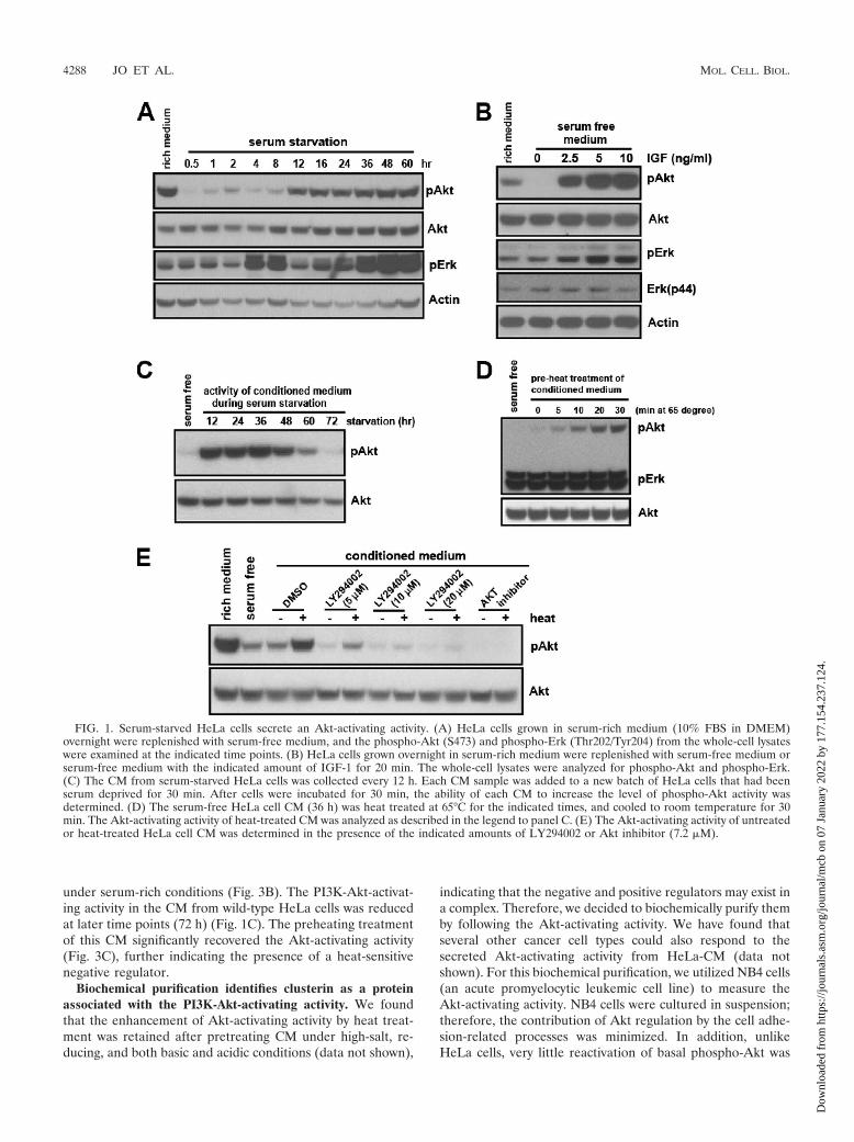
Serum-starved HeLa cells secrete an Akt-activating activity.A number of signaling receptors rely on the PI3K-Akt pathwayto control a wide range of cellular responses. However, howthis pathway is regulated under cellular stress conditions is notwell defined. We hypothesized that cancer cells under growthconstraint conditions secrete soluble factors to modulate thePI3K-Akt pathway. To identify such regulatory factors, weestablished a biochemical system. We chose serum starvationas one of the paradigms for cellular stress. It is known thatcellular stresses caused by serum deprivation cross talk withhypoxic responses (3, 11, 51), analogous to that in in vivocancer cells. Also, excluding other serum factors allowed us toidentify the cancer cell-derived signaling modulators.
Akt is recruited onto the plasma membrane through itsspecific binding to PIP3, a product of PI3K, and is activated byphosphorylation; thus, Akt phosphorylation has been usedwidely as an indicator of PI3K-Akt pathway activation (8, 9, 32,39, 46). HeLa cells grown in serum-rich medium were replen-ished with serum-free medium, and the level of phospho-Aktwas examined at the indicated time points. Interestingly, theprofile of the phospho-Akt exhibited a biphasic nature, that is,the lowest level appeared at first, followed by a gradual in-crease, and moderate levels were sustained at later time points(Fig. 1A). While the kinetics and levels of reactivation wereslightly variable between experiments, the biphasic nature ofphospho-Akt was highly reproducible (Fig. 1A and see Fig.S1A in the supplemental material). The initial deactivation(dephosphorylation) of Akt was rapid and came about withinone-half hour of serum removal (see Fig. S1B in the supple-mental material). The presence of growth factors, such as IGF,completely prevented this initial deactivation (Fig. 1B). Underthe identical conditions, the initial deactivation of phospho-Erk was not obvious, and its response to IGF was much lessrobust than that of phospho-Akt (Fig. 1A and B and see Fig.S1A in the supplemental material).
The reactivation of Akt following the initial deactivationcould be due to restimulation by an extracellular Akt activa-tor(s) secreted by the starved HeLa cells. If this is the case, theserum-free culture medium in which HeLa cells were grown(the CM) should contain the Akt-activating activity. To testthis possibility, the serum-free CM from HeLa cells was col-lected at 12-h intervals for 3 days, and the ability to activateAkt (i.e., to prevent the initial deactivation upon serum re-moval) was examined in a new batch of cells. The serum-freeCM, indeed, contained the Akt-activating activity. This activitywas increased up until 36 h, followed by a gradual decrease,leading to an almost complete lack of activity by 72 h (Fig. 1C).
To better understand the molecular nature of the secreted
Akt-activating activity, we first determined its physical proper-ties. Interestingly, the preheating treatment at 65°C signifi-cantly enhanced the Akt-activating activity, while it had noeffect on the level of phospho-Erk (Fig. 1D). To assess themolecular weight, we applied the CM to a filtration membranedesigned to separate macromolecules based on their size. Theactivity was found to be bigger than 50 kDa, even after thepreheating treatment (see Fig. S1C and S1D in the supplemen-tal material). The activation of Akt by either the heat-treatedor the untreated CM was blocked in the presence of Akt orPI3K inhibitor, suggesting that this activity acts upstream ofPI3K (Fig. 1E).
The PI3K-Akt signaling pathway mediates the secretion ofAkt-activating activity. The expression of signaling ligands andantagonists is often under the control of their cognate signalingpathways. To determine if the level of Akt-activating activity inHeLa-CM could be regulated by the PI3K-Akt pathway, wetreated HeLa cells with a sublethal amount of PI3K or an Aktinhibitor. Residual chemicals from the CM were removed byfiltration (see Materials and Methods), and their absence fromthe CM was confirmed (Fig. 2A). Each CM sample was thentested for Akt-activating activity. Compared with the controlCM, the drug-treated CM showed a significantly reduced ac-tivity level (Fig. 2B). To substantiate this finding, we generatedvariant HeLa cells with compromised PI3K signaling. The el-evation of PIP3, a product of PI3K, and its recognition byvarious cellular proteins are the key steps for the PI3K-elicitedcellular processes. We reasoned that the ectopic expression ofthe PIP3-binding PH domain would sequester the endogenousPIP3, hence attenuating the PI3K signaling. We generated twoindependent stable HeLa cell lines expressing a PH domain ofAkt fused to EGFP, HeLa-PH1 and HeLa-PH2. As expected,compared to the wild-type HeLa cells, the HeLa-PH cellsshowed a reduced basal level of phospho-Akt (Fig. 2C). Whenthe CM from the variant HeLa cells was tested for secretedAkt-activating activity, the variant CM, unlike wild-type HeLa-CM, showed no activity (for HeLa-PH1) or a much reducedactivity (for HeLa-PH2) level at all time points (Fig. 2D). Thelack of Akt-activating activity in the CM from HeLa-PH cells(HeLa-PH-CM) was indeed due to its inability to activate thePI3K, as it failed to induce the membrane translocation ofPH-EGFP, a process mediated by PIP3 (Fig. 2E). Togetherwith the previous results using chemical inhibitors, these find-ings suggest that the secretion and/or synthesis of Akt-activat-ing activity is mediated by the PI3K-Akt signaling pathway.
Serum-starved HeLa cells also secrete a negative regulatorof PI3K-Akt pathway. Heat activation is a unique feature of theHeLa cell-secreted PI3K-Akt-activating activity. We foundthat a preheating treatment of purified growth factors, such asIGF, had no differential effect (Fig. 3A). One mechanism thatmay explain this finding is the presence of a heat-sensitivenegative regulator. The heat treatment may lead to conforma-tional changes of this regulator and thus suppress its inhibitoryeffect on Akt activation. Supporting this idea, the CM fromHeLa-PH cells, which do not secrete the Akt-activating activ-ity, further decreased the basal level of phospho-Akt underserum-free conditions. The preheating treatment of CM fromHeLa-PH cells abolished this inhibitory effect (Fig. 3A). Con-sistent with this finding, the concentrated CM from HeLa-PHcells reduced the level of phospho-Akt in HeLa cells, even
VOL. 28, 2008 CLUSTERIN AS A REGULATOR OF THE PI3K-AKT PATHWAY 4287
Dow
nloa
ded
from
http
s://j
ourn
als.
asm
.org
/jour
nal/m
cb o
n 07
Jan
uary
202
2 by
177
.154
.237
.124
.
under serum-rich conditions (Fig. 3B). The PI3K-Akt-activat-ing activity in the CM from wild-type HeLa cells was reducedat later time points (72 h) (Fig. 1C). The preheating treatmentof this CM significantly recovered the Akt-activating activity(Fig. 3C), further indicating the presence of a heat-sensitivenegative regulator.
Biochemical purification identifies clusterin as a proteinassociated with the PI3K-Akt-activating activity. We foundthat the enhancement of Akt-activating activity by heat treat-ment was retained after pretreating CM under high-salt, re-ducing, and both basic and acidic conditions (data not shown),
indicating that the negative and positive regulators may exist ina complex. Therefore, we decided to biochemically purify themby following the Akt-activating activity. We have found thatseveral other cancer cell types could also respond to thesecreted Akt-activating activity from HeLa-CM (data notshown). For this biochemical purification, we utilized NB4 cells(an acute promyelocytic leukemic cell line) to measure theAkt-activating activity. NB4 cells were cultured in suspension;therefore, the contribution of Akt regulation by the cell adhe-sion-related processes was minimized. In addition, unlikeHeLa cells, very little reactivation of basal phospho-Akt was
FIG. 1. Serum-starved HeLa cells secrete an Akt-activating activity. (A) HeLa cells grown in serum-rich medium (10% FBS in DMEM)overnight were replenished with serum-free medium, and the phospho-Akt (S473) and phospho-Erk (Thr202/Tyr204) from the whole-cell lysateswere examined at the indicated time points. (B) HeLa cells grown overnight in serum-rich medium were replenished with serum-free medium orserum-free medium with the indicated amount of IGF-1 for 20 min. The whole-cell lysates were analyzed for phospho-Akt and phospho-Erk.(C) The CM from serum-starved HeLa cells was collected every 12 h. Each CM sample was added to a new batch of HeLa cells that had beenserum deprived for 30 min. After cells were incubated for 30 min, the ability of each CM to increase the level of phospho-Akt activity wasdetermined. (D) The serum-free HeLa cell CM (36 h) was heat treated at 65°C for the indicated times, and cooled to room temperature for 30min. The Akt-activating activity of heat-treated CM was analyzed as described in the legend to panel C. (E) The Akt-activating activity of untreatedor heat-treated HeLa cell CM was determined in the presence of the indicated amounts of LY294002 or Akt inhibitor (7.2 �M).
4288 JO ET AL. MOL. CELL. BIOL.
Dow
nloa
ded
from
http
s://j
ourn
als.
asm
.org
/jour
nal/m
cb o
n 07
Jan
uary
202
2 by
177
.154
.237
.124
.

found in NB4 cells during serum deprivation, making the back-ground level consistently very low in each assay. To trace theAkt-activating activity level that can be enhanced by heat treat-ment, each fraction was treated at 65°C for 20 min and testedfor its ability to increase the phospho-Akt level in serum-starved NB4 cells (see methods in the supplemental material).We showed that the Akt-activating activity existed in theflowthrough fractions of the hydrophobic (butyl-S-Sepharose)and anion-exchange columns (High Q). This activity could beretained on the cation-exchange (High S) column and recov-ered by elution with a pH gradient (see Fig. S2A, B, and C inthe supplemental material). We then attempted to identify the
proteins from this intermediate purification by mass spectrom-etry analysis.
The final fractions from two independent purifications weretested for Akt-activating activity. Consistent with the previousobservation that this activity acts upstream of the PI3K path-way (Fig. 1E and 2E), phosphorylation of both Akt-T308 andAkt-S473 was enhanced by the purified activity (Fig. 4A). Us-ing the Coomassie staining of these fractions, we identifiedeight bands that appeared to be correlated with the activity(Fig. 4B, enriched in the E1-P and E2-fraction 11 but not in theE2 fractions 9 and 10; see also Fig. S2C in the supplementalmaterial), and these were subjected to MALDI analysis. In-
FIG. 2. The PI3K-Akt signaling pathway mediates the secretion of Akt-activating activity. (A) HeLa cells were cultured in serum-free mediumfor 24 h in the presence of dimethyl sulfoxide (DMSO), LY294002 (5 �M), wortmannin (WM, 0.1 �M), or Akt inhibitor (3.6 �M). The CM wasfiltered through a Centriprep-10K column to remove the residual chemicals (see Materials and Methods). The filtered CM was mixed with IGF-1(1 ng/ml) and tested for the removal of residual drug. (B) Each CM sample from the experiment described in the legend to panel A was untreatedor heat treated, and its ability to activate Akt was assayed in HeLa cells. (C) The whole-cell lysates of HeLa cells or HeLa-PH cells grown inserum-rich medium were analyzed for phospho-Akt level. (D) The serum-free CM from HeLa, HeLa-PH1, or HeLa-PH2 cells was collected at12-h intervals and tested for its ability to activate Akt. (E) The CM from HeLa or HeLa-PH cells was added to HeLa-PH cells that had been serumstarved for 12 h. After the HeLa-PH cells were incubated for 30 min, they were fixed in 3% paraformaldehyde, and the membrane translocationof PH-EGFP was examined using fluorescence microscopy (20� objective). Arrows indicate the membrane translocation of PH-EGFP.
VOL. 28, 2008 CLUSTERIN AS A REGULATOR OF THE PI3K-AKT PATHWAY 4289
Dow
nloa
ded
from
http
s://j
ourn
als.
asm
.org
/jour
nal/m
cb o
n 07
Jan
uary
202
2 by
177
.154
.237
.124
.

triguingly, one of the proteins identified, clusterin, was de-tected in all eight samples, with a high percentage of peptidecoverage (Fig. 4C). The Western blotting analysis confirmed abroad range of clusterin immunoreactivity that correspondedto the analyzed bands (Fig. 4D).
Clusterin is a multifunctional protein involved in variousbiological processes, including tumorigenesis (17, 41), al-though the underlying mechanisms are still poorly charac-terized. Its function as an extracellular chaperon and itsregulation by cellular stresses prompted us to evaluate thisprotein further. First, we found that clusterin was accumu-lated in the CM during serum starvation (Fig. 4E). Since theAkt-activating activity in CM can be enhanced by heat treat-ment, we determined if heat treatment could affect theconformation of clusterin. The heat treatment led to a high-molecular-weight complex of clusterin, indicating a struc-tural change(s) (Fig. 4F). Longer periods of treatment at ahigher temperature (80°C over 3 h) led to its degradation ina manner corresponding to the loss of Akt-activating activity
(Fig. 4F). When the High S-bound activity was subjected toelution by a fine gradient of pH, clusterin was coeluted withthe Akt-activating activity (see Fig. S2D in the supplementalmaterial). In addition, the immunodepletion of clusterinfrom the CM led to a reduced Akt-phosphorylating activity,indicating that clusterin is associated with the Akt-activatingactivity (Fig. 4G).
Clusterin is a secreted negative regulator of the PI3K-Aktsignaling pathway. We showed that serum-starved HeLa cellssecrete both positive and negative regulators of the PI3K-Aktpathway (Fig. 3). Clusterin is associated with the Akt-activatingactivity, but whether it acts as a positive or a negative regulatorneeds to be determined. First, we took advantage of thoseHeLa-PH cells whose CM lacked the Akt-activating activity(Fig. 2C and D). Equal amounts of CM from wild-type HeLa(HeLa-CM) and HeLa-PH-CM were subjected to the High Scolumn, and bound proteins were eluted by a pH gradient. Thelevel of clusterin in each fraction of HeLa-PH-CM was notdifferent from that of corresponding fractions of HeLa-CM.
FIG. 3. Serum-starved HeLa cells also secrete a negative regulator of the PI3K-Akt pathway. (A) Serum-free medium, IGF-1, or CM fromHeLa or HeLa-PH cells was untreated or heat treated, and their abilities to activate Akt were tested. (B) The CM from HeLa-PH or its columnfraction was added to HeLa cells grown in serum-rich medium (20% volume of the medium) and incubated for 30 min prior to analysis forphospho-Akt. For the Centriprep column retentate (RT), the CM was concentrated with the Centriprep 10K or 50K filter column. Eachconcentrate (10�) was added directly to HeLa cells grown in serum-rich medium. (C) The HeLa cell CM collected after 48 h or 72 h of serumstarvation was untreated or heat treated, and its ability to activate Akt was assayed.
4290 JO ET AL. MOL. CELL. BIOL.
Dow
nloa
ded
from
http
s://j
ourn
als.
asm
.org
/jour
nal/m
cb o
n 07
Jan
uary
202
2 by
177
.154
.237
.124
.

FIG. 4. Biochemical purification identified clusterin as a protein associated with the PI3K-Akt-activating activity. (A) The active fractions eluted fromthe High S column from two independent purification experiments (see Fig. S3 in the supplemental material) were mixed (1:1) with serum-freemedium. After heat treatment, their ability to phosphorylate S473 and T308 of Akt was assayed in NB4 cells. Exp1 and Exp2 indicate twoindependent purification experiments. P indicates the pooled activity from the first purification. NS indicates a nonspecific band. (B) Thecorresponding fractions were concentrated with a Centriprep 10K column. Proteins were resolved on a NuPAGE 4 to 12% gel stained withCoomassie blue. Arrows indicate the protein bands analyzed by MALDI mass spectrometry. (C) Human clusterin peptides identified by MALDIanalysis are underlined. (D) Western blotting (WB) of the corresponding fractions, using C-terminal-specific monoclonal clusterin antibody.(E) Serum-free CM from HeLa cells collected at the indicated time points was analyzed for clusterin. (F) CM from HeLa cells was heat treatedfor the indicated times, and proteins were analyzed with the C-terminal-specific clusterin antibody. The bracket indicates the high-molecular-weightcomplexes induced by heat treatment (top). Akt-activating activity of the corresponding CM was tested in NB4 cells (bottom). (G) CM wasimmunodepleted with the protein G/A beads alone (control), the control antibody (monoclonal anti-GSK), or the C-terminal-specific clusterinantibody. The supernatant was analyzed for clusterin (at left). Their abilities to activate Akt were tested in NB4 cells (at right).
VOL. 28, 2008 CLUSTERIN AS A REGULATOR OF THE PI3K-AKT PATHWAY 4291
Dow
nloa
ded
from
http
s://j
ourn
als.
asm
.org
/jour
nal/m
cb o
n 07
Jan
uary
202
2 by
177
.154
.237
.124
.

However, all fractions from HeLa-PH-CM lacked the Akt-activating activity when they were tested in NB4 cells (Fig. 5A),indicating that clusterin by itself does not function as an Akt-activating factor.
Previously, we observed that concentrated HeLa-PH-CMinhibited Akt phosphorylation (Fig. 3B). Therefore, we exam-ined whether HeLa-PH-CM could inhibit the Akt-activatingactivity present in HeLa-CM. In the presence of equal amountsof HeLa-PH-CM, Akt phosphorylation by HeLa-CM wasstrongly inhibited, and, moreover, HeLa-PH-CM fractionsfrom the High S column also reduced Akt phosphorylation ina manner that correlated with its clusterin level (Fig. 5B). Thisfinding indicates the possibility that clusterin may play an in-hibitory function. If this is the case, then the reduction ofclusterin in HeLa-PH-CM should attenuate its ability to inhibitHeLa-CM-mediated Akt activation. To test this possibility, weknocked down the endogenous clusterin in HeLa-PH cells byRNA interference (Fig. 5C), and then the CM from these cellswas tested for its ability to inhibit the Akt phosphorylation byHeLa-CM. We found that a significant reduction of clusterin inHeLa-PH cells, in which prosurvival IGF-1 signaling was com-promised, led to apoptosis under serum-deprived conditions(see Fig. S7 in the supplemental material). Thus, we relied on
a moderate knockdown that did not cause apoptosis. Com-pared to CM from empty vector- or control shRNA-expressingHeLa-PH cells, the CM from the clusterin-shRNA-expressingHeLa-PH cells manifested a reduced inhibitory activity whentested in both the NB4 (Fig. 5D) and the HEK293 cells (Fig.5E), strongly indicating a negative role for clusterin.
We then asked if the ectopic expression of clusterin couldconfer a similar inhibitory activity in a heterologous system.For this end, we used HEK293T cells, which almost completelylack endogenous clusterin and do not secrete Akt-activatingfactors during serum starvation (Fig. 6A). We transfectedHEK293T cells with the empty or clusterin-expressing (Clu)vector, and the serum-free CM was collected. Neither thevector control nor clusterin-containing HEK293T-CM showedany effects on the Akt phosphorylation when tested with NB4cells (Fig. 6A). However, compared with the control CM, theCM from clusterin-transfected cells (HEK293T-Clu) markedlyinhibited the Akt phosphorylation induced by HeLa-CM whentested with both NB4 cells and HeLa cells (Fig. 6B and C).
Activation of the PI3K-Akt pathway promotes cell prolif-eration (31, 42). To determine if clusterin could exert asustained inhibitory effect on the PI3K-Akt pathway, weexamined its effects on cell proliferation. The same number
FIG. 5. Clusterin is a secreted negative regulator of the PI3K-Akt signaling pathway. (A) The High S column-bound proteins from HeLa cellCM (HeLa-CM) or HeLa-PH cells (HeLa-PH-CM) were eluted by the pH gradient. The eluted fractions were analyzed for clusterin (top), andthe corresponding fractions were tested for their ability to activate Akt in NB4 cells (bottom). (B) HeLa-PH-CM inhibits the Akt activation elicitedby HeLa-CM. HeLa-PH-CM and its fractions shown in panel A were mixed (1:1) with HeLa-CM and tested for their abilities to inhibitHeLa-CM-elicited Akt activation in NB4 cells. (C) The shRNA-mediated knockdown of clusterin in HeLa-PH1 cells is shown. HeLa-PH1 cellswere transfected with the empty vector, the control shRNA vector, or two different clusterin shRNA vectors, and then the serum-free CM (24 h)and the whole-cell lysates of each transfectant were analyzed for clusterin expression. (D and E) Each HeLa-PH-CM was mixed (1:1) withHeLa-CM and tested as described in the legend to panel B for the ability to inhibit HeLa-CM-mediated Akt phosphorylation in NB4 or HEK293cells. The representative Western blotting of at least three independent experiments is shown.
4292 JO ET AL. MOL. CELL. BIOL.
Dow
nloa
ded
from
http
s://j
ourn
als.
asm
.org
/jour
nal/m
cb o
n 07
Jan
uary
202
2 by
177
.154
.237
.124
.

of HEK293 cells were plated using the reduced serum con-ditions (1% and 2.5%) in the presence of control or clus-terin-containing HEK293T-CM and cultured for 3 days. Asrevealed by the relative cell proliferation, compared to thatof the control, the presence of clusterin led to a 28 to 30%reduction in cell proliferation (Fig. 6D). Taken together,these data support a role for clusterin in the negative reg-ulation of the PI3K-Akt pathway.
Clusterin inhibits the IGF-1- but not the EGF-mediatedsignaling pathway. Next, we decided to identify the Akt acti-vator(s) that is inhibited by clusterin. First, we found that theheat-treated HeLa-CM did not enhance G protein-coupledreceptor signaling as determined by Akt activation and che-motactic migration of mouse peripheral neutrophils (see Fig.S3A and B in the supplemental material). We then checked forthe growth factor receptor signaling. NB4 cells lack both thePDGF and the EGF receptors (data not shown). This findingled us to examine IGF receptor signaling. Using a phosphory-lation-specific antibody, we determined the kinetics of IGF-1receptor activation by HeLa-CM with or without heat treat-ment. HeLa-CM activated the IGF-1 receptor, and its activa-tion preceded that of Akt phosphorylation. Importantly, theheat treatment of HeLa-CM significantly enhanced the activa-tion of the IGF receptor (Fig. 7A). Next, we asked if theactivation of the IGF receptor by HeLa-CM was responsiblefor Akt activation. For this purpose, we took advantage of aneutralizing antibody against the IGF-1 receptor (27). Prein-cubation of NB4 cells with the IGF-1 receptor-neutralizingantibody, but not the control antibody, markedly inhibited theAkt activation by the heat-treated HeLa-CM (Fig. 7B). Acti-vation of the IGF-1 receptor strongly indicates the presence ofits ligands, such as IGF-1, in HeLa-CM. However, its activation
could be also due to cross talk between signaling receptors (12,25). Therefore, we determined if similar neutralization ofIGF-1 could affect the Akt-activating activity of HeLa-CM.The inhibitory activity of the IGF-1-neutralizing antibody hasbeen well documented (34). We also confirmed that this anti-body blocked the activity of both purified and serum IGF-1(see Fig. S3C in the supplemental material). The IGF-1-neu-tralizing antibody, but not the control antibody, almost com-pletely abolished Akt activation by heat-treated HeLa-CM(Fig. 7C). Next, we determined if IGF-1 was indeed secreted byHeLa cells during serum starvation. We showed that serum-free CM from HeLa-PH cells lacked Akt-activating activity(Fig. 2D). Therefore, we measured the amount of IGF-1 inHeLa-CM and HeLa-PH-CM with or without heat treatment,by ELISA. The results showed that IGF-1 was indeed presentin HeLa-CM and its level peaked at 36 h and decreased there-after. Importantly, the heat treatment significantly increasedthe detectable level of IGF-1, and the amount of IGF-1 de-tected in 72-h HeLa-CM was comparable (slightly higher) tothat detected in 36-h HeLa-CM (Fig. 7D). As expected, verylittle IGF-1 was detected in untreated and heat-treated HeLa-PH-CM (Fig. 7D), confirming that the lack of IGF-1 wasresponsible for its inability to activate Akt.
We showed that ectopic clusterin inhibited the Akt-activat-ing activity of HeLa-CM (Fig. 6B and C), indicating that clus-terin may attenuate IGF-1 function. To test this possibility, wepreincubated the purified IGF-1 with the control or the clus-terin-containing HEK293T-CM and then compared their ac-tivity levels. Consistent with our prediction, the presence ofclusterin led to a functional inhibition of IGF-1, as revealed bya decreased activation of the IGF-1 receptor and Akt (Fig. 7E).To further determine if the inhibitory function of secreted
FIG. 6. Ectopic expression of clusterin inhibits PI3K-Akt activation and cell proliferation. (A) The empty control (Con) vector or theclusterin-expressing plasmid (Clu) was transfected into HEK293T cells. The resulting serum-free CM (30 h) fractions were analyzed for clusterinexpression (left) and their abilities to affect the Akt phosphorylation in NB4 cells (right). (B) The Con- or Clu-containing HEK293T-CM sampleswere mixed (1:1) with HeLa-CM and left untreated or were heat treated and tested for their abilities to affect HeLa-CM-mediated Aktphosphorylation in NB4 cells. (C) Effects of clusterin on HeLa-CM-elicited Akt phosphorylation in HEK293 cells. (D) The Con- or Clu-containingHEK293T-CM was mixed (1:1) with different amounts of serum and added to HEK293 cells in triplicate. Cell proliferation was measured by MTTassay after 72 h. Results are the means � standard errors of three independent experiments.
VOL. 28, 2008 CLUSTERIN AS A REGULATOR OF THE PI3K-AKT PATHWAY 4293
Dow
nloa
ded
from
http
s://j
ourn
als.
asm
.org
/jour
nal/m
cb o
n 07
Jan
uary
202
2 by
177
.154
.237
.124
.

clusterin is preferential for IGF-1 or if it functions generally toinhibit other growth factors, we examined its effects on EGFsignaling.
We found that the preheating treatment of HeLa-CM failedto enhance the activity of the EGF receptor, while it markedly
increased that of the IGF-1 receptor (see Fig. S3D in thesupplemental material). Next, we determined if clusterin couldaffect the activity of purified EGF. Within 5 min, the stimula-tion of starved HeLa cells with EGF led to a robust activationof the EGF receptor, Akt, and Erk (see Fig. S3E in the sup-
FIG. 7. Clusterin inhibits the IGF-1-mediated, but not the EGF-mediated, signaling pathway. (A) Serum-starved NB4 cells were incubated withthe untreated [heat(�)] or heat-treated [heat(�)] HeLa-CM for the indicated times, and the activation of the IGF-1 receptor and Akt was analyzedin whole-cell lysates. (B) NB4 cells were preincubated with different amounts of control or IGF-1 receptor-neutralizing antibody in serum-freemedium and then incubated with heat-treated HeLa-CM for 30 min before the HeLa-CM was analyzed for phospho-Akt level. (C) TheIGF-1-neutralizing antibody blocked the HeLa-CM-elicited Akt activation. The heat-treated HeLa-CM was preincubated with the control orIGF-1-neutralizing antibody, and then the ability to activate Akt was tested in NB4 cells. (D) Secretion of IGF-1 by serum-starved HeLa cells. Thelevels of IGF-1 in untreated or heat-treated CM collected from HeLa cells or HeLa-PH cells at different time points of serum starvation weremeasured by ELISA. (E) Functional inhibition of IGF-1 by clusterin. The purified IGF-1 was preincubated with the control (HEK293T-Con-CM)or clusterin-containing CM (HEK293T-Clu-CM) and assayed for the activation of IGF-1 receptor and Akt in NB4 cells. (F) The purified EGF (5ng/ml) was preincubated with the control or clusterin-containing CM as described in the legend to panel E and assayed for the activation of theEGF receptor, Akt, and Erk in serum-starved HeLa cells at the indicated time points.
4294 JO ET AL. MOL. CELL. BIOL.
Dow
nloa
ded
from
http
s://j
ourn
als.
asm
.org
/jour
nal/m
cb o
n 07
Jan
uary
202
2 by
177
.154
.237
.124
.
plemental material). Unlike clusterin’s inhibitory function to-ward IGF-1 (Fig. 7E and Fig. 8A), at all time points, thepresence of clusterin had little effect on the activation of theEGF receptor and its downstream components (Fig. 7F).
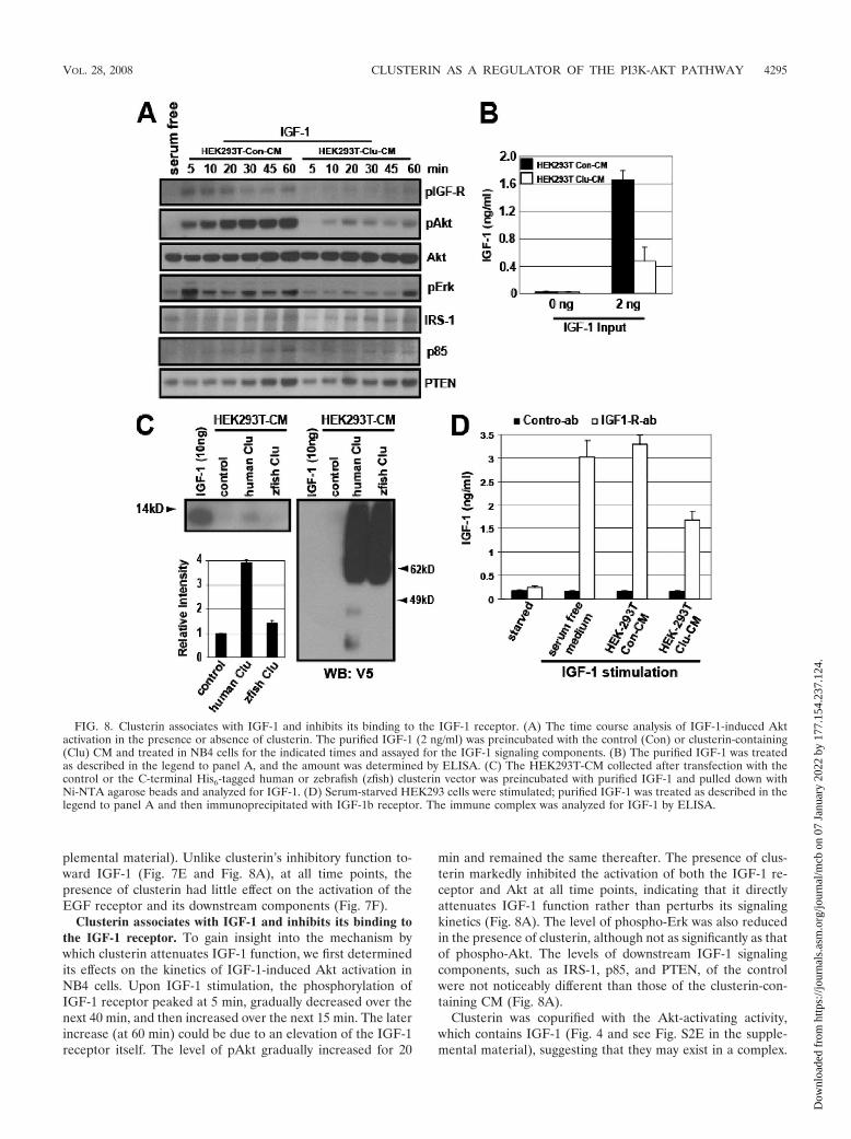
Clusterin associates with IGF-1 and inhibits its binding tothe IGF-1 receptor. To gain insight into the mechanism bywhich clusterin attenuates IGF-1 function, we first determinedits effects on the kinetics of IGF-1-induced Akt activation inNB4 cells. Upon IGF-1 stimulation, the phosphorylation ofIGF-1 receptor peaked at 5 min, gradually decreased over thenext 40 min, and then increased over the next 15 min. The laterincrease (at 60 min) could be due to an elevation of the IGF-1receptor itself. The level of pAkt gradually increased for 20
min and remained the same thereafter. The presence of clus-terin markedly inhibited the activation of both the IGF-1 re-ceptor and Akt at all time points, indicating that it directlyattenuates IGF-1 function rather than perturbs its signalingkinetics (Fig. 8A). The level of phospho-Erk was also reducedin the presence of clusterin, although not as significantly as thatof phospho-Akt. The levels of downstream IGF-1 signalingcomponents, such as IRS-1, p85, and PTEN, of the controlwere not noticeably different than those of the clusterin-con-taining CM (Fig. 8A).
Clusterin was copurified with the Akt-activating activity,which contains IGF-1 (Fig. 4 and see Fig. S2E in the supple-mental material), suggesting that they may exist in a complex.
FIG. 8. Clusterin associates with IGF-1 and inhibits its binding to the IGF-1 receptor. (A) The time course analysis of IGF-1-induced Aktactivation in the presence or absence of clusterin. The purified IGF-1 (2 ng/ml) was preincubated with the control (Con) or clusterin-containing(Clu) CM and treated in NB4 cells for the indicated times and assayed for the IGF-1 signaling components. (B) The purified IGF-1 was treatedas described in the legend to panel A, and the amount was determined by ELISA. (C) The HEK293T-CM collected after transfection with thecontrol or the C-terminal His6-tagged human or zebrafish (zfish) clusterin vector was preincubated with purified IGF-1 and pulled down withNi-NTA agarose beads and analyzed for IGF-1. (D) Serum-starved HEK293 cells were stimulated; purified IGF-1 was treated as described in thelegend to panel A and then immunoprecipitated with IGF-1b receptor. The immune complex was analyzed for IGF-1 by ELISA.
VOL. 28, 2008 CLUSTERIN AS A REGULATOR OF THE PI3K-AKT PATHWAY 4295
Dow
nloa
ded
from
http
s://j
ourn
als.
asm
.org
/jour
nal/m
cb o
n 07
Jan
uary
202
2 by
177
.154
.237
.124
.
To determine if clusterin could form a complex with IGF-1 invitro, we employed two independent approaches. First, weincubated the purified IGF-1 with the control or the clusterin-containing CM and determined if clusterin could affect theaccessibility of IGF-1. We found that preincubation with clus-terin reduced the amount of IGF-1 detected by ELISA (Fig.8B). Next, we performed a biochemical pull-down experiment.We found that, under a nonreducing condition, the purifiedIGF-1 could be readily detected by Western blotting (see Fig.S4A in the supplemental material). For the pull-down experi-ment, the C-terminally tagged (V5 and His6) clusterin wasexpressed in HEK293T cells. As a control, we used the ze-brafish clusterin, which shares less than 40% identity withhuman clusterin but is readily glycosylated and secreted whenexpressed in HEK293T cells (see Fig. S4B and C in the sup-plemental material). The purified IGF-1 was incubated withthe control or the human clusterin- or the zebrafish clusterin-containing CM and pulled down with Ni-NTA beads. This
experiment revealed that the purified human IGF-1 could forma complex with human clusterin but not with zebrafish clusterin(Fig. 9C). Next, we determined if clusterin prevents binding ofIGF-1 to its receptor. For this end, serum-starved HEK293cells were stimulated with IGF-1 that had been preincubatedwith control or clusterin-containing CM and then immunopre-cipitated with the nonneutralizing IGF-1 receptor antibody.Given the fact that the purified IGF-1 is heat stable (Fig. 3A),we heat treated the immunocomplex to release the boundIGF-1, and its level was measured by ELISA. Consistent withthe previous results, the presence of clusterin reduced theamount of IGF-1 bound to its receptor (Fig. 8D). Together,these results suggest that clusterin attenuates IGF-1 signalingby interfering with its binding to the receptor.
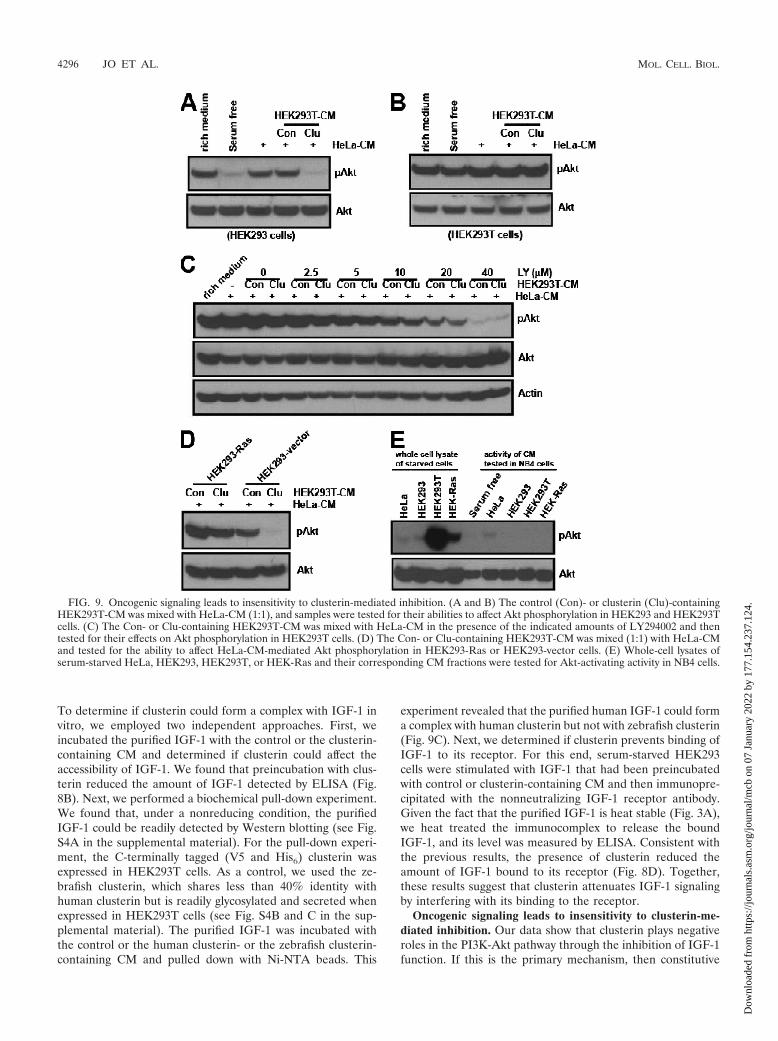
Oncogenic signaling leads to insensitivity to clusterin-me-diated inhibition. Our data show that clusterin plays negativeroles in the PI3K-Akt pathway through the inhibition of IGF-1function. If this is the primary mechanism, then constitutive
FIG. 9. Oncogenic signaling leads to insensitivity to clusterin-mediated inhibition. (A and B) The control (Con)- or clusterin (Clu)-containingHEK293T-CM was mixed with HeLa-CM (1:1), and samples were tested for their abilities to affect Akt phosphorylation in HEK293 and HEK293Tcells. (C) The Con- or Clu-containing HEK293T-CM was mixed with HeLa-CM in the presence of the indicated amounts of LY294002 and thentested for their effects on Akt phosphorylation in HEK293T cells. (D) The Con- or Clu-containing HEK293T-CM was mixed (1:1) with HeLa-CMand tested for the ability to affect HeLa-CM-mediated Akt phosphorylation in HEK293-Ras or HEK293-vector cells. (E) Whole-cell lysates ofserum-starved HeLa, HEK293, HEK293T, or HEK-Ras and their corresponding CM fractions were tested for Akt-activating activity in NB4 cells.
4296 JO ET AL. MOL. CELL. BIOL.
Dow
nloa
ded
from
http
s://j
ourn
als.
asm
.org
/jour
nal/m
cb o
n 07
Jan
uary
202
2 by
177
.154
.237
.124
.

activation of downstream components of IGF-1 receptor shouldconfer insensitivity to clusterin. To investigate this possibility,we examined the effects of clusterin on cell lines containing theimpaired IGF-1 receptor signaling. Consistent with previousreports (14, 38), we found that compared to the level of PI3Kactivation found with wild-type HEK293 cells, HEK293T cellsin which simian virus 40 large T (SV40T) antigen is overex-pressed had a high level of PI3K activation, as demonstrated bya prominent augmentation of PIP3-mediated membrane trans-location of PH-EGFP (see Fig. S5A in the supplemental ma-terial). Unlike that in wild-type HEK293 cells, clusterin failedto affect Akt phosphorylation in HEK293T cells (Fig. 9A andB). This insensitivity was not caused by a “high basal level ofphospho-Akt” per se, as lowering the basal level of phospho-Akt by cotreatment with LY294002 still failed to manifest anyadditional inhibitory effects (Fig. 9C).
To determine whether any other oncogenic events could in-duce a similar insensitivity to clusterin, we transfected HEK293cells with an empty vector or an oncogenic form of H-Ras,H-Ras(V12). The oncogenic H-Ras(V12) protein constitu-tively elicits downstream signaling pathways, including that ofthe PI3K, independently of upstream receptor tyrosine ki-nases. In the control HEK293 vector, clusterin almost com-pletely blocked HeLa-CM-mediated Akt phosphorylation. Incontrast, in HEK293-Ras cells, clusterin showed only a mar-ginal inhibitory effect (Fig. 9D). The inhibition of H-Ras pro-tein in these cells, with a farnesyl transferase inhibitor, signif-icantly restored their sensitivity to clusterin (see Fig. S5B in thesupplemental material). To determine if this insensitivity wasdue to the uncontrolled activation of downstream IGF-1 sig-naling or to transformed cells secreting other factors that actedupstream of Akt, the serum-free CM from these cells weretested for their abilities to activate Akt. Compared with HeLaor wild-type HEK293 cells, the basal levels of phospho-Akt inboth HEK293T and HEK-Ras cells were much higher underserum deprivation conditions. However, unlike HeLa-CM, theCM from HEK or its transformed counterparts failed to acti-vate Akt when tested with NB4 cells. These results suggest thatoncogenic activation downstream of IGF-1 signaling can leadto insensitivity to clusterin-mediated control without the secre-tion of other activating factors.
Synthesis of clusterin during serum deprivation is indepen-dent of IGF-1 signaling. It has been reported that IGF-1 re-ceptor signaling regulates clusterin expression (10), and weshowed that clusterin was secreted during serum deprivation(Fig. 4E). We therefore determined if IGF-1 signaling couldaffect the synthesis and/or secretion of clusterin during serumdeprivation. Clusterin undergoes several steps of posttransla-tional modifications, including glycosylation, proteolytic cleav-age, and disulfide bond formation (28, 41). The precursor andmature (to-be-secreted) forms could be differentiated easily bytreatment with the reducing agents that disrupt the intermo-lecular (between � and � chains) disulfide bonds present onlyin the mature form. We analyzed the whole-cell lysates ofHeLa cells grown in serum-rich or serum-free medium in thepresence or absence of a reducing agent. Both the total and thesecreted clusterin levels were found to be elevated during se-rum starvation (see Fig. S6A in the supplemental material),and the time course analysis further revealed that the level ofthe proteolytically processed (mature) form could be increased
within 12 h of serum deprivation (see Fig. S6B in the supple-mental material). This initial elevation in protein level ap-peared not to be due to its transcripts, which began to increase24 h after serum deprivation (data not shown). It has beenreported that there are two isoforms of clusterin (nuclear andsecreted) present inside cells (41). To determine which isoformwas accumulated during serum deprivation, HeLa cells weretreated with tunicamycin, an inhibitor of N glycosylation. Thistreatment revealed a nonglycosylated precursor form of clus-terin (approximately 55 kDa) with a concomitant loss of the62-kDa precursor form, indicating that only the secreted (ornonnuclear) isoform was present during serum deprivation(see Fig. S6C in the supplemental material). Next, we deter-mine if the perturbation of IGF-1 signaling could affect thelevel of clusterin during serum deprivation. The addition ofIGF-1 or IGF-1 receptor-neutralizing antibody failed to influ-ence the level of clusterin (see Fig. S6D and E in the supple-mental material). We then determined if restimulation ofstarved cells with serum or IGF-1 could affect its level. Re-stimulation with serum led to a reduction in the clusterin levelwithin 2 h, but IGF-1 failed to exert any effects at later timepoints and higher concentrations (see Fig. S6F in the supple-mental material). These results showed that the synthesis ofclusterin during serum deprivation is independent of IGF-1signaling. Reduction of the clusterin level within 2 h afterserum restimulation suggests that its level could be controlledby protein translation and/or turnover. To test this possibility,we treated HeLa cells grown under serum-rich or -starvedconditions with cycloheximide and examined the level of clus-terin over a time course. Within 2 h of treatment, only a smallfraction (approximately 10%) of the precursor form was de-tected under both serum-rich and -starved conditions. Intrigu-ingly, while around 35% of the mature form was detectedunder serum-rich conditions (see Fig. S6G in the supplementalmaterial), a significant fraction (75%) of the mature form stillremained under serum-starved conditions, indicating a post-translational regulation of clusterin.
DISCUSSION
The ability of cancer cells to modulate the cellular signalingpathways in accordance with the changing local environmentrepresents one of the molecular mechanisms underlying theprogression of malignancy and resistance to anticancer drugs.The PI3K-Akt pathway serves as a converging point for manyupstream signaling receptors. However, the cellular mecha-nisms that modulate this pathway under growth-constrainingconditions are not clearly defined. In the present study, weshow that serum-deprived cancer cells secrete a PI3K-Akt-regulating activity that consists of both positive and negativefactors. Via biochemical purification, we reveal that cancercell-derived clusterin and IGF-1 constitute this regulatory ac-tivity. We demonstrate furthermore that clusterin negativelymodulates the PI3K-Akt pathway through inhibition of IGF-1,a major growth factor secreted by serum-starved cancer cells,thus identifying a novel function for clusterin.
Previous studies with clusterin, including overexpression andknockdown in cancer cells, implicated it in different aspects ofthe tumorigenic process (5, 10, 29, 41, 49). Clusterin exists inan intracellular form and an extracellular form, and its func-
VOL. 28, 2008 CLUSTERIN AS A REGULATOR OF THE PI3K-AKT PATHWAY 4297
Dow
nloa
ded
from
http
s://j
ourn
als.
asm
.org
/jour
nal/m
cb o
n 07
Jan
uary
202
2 by
177
.154
.237
.124
.

tional roles in biological processes have been enigmatic. Dif-ficulties in delineating its function as an intracellular or anextracellular factor may explain many of its promiscuous func-tions. The two isoforms (nuclear and secreted) of clusterinwere shown to play opposing functions for cell survival, inwhich the nuclear form was proapoptotic, and the secretedform was prosurvival (30, 41). We confirmed that during serumdeprivation in HeLa cells, only the secreted form of clusterincould be found (see Fig. S6A to C in the supplemental mate-rial), and its knockdown in HeLa-PH cells, in which prosurvivalIGF-1 signaling was compromised, led to apoptosis (see Fig. S7in the supplemental material). This suggests that the secretoryforms, while they are still inside the cell, play a prosurvivalfunction, in agreement with other studies (41). Relatively fewstudies have focused on the extracellular functions of clusterinin relation to tumorigenesis. Nevertheless, several experimen-tal results identified its inhibitory roles in cell proliferation (5,45, 50), which is consistent with our finding (Fig. 6D). Forinstance, it has been shown that recombinant human clusterinsuppressed epithelial cell proliferation, and a high incidence ofpapilloma was found in clusterin-null mice in a chemical-in-duced skin carcinogenesis model (45). However, the underly-ing mechanism remains unknown. This is partly due to itscapacity to bind to a wide range of molecules, including lipidsand protein aggregates. Thus, clusterin is thought to play pro-miscuous functions (41), often resulting in conflicting results(18, 48). Despite this view, we found that the inhibitory effectof clusterin on the PI3K-Akt pathway could be attributed to itspreferential attenuation of IGF-1, not to other growth factorssuch as EGF (Fig. 7).
Cancer cells under stress conditions may secrete many au-tocrine and/or paracrine factors that feed into the PI3K-Aktpathway to support cell growth and survival (4, 6, 24, 36).Surprisingly, however, we found that the inhibition of IGF-1 orits receptor alone almost completely abolished the Akt activa-tion induced by CM from the serum-starved cancer cells (Fig.7B and C). We showed that, under identical serum deprivationconditions, HeLa cells with an impaired PI3K-Akt pathwayfailed to secrete the Akt-activating activity (Fig. 2D) and IGF-1(Fig. 7), identifying a crucial role for this pathway in producingits own upstream activator. Whether this finding can be ex-tended to other cancer cell types and different stress conditionsneeds to be determined. Nevertheless, this result further ex-plains why the deactivation of the PI3K-Akt pathway could beso effective in managing human cancers.
While the presence of IGF-1 in the tumor microenviron-ment is known (37), its secretion by serum-starved cancer cellshas not been readily observed. One potential reason might beits biological accessibility. For example, we showed that, asmeasured by ELISA, the levels of IGF-1 in serum-free CMwere marginal, especially at later time points (i.e., 72 h com-pared to 36 h). However, as revealed by ELISA following heattreatment, this was not due to a lack of its secretion (Fig. 7D).Instead, this result demonstrates that a large portion of se-creted IGF-1 exists in a nonaccessible complex, indicating animportant role for extracellular factors in modulating the bio-availability of IGF-1 (37). The current study identifies a novelrole for cancer cell-derived clusterin in this process.
Extracellular clusterin manifests a growth suppressive func-tion (Fig. 6D). Why would tumor cells secrete a factor that
could potentially suppress their own growth? The level of clus-terin is tightly associated with various cellular stress responses(7, 41). Therefore, its secretion may reflect the cellular adap-tive responses to endure adverse environmental conditions(i.e., by suppressing its own growth and that of surroundingcells). In a mouse model of prostate cancer, it has been shownthat epithelial cancer cells initiate and promote the clonalexpansion of stromal fibroblasts that lack the p53 tumor sup-pressor gene (23), indicating that the cancer cell-derived fac-tors initially impose selective pressures (i.e., antiproliferation)on neighboring cells. The presence of clusterin in the tumormicroenvironment can contribute to such selective pressures.For example, the availability of glucose is often limited in thetumor microenvironment (16), and its uptake, a process medi-ated by the PI3K-Akt pathway (13, 22), can be a decidingfactor for cell proliferation. Under this condition, those cellswith a higher resistance (or insensitivity) to clusterin wouldgain growth advantages and thus would be positively selected.Our demonstration that constitutive activation of oncogenicsignaling led to insensitivity to clusterin is consistent with thisidea (Fig. 9).
The cellular adaptive response to environmental stresses isrecognized as an important mechanism that facilitates tumor-igenic progression (20, 43). We identified an extracellular reg-ulatory system for the PI3K-Akt pathway in cancer cells underserum deprivation stress. We demonstrated that cancer cell-derived clusterin, an extracellular stress protein, and IGF-1constituted this regulatory system, thus providing a genetic toolwith which to better understand the complex signaling inter-plays between cancer cells and their environment.
ACKNOWLEDGMENTS
We thank Leslie Silberstein, James Campbell, John Manis, and LiCai for helpful discussions.
H. Luo is supported by NIH grants NS052200 and GM076084 and aresearch scholar grant from the American Cancer Society.
REFERENCES
1. Alt-Holland, A., W. Zhang, A. Margulis, and J. A. Garlick. 2005. Microen-vironmental control of premalignant disease: the role of intercellular adhe-sion in the progression of squamous cell carcinoma. Semin. Cancer Biol.15:84–96.
2. Bader, A. G., S. Kang, L. Zhao, and P. K. Vogt. 2005. Oncogenic PI3Kderegulates transcription and translation. Nat. Rev. Cancer 5:921–929.
3. Baek, J. H., J. E. Jang, C. M. Kang, H. Y. Chung, N. D. Kim, and K. W. Kim.2000. Hypoxia-induced VEGF enhances tumor survivability via suppressionof serum deprivation-induced apoptosis. Oncogene 19:4621–4631.
4. Barcellos-Hoff, M. H., and S. A. Ravani. 2000. Irradiated mammary glandstroma promotes the expression of tumorigenic potential by unirradiatedepithelial cells. Cancer Res. 60:1254–1260.
5. Bettuzzi, S., F. Scorcioni, S. Astancolle, P. Davalli, M. Scaltriti, and A. Corti.2002. Clusterin (SGP-2) transient overexpression decreases proliferationrate of SV40-immortalized human prostate epithelial cells by slowing downcell cycle progression. Oncogene 21:4328–4334.
6. Bhowmick, N. A., E. G. Neilson, and H. L. Moses. 2004. Stromal fibroblastsin cancer initiation and progression. Nature 432:332–337.
7. Bjork, J. K., and L. Sistonen. 2006. Clustering of heat-shock factors. Bio-chem. J. 395:e5–e6.
8. Brazil, D. P., and B. A. Hemmings. 2001. Ten years of protein kinase Bsignalling: a hard Akt to follow. Trends Biochem. Sci. 26:657–664.
9. Cantley, L. C. 2002. The phosphoinositide 3-kinase pathway. Science 296:1655–1657.
10. Criswell, T., M. Beman, S. Araki, K. Leskov, E. Cataldo, L. D. Mayo, andD. A. Boothman. 2005. Delayed activation of insulin-like growth factor-1receptor/Src/MAPK/Egr-1 signaling regulates clusterin expression, a pro-survival factor. J. Biol. Chem. 280:14212–14221.
11. Das, B., H. Yeger, R. Tsuchida, R. Torkin, M. F. Gee, P. S. Thorner, M.Shibuya, D. Malkin, and S. Baruchel. 2005. A hypoxia-driven vascular en-dothelial growth factor/Flt1 autocrine loop interacts with hypoxia-inducible
4298 JO ET AL. MOL. CELL. BIOL.
Dow
nloa
ded
from
http
s://j
ourn
als.
asm
.org
/jour
nal/m
cb o
n 07
Jan
uary
202
2 by
177
.154
.237
.124
.
factor-1alpha through mitogen-activated protein kinase/extracellular signal-regulated kinase 1/2 pathway in neuroblastoma. Cancer Res. 65:7267–7275.
12. Desbois-Mouthon, C., W. Cacheux, M. J. Blivet-Van Eggelpoel, V. Barbu, L.Fartoux, R. Poupon, C. Housset, and O. Rosmorduc. 2006. Impact of IGF-1R/EGFR cross-talks on hepatoma cell sensitivity to gefitinib. Int. J. Cancer119:2557–2566.
13. Engelman, J. A., J. Luo, and L. C. Cantley. 2006. The evolution of phospha-tidylinositol 3-kinases as regulators of growth and metabolism. Nat. Rev.Genet. 7:606–619.
14. Fei, Z. L., C. D’Ambrosio, S. Li, E. Surmacz, and R. Baserga. 1995. Asso-ciation of insulin receptor substrate 1 with simian virus 40 large T antigen.Mol. Cell. Biol. 15:4232–4239.
15. Gatenby, R. A., E. T. Gawlinski, A. F. Gmitro, B. Kaylor, and R. J. Gillies.2006. Acid-mediated tumor invasion: a multidisciplinary study. Cancer Res.66:5216–5223.
16. Gatenby, R. A., and R. J. Gillies. 2004. Why do cancers have high aerobicglycolysis? Nat. Rev. Cancer 4:891–899.
17. Gleave, M., and K. N. Chi. 2005. Knock-down of the cytoprotective gene,clusterin, to enhance hormone and chemosensitivity in prostate and othercancers. Ann. N. Y. Acad. Sci. 1058:1–15.
18. Han, B. H., R. B. DeMattos, L. L. Dugan, J. S. Kim-Han, R. P. Brendza, J. D.Fryer, M. Kierson, J. Cirrito, K. Quick, J. A. Harmony, B. J. Aronow, andD. M. Holtzman. 2001. Clusterin contributes to caspase-3-independent braininjury following neonatal hypoxia-ischemia. Nat. Med. 7:338–343.
19. Hanada, M., J. Feng, and B. A. Hemmings. 2004. Structure, regulation andfunction of PKB/AKT: a major therapeutic target. Biochim. Biophys. Acta1697:3–16.
20. Hanahan, D., and R. A. Weinberg. 2000. The hallmarks of cancer. Cell100:57–70.
21. Hennessy, B. T., D. L. Smith, P. T. Ram, Y. Lu, and G. B. Mills. 2005.Exploiting the PI3K/AKT pathway for cancer drug discovery. Nat. Rev. DrugDiscov. 4:988–1004.
22. Hill, M. M., S. F. Clark, D. F. Tucker, M. J. Birnbaum, D. E. James, andS. L. Macaulay. 1999. A role for protein kinase B�/Akt2 in insulin-stimu-lated GLUT4 translocation in adipocytes. Mol. Cell. Biol. 19:7771–7781.
23. Hill, R., Y. Song, R. D. Cardiff, and T. Van Dyke. 2005. Selective evolutionof stromal mesenchyme with p53 loss in response to epithelial tumorigenesis.Cell 123:1001–1011.
24. Hurbin, A., J. L. Coll, L. Dubrez-Daloz, B. Mari, P. Auberger, C. Brambilla,and M. C. Favrot. 2005. Cooperation of amphiregulin and insulin-likegrowth factor-1 inhibits Bax- and Bad-mediated apoptosis via a proteinkinase C-dependent pathway in non-small cell lung cancer cells. J. Biol.Chem. 280:19757–19767.
25. Hurbin, A., L. Dubrez, J. L. Coll, and M. C. Favrot. 2003. Inhibition ofapoptosis by amphiregulin via an insulin-like growth factor-1 receptor-de-pendent pathway in non-small cell lung cancer cell lines. Ann. N. Y. Acad.Sci. 1010:354–357.
26. Jo, H., R. Zhang, H. Zhang, T. A. McKinsey, J. Shao, R. D. Beauchamp,D. W. Ballard, and P. Liang. 2000. NF-kappa B is required for H-ras onco-gene induced abnormal cell proliferation and tumorigenesis. Oncogene 19:841–849.
27. Karey, K. P., and D. A. Sirbasku. 1988. Differential responsiveness of humanbreast cancer cell lines MCF-7 and T47D to growth factors and 17 beta-estradiol. Cancer Res. 48:4083–4092.
28. Lakins, J., S. A. Bennett, J. H. Chen, J. M. Arnold, C. Morrissey, P. Wong,J. O’Sullivan, and M. Tenniswood. 1998. Clusterin biogenesis is alteredduring apoptosis in the regressing rat ventral prostate. J. Biol. Chem. 273:27887–27895.
29. Lau, S. H., J. S. Sham, D. Xie, C. H. Tzang, D. Tang, N. Ma, L. Hu, Y. Wang,J. M. Wen, G. Xiao, W. M. Zhang, G. K. Lau, M. Yang, and X. Y. Guan. 2006.Clusterin plays an important role in hepatocellular carcinoma metastasis.Oncogene 25:1242–1250.
30. Leskov, K. S., D. Y. Klokov, J. Li, T. J. Kinsella, and D. A. Boothman. 2003.Synthesis and functional analyses of nuclear clusterin, a cell death protein.J. Biol. Chem. 278:11590–11600.
31. Liang, J., J. Zubovitz, T. Petrocelli, R. Kotchetkov, M. K. Connor, K. Han,
J. H. Lee, S. Ciarallo, C. Catzavelos, R. Beniston, E. Franssen, and J. M.Slingerland. 2002. PKB/Akt phosphorylates p27, impairs nuclear import ofp27 and opposes p27-mediated G1 arrest. Nat. Med. 8:1153–1160.
32. Luo, H. R., H. Hattori, M. A. Hossain, L. Hester, Y. Huang, W. Lee-Kwon,M. Donowitz, E. Nagata, and S. H. Snyder. 2003. Akt as a mediator of celldeath. Proc. Natl. Acad. Sci. USA 100:11712–11717.
33. Ma, Y., and L. M. Hendershot. 2004. ER chaperone functions during normaland stress conditions. J. Chem. Neuroanat. 28:51–65.
34. Maiorano, E., A. Ciampolillo, G. Viale, P. Maisonneuve, A. Ambrosi, V.Triggiani, E. Marra, and E. Perlino. 2000. Insulin-like growth factor 1expression in thyroid tumors. Appl. Immunohistochem. Mol. Morphol.8:110–119.
35. Mueller, M. M., and N. E. Fusenig. 2004. Friends or foes: bipolar effects ofthe tumour stroma in cancer. Nat. Rev. Cancer 4:839–849.
36. Orimo, A., P. B. Gupta, D. C. Sgroi, F. Arenzana-Seisdedos, T. Delaunay, R.Naeem, V. J. Carey, A. L. Richardson, and R. A. Weinberg. 2005. Stromalfibroblasts present in invasive human breast carcinomas promote tumorgrowth and angiogenesis through elevated SDF-1/CXCL12 secretion. Cell121:335–348.
37. Pollak, M. N., E. S. Schernhammer, and S. E. Hankinson. 2004. Insulin-likegrowth factors and neoplasia. Nat. Rev. Cancer 4:505–518.
38. Prisco, M., F. Santini, R. Baffa, M. Liu, R. Drakas, A. Wu, and R. Baserga.2002. Nuclear translocation of insulin receptor substrate-1 by the simianvirus 40 T antigen and the activated type 1 insulin-like growth factor recep-tor. J. Biol. Chem. 277:32078–32085.
39. Sarbassov, D. D., D. A. Guertin, S. M. Ali, and D. M. Sabatini. 2005.Phosphorylation and regulation of Akt/PKB by the rictor-mTOR complex.Science 307:1098–1101.
40. Shachaf, C. M., and D. W. Felsher. 2005. Tumor dormancy and MYCinactivation: pushing cancer to the brink of normalcy. Cancer Res. 65:4471–4474.
41. Shannan, B., M. Seifert, K. Leskov, J. Willis, D. Boothman, W. Tilgen, andJ. Reichrath. 2006. Challenge and promise: roles for clusterin in pathogen-esis, progression and therapy of cancer. Cell Death Differ. 13:12–19.
42. Shin, I., F. M. Yakes, F. Rojo, N. Y. Shin, A. V. Bakin, J. Baselga, and C. L.Arteaga. 2002. PKB/Akt mediates cell-cycle progression by phosphorylationof p27(Kip1) at threonine 157 and modulation of its cellular localization.Nat. Med. 8:1145–1152.
43. Spencer, S. L., R. A. Gerety, K. J. Pienta, and S. Forrest. 2006. Modelingsomatic evolution in tumorigenesis. PLoS Comput. Biol. 2:8.
44. Subramanian, K. K., Y. Jia, D. Zhu, B. T. Simms, H. Jo, H. Hattori, J. You,J. P. Mizgerd, and H. R. Luo. 2007. Tumor suppressor PTEN is a physiologicsuppressor of chemoattractant-mediated neutrophil functions. Blood 109:4028–4037.
45. Thomas-Tikhonenko, A., I. Viard-Leveugle, M. Dews, P. Wehrli, C. Sevig-nani, D. Yu, S. Ricci, W. el-Deiry, B. Aronow, G. Kaya, J. H. Saurat, and L. E.French. 2004. Myc-transformed epithelial cells down-regulate clusterin,which inhibits their growth in vitro and carcinogenesis in vivo. Cancer Res.64:3126–3136.
46. Vivanco, I., and C. L. Sawyers. 2002. The phosphatidylinositol 3-kinase AKTpathway in human cancer. Nat. Rev. Cancer 2:489–501.
47. Watton, S. J., and J. Downward. 1999. Akt/PKB localisation and 3� phos-phoinositide generation at sites of epithelial cell-matrix and cell-cell inter-action. Curr. Biol. 9:433–436.
48. Wehrli, P., Y. Charnay, P. Vallet, G. Zhu, J. Harmony, B. Aronow, J.Tschopp, C. Bouras, I. Viard-Leveugle, L. E. French, and P. Giannakopou-los. 2001. Inhibition of post-ischemic brain injury by clusterin overexpres-sion. Nat. Med. 7:977–979.
49. Zhang, H., J. K. Kim, C. A. Edwards, Z. Xu, R. Taichman, and C. Y. Wang.2005. Clusterin inhibits apoptosis by interacting with activated Bax. Nat. CellBiol. 7:909–915.
50. Zhou, W., L. Janulis, I. I. Park, and C. Lee. 2002. A novel anti-proliferativeproperty of clusterin in prostate cancer cells. Life Sci. 72:11–21.
51. Zhu, W., J. Chen, X. Cong, S. Hu, and X. Chen. 2006. Hypoxia and serumdeprivation-induced apoptosis in mesenchymal stem cells. Stem Cells 24:416–425.
VOL. 28, 2008 CLUSTERIN AS A REGULATOR OF THE PI3K-AKT PATHWAY 4299
Dow
nloa
ded
from
http
s://j
ourn
als.
asm
.org
/jour
nal/m
cb o
n 07
Jan
uary
202
2 by
177
.154
.237
.124
.


![Advances in Cancer Research [Vol 104] (Clusterin) - S. Bettuzzi, et. al., (AP, 2009) WW](https://static.fdocuments.us/doc/165x107/613caa549cc893456e1e96f8/advances-in-cancer-research-vol-104-clusterin-s-bettuzzi-et-al-ap-2009.jpg)


