
Cancer
81
KEY WORDS AND DEFINITIONS Cancer: A relatively autonomous growth of tissue. Cancer Staging: The process by which cancer is divided into groups of early and late disease; useful for prognosis and guiding therapy. Carbohydrate Tumor Marker; Antigens containing a major carbohydrate component usually found on the surface of cells or secreted by cells (e.g., mucins or blood group antigens).
-
Upload
ishah-khaliq -
Category
Science
-
view
77 -
download
0
Transcript of Cancer

KEY WORDS AND DEFINITIONS
Cancer: A relatively autonomous growth of tissue.
Cancer Staging: The process by which cancer is divided into groups of early and late disease;
useful for prognosis and guiding therapy. Carbohydrate Tumor Marker; Antigens
containing a major carbohydrate component usually found on the surface of cells or secreted by cells (e.g., mucins or blood group antigens).

KEY WORDS AND DEFINITIONS
Ectopic Syndrome: Production of a hormone by nonendocrine cancerous tissue that normally does not produce the hormone (e.g., ADH production by small-cell lung carcinoma).

KEY WORDS AND DEFINITIONS
Oncofetal Antigen: Proteins produced during fetal life that decrease to low or undetectable concentrations after birth.
They reappear in some forms of cancer because of the reactivation of a gene in the transformed malignant cells.
KEY WORDS AND DEFINITIONS
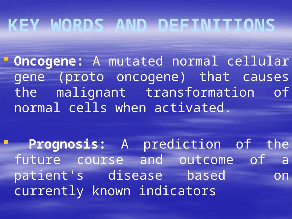
Oncogene: A mutated normal cellular gene (proto oncogene) that causes the malignant transformation of normal cells when activated.
Prognosis: A prediction of the future course and outcome of a patient's disease based on currently known indicators

KEY WORDS AND DEFINITIONS
A carcinogen :is an agent that causes cancer. A carcinogen may be physical (e.g., radiation), chemical (e.g., a polycyclic hydrocarbon), or biological (e.g., a virus).

KEY WORDS AND DEFINITIONS
Exposure to such an agent may cause cancer either by producing
direct genotoxic effects on deoxyribonucleic acid (DNA) (e.g., as with radiatation) or
by increasing cell proliferation (e.g., by a hormone), or
both (e.g., through the use of tobacco).
KEY WORDS AND DEFINITIONS
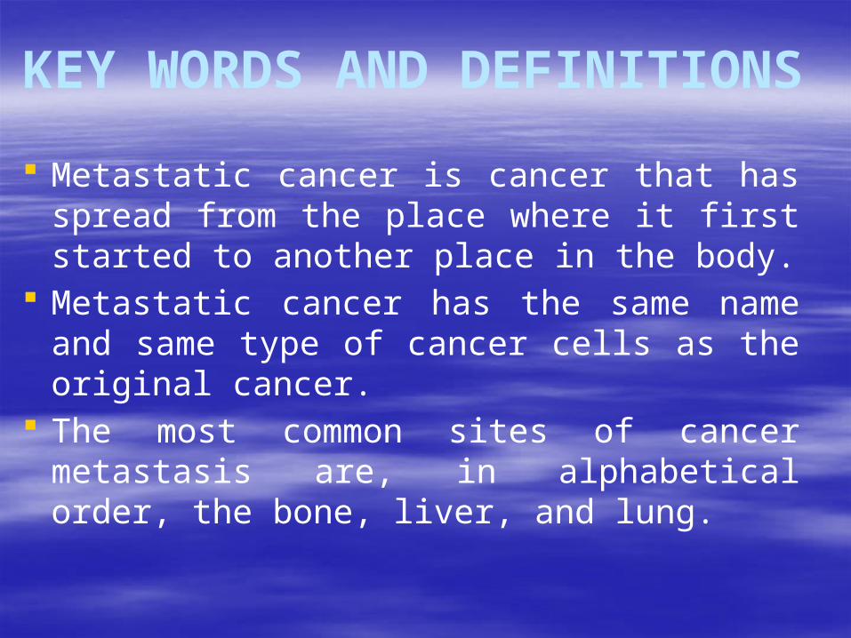
Metastatic cancer is cancer that has spread from the place where it first started to another place in the body.
Metastatic cancer has the same name and same type of cancer cells as the original cancer.
The most common sites of cancer metastasis are, in alphabetical order, the bone, liver, and lung.

What is Cancer ?Application of Proteomics in Cancer Research
Tumors: loss of cell cycle control
de-differentiation and proliferation
benign: encapsulated by connective tissue rarely life-threatening
malignant: invasive growth cell shedding metastasis cancer
life-threatening

Application of Proteomics in Cancer Research
Molecular basis of cancer Causes of cancer: - carcinogens, radiation, viruses, random - hereditary vs. spontaneous tumors - multi step process Genes and gene products involved in cancer: activation of proto-oncogenes to oncogenes
(gain-of-function) inactivation of tumor suppressor genes (loss-of-function)
altered activity of modulator genes

Characteristics of cancer cellsApplication of Proteomics in Cancer Research
• general changes: - loss of division limits (immortality)- uncontrolled proliferation
• genetic changes: - point mutations …- chromosomal changes
• structural changes: - less organized cytoskeleton- increased membrane fluidity
• biochemical changes: - altered protein expression- altered protein modification

Cancer facts and treatmentApplication of Proteomics in Cancer Research
• > 100 different types of human cancer• 20 % of the mortalities in industrialized nations
detection classification and localization
imaginghistology
biomarkers
surgery radiation
chemotherapy
therapy

What is a biomarker? Gives us the ability to analyze organ function,
diagnose diseases in a non-invasive way.
Biomarkers can be any molecule (organic or inorganic) that acts at the test subject while the patient is the host to a biological process.
Biomarkers can be tested from bodily fluids (blood, urine) or from tissues.

Importance
Biomarkers give scientists and doctors the ability to ‘work backwards’ and asses organ function.
Cancer biomarkers can identify genetic variations or mutations as well as changes in gene or protein expression that can be linked to a disease state or a response to a medical intervention

Uses
Biomarkers can be used to:
• confirm diagnosis of acute or chronic disease
• assess the effectiveness of treatment
• evaluate the prognosis of individual cases.

Biomarkers in Cancer Detection
The Early Detection Research Network has put growing focus on discovering and validating biomarkers in their use to diagnose cancer in its early stages.
Many patients are diagnosed in late stages of cancer and it may be too late.
Could be a huge breakthrough for science if this non-invasive method can test for cancer.
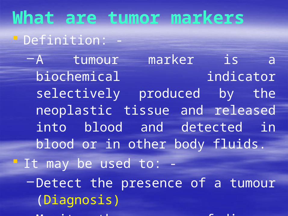
What are tumor markers Definition: -
–A tumour marker is a biochemical indicator selectively produced by the neoplastic tissue and released into blood and detected in blood or in other body fluids.
It may be used to: - –Detect the presence of a tumour (Diagnosis)–Monitor the progress of disease –Monitor the response to treatment–Prognosis
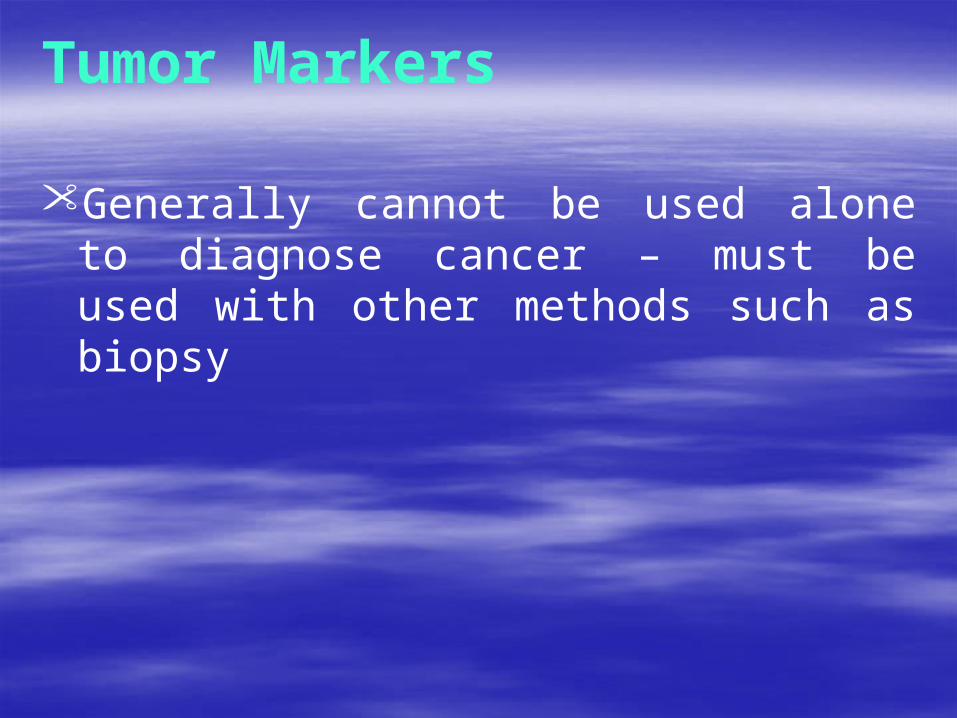
Tumor Markers
• Generally cannot be used alone to diagnose cancer – must be used with other methods such as biopsy

Characteristics Produced exclusively by a cancer cell as a
response to tumor development–Sensitivity
Not exclusively by a cancer cell, but has a sufficient quantities to be distinguished from production by a normal tissue cell–Specificity

An ideal tumor marker The quality should be included
–High sensitivity –High specificity–Can be quantified –Safe –Convenience–Low price

How to identify tumor marker ? On cell
– Cytochemistry, Flow cytometry
On tissue– Histochemistry, Cytosol assays
In body fluids– Blood, urine, CSF, Amniotic fluid

Tumor marker in Oncology
Screening
Diagnosis
Staging
Prognosis

Screening Tumor markers play a limited role for tumor
screening, just because….– relatively low sensitivity– lack of specificity and relation to tumor size
Inappropriate for the detection of small in situ cancer
In some cases, tumor markers can be equal to other examinations envisioned for screening– PSA & prostate cancer– calcitonin & medullary thyroid cancer

Diagnosis Tumor is not the key diagnostic examination, but
can be a complementary sign to clinical finding or medical imaging–AFP & hepatoma
Sometimes implicate the existence within the tumor of an exclusive secretary histological contingent–NSE (neurone specific enolase) &
SCLC (small cell lung cancer)

Staging
The tumor markers and medical imaging are complementary in the pre-therapeutic and post-therapeutic staging

Prognosis
The pre-therapeutic level of certain tumor marker can contributes a prognostic factor because of links with... – Metabolic activity– Tumor size– Invasion
More valuable in that it is independent or other usual prognostic factors for the pathology

Allow doctors to refine therapeutic strategy by selecting groups with risk of failure to response to treatment
This property is one of the major aspects of current use of the tumor marker – CEA & colon cancer– CA19-9 & pancreatic cancer– CYFRA 21-1(Cytokeratin 19 Fragment) & lung
squamous cell cancer

During treatment High markers level before treatment generally
– Not only correlate very well with the therapeutic result but are sometimes superior to this result in the assessment of complete remission
The assay must be taking into account the marker half-life when during treatment and all post-therapeutic re-evaluation
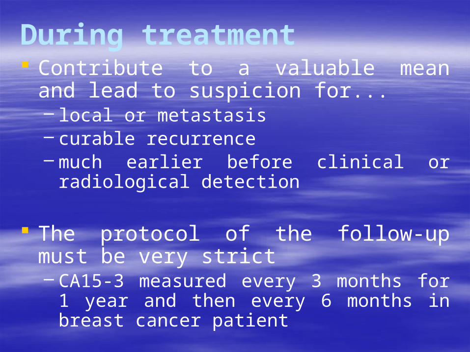
During treatment Contribute to a valuable mean and lead to
suspicion for...– local or metastasis– curable recurrence– much earlier before clinical or radiological detection
The protocol of the follow-up must be very strict– CA15-3 measured every 3 months for 1 year and
then every 6 months in breast cancer patient
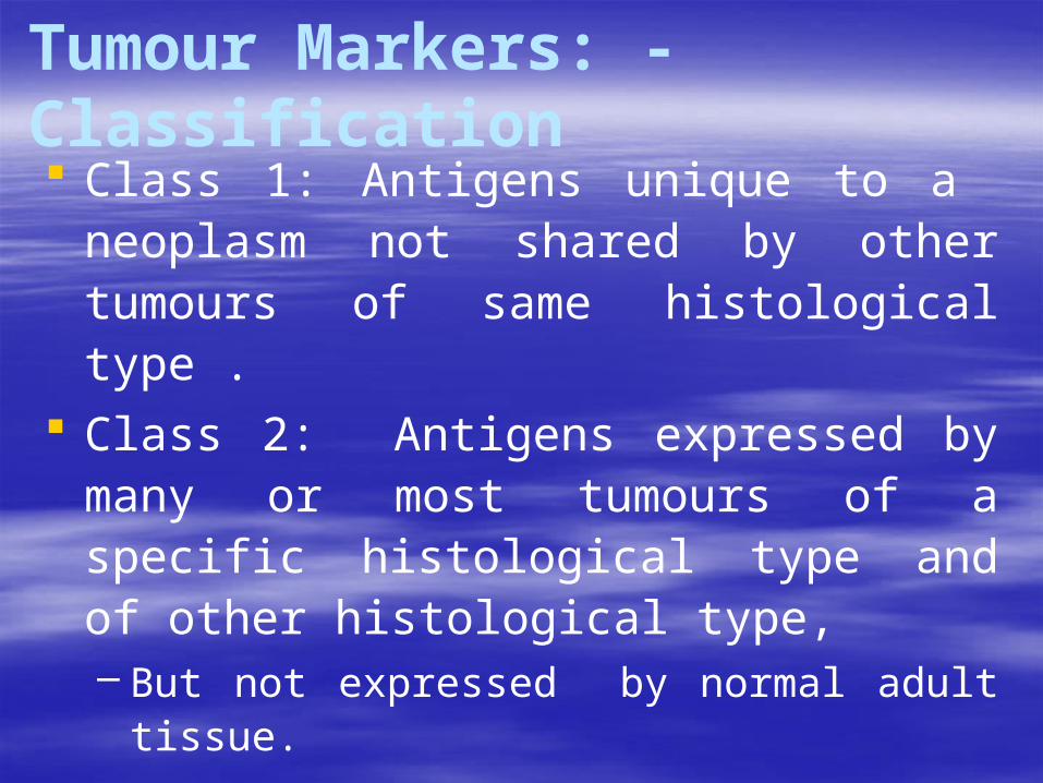
Tumour Markers: - Classification Class 1: Antigens unique to a neoplasm not
shared by other tumours of same histological type .
Class 2: Antigens expressed by many or most tumours of a specific histological type and of other histological type, – But not expressed by normal adult tissue.
Class 3: Antigens expressed by both cancer and normal adult tissue.

NATURE OF TUMOUR MARKERS 1.Oncofetal antigens
– Alpha Feto Protein – CEA – Pancreatic Oncofoetal Antigen
2.Proteins – Casein – By breast carcinoma– Ferritin- Leukaemia
3.Enzymes– Creatine kinase (BB) – Prostate tumour– Alkaline Phosphatase – Lungs tumour– Acid Phosphatase – Prostate tumour

4. Receptors– Oestrogen, Progesterone, Androgen
5. Polyamines – Spermine, Spermidine, Putridine – leukemia,
lymphoma, colorectal CA 6. Cell Markers
– T cell marker, B cell marker-lymphoma7. Ectopic Hormones
– HCG, GH, Erythropoetin, Renin

Common Tumor Markers Alpha-fetoprotein
CEA
CA-19.9
PSA
CA-125
-hCG
VMA
CA-15.3
Estrogen receptor
Progesterone receptor
HER-2/NEU
BRCA1 BRCA2 p53

2007EKUSILENI CLINICAL
LABORATORY 33

Alfa Feto Protein:- After birth AFP usually falls, within 8 to 12
months of delivery to a very low conc. of 10mcg/ml and persists at this low level throughout life.
Unexplained and persistent elevation of AFP in nonpregnant state should be screened, as it may be due to-– Hepatocellular Ca, germ cell tumour, hereditary
persistence of AFP, viral hepatitis and cirrhosis . In addition to its role in prenatal diagnosis, it is
also widely used in the diagnosis, therapeutic monitoring and follow up of patients in germ cell tumours.

Germ Cell Tumours Producing AFP
AFP
1. Dysgerminoma --
2. Endodermal Sinus tumour / +
yolk sac tumour
3. Immature tetratoma +/-
4. Mixed germ cell tumour
+/-
5. Choreocarcinoma --
6. Embryonal CA --

Alpha feto protein (-FP)
Introduction:
–Oncofetal antigen
–Abundant serum protein normally synthesized by the fetal liver
–Re-expressed in certain types of tumors

AFP continued… Clinical Applications:
–Diagnosis, prognosis, and treatment monitoring of hepatocellular carcinoma (HCC; hepatoma)
–Screening (High-risk; HBV or HCV patients)
–AFP is not completely specific for HCC
–AFP might be increased in pregnancy & benign liver disease

AFP continued… AFP be used in conjunction with ultrasound every
6 months in patients at high risk of developing HCC
Patients with hepatitis B virus- and/or hepatitis C virus-induced liver cirrhosis
Lead period i.e., early detection which is ~ 6 months before clinical manifestations

AFP continued… A tumor marker for classification and
monitoring therapy for nonseminomatous testicular cancer
“in combination with -human chorionic gonadotropin (-hCG)”

Cancer Antigen 125 (CA-125)–Detection of ovarian tumors at an early
stage
–monitoring treatments without surgical restaging
–CA-125 is not specific for ovarian cancer, as it may be elevated in:
Menstruation First trimester of pregnancy Endometriosis

CA-125, continued…
–Currently, CA-125 is the only clinically accepted serologic marker of ovarian cancer

Carcinoembryonic Antigen (CEA)
Introduction:
–CEA is an oncofetal antigen
– It is expressed druing development and then re-expressed in tumors
– It is the most widely used tumor marker for colorectal cancer

CEA, continued…
Clinical Applications:
–The main clinical use of CEA is as a tumor marker for colorectal cancer
– In colon cancer, CEA is used for prognosis, in postsurgery surveillance and to monitor response to chemotherapy

CEA:- It is a glycoprotein of mol.wt 200kda. Though it is a tumour marker for GI cancers, it
is also expressed by – malignant mucinous tumor (100%), – 100% cases of atypical hyperplasia of endometrium, – 60% cases of endometrial Ca.– 50-80% cases of squamous cell of Cx, – 75-100% cases of adenocarcinoma of Cx.
It is also produced in pneumonia, hypothyroidism and pancreatic tumours.

2007EKUSILENI CLINICAL
LABORATORY 45

Human Chorionic Gonadotropin (hCG) Introduction:
– hCG is a hormone normally secreted by trophoblasts in the placenta during pregnancy
– It is a glycoprotein consisting of - and -subunits

hCG, continued… Clinical Applications:
–Detection and follow-up of gestational trophoblastic diseases (GTDs)
–GTDs include:
Hydatiform mole (vesicular mole)
Choriocarcinoma
– It is also elevated in nonseminomas testicular cancers

Molar pregnancy
Molar pregnancy is an abnormal form of pregnancy in which a non-viable fertilized egg implants in the uterus and converts a normal pregnancy into an abnormal one (which will fail to come to term.
A molar pregnancy grows into a mass in the uterus that has swollen chorionic villi.

Molar pregnancy
These villi grow in clusters that resemble grapes.
A molar pregnancy can develop when an egg that is missing its nucleus is fertilized and that may or may not contain fetal tissue

Choriocarcinoma
Choriocarcinoma is a malignant, trophoblastic
cancer, usually of the placenta
It is characterized by early hematogenous spread to the lungs

Prostate Specific Antigen (PSA)
Introduction:
–PSA is a glycoprotein produced by the epithelial cells of the acini and ducts of the prostatic ducts in the prostate
–PSA is a serine protease

PSA, continued…
–There are 2 major circulating forms of PSA:
Free
Complexed:
–Complexed to 1-antichymotrypsin or 2-macroglobulin

PSA, continued… Annual PSA for screening of prostate cancer:
– in men over 50 years old– in younger men at high risk: e.g.,
Those with a family history of prostate cancer
–Total PSA: Screening for and in monitoring of prostate cancer
–Free PSA: Differentiate levels of PSA that are in the grey zonePatient with cancer prostate have a lower % of free PSA
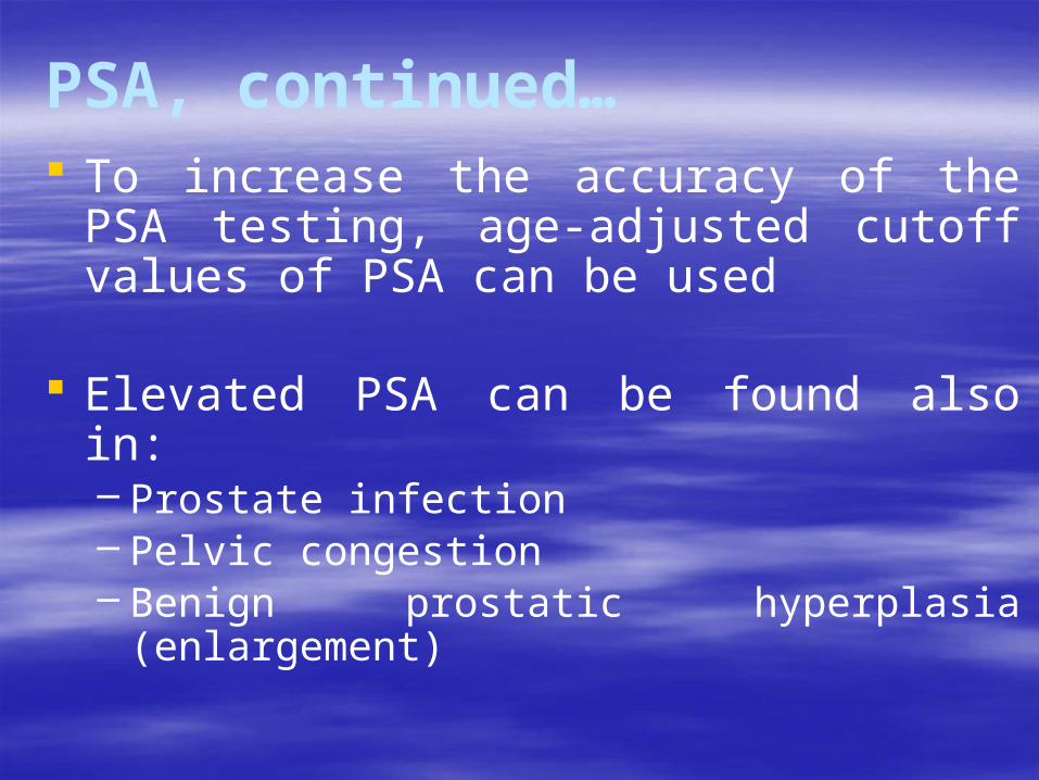
PSA, continued… To increase the accuracy of the PSA testing, age-
adjusted cutoff values of PSA can be used
Elevated PSA can be found also in:– Prostate infection– Pelvic congestion– Benign prostatic hyperplasia (enlargement)

Common Cancer TermsAngiogenesis Development of new blood vessels to supply
oxygen and nutrients to cells
Physiological PathologicalThe process is transient and tightly regulated
e.g., Wound healing, Pregnancy, Menstruation, development
The process is persistent and out of controle.g., tumorogenesis & Metastasis
Marker for angiogenesis: e.g., Vascular Endothelial Growth Factor (VEGF)
Follow-up & treatment of angiogenic cancer
Treatment can target more than one tumor type

HER-2/NEU Encodes an Epidermal Growth Factor Receptor
(EGF-R)
HER2 (from human epidermal growth factor receptor 2).
A proto-oncogene that is converted to oncogene by:– Mutation (especially point mutation) or – Altered (over) expression
Marker for breast and ovarian cancers

HER-2/NEU It is now routinely measured in breast cancer (IHC
and FISH) to determine the type of therapy:– Breast cancer positive for HER-2/NEU is responsive to
treatment (Herceptin)
– Tests are usually performed on biopsy samples obtained by either fine-needle aspiration, core needle biopsy, vacuum-assisted breast biopsy, or surgical excision.
– Immunohistochemistry is used to measure the amount of HER2 protein present in the sample. Alternatively, fluorescence in situ hybridisation (FISH) can be used to measure the number of copies of the gene which are present.

Tumor suppressor genes, e.g., p53Tumor suppressor gene
Encodes a protein involved in protecting cells from unregulated growth
The gene is located on chromosome 17 (Plus the genes of BRCA1 and HER-2/NEU)
Encodes a protein of 53 kDa
Encodes a protein that normally result in cell cycle arrest and induces apoptosis
Upon mutation: loss of function mutation cancer

Estrogen and Progesterone Receptors Estrogen and progesterone receptors are used in
breast cancer as indicators for hormonal therapy.
Patients with positive estrogen and progesterone receptors tend to respond to hormonal treatment.

Estrogen and Progesterone Receptors Those with negative receptors will be treated
using other therapies, such as chemotherapy.
Hormone receptors also serve as prognostic factors in breast cancer.
Patients positive for estrogen and progesterone receptors have a better prognosis.

Estrogen and Progesterone Receptors: Analytical Methodology
Immunocytochemical assays are used to measure steroid hormone receptors.
Immunocytochemical assays use monoclonal antibodies to detect steroid receptor proteins in;
frozen tissue sections, paraffin imbedded tissue, fine,needle aspirates, and malignant effu sions.

CA125 cancer-antigen 125 Reference value
– 95% general population < 35 U/ml Indication: ovarian cancer
–High sensitivity to serous adenocarcinoma, lower to mucinous adenocarinoma (associated with CEA and CA72-4)
–Screening not suggested for ovarian cancer but for ovarian
tumor –Follow-up:
Post-op: tumor residues is good response to CA125

Second look surgery: CA125 increase means bulky peritoneal residues or metastasis, but normal CA125 does not exclude the second look surgery
Early detection of recurrence: increased more than 50% of CA125 level precedes the clinical diagnosis of recurrence
Non-specific increases– Liver cirrhosis with ascites– Pleural effusion– Peritonitis and Pericarditis– During menstruation– Third trimester– Endometriosis– Ovarian cysts

CA15-3 cancer antigen 12-3 Reference value
– 98.7% general population < 30 U/ml
Indication: breast cancer– Most specific tumor marker– At the time of suspected breast cancer
Unable to detect localized or metastatic breast cancer
– Prognostic value CA15-3 > 50 U/ml = high suspicion of metastasis with poor
prognosis

–Follow-up: 6 weeks after surgery–Clinical follow-up
3yrs a year then every 6 months > 50% of reference value predict reccurence or metastasis The association of CA15-3 and CEA assays = increase
sensitivity by 10% Monthly assay during chemotherapy in metastasis stages High correlation with the clinical response to treatment
Non-specific increases– Liver cirrhosis, acute hepatitis, severe chronic
hepatitis (< 50 U/ml)– Other metastasis: pancreas, ovary, colorectal, lung,
stomach and uterus = rarely > 50 U/ml except pancreas adenocarcinoma

CA19-9 carbohydrate antigen 19-9 Reference value
–99.6% general population < 37 U/ml Indication
–Digestive tract carcinoma Pancreatic and biliary tract cancer: sensitivity 85%,
specificity 95% Colorectal cancer: associated with CEA Gastric cancer: associated with CEA and CA72-4
–Follow-up Monthly assay during the first year, then every two months
during two years, then every six months CA19-9 > 1000 ng/ml indicates the metastasis

–Remarks Combination of CEA and CA19-9 increase the early
diagnostic rate to 90% in patient with high risk with a mean lead time of 4-6 months before clinical response
No relation associated with tumor size
Non-specific increases: benign pathology– Lung: acute cystic fibrosis– Digestive tract:
10% of cholecystitis and 8% of pancreatitis (< 3 times of normal value)
Liver cirrhosis
–Other metastatic adenocarcinoma usually < 3 times of normal value

SCC squamous cell carcinoma associated antigen Known as TA-4 (SCC antigen) Origin
– Separate and purify from cervical epithelial cell
Reference value– < 1.5 ng/ml
Indication: SCC, especial in cervical cancer – > 2.5 ng/ml in 53.6% of cervical cancer

– Increase according to the disease progression an d stage
–Follow-upShould downhill to normal range within 72
hours after operation Increasing persist indicating incomplete
resection–Remark
TA-4 in Lung SCC is 3-4 times to normal range, but is normal in other types of lung cancer
Helping tracing tumor and early diagnose in recurrence

CT Calcitonin Reference value
–99% general population < 10 ng/ml Indication: Medullary thyroid cancer
–Screening and diagnosis very sensitive in screening and early diagnosis in high risk
group ( familial and multiple endocrine neoplasia)
–Follow-up Therapy follow-up: repeat assay after operation,
high level indicates incomplete resection or metastasis
Clinical follow-up: monthly assay, then every three months

Non-specific increases –Neuroendocrine tumors: pheochromocytomas,
carcinoid tumors–Digestive tract and pancreatic endocrine
tumors–SCLC (Small Cell Lung Cancer)
–Differentiated thyroid cancer (< 5% of cases)–Benign condition
CRF, hyperparathyroidism, paget’s bone disease

Conclusion The tumor markers contribute to cancer
detection, diagnosis and prognosis is unquestionable, but they need to be estimated considerably
The tumor markers in oncology should be used depending on knowledge and clinical experience

Recommended Tumor Markers for Specific type of Cancers
Tumor Tumor markers1. Hepatoma
(HCC)AFP
2. Ovarian Cancer
CA-125 Inherited ovarian cancer: BRCA1
3. Breast Cancer
CA15-3 CEA HER-2/NEU Estrogen and progesterone receptorsIf inherited: BRCA1, and BRCA2 (on
chromosome 13)

Recommended Tumor Markers for Specific type of Cancers….continued
Tumor Tumor markers
4. Cancer head of the pancreas CA 19-9CEA
5. Colorectal carcinoma CEACA 19-9
6. Pheochromocytoma Vanillylmandelic Acid (VMA) in urine

Recommended Tumor Markers for Specific type of Cancers….continued
Tumor Tumor markers
7. Nonseminomatous testicular cancer
AFP-hCGCEA
8. Vesicular mole & Choriocarcinoma
-hCG
9. Prostate cancer PSA
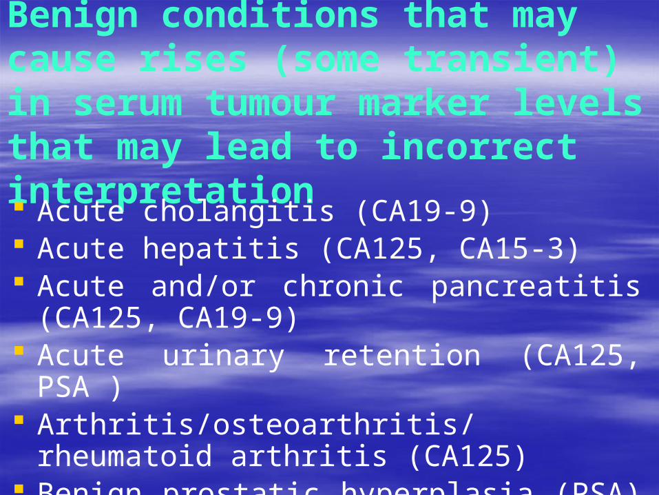
Benign conditions that may cause rises (some transient) in serum tumour marker levels that may lead to incorrect interpretation Acute cholangitis (CA19-9) Acute hepatitis (CA125, CA15-3) Acute and/or chronic pancreatitis (CA125, CA19-
9) Acute urinary retention (CA125, PSA ) Arthritis/osteoarthritis/rheumatoid arthritis
(CA125) Benign prostatic hyperplasia (PSA)

Cholestasis (CA19-9)
Chronic liver diseases such as cirrhosis, chronic active hepatitis (CA125, CA15-3,CA19-9, carcinoembryonic antigen (CEA))
Chronic renal failure (CA125, CA15-3, CEA, human chorionic gonadotrophin)
Colitis (CA125, CA15-3, CEA)
Congestive heart failure (CA125)

Cystic fibrosis (CA125)
Dermatological conditions (CA15-3)
Diabetes (CA125, CA19-9)
Diverticulitis (CA125, CEA)
Endometriosis (CA125)
Heart failure (CA125)

Irritable bowel syndrome (CA125, CA19-9, CEA)
Jaundice (CA19-9, CEA)
Leiomyoma (CA125)
Liver regeneration (α fetoprotein)
Menopause (human chorionic gonadotrophin)
Menstruation (CA125)

Non-malignant ascites (CA125)
Ovarian hyperstimulation (CA125)
Pancreatitis (CA125, CA19-9)
Pericarditis (CA125)
Peritoneal inflammation (CA125)
Pregnancy (α fetoprotein, CA125, human chorionic gonadotrophin) Prostatitis (PSA)

Recurrent ischaemic strokes in patients with metastatic cancer (CA125)
Respiratory diseases such as pleural inflammation,
pneumonia (CA125, CEA)
Sarcoidosis (CA125)
Systemic lupus erythematosus (CA125)
Urinary tract infection (PSA)


![[Ghiduri][Cancer]Gastric Cancer](https://static.fdocuments.us/doc/165x107/55cf9399550346f57b9de771/ghiduricancergastric-cancer.jpg)
![[Ghiduri][Cancer]Esophageal Cancer](https://static.fdocuments.us/doc/165x107/577cc7761a28aba711a10585/ghiduricanceresophageal-cancer.jpg)

