CanadianPathology Journal of Volume 3, Issue 1 … final.pdf · proposera des lignes directrices...
44
Canadian Laboratory Physician Supply: Falling Behind Interpretation of Diagnostic Muscle Biopsies Canadian Journal of Pathology Publications Agreement Number 40025049 • ISSN 1918-915X Official Publication of the Canadian Association of Pathologists www.cap-acp.org Volume 3, Issue 1 • Spring 2011
Transcript of CanadianPathology Journal of Volume 3, Issue 1 … final.pdf · proposera des lignes directrices...

Canadian Laboratory Physician Supply: Falling Behind
Interpretation of Diagnostic Muscle Biopsies
Canadian Journal of
Pathology
Publications Agreement Number 40025049 • ISSN 1918-915X
Official Publication of the Canadian Association of Pathologists
www.cap-acp.org
Volume 3, Issue 1 • Spring 2011

APR10
The Automatic Choice for H&EDako CoverStainerThe new Dako CoverStainer completely automates H&E performance with a highly flexible system that meets the special needs of laboratories. This comprehensive, fully integrated H&E solution delivers consistent, high-quality results while maximizing productivity and optimizing resource utilization.
Make Dako CoverStainer your automatic choice for H&E performance.
www.dako.com
Up to 240 H&E Slides Per Hour
Bake Dewax DehydrateStain Coverslip Dry

VOLUME 3, ISSUE 1 2011
About the Cover
4 Editorial: Two Years LaterJ. Godfrey Heathcote, MA, MB BChir, PhD, FRCPC
Éditorial : Deux ans plus tardJ. Godfrey Heathcote, MA, MB BChir, PhD, FRCPC
EDITOR-IN-CHIEFJ. Godfrey Heathcote, MA, MB BChir, PhD, FRCPC
EDITORIAL BOARD
Manon Auger, MD, FRCPC, Cytopathology; Calvino Cheng, BSc, MD, FRCPC, Pathology Informatics
and Quality Management; Eleftherios Diamandis, BSc, MD, PhD, FRCPC, Medical Biochemistry;
David K. Driman, MB ChB, FRCPC, Anatomical Pathology; Todd F. Hatchette, BSc, MD, FRCPC, Medical Microbiology;
Michael J. Shkrum, MD, FRCPC, Forensic Pathology; Louis D. Wadsworth, MB ChB, FRCPath, FRCPC, Hematopathology
C AP A SSOCIATION MANAGER Danièle Saintonge
MANAGING EDITORSusan Harrison
PROOFREADERScott Bryant
ART DIRECTORAndrea Brierley, [email protected]
TR ANSL ATORMarie Dumont
SALE S AND CIRCUL ATION COORDINATORBrenda Robinson, [email protected]
ACCOUNTINGSusan McClung
GROUP PUBLISHERJohn D. Birkby, [email protected]
––––––––––––––––––––––––––––––––––––––––––––––––––––––––––––Canadian Journal of Pathology is published four times annually by
Andrew John Publishing Inc., with offices at 115 King Street West, Dundas, ON, Canada L9H 1V1.
We welcome editorial submissions but cannot assume responsibility or commitment for unsolicited material. Any editorial material, including photographs that are accepted from an unsolicited
contributor, will become the property of Andrew John Publishing Inc.
FEEDBACKWe welcome your views and comments. Please send them to
Andrew John Publishing Inc., 115 King Street West, Dundas, ON, Canada L9H 1V1.
Copyright 2011 by Andrew John Publishing Inc. All rights reserved.Reprinting in part or in whole is forbidden without express
written consent from the publisher.
Publications Agreement Number 40025049 • ISSN 1918-915XReturn undeliverable Canadian Addresses to:
115 King Street West, Suite 220, Dundas, ON L9H 1V1
Canadian Journal of Pathology • Volume 3, Issue 1, 2011 Contents
Original Articles
For Instructions to Authors, please visit http://www.andrewjohnpublishing.com/CJP/instructionstoauthors.html
This image depicts skeletal muscle fibres showing widespread central nuclei in myotubular myopathy.
12
18
25
CAP-ACP News
6
5
Message from the PresidenttLaurette Geldenhuys, MBBCH, FFPATH, MMED, FRCPC, FIAC, MAEd
Canadian Laboratory Physician Supply: Falling BehindAaron F. Pollett, MD, Ginette Lajoie, MD, Terence J. Colgan, MD
The Development and Delivery of a Pathology Objective Structured Clinical Examination for Postgraduate Pathology TraineesMarla Nayer, PhD, William Chapman, MD, FRCPC
Malignant Mixed Germ Cell Tumour in a 13-Year-Old Boy with Familial TestotoxicosisSusanne M. Chan, MD, Jose A. Gomez-Lemus, MD, FRCPC
Mathematical Modelling of Lymph Node Grossing TechniquesEtienne Mahe, BSc, MD
27
COPY EDITORSSusan Harrison, Michael Peebles
Opinion
10 In Praise of the Part-Time PathologistAnn Hall, MD, FRCPC
Invited Review
33 Interpretation of Diagnostic Muscle BiopsiesMichael Farrell, FRCPI, FRCPC, FRCPath
FOUNDING EDITORJagdish Butany, MBBS, MS, FRCPC
Canadian Journal of P athology 3Spring 2011

Spring 20114 Canadian Journal of P athology
EDITORIAL
With Canadian Journal of Pathology (CJP)now entering its
third year, it is a good time to examine the journal’s
development over the 2 years since its inception. To assist in the
evaluation, all members of the Canadian Association of
Pathologists (CAP-ACP) were invited in November 2010 to
participate in an online survey, although at that time only four
scientific issues had been released. We received 113 responses
out of 1,032 members surveyed (11%), and most questions were
answered by over 94% of respondents.
It was encouraging to learn from the survey that 55% of
respondents rated the overall content of the journal as very good
or excellent, and only 3% rated it as poor. Similarly, 67% of
respondents rated the overall appearance of the journal as very
good or excellent, and only 3% regarded it as poor. Over 83%
considered the journal to be relevant to their practice. Between
85 and 90% of respondents indicated that articles were well
written and informative with images and illustrations of high
quality.
Although this “snapshot” of readers’ opinions was indeed
gratifying, the request for lists of topics that should be covered
in the journal provided evidence to support these opinions.
Among the topics suggested, quality assurance, particularly in
anatomical pathology, and how it might be achieved in smaller
community hospitals, was clearly a major interest. Other
suggestions focused on areas of professional practice, including
workload, human resources, standardized reporting, and
laboratory management in general, as well as the history of
Canadian pathology. Articles on some of these topics have
already appeared in the early issues of the journal and others
have been accepted for publication.
There was general support for the continuation of regular
invited reviews, dealing with both broad areas of systemic
pathology and difficult or problem topics, as well as reviews of
new technologies and updates in pathobiology written for a
broad readership. This issue contains a masterful and
comprehensive review on the interpretation of muscle biopsies,
not perhaps a matter of daily concern to most, and the next will
include a guide to the use of immunohistochemistry in
dermatopathology that will be of practical value to all surgical
pathologists.
The survey indicates that for many readers the journal is heading
in the right direction, and perhaps the silent majority agree.
Constructive criticisms are always more useful than vague
plaudits, however, and there were some negative comments that
made me, as editor-in-chief, pause to re-evaluate the goals of
CJP. One anonymous respondent commented: “There is no
need for a Canadian pathology journal. It has no impact, and I
would rather send my contributions to well-established and read
journals.”
Clearly, it is good that Canadian pathologists, whose research is
of sufficient calibre, should strive to have their work published
in journals such as Nature or The American Journal of Pathology.
For most Canadian pathologists, that is not an option; and
valuable contributions to the understanding and practice of
pathology are not confined to the pages of such prestigious
journals. We can learn much from the practice of pathology and
laboratory medicine in other countries, but ultimately we will
have to solve our own particular problems. One of the major
weaknesses of Canadian pathology and laboratory medicine has
been its fragmentation within a provincially funded health care
system. That this fragmentation has been to nobody’s benefit is
recognized by the recent decision to create a Canadian Pathology
and Laboratory Medicine Leadership Council, which the
president of CAP-ACP discusses in her letter on page 6. Whether
or not this initiative is successful remains to be seen, but CJP
offers us a way to share the developments and experiences in our
fragmented system in a way that can benefit the whole country.
At this time, CJP is not indexed in PubMed and therefore has
no impact factor. Whether it has a real impact depends on the
usefulness of its contents to practising pathologists across
Canada. The editorial board did not set out to create a Canadian
journal of surgical pathology but, rather, to model our
publication more on Journal of Clinical Pathology, with its
discipline-wide scope. We recognize that it will take years,
perhaps decades, to develop a significant impact factor for a new
general specialty journal such as ours.
I would like to thank those readers who completed the survey
and encourage all of you to consider submitting your studies to
the journal. Your comments on published articles and matters
of concern to Canadian pathologists, laboratory physicians, and
scientists will always find space in our correspondence section.
J. Godfrey HeathcoteEditor-in-Chief
Two Years Later

Canadian Journal of P athology 5Spring 2011
ÉDITORIAL
Alors que la Revue canadienne de pathologie (RCP) amorce sa
troisième année d’existence, il semble opportun d’examiner
son parcours depuis sa création. En prévision de cette évaluation,
l’Association canadienne des pathologistes (CAP-ACP) a invité ses
membres à répondre à un sondage en ligne en novembre dernier
après la publication de quatre numéros scientifiques seulement. En
tout et pour tout, 113 des 1 032 membres sollicités ont répondu
au questionnaire (taux de réponse de 11 %), et 94 % des
répondants ont répondu à la plupart des questions.
L’analyse des résultats révèle que 55 % des répondants estiment
que le contenu de la revue est soit très bon, soit excellent, que
seulement 3 % le jugent passable. De même, 67 % des répondants
sont d’avis que l’apparence générale de la revue est soit très bonne,
soit excellente, tandis que 3 % des répondants la trouvent médiocre.
Pour plus de 83 % des répondants, la revue s’avère pertinente dans
leur pratique. Enfin, de 85 % à 90 % des répondants affirment que
les articles sont de bonne tenue et instructifs, qu’ils s’accompagnent
d’images et d’illustrations de grande qualité.
Bien que cet « instantané » de l’opinion du lectorat soit gratifiant,
la liste des sujets proposés en réponse à notre demande de sujets
qui devraient faire parler d’eux dans la revue vient étayer ces
propos. Le sujet de l’assurance de la qualité, particulièrement en
anatomopathologie, et du mécanisme d’assurance de la qualité
approprié dans le petit hôpital communautaire, est indéniablement
un sujet d’intérêt majeur. La liste comprend en outre des sujets
ayant trait à l’exercice de la profession, notamment la charge de
travail, les ressources humaines, le compte rendu normalisé et la
gestion du laboratoire en général, ainsi qu’à l’histoire de la pathologie
au Canada. Des articles sur certains de ces sujets ont déjà paru dans
les premiers numéros de la revue, d’autres sont prévus.
En général, les lecteurs souhaitent retrouver des articles de fond de
collaborateurs invités, que ce soit sur des sujets de vaste portée
relevant de la pathologie systémique, sur des sujets problématiques
ou encore sur de nouvelles technologies ou sur les percées de la
biopathologie d’intérêt général. Le présent numéro renferme un
article de fond éminent et exhaustif sur l’interprétation de la biopsie
musculaire, un sujet qui déborde peut-être du cadre de la pratique
courante de la plupart d’entre nous, tandis que le prochain
proposera des lignes directrices sur l’immunohistochimie en
dermatopathologie qui se révéleront certes d’utilité pratique pour
tous les pathologistes spécialisés en pathologie chirurgicale.
Les résultats du sondage indiquent que, pour beaucoup de lecteurs,
la revue est sur le bon chemin, et peut-être que la majorité
silencieuse en convient également. Néanmoins, la critique
constructive demeure sans doute plus utile que les acclamations
générales, et certaines observations défavorables m’ont porté à
réfléchir, en tant que rédacteur en chef, sur les buts de la RCP.Un
répondant a dit ceci dans l’anonymat : « Il n’y a pas lieu d’avoir une
revue canadienne sur la pathologie. Ça n’a aucun impact, et
j’aimerais mieux que mes articles paraissent dans des revues
réputées qui font autorité. »
Il est clair que le pathologiste canadien dont les projets de recherche
sont d’envergure a tout intérêt à voir ses travaux publier dans des
revues telles Nature ou The American Journal of Pathology.
Cependant, pour la plupart des pathologistes au pays, cela n’est pas
possible, et les communications qui contribuent à l’avancement
des connaissances sur la pathologie et de l’exercice de la profession
ne sont pas réservées qu’à ces revues prestigieuses. Bien sûr, nous
pouvons tirer des enseignements utiles sur la pathologie et la
biologie médicale d’autres pays, mais, au bout du compte, nous
aurons à régler nos problèmes particuliers. La compartimentation
de l’exercice de cette branche de la médecine dans les réseaux de
santé publics provinciaux en constitue l’une des grandes lacunes.
La récente création du Conseil canadien de la pathologie et de la
biologie médicale, sujet abordé par la présidente de l’Association à
la page 6, illustre à quel point cette compartimentation est
désavantageuse. Reste à savoir si cette initiative sera fructueuse, mais
la RCPnous permet d’échanger entre nous sur les faits nouveaux
et les expériences dans notre système compartimenté afin que tous
au pays puissent en bénéficier.
Actuellement, étant donné que la RCP n’est pas indexée dans
PubMed, nous ne connaissons pas son facteur d’impact, soit
l’indicateur mesurant son influence. Son influence réelle dépend
du caractère utile de son contenu pour les pathologistes en exercice
au pays. Le comité de rédaction n’avait pas l’intention de diffuser
une revue canadienne sur la pathologie chirurgicale, mais bien de
publier une revue sur le modèle du Journal of Clinical Pathology
qui s’intéresse à tous les champs de pratique de la discipline. Nous
savons fort bien qu’il faudra des années, plusieurs dizaines d’années
peut-être, avant qu’une nouvelle revue spécialisée d’ordre général
comme la nôtre exerce un facteur d’impact important.
Je tiens à remercier les lecteurs qui ont répondu au sondage et je
vous invite à faire publier vos études dans les pages de la revue.
Sachez que la rubrique du courrier des lecteurs réservera toujours
un bon accueil à vos observations sur les articles et sur les sujets
d’intérêt pour les pathologistes, les médecins biologistes et les
scientifiques canadiens.
J. Godfrey HeathcoteRédacteur en chef
Deux ans plus tard

Spring 20116 Canadian Journal of P athology
Message from the President
CAP-ACP NEWS
Dear CAP-ACP Members,
It is my pleasure to provide an update on the activities of the
CAP-ACP since our very successful Annual Meeting in
Montreal in July 2010. The executive met in Toronto in
November and moved a number of projects forward. Some
of these projects, as well as other activities of the CAP-ACP,
are highlighted below.
CAP-ACP OfficeThe CAP-ACP Office with the executive are reviewing the
bylaws and terms of reference for committees and working
groups, as well as fine-tuning an organizational chart and
job descriptions for positions in the organization.
SurveyWe thank members for participating in a recent survey. A
detailed report will be shared with members, but, overall,
members appear satisfied with the work of the CAP-ACP.
There was strong encouragement to develop online self-
assessment programs (SAPs) in line with the new Royal
College Maintenance of Certification (MOC) Program,
which now awards three credits per hour for participation
in SAPs. There will be a session on the new MOC Program
at the upcoming Annual Meeting in Vancouver in June.
GuidelinesWork has begun to update guidelines, including those for
retention of pathology material, led by Vina Alexopoulou,
and telepathology, led by Bernard Têtu. Guidelines for
quality assurance, including turnaround times, will be
developed in the near future.
Continuing Professional DevelopmentWe successfully renewed our status as a Royal College
Accredited Provider of Continuing Professional
Development (CPD), and accreditation is being managed
by a new Accreditation Subcommittee. In addition to
accrediting our Annual Meeting and our
Immunohistochemistry Meeting, we co-developed and
accredited a series of distributed education sessions on the
College of American Pathologists (CAP) cancer checklists
with Cancer Care Ontario and the Canadian Partnership
against Cancer (CPAC). A session on the TNM system is to
be offered in April. The sessions can be accessed live or
online after the event. We also co-developed and accredited
an ongoing educational series on molecular pathology. We
are working with CAP to offer a Canadian version of a
course on breast pathology that is currently being offered in
the United States. The course is composed of on-site group
learning and online self-assessment modules, and these are
being accredited for the Royal College MOC Program.
An immunohistochemistry course will be scheduled later in
the year, and a molecular pathology course will precede the
Annual Meeting in June. The CAP-ACP is endorsing a
conference being organized by Emina Torlakovic in
collaboration with the University Health Network titled
Redefining the Crucial Role of the Pathologist in the Era of
Personalized Medicine: The Development of a National
Strategy.
The CPD Committee, under Joan Sweet, continues to
expand the offerings of online CPD, and we urge members
to contribute cases.
WebsiteThe website is very active, providing regular updates on
CAP-ACP activities. It also provides an online venue for
discussion.
Canadian Journal of PathologyCanadian Journal of Pathology is entering its third year.
Founded under the leadership of past president Jagdish
Butany, it is starting to flourish, featuring articles of high
quality and interest. We urge members to contribute suitable
papers. The Journal published the abstracts from the 2010
Annual Meeting online and will do so again for the
upcoming Annual Meeting.
Annual MeetingThe Annual Meeting will be held from June 4 to 8 in
Vancouver and will occur in partnership with the Canadian
Society of Clinical Chemists. The Annual Meetings
Committee, under Avrum Gotlieb, the Local Organizing
Committee, under Diponkar Banerjee, and the CPD
Committee, under Joan Sweet, are working hard to make
this one of the most successful meetings ever.
Also mark your calendars for upcoming Annual Meetings

Canadian Journal of P athology 7Spring 2011
CAP-ACP NEWS
in Calgary, July 21–25, 2012, and in Quebec City, June 8–12,
2013, in partnership with the World Association of Societies
of Pathology and Laboratory Medicine.
Andrew Herzenberg AwardAt the Executive Meeting in Toronto in November, the
memory of Dr. Andrew Herzenberg, who tragically passed
away just before the Annual Meeting, was honoured by a
gathering with some of his family members and friends, and
a presentation of the Junior Scientist Award to his family. At
the same event, we announced the founding by the executive
of the Andrew Herzenberg Award for the best paper by a
resident on nephropathology or transplant pathology at the
Annual Meeting. Members will have an opportunity to
contribute to the fund for the award at the time of
membership renewal, or at any other time, through the
CAP-ACP Office.
Resident Review CourseUnder the leadership of Vina Alexopoulou, Jagdish Butany,
and a board and faculty of CAP-ACP members, the first
Resident Review Course will be offered in Hamilton from
March 25 to 27 (see details on page 9). Registration numbers
are high, and this event promises to be an ongoing and
valuable service to our junior colleagues.
CAP-ACP SectionsThe Canadian Society of Cytopathology and the sections of
Haematological Pathology, Autopsy Pathology,
Neuropathology, Advanced Diagnostics and Pathology
Assistants, among others, continue work on a number of
issues and a more detailed update of their activities will be
provided at the time of the Annual Meeting.
Patient Safety and Quality AssuranceThe Task Force on Guidelines on Investigation of Laboratory
Medicine Irregularities, led by Diponkar Banerjee,
developed Guidelines on Investigation of Laboratory
Medicine Irregularities, and these are now posted on the
website.
The National Standards Committee on Immuno-
histochemistry Testing, under Emina Torlakovic, produced
class I and II checklists on immunohistochemistry, posted
on the website, and expanded to become the National
Standards Committee on High Complexity Laboratory
Testing, which will also develop guidelines on molecular
testing.
Anatomical PathologyPat Shaw, in collaboration with the Anatomical Pathology
section chair, Anne O’Brien, formed a working group that
will produce a statement on the pathologist as custodian of
tissue.
Special Interest GroupsJoining five other special interest groups (SIGs), the Semen
Analysis SIG was formed under the leadership of David
Moore.
Collaboration with Other OrganizationsCanadian Partnership against CancerIn addition to the educational efforts mentioned above, we
continue working with CPAC on many other fronts,
including establishing Canadian cancer committees to
provide Canadian input into the CAP cancer checklists and
the national pathology synoptic reporting effort. The CAP-
ACP was involved in the preparation for a CPAC press
release earlier this year on the National Staging Initiative.
CPAC continues to provide funding for a number of CAP-
ACP activities, including CPD.
Royal CollegeThe CAP-ACP is represented on the Royal College Specialty
Committees for Anatomical Pathology by Vina Alexopoulou
and General Pathology by Chris Naugler. Marciano Reis
chairs both the CAP-ACP Section of Haematological
Pathology and the Royal College Specialty Committee for
Haematological Pathology. We urge members to
communicate any comments or concerns they have about
residency training issues to our representatives so that they
can bring them to the attention of the Royal College.
The Royal College organized a Consensus Conference on
Laboratory Medicine on November 15, 2010. The CAP-ACP
was a member of the planning committee, and many CAP-
ACP members were chairs, speakers, and participants in the
conference. The press release that followed the conference

Spring 20118 Canadian Journal of P athology
CAP-ACP NEWS
created much interest in the media, as well as from other
laboratory medicine national specialty societies, and it was
clear that the CAP-ACP is regarded by the media and the
public as the spokesperson for laboratory medicine in
Canada. There were articles in newspapers and online. The
news release was published in the Royal College Dialogue,
and a very good overview of the activities of the CAP-ACP
was published in the December 15 issue of theMedical Post.
It was resolved at the conference to form a Canadian
Leadership Council on Laboratory Medicine (CLCLM) and
committees of the council to work on areas within
laboratory medicine, including workload, quality assurance,
and education.
The CAP-ACP is currently part of a small working group
developing terms of reference for the proposed council. The
executive developed a CAP-ACP position statement on the
CLCLM and requested input on the document from the
CAP-ACP Council. The CAP-ACP Council represents a
broad spectrum of groups in Canadian laboratory medicine.
The final document was forwarded to the working group
developing terms of reference. The position statement
includes the following principles:
1. Laboratory physicians should lead the proposed
council.
2. There should be broad and equal representation of
laboratory physicians, including from all laboratory
physician national specialty societies and CAP-ACP
sections, and geographic representation from
provincial societies and academic pathology
departments.
3. There should be participation from organizations that
impact laboratory medicine such as the Royal College,
Canadian Medical Association, and others yet to be
decided.
4. There should be patient representation.
5. It is suggested that at least one of the sessions of the
proposed council occur at the CAP-ACP Annual
Meeting since many members of the proposed CLCLM
would be members of the CAP-ACP Council.
International Liaison Committee of PresidentsThe CAP-ACP is represented on the International Liaison
Committee of Presidents of Pathology Organizations. It is
reassuring to learn that most of the issues that Canadian
pathologists struggle with are the same issues that our
colleagues in other countries face. Membership of this
committee is also an opportunity to learn from the
experience of our international colleagues.
The CAP-ACP contributed an article on Canadian
pathology in the Bulletin of the Royal College of Pathology in
Britain.
Thank YouAs always, thank you for the important contribution you
make as CAP-ACP members to the Canadian community of
pathologists and the Canadian public. Above all, thank you
to Daniele Saintonge and Heather Wieland in the CAP-ACP
Office, without whom none of the above would happen.
A very happy and healthy 2011 to all!
Laurette GeldenhuysPresident

1st Annual Canadian Association of Pathologists’ Association canadienne des pathologistes
Residents Review Course
Friday, March 25 to Sunday, March 27, 2011 McMaster University - Hamilton, ON
The Canadian Association of Pathologists’/Association canadienne des pathologistes is pleased to announce and invite all Canadian trainees, US trainees, CAP-ACP resident members as well as non-members and pathologists to the 1st Canadian Association of Pathologists’ Residents Review Course in Anatomical Pathology to be held in Hamilton, Ontario at McMaster University’s Michael G. DeGroote School of Medicine (MDCL-3020).
Friday - March 25, 2011
1300-1315 Welcome Dr. Laurette Geldenhuys
1315-1340 The RCPSC AP Exam - Format and General Information Dr. David Driman
1340-1400 The Oral Exam – How to Perform at Your Best Dr. David Driman
1400-1500 Gastro Intestinal Dr. Jeremy Parfitt
1500-1515 Health Break
1515-1545 Quality Assurance in Anatomical Pathology Dr. Julianne Klein
1545-1615 Lab Administration - What the Junior Pathologist Needs to Know Dr. Jagdish Butany
1615-1645 Nervous System Dr. Robert Macaulay
1645-1715 Cardiovascular Surgical and Autopsy Pathology – An Overview Dr. Vidhya Nair
1715-1745 Pediatric Pathology Review Dr. Jorge Arredondo
Saturday - March 26, 2011
0730-0800 Continental Breakfast
0800-0830 Diagnostic Immunohistochemistry Dr. Emina Torlakovic
0830-0930 Head and Neck Dr. Ilan Weinreb
0930-0945 Health Break
0945-1000 Kidney Pathology – Medical Dr. Vina Alexopoulou
1000-1045 Kidney, Ureter and Bladder – Surgical Dr. Andrew Evans
1045-1145 Prostate and Testis Dr. Andrew Evans
1145-1215 Skin Pathology Dr. Martin Trotter
1215-1315 Lunch
1315-1415 Pancreatic Pathology Review Liver Pathology: Understanding the Clinical Process
Dr. Jasim Radhi (pancreas) Dr. Oyedele Adeyi (liver)
1415-1445 Gross Pathology Dr. Michele Weir
1445-1515 Breast Pathology Review Dr. Penny Barnes
1515-1530 Health Break
1530-1630 Pathology of the Female Genital Tract Dr. Maire Duggan
1630-1700 Molecular Pathology for the Practicing Pathologist Dr. Kenneth Craddock
1700-1730 Lung and Pleura Dr. Jean-Claude Cutz
Sunday - March 27, 2011
0730-0800 Continental Breakfast
0800-0900 Forensic Pathology - What the Junior Pathologist Needs to Know Dr. Matt Bowes
0900-1000 Cytology – Review of Basic Principles Dr. Linda Kapusta
1000-1030 Health Break
1030-1130 General Principles of Approach to Interpretation of Bone and Soft Tissue Pathology
Dr. Snezana Popovic
1130-1230 Overview of Benign and Malignant Haematological Disorders and Application of Ancillary Tests in Diagnosis and Prognosis
Dr. Monalisa Sur
1230-1330 Lunch
1330-1430 Wrap-up and Self-Assessment
Dr Vina Alexopoulou and Dr. Jagdish Butany
Accreditation The 1
st Annual Canadian Association of Pathologists’/Association canadienne des pathologistes Residents Review Course is accredited by the
Canadian Association of Pathologists. This event is an Accredited Group Learning Activity (Section 1) as defined by the Maintenance of Certification program of The Royal College of Physicians and Surgeons of Canada.
FOR FULL COURSE DETAILS AND REGISTRATION PLEASE VISIT WWW.CAP-ACP.ORG

Spring 201110 Canadian Journal of P athology
In Praise of the Part-Time Pathologist
Ann Hall, MD, FRCPC, is a general pathologist, associate staff, at the Brant Community Healthcare System, Brantford GeneralHospital, in Brantford, Ontario; and a peer assessor for the College of Physicians and Surgeons of Ontario. She can be contacted at [email protected]. This article has been peer reviewed.Competing interests: Dr. Hall works part-time at Brantford General Hospital. Until recently, she worked part-time at Cambridge Memorial Hospital and continues to provide locum coverage there.
Ann Hall, MD, FRCPC
The recent increases in pathologists’ remuneration are a
double-edged sword.1 Many would argue that they
represent a long overdue game of “catch-up” for a group of
highly skilled and under-valued professionals facing a
diminishing recruitment pool.2 As a group that has little in
the way of personal overhead commitment, this can be
reflected in a degree of departmental staffing instability
worthy of the installation of a revolving door. Many
pathologists are paid under the global budget of the
institution for which they provide services. This may come
as a salary with benefits, contract, or fee-for-service
arrangement with a hard cap. Regardless of the payment
system, with the increased monetary value assigned, there is
heightened scrutiny of workload. Since 2005, in the province
of Ontario, many pathologists work under a Laboratory
Medicine Funding Framework Agreement (LMFFA), which
determines a uniform minimum level of compensation
(UMLC) that increases each fiscal year. This has been a
successful technique that has brought more uniform
compensation to the profession. However, the ultimate goal
is to link workload to compensation. Workload is the new
currency in areas where there is levelling of the remuneration
playing field. In conjunction with this greater value placed
on pathologists’ services, there has been an augmentation in
case complexity and scrutiny and a push for standardization
with rigorous quality assurance.3 In general, workload seems
to have increased, and pathologists now have empirical tools
to measure this.4,5
When faced with a greater and greater workload, the
difficulty lies in managing this volume. This is especially
problematic in a small department, where there are fewer
pathologists to carry the extra load. In general, the full-time
pathologists absorb the extra workload, resulting in longer
hours; this can contribute to burnout and longer turnaround
times. This continues until an extra position is approved by
the hospital administrators. The irony is that the busier one
is with patient care, the more management issues – such as
tracking workload, monitoring turnaround time, and
engaging in quality assurance initiatives – take a back seat.
A way to alleviate this situation is to anticipate and plan
when staffing a department. If a department is staffed with
a core of full-time pathologists and a few part-time
pathologists, managing the workload becomes dynamic.
Flexible scheduling can accommodate slower periods, such
as operating room shutdowns during the summer and peak
holiday times. Likewise, increased staffing can be introduced
during times of volume growth, such as the opening of a new
endoscopy room or operating room.
When a department has a flexible pool of part-time
pathologists, it can negate the need to ask, cap in hand, the
administration for the funding to support a new full-time
position. An incremental funding increase justified with
workload data is more readily attainable. If a department
commits to this data collection and can use it this way,
administrators may even anticipate and budget for it over
time. This funding increase can support extra days of
coverage by part-timers, particularly when others are sick,
on vacation, or on educational leave. This is an effective
strategy to prevent stress and fatigue during times of
increased caseload. Part-time staff offer other advantages to
a department. They help promote a family-friendly
atmosphere that can benefit recruitment and retention. They
may also provide locums for other institutions, thus
promoting networking, exposure to new ideas, and
environmental scanning.
However, when one looks around the country, one sees many
departments that are designed on the assumption that every
pathologist must be full-time. I believe that part-time
professionals are often viewed as less than dedicated to their
profession. I have been fortunate to experience the opposite.
OPINION

Canadian Journal of P athology 11Spring 2011
HALL
For the last eight years of my professional career, I have been
proudly working part-time in a small community hospital
laboratory. We currently have six pathologists for four full-
time positions. I have ranged from providing 0.3 to 0.7
full-time equivalents during this period, depending on the
needs of the department. My level of professional
enthusiasm is higher than when I was full-time. Working
part-time forces me to scrutinize every report I issue to
ensure clarity as I may not be at the hospital if a clinician
questions my wording. My part-time colleagues and I have
facilitated surge capacity and departmental growth.
I believe that greater consideration should be given to hiring
part-time pathologists when planning departmental staffing
needs. Before embarking on this approach, it might be wise
to consider discussion with pathologists who have
successfully integrated part-time colleagues. There is no
need to reinvent the wheel. Accept that every department
has different workflow priorities and that flexibility is
required.
References1. Ontario Ministry of Finance. Public sector salary disclosure 2010. Toronto
(ON): The Ministry, 2010; http://www.fin.gov.on.ca/en/publications
/salarydisclosure/2010/. Accessed February 2, 2011.
2. Physician Working Group, Provincial Advisory Group, Ontario Regional
Laboratory Services Planning Program. A Discussion paper on laboratory
physician human resources issues in Ontario. Why is the shortage of
laboratory physicians/pathologists different from other physician shortages?
Toronto, ON: The Group, 2002; https://www.oma.org/Member/Community/
Sections/LAB/Documents/2002LabPhysicianShortages.pdf.
3. de Baca ME, Madden JF, Kennedy M. Electronic pathology reporting digitizing
the College of American Pathologists cancer checklists. Arch Pathol Lab Med
2010;134:663–4.
4. Maung R. What is the best indicator to determine anatomic pathology
workload? Canadian experience. Am J Clin Pathol 2005;123:45–55.
5. Maung R. Canadian Association of Pathologists guidelines for measurement
of workload. Canadian Journal of Pathology 2010;2(1):8–19.

Spring 201112 Canadian Journal of P athology
Canadian Laboratory Physician Supply: Falling Behind
Aaron F. Pollett, MD, and Terence J. Colgan, MD, are members of the Department of Pathology and Laboratory Medicine,Mount Sinai Hospital, University of Toronto, in Toronto, Ontario. Ginette Lajoie, MD, is a member of the Department of Laboratory Medicine, Brampton Civic Hospital, William Osler Health System, in Brampton, Ontario. Correspondence may bedirected to Terence J. Colgan at [email protected] article has been peer reviewed.Competing interests: The authors are practising pathologists in Ontario.
Aaron F. Pollett, MD, Ginette Lajoie, MD, Terence J. Colgan, MD
ABSTRACTPurpose: High-quality pathology and laboratory medicine require a sufficient supply of
laboratory physicians. This study sought to identify trends in the supply of laboratory physicians
over the past decade.
Methods: Physician supply data were retrieved from the “Supply, Distribution and Migration of
Canadian Physicians” of the Canadian Institute for Health Information for the years 1998 and
2008. Three measures of laboratory physician and/or pathologist supply were defined and then
calculated: (1) population-to-laboratory physician and population-to-pathologist ratios;
(2) clinical physician-to-laboratory physician ratio; and (3) comparison of population-to-
pathologist and population-to-radiation oncologist ratios.
Results:All three of the chosen demographic parameters indicate that the supply of laboratoryphysicians and/or pathologists has diminished in the past decade, relative to population, clinical
physician, and radiation oncologist numbers. Supply trends varied by province and parameter,
but the supply of laboratory physicians for clinical physicians fell in most provinces.
Conclusions: Current trends in the supply of laboratory physicians give rise to concerns. Ifthese trends continue, an adverse impact on Canadian health care can be expected.
RÉSUMÉBut : L’offre de services de qualité dans les domaines de la pathologie et de la biologie médicaleest une question notamment d’effectifs médicaux suffisants dans ces disciplines. La présente
étude a pour objectif de cerner les tendances qui marquent les effectifs de pathologistes et de
médecins biologistes dans les dix dernières années.
Méthode : L’information sur la main-d’œuvre médicale des années 1998 et 2008 provient de lapublication Nombre, répartition et migration des médecins canadiens de l’Institut canadien
d’information sur la santé. Nous avons calculé la proportion des effectifs de médecins biologistes
et de pathologistes par rapport à la population (ratio médecins biologistes-population et ratio
pathologistes-population) et le rapport entre les médecins cliniciens et les médecins biologistes;
enfin, nous comparons entre eux les ratios pathologistes-population et radio-oncologues-
population.
ORIGINAL ARTICLE

Canadian Journal of P athology 13Spring 2011
POLLETT ET AL.
High-quality clinical and pathology laboratories are
essential to Canadian health care. Indeed, in the past
decade there has been renewed interest in improving the
quality of laboratory testing and consultation.1–3 Medical
leadership and consultation by laboratory physicians are key
components in establishing and maintaining these high-
quality laboratories. There are no Canadian studies on
trends in the supply of laboratory physicians even though it
is generally recognized that an increased supply of health
care professionals is required to meet the demographic
challenge of Canada’s aging population. The purpose of this
Canada-wide study was to determine whether there has been
any demonstrable trend in laboratory physician and
pathologist supply in the past decade.
Materials and MethodsThis study used data exclusively from the Supply,
Distribution, and Migration of Canadian Physicians reports
of the Canadian Institute for Health Information (CIHI).4
The CIHI is a not-for-profit independent organization
created by the federal, provincial, and territorial
governments and is dedicated to forging a common
approach to Canadian health information. Physician data
were derived primarily from Scott’s Medical Database5 and
then verified or supplemented through cross-reference with
data from the Royal College of Physicians and Surgeons of
Canada, the College of Family Physicians, and the Collège
des médecins du Québec. Active physicians include all those
in clinical practice or those with a valid address. Residents
and military, non-licensed, and semi-retired physicians are
excluded from the database.
Although the CIHI database recognizes six laboratory
specialties, two specialist groupings were defined for the
purposes of this study. The first group consisted of
laboratory physicians working in all six laboratory
specialties (i.e., anatomical pathologists, general
pathologists, neuropathologists, hematopathologists,
medical microbiologists, and medical biochemists). The
second group consisted of pathologists only, a subset of
laboratory physicians that encompasses anatomical and
general pathologists and neuro- and hematopathologists. All
non-laboratory physicians were designated “clinical
physicians”; these included family practitioners, clinical
specialists of internal medicine, and surgical specialists.
Many microbiologists are also qualified in infectious disease
and consequently could be listed in either the clinical
medicine or the laboratory medicine category. Any change
in the listing of this group between the years 1998 and 2008
could potentially impact trends. An assessment of the CIHI
database suggested that this had not happened since
microbiologists made up a similar proportion of laboratory
physicians in 1998 (16.3%) and 2008 (16.8%).
The following three parameters were defined and then used
to measure the supply of laboratory physicians and
pathologists:
1. Population-to-laboratory physician or pathologist ratio. This parameter is an established measure of laboratory physician supply and is based upon the
assumption that the need for laboratory services and
consultations is directly correlated with population size.
2. Clinical physicians-to-laboratory physician ratio. Laboratory testing and pathology consultations are
performed at the request of clinical specialists and
family practitioners. In both institutional and
community laboratory settings, each laboratory
physician or pathologist supports a number of clinical
specialists and family practitioners in their daily
practice. In this parameter, the number of clinical
physicians (i.e., both clinical specialists and family
practitioners) per laboratory physician was calculated.
3. Comparison of population-to-pathologist and
Résultats : Les trois paramètres démographiques choisis indiquent que les effectifs de médecinsbiologistes et de pathologistes ont diminué dans la dernière décennie, par rapport à la
population, aux médecins cliniciens et aux radio-oncologues. Les tendances de l’offre varient
selon la province et le paramètre; à noter, cependant, que l’effectif de médecins biologistes par
rapport à l’effectif de médecins cliniciens a reculé dans la plupart des provinces.
Conclusion : Les tendances qui caractérisent l’effectif de médecins biologistes à l’heure actuellesont préoccupantes. Si elles se maintenaient, la situation pourrait entraîner des répercussions
de taille sur le système de santé au pays.

Spring 201114 Canadian Journal of P athology
CANADIAN LABORATORY PHYSICIAN SUPPLY: FALLING BEHIND
population-to-radiation oncologist ratios. Cancer diagnosis and monitoring of cancer management is a
major component of pathology practice. Owing to the
aging of Canada’s population, the increase in the
incidence of cancer exceeds that of the population.
Consequently there is a growing need for the provision
of oncologic diagnosis and treatment. The only clinical
oncology specialty that can be identified in the CIHI
database is radiation oncology. This specialty was used
as a measure of the changes in oncologist supply to meet
the growing demand for cancer management.
Provincial and Canadian data from the CIHI database were
extracted for 1998 and 2008. Subsequently the three
parameters were calculated for each of the 2 years. The
difference between 1998 and 2008 was calculated, followed
by the proportional (percentage) change from the base year,
1998. The year 1998 was used as the study’s reference point,
but this was an arbitrary choice and does not imply that
1998 represents an ideal or optimal state for supply. A ratio
that increased between 1998 and 2008 signified a decreased
supply. Conversely, a ratio that decreased between 1998 and
2008 signified an increased supply.
ResultsThe supply of Canadian physicians as reflected in the
population-to-physician ratio is shown in Table 1. In brief,
the overall supply of physicians increased by 4.8% between
1998 and 2008. This increased supply was entirely
attributable to family practice and to clinical medical
specialties since both surgical and laboratory specialties
registered a decreased supply (i.e., an increase in their
population-to-physician ratios). Of the nine surgical
specialties, the following six experienced an increased
population-to-physician ratio (i.e., a decreased supply):
general surgery, cardiothoracic surgery, ophthalmology,
otolaryngology, plastic surgery, and urology. The remaining
three (neurosurgery, obstetrics, and orthopedics)
experienced decreased population-to-physician ratios.
Changes in laboratory physician supply varied across
Canada. Five provinces (Alberta, Manitoba, New Brunswick,
Prince Edward Island, and Nova Scotia) did show a stable
or decreased population to-laboratory physician ratio, but
the three largest provinces by population (Ontario, Quebec,
and British Columbia), along with Newfoundland and
Saskatchewan, all experienced a decline in laboratory
physician supply, ranging from 1.2 to 31.6% from 1998 to
2008 (Table 2).
Table 1. Supply of Canadian Physicians: Population-to-Practitioner Ratio by Specialty Groups, 1998 versus 2008
1998 2008 Change in Supply (%)Family practitioners 1,060 994 ↑ 6.2 Clinical medical specialists 1,635 1,529 ↑ 6.5 Surgical specialists 3,912 4,068 ↓ 4.0Laboratory physicians 21,311 21,686 ↓ 1.8Pathologists 27,612 27,991 ↓ 1.4All physicians 538 512 ↑ 4.8
Table 2. Provinces Showing Decrease in the Supply of Laboratory Physicians by Population-to-Laboratory Physician Ratio,1998 versus 2008
1998 2008 Change in Supply (%)Saskatchewan 19,943 26,251 ↓ 31.6British Columbia 18,498 19,644 ↓ 6.2Ontario 23,744 24,784 ↓ 4.4Quebec 18,276 18,753 ↓ 2.6Newfoundland 16,769 16,967 ↓ 1.2Canada 21,311 21,686 ↓ 1.8

Similar trends were evident with respect to the supply of
pathologists during the same period (Table 3). Only a minor
increase in the population-to-pathologist ratio was noted
for Canada overall (1.4%). This decreased pathologist
supply was entirely attributable to four provinces whereas
the six other provinces had stable or increased pathologist
numbers. The four provinces experiencing declines in
pathologist supply were Saskatchewan, British Columbia,
Ontario, and Quebec; declines ranged from 3.0 to 31.2%.
The second parameter, the ratio of clinical physicians to
laboratory physicians, is shown in Table 4. In 1998 there
were 38.5 clinical physicians for each Canadian laboratory
physician. By 2008, this ratio had increased to 41.4, an
increase of 7.5%. In seven provinces, the ratio increased by
0.5 to 43.0% (Table 5).
The third parameter, the ratio of pathologists to radiation
oncologists, is shown in Table 6. From 1998 to 2008, the ratio
of the Canadian population to radiation oncologists
decreased by 11.9%, indicating a significant improvement
in supply. During the same time period, the supply of both
laboratory physicians and pathologists decreased slightly, as
indicated by increases of less than 2% in the population-to-
practitioner ratio.
DiscussionFrom 1998 to 2008, the Canadian population grew by
10.7%, presenting a challenge to Canada’s physician supply.
Nevertheless, the overall supply of physicians met this
challenge, leading to an improved population-to-physician
ratio of almost 5% (from 538 to 512). In contrast, there has
been a slight decrease in both the laboratory physician
(1.8%) and pathologist (1.4%) supply. The overall supply of
surgical specialists also dropped (see Table 1). This study
cannot identify the reason or reasons for the increased ratio
of population to surgical specialists or whether such a
decreased supply could be appropriate with shifting
technologies and emerging subspecialties in clinical internal
medicine.
Not surprisingly, there is variability among the provinces
with respect to any change in laboratory physician supply
(see Table 2). With the exception of Newfoundland, the
same provinces that experienced a drop in laboratory
physician supply also showed a drop in pathologist supply
(see Table 3).
In addition to overall population growth, health care faces
Canadian Journal of P athology 15Spring 2011
POLLETT ET AL.
Table 3. Provinces Showing Decrease in the Supply of Pathologists by Population-to-Pathologist Ratio, 1998 versus 2008
1998 2008 Change in Supply (%)Saskatchewan 23,653 31,024 � ↓31.2British Columbia 22,322 23,636 � ↓5.9Ontario 27,587 28,418 � ↓3.0Quebec 33,844 37,237 � ↓10.0Canada 27,612 27,991 � ↓1.4
Table 4. Clinical and Laboratory Physicians in Canada, 1998versus 2008
1998 2008Clinical physicians 54,722 63,865Laboratory physicians 1,420 1,545Ratio of clinical to 38.5 41.4 (↑7.5%)laboratory physicians
Table 5. Provinces Showing Increased Ratio of Clinical Physicians to Laboratory Physicians, 1998 versus 2008
1998 2008 % IncreaseSaskatchewan 29.0 41.5 43.0Newfoundland 27.9 36.0 29.0British Columbia 34.7 39.3 13.3Alberta 38.7 43.7 12.9Quebec 37.6 40.1 6.6Ontario 41.5 43.0 3.9New Brunswick 40.1 40.3 0.5Canada 38.5 41.4 7.5
Table 6. Supply of Canadian Pathologists and LaboratoryPhysicians Compared with Radiation Oncologists: Population-to-Practitioner Ratios, 1998 versus 2008
Change in1998 2008 Supply (%)
Radiation oncologists 99,547 87,709 ↑ 11.9 Pathologists 27,612 27,991 ↓ 1.4All laboratory physicians 21,311 21,686 ↓ 1.8

Spring 201116 Canadian Journal of P athology
CANADIAN LABORATORY PHYSICIAN SUPPLY: FALLING BEHIND
increased demand for clinical services by an aging
population. The second parameter, the ratio of clinical
physicians to laboratory physicians, examines whether the
growth in clinical physicians is matched by a similar growth
in laboratory physicians since laboratory testing and
consultations underpin clinical practice and diagnosis. The
data clearly demonstrate that each laboratory physician is
“supporting” more clinical physician practitioners in
Canada overall (see Table 4) and in seven of Canada’s 10
provinces, including the four most populous provinces (see
Table 5). Notably, even Alberta, which had maintained a
stable or slightly improved laboratory physician supply,
showed a 12.9% increase in the ratio of clinical physicians
to laboratory physicians during the same period. To
maintain the 1998 ratio of clinical physicians to laboratory
physicians (38.5) would have required an increase of 114
laboratory physicians across Canada in 2008. This is
approximately the total number of laboratory physicians
practising in the three provinces of Nova Scotia,
Newfoundland, and New Brunswick in 2008.
From 1998 to 2008, the supply of radiation oncologists
increased by almost 12% (see Table 6) while that of both
pathologists and laboratory physicians registered a slight
decrease. Undoubtedly, one of the drivers of the increased
supply of radiation oncologists is the federal wait-times
strategy, which rewards provinces that meet benchmarks for
cancer treatment wait times. These benchmarks do not
include a wait time for pathological diagnosis, thus
providing no incentive for bolstering pathology capacity.6
What is the appropriate laboratory physician supply for the
Canadian health care system? The data from this study
cannot address this central question. Laboratory
automation, which has greatly improved productivity in the
technical components of laboratory testing, does not
provide opportunities for improved efficiencies in the
medical practice of laboratory medicine and pathology.
Both the regionalization of laboratory services (which
occurred throughout most Canadian provinces either prior
to or during the period of this study) and the movement
toward subspecialty practice in all areas of laboratory
medicine may have provided opportunities for improving
professional efficiency. On the other hand, laboratory
physicians and pathologists must meet the demands of a
growing list of new or expanding activities, including
infectious epidemic response and planning, increasing test
menus, forensic pathology, case complexity, quality
management, novel screening programs, and “personalized
medicine” with requisite molecular pathology and
“pharmacopathology.” These growing demands likely
outweigh the efficiencies that may have been realized
through laboratory restructuring and subspecialization.
Why have clinical physicians been able to meet the Canadian
demographic challenge whereas laboratory physicians have
not? In most provinces, the vast majority of clinical services
are performed on a fee-for-service basis, thus providing a
link between demand and remuneration. In contrast to these
clinical physicians, most Canadian laboratory physicians are
remunerated largely or entirely through salary or contract
with the hospital, regional system, or province. Within this
arrangement, laboratory, regional, and/or governmental
administrators are often reluctant to recognize the need for
more laboratory physician positions or to enhance the
attractiveness of current unfilled positions. As more
physicians are encouraged to adopt salaried or contractual
arrangements similar to those for laboratory physicians, the
supply trends of laboratory physicians should become of
greater interest to the medical profession as a whole.
Although workload systems for some areas of laboratory
medicine have been devised, acceptance by both laboratory
physicians and administrators in light of the funding
implications has often been difficult.7–9
In conclusion, all three of the selected demographic
parameters indicate that the supply of laboratory physicians
and/or pathologists relative to population, clinical
physicians, or radiation oncologists has diminished in the
past decade. Some provinces have been more affected than
others. The impact of a diminishing supply of Canadian
laboratory physicians, if any, needs to be identified. At a
minimum, it will likely have an effect on the ability to
institute new (or maintain existing) quality management
programs.
References1. Lett D. Canada lags in standardized protocols for medical labs [news]. CMAJ
2008;179:125–6.
2. Message from the CEO, July 2008 – report on Royal College Activities. The
Royal College of Physicians and Surgeons of Canada, 2008;

Canadian Journal of P athology 17Spring 2011
POLLETT ET AL.
http://rcpsc.medical.org/about/CEO/Message_CEO_July_2008_e.pdf.
Accessed June 15, 2010.
3. Chorneyko K, Butany J. Canada’s pathology [Editorial]. CMAJ
2008;178:1523–4.
4. Supply, distribution, and migration of Canadian physicians. Canadian Institute
for Health Information, 2008; http://secure.cihi.ca/cihiweb/dispPage.jsp
?cw_ page=PG_2510_E&cw_topic=2510&cw_rel=AR_14_E.
Accessed June 24, 2010.
5. Scott’s Medical Database; http://secure.cihi.ca/cihiweb/dispPage.jsp?cw_page=
hhrdata_smdb_e. Accessed September 30, 2010.
6. Colgan TJ. Has Canada’s wait list strategy made pathologists the “Cinderella”
of oncologists? Can J Pathol 2009;1:33–4.
7. Horne GJ, Barber DF, Bruecks AK, et al. Workload measurement in
subspecialty dermatopathology. J Clin Pathol 2009;62:385–6.
8. Maung RT. What is the best indicator to determine anatomic pathology
workload? Canadian experience. Am J Clin Pathol 2005;123:45–55.
9. Carr RA, Sanders DS, Stores OP, et al. The Warwick system of prospective
workload allocation in cellular pathology – an aid to subspecialisation: a
comparison with the Royal College of Pathologists’ system. J Clin
Pathol 2006; 59:835–9.

Spring 201118 Canadian Journal of P athology
The Development and Delivery of a PathologyObjective Structured Clinical Examination for
Postgraduate Pathology Trainees
Marla Nayer, PhD, is a member of the Centre for the Evaluation of Health Professionals Educated Abroad, in Toronto, Ontario.William Chapman, MD, FRCPC, is a member of the Department of Laboratory Medicine and Pathobiology, University ofToronto, in Toronto, Ontario. Correspondence may be directed to [email protected]. This article has been peer reviewed.Competing interests: None declared
Marla Nayer, PhD, William Chapman, MD, FRCPC
ABSTRACTPreviously, a slide-based oral examination was used by Ontario pathology residency program
directors to select international medical graduate pathologists for residency positions in
Ontario. The primary focus of this examination was the CanMEDS Medical Expert role. The
directors decided to expand the scope of the examination to address the range of competencies
and skills required by pathologists and to develop an examination with acceptable reliability
and validity and that was easy to administer. This article describes the evolution from a
traditional slide-based oral test to an objective structured clinical examination (OSCE). The
new examination addresses the Medical Expert, Collaborator, Communicator, Professional, and
Manager roles. The OSCE format and results obtained were validated with a group of Canadian
residents. This format is appropriate for assessing pathology residents in Canadian residency
programs.
RÉSUMÉPar le passé, les directeurs de programme de résidence en pathologie de l’Ontario ont eu
coutume de sélectionner les médecins diplômés à l’étranger candidats à un poste de résidence
en leur faisant subir une épreuve orale prenant la forme d’un diaporama. L’épreuve était centrée
principalement sur le rôle d’expert médical selon le cadre de référence CanMEDS. Puis, les
directeurs ont convenu d’élargir la portée de l’épreuve à la gamme des compétences et des
aptitudes que doit posséder le pathologiste et de concevoir un examen de fiabilité et de validité
raisonnables, facile à administrer. L’article retrace l’évolution de l’épreuve orale classique qui
s’est transformée en un examen clinique objectif structuré (ECOS). Ce nouvel examen porte
sur les rôles d’expert médical, de collaborateur, de communicateur, de professionnel et de
gestionnaire. La forme et les résultats de l’ECOS ont été validés auprès d’un groupe de médecins
résidents canadiens. L’examen est tout à fait propre à évaluer les médecins résidents en
pathologie au pays.
ORIGINAL ARTICLE

Canadian Journal of P athology 19Spring 2011
NAYER AND CHAPMAN
In 2002, the Canadian Task Force on Licensure ofInternational Medical Graduates was established with a
mandate to aid in the integration of international medical
graduates (IMGs) into the Canadian workforce of
physicians. The Task Force presented its recommendations
in 2003. The first recommendation was to “increase the
capacity to assess and prepare IMGs for licensure.”1
International Medical Graduates Ontario (IMGO) was
established by the Ministry of Health and Long-Term Care
in 2004 in response to this recommendation, and
assessments were developed for family medicine and for 11
specialties, including pathology. In 2007, IMGO was closed,
and the Centre for the Evaluation of Health Professionals
Educated Abroad (CEHPEA) was established. CEHPEA’s
mandate is twofold: (1) to assess IMGs in order to determine
their eligibility for placement in postgraduate year 1 (PGY1),
postgraduate year 2 (PGY2), or practice-ready assessment
(PRA) positions, and (2) to provide an orientation program
for IMGs who have been accepted into residency positions.
Pathology was a highly sought-after specialty during the
early years of IMGO and CEHPEA. In the first 3 years, more
than 30 candidates applied for pathology (either anatomical
or general) assessment (Table 1) although numbers declined
during the past two application cycles. The format used for
assessing the suitability of IMG pathologists for residency
positions in the first few years of IMGO and CEHPEA was
a slide-based oral examination that primarily tested the
CanMEDS Medical Expert competency. This format was
time consuming, given the size of the group, and did not
address other competencies. The Ontario Pathology
Residency Program directors expressed a desire to improve
the reliability and validity of the examination, to expand the
scope beyond the Medical Expert competency, and to
simplify the administrative process.
This article describes the evolution of the advanced
placement assessment process for applicants to general or
anatomical pathology, from a traditional oral examination
to an objective structured clinical examination (OSCE)
format that specifically focuses on the broader range of
competencies and skills required by pathologists.
The Pathology Assessment ProcessPathology applicants have been assessed for advanced
placement at the PGY2 or PRA levels since the 2004–2005
application cycle. These two entry points are based on an
agreement between the Royal College of Physicians and
Surgeons of Canada (RCPSC) and the IMG assessment
programs used in Ontario since the 2004–2005 cycle. This
agreement allows candidates who have been assessed as
ready to enter a residency at the PGY2 level to be granted an
exemption by the RCPSC from completing PGY1. Similarly,
those who have been assessed as ready to enter a PRA
position are granted an exemption from completing the
entire residency. To be eligible for the PGY2 assessment,
candidates must have completed at least 1 year of
postgraduate education in pathology. (In most cases, this
would have been in the candidate’s home country.) To be
eligible for consideration at the PRA level, candidates must
have been in independent pathology practice within the
previous 5 years.
The assessment includes both a written and a clinical
examination. Program directors from the various faculties
of medicine in Ontario oversee the examination processes.
These individuals provide expertise in specialty-specific
content as well as in various contextual factors
(environmental, educational, and political) related to the
assessment and selection of applicants for advanced entry
positions. The purpose of the written examination is to
assess the candidate’s basic and applied knowledge of the
specialty. During the initial discussions, the oversight group
for pathology was consulted on the selection of an
appropriate written examination. The examination selected
was the Resident-In-Training Examination administered
by the American Society for Clinical Pathology. The
oversight group confirmed that this examination was
appropriate for the assessment of candidates’ basic and
applied knowledge of pathology.
Table 1. Numbers of Applicants for Assessment in Pathology
Application Cycle Number of Applicants
2004–2005 332005–2006 302006–2007 382007–2008 92008–2009 10

Spring 201120 Canadian Journal of P athology
THE DEVELOPMENT AND DELIVERY OF A PATHOLOGY OSCE FOR POSTGRADUATE PATHOLOGY TRAINEES
The purpose of the clinical examination is to evaluate
specialty-specific medical knowledge, skills, attitudes, and
behaviour. For pathology, the skills evaluated may include
aspects of medical expert/clinical problem solving but
should also include aspects of other CanMEDS
competencies, such as Communicator, Collaborator, Health
Advocate, Manager, and Professional. The format originally
chosen for this examination was a slide review and an oral
examination based on the slide review. This format assesses
the Medical Expert and clinical decision-making
competencies and (to some extent) the Communicator
competency but fails to address the other competencies
necessary for the practice of pathology.
Development of the Pathology OSCEFrom 2004 to 2006, the clinical examination for pathology
consisted of a review of two or three slides. Candidates were
given half an hour to review the slides, after which they
underwent an oral examination with questions about the
slides and about clinical scenarios. Two examiners were
present for this oral examination, and scoring was based on
a specified answer key for the particular questions asked.
The advantages of this format were that (1) all of the
candidates were presented with the same slides and clinical
scenarios, (2) two examiners evaluated each candidate,
which is an improvement over having only a single assessor,
and (3) the format was similar to that of the oral
examination for pathology prescribed by the RCPSC. The
disadvantages were the following:
• Only two examiners evaluated each candidate,
compared with an OSCE, which could provide multiple
assessor ratings.
• Candidates were evaluated by different examiners.
• There was no specific standard-setting process.
• Scores tended to rank candidates in a particular order
rather than indicating a readiness for residency.
• The format primarily addressed only the CanMEDS
Medical Expert and (to some extent) Communicator
competencies.
At the start of the 2007–2008 application cycle, five
pathologists (two of whom were program directors)
attended a workshop on examination development at which
they discussed the content of the examination and the
possibility of creating an OSCE, a format that had not
previously been used for pathology examinations.
Developed in the 1970s, the OSCE is generally thought of as
a method of assessment that includes interactions with
standardized patients. However, the original premise was
that the OSCE is merely an examination format and that
different types of stations can be included. For example,
while the majority of OSCEs use standardized patients, other
stations may consist of written components (including the
writing of admission orders) and reviews of radiography or
laboratory test results.
The examination that was developed from this workshop in
2007 was a nine-station OSCE. The nine stations included
the following:
• Four stations with a slide review and written responses
(including report preparation)
• One oral station with a slide review
• Two oral stations that assessed both the Medical Expert
and Communicator competencies
• Two stations with “standardized colleagues” to assess
the Medical Expert, Communicator, Collaborator, and
Professional competencies
The “standardized colleagues” in the last two stations were
individuals who portrayed colleagues of the physician
candidates (for example, laboratory employees or other
physicians with whom the candidate had to interact).
Standardized colleagues were played either by physician
examiners or by nonmedical individuals who were trained
for the specific role of medical colleague.
The time spent at each station was 15 minutes. Eight
examiners participated: five were examiners at stations, two
marked the written responses, and the lead program director
served as the chief examiner.
The scoring format depended on the station. The
candidates’ responses in the four stations with slide reviews
and written responses were scored by two examiners
working independently. The two examiners used scoring
guides with explicit criteria for awarding marks to the
written responses. The examiners then provided an overall

Canadian Journal of P athology 21Spring 2011
NAYER AND CHAPMAN
opinion of each candidate’s performance at that station by
using the following rating scale: unsatisfactory; comparable
to PGY1, PGY2, or PGY2+; or PRA. Descriptions of each
level of training were provided to the examiners (Table 2).
These descriptions were generic in nature as they were used
across multiple specialties assessed at CEHPEA. On the
morning of the examination, the chief examiner led a
discussion on how these descriptions related specifically to
pathology residents.
Candidates’ performances at the oral stations and at one of
the two stations for interaction with a standardized
colleague were scored by one examiner, again with explicit
scoring criteria. The examiner also provided an overall
opinion of each candidate’s performance at that station,
using the rating scale in Table 2. At the second standardized-
colleague station, one examiner rated the candidate on six
domains (Table 3) and also provided an overall opinion,
using the global ratings listed earlier. Each rating level
(PGY1, PGY2, etc.) was assigned a point value as shown in
Table 2.
The total number of marks obtained at each station was
converted into a percentage score to provide a total score for
that station. The total scores for the nine stations were
averaged to derive the final test score for each candidate.
Following the examination, the examiners for each station
were asked to identify the scores that would designate a
candidate at a PGY2 level and a PRA level. The chief
examiner conducted this exercise for all of the stations
because he was familiar with the entire examination. For the
written-response stations, the two examiners who had
marked each station conducted this exercise; these two
scores were then averaged to produce a passing score for
each station. Finally, the passing scores for all stations were
combined to produce a passing score for the overall
examination. The final passing scores for this examination
were 55% for the PGY2 level and 85% for the PRA level.
The advantages of this OSCE approach were that (1) all of
the candidates were presented with the same slides and
clinical scenarios, (2) there were eight examiners (instead of
two) involved per candidate, and (3) a specific standard was
described. Data for inter-rater reliability were collected for
the written-response stations by having two examiners mark
Table 2. Descriptions of Performance Levels
Level Description Point Value
Unsatisfactory Below PGY1 1PGY1 Could function as a resident. Demonstrates skill in judgment, synthesis, caring, 2
effectiveness. In addition to what a clerk can do, able to generate a treatment plan that will become increasingly comprehensive and detailed. Is efficient and organized in history taking and physical examination. Skilled and selective in treatment planning, understanding of limits, and assuredness. Balanced demonstration of technical and humanistic skills. Able to deal with common conditions with typical presentation.
PGY2 Could function as a resident. Demonstrates skill in judgment, synthesis, flexibility, 3caring, effectiveness. Able to individualize the treatment plan for the patient, taking into account contraindications, patient preferences, cost, etc. Is increasingly efficient and organized in history taking. Basic competence in treatment planning. Understanding of limits and abilities as a professional. Increasingly acting as if this is their patient (i.e., not just the staff doctor’s patient). Increasingly able to deal with common and uncommon conditions with typical presentations. Demonstrates openness, confidence, and assuredness as a communicator. Balanced demonstration of competence as a medical expert and professional.
PGY2+ Senior resident; above PGY2 level but not yet PRA. 4Practice ready Could practice without supervision. Could function as an independent practitioner. 5
Professionally sophisticated; able to integrate adequate knowledge, interpersonal skills, assumption of responsibility. Able to deal with complexity and indeterminacy. At an exemplary level, enough competence to act as a resource to other health careprofessionals would be implied.
PGY = postgraduate year; PRA = practice-ready assessment.

Spring 201122 Canadian Journal of P athology
THE DEVELOPMENT AND DELIVERY OF A PATHOLOGY OSCE FOR POSTGRADUATE PATHOLOGY TRAINEES
each candidate’s score sheet. Overall, the examination
provided greater coverage of the CanMEDS competencies.
The disadvantages of this approach were that (1) it was
operationally challenging, (2) the examiners lacked
familiarity with the format for this specialty, and (3) there
were different scoring structures for written-response
stations and interactive stations.
During the 2007–2008 cycle, five candidates were assessed
with this OSCE. Analysis was conducted with the Statistical
Package for the Social Sciences (SPSS) version 16. The
interstation alpha coefficient was 0.57. (Alpha is a measure
of a test’s internal consistency; it signifies the extent to which
the items –in this case, OSCE stations – measure the same
construct, namely, expertise in pathology. The higher the
correlations between the scores on each station, the higher
the value of alpha.) Given the small sample size, this
reliability must be interpreted with caution. All five IMG
candidates passed at the PGY2 level, with scores ranging
from 57% to 72%. Despite the challenges, the examiners
(three of whom were program directors) were pleased with
the information obtained from the examination and from
the interactions with the candidates and recommended that
the development of this format should continue.
For the 2008–2009 application cycle, the pathologists who
attended the workshop on station development created a
more comprehensive blueprint. Eight competencies or roles
and five subdomains were identified for assessment at all
future examinations (Table 4). The station developers then
created stations that would address these competencies,
roles, and subdomains. The comprehensive blueprint
Table 3. Domain Descriptions for Interactive Station, 2007–2008
Colleague Encounter Notes
Verbal communication skills Demonstrates fluency in verbal communications (e.g., grammar, vocabulary, tone, volume).
Nonverbal communication skills Demonstrates responsiveness. Demonstrates appropriate nonverbal communications (e.g., eye contact, gesture, posture, use of silence).
Response to colleague’s feelings, needs, values Shows respect, establishes trust; attends to patient’s needs of comfort, modesty, confidentiality, information.
Organization Maintains a coherent and succinct approach.Management of error resolution Explains rationale for test/treatment/approach, obtains patient consent,
educates/counsels regarding management; performs appropriate management; considers risks, benefit.
Post-Encounter Probe Notes
Understanding of responsibilities It is the responsibility of a pathologist to report errors found in colleagues’ reports and deal with them in a collegial manner.
Table 4. Pathology OSCE Blueprint
Subdomains of PathologyCompetencies or Roles Surgical/Histology/ Clinical Pathology Gross Cytology Forensic Autopsy
Ancillary Studies Pathology
ErrorCommunicationManager/safetyWritten/synoptic reportDiagnostic accuracyQuality assuranceEthics/professionalismMedical expert

Canadian Journal of P athology 23Spring 2011
NAYER AND CHAPMAN
resulted in a 12-station OSCE consisting of two stations with
written assessment, six stations with slides or images with
oral questions, and four stations with standardized
colleagues. The interactive stations with standardized
colleagues required communication with clinicians
(surgeons and family physicians). Topics included
addressing medical error (colleague error), educating a
colleague, and assessing the Manager competency
(laboratory safety issues).
Once again, scoring schemes were clearly articulated for all
written questions (e.g., two points for question 1, five points
for question 2); however, standardized ratings (from
unsatisfactory to PRA) were used for all stations regardless
of whether the format was written, oral, or interactive. Eight
domains for assessment were standardized for all stations
(Table 5). The written stations retained the specific
numerical scoring for the responses but added these eight
domain ratings plus an overall rating based on the five-point
scale ranging from unsatisfactory to PRA. Only domains
relevant to the station were scored; for example, verbal
communication skills were not evaluated at the written-
response stations. The total scores for a station were
calculated as the mode of scores on all domains at that
station. The total test score was calculated as the mode of
the 12 station scores on the five-point scale. The feedback
provided to candidates included ratings for each separate
domain. These were the mode of all scores for that domain
across all 12 stations (Appendix A).
The administration of this examination also included a
validation study wherein 11 Canadian medical graduates
(CMGs) intermingled with the nine IMGs. The CMGs were
from PGY1, PGY3, PGY4, and PGY5. There were no PGY2
candidates owing to the scheduling of this examination close
to the administration of Medical Council of Canada
Qualifying Examination Part II.
Based on 20 candidates (9 IMGs and 11 CMGs), the
intraclass coefficient for the 12-station OSCE was 0.82. The
intraclass correlation signifies the extent to which the 12
examiners agree in their ratings of the candidates across the
12 stations. The higher the value of the intraclass correlation,
the greater the agreement among examiners. All nine IMGs
received passing scores at the PGY2 level. Eighty-eight
ratings were generated among the 11 CMGs, one in each of
Table 5. Domain Descriptions for Interactive Stations, 2008–2009
Domain Description Notes
Data collection Collects appropriate information prior to answering questions.
Verbal communication skills Demonstrates fluency in verbal communications (e.g., grammar, vocabulary, tone, volume).
Working diagnosis Obtains correct diagnosis.Differential diagnoses Identifies most likely causes of the
disorder. Investigation Selects appropriate diagnostic
studies/treatments; considers risks, benefits, and costs.
Organization Uses an approach that is coherent and succinct.
Case management Explains rationale for test/treatment/approach at appropriate level for colleagues; educates/counsels regarding management.
Content knowledge Demonstrates knowledge of topic content appropriately.
100 –
80 –
60 –
40 –
20 –
0 –
Perc
ent o
f Dom
ain
Ratin
gs a
t Lev
el
PGY1 PGY2 PGY3 PGY4 PGY5
Year of Training
– – – – –
PGY1 ratingPGY2 ratingPGY2+ ratingPRA rating
Figure 1. Higher-level residents receive higher-level ratings. (PGY = postgraduate year; PRA = practice-ready assessment)

Spring 201124 Canadian Journal of P athology
THE DEVELOPMENT AND DELIVERY OF A PATHOLOGY OSCE FOR POSTGRADUATE PATHOLOGY TRAINEES
the eight competency domains for each candidate. The only
group that received PGY1 domain ratings were the PGY1
residents (Figure 1). All but one of the PGY2 domain ratings
was given to the PGY1 residents. One PGY5 CMG received
one domain rating of PGY2 in the management of a case.
Although a few PGY2+ ratings were given to PGY1
residents, almost all ratings given to PGY3 residents were at
the PGY2+ level. As residents progressed in their training,
examiner ratings increased. Although 95% of the PGY3
resident ratings were at the PGY2+ level, 37.5% of the
ratings for residents in PGY5 were at the PRA level. This
pattern of increasing levels of ratings with increasing years
of education demonstrates that the OSCE was able to
discriminate between lower- and upper-level residents.
DiscussionTraditional oral examinations can assess a component of the
competencies expected of a pathology resident. Residents
have pointed out that some areas of importance in residency,
such as laboratory management, are inadequately covered.2
There is a need to enhance education in working with
others, and the CanMEDS framework is useful in guiding
this process.3 Broadening the clinical examinations to
include standardized assessments of additional CanMEDS
competencies may also ensure that education for residents
in these areas will be increased.
It is possible to develop an OSCE for pathology residents
that includes multiple CanMEDS competencies such as
Medical Expert, Collaborator, Communicator, Professional,
and Manager. Station development in the past 2 years has
covered all but two objectives of the blueprint. A future
workshop on station development will focus on the two
remaining areas, ensuring that all examinations can meet
the blueprint requirements. The program directors and
examiners were positive about the assessment of additional
competencies and recommended that this examination
format continue for future application cycles.
AcknowledgementThe validation study discussed formed a part of the Quality
Assurance Program at CEHPEA.
References1. Canadian Task Force on Licensure of International Medical Graduates. Report
of the Canadian Task Force on Licensure of International Medical Graduates,
2004.
2. Chen ZW, Morgen EK. Anatomical pathology training in Canada: perspectives
from junior residents. Can J Pathol 2009;1:39–40.
3. Butany J. Pathology in Canada: today and tomorrow. Can J Pathol
2009;1:11–3.
Appendix A. Sample Candidate Rating Calculation
Sample Station Rating for Candidate 1Colleague Encounter Rating*Data collection PGY2Verbal communication skills PGY2Working diagnosis PGY1Differential diagnoses PGY2Investigation PGY2+Organization PGY2+Case management PGY2Content knowledge PGY2PGY = postgraduate year.*Candidate station rating = mode of all ratings at the station =PGY2 for this station.
Sample Examination Rating* for Candidate 1Station Station Rating1 PGY12 PGY23 PGY24 PGY2+5 PGY26 PRA7 PGY28 PGY2+9 PGY210 PGY211 PGY212 PGY2+PGY = postgraduate year; PRA = practice-ready assessment.*Candidate examination rating = mode of all station ratings = PGY2. Similarly, do-main ratings were calculated as the mode of all ratings for that domain across allstations.
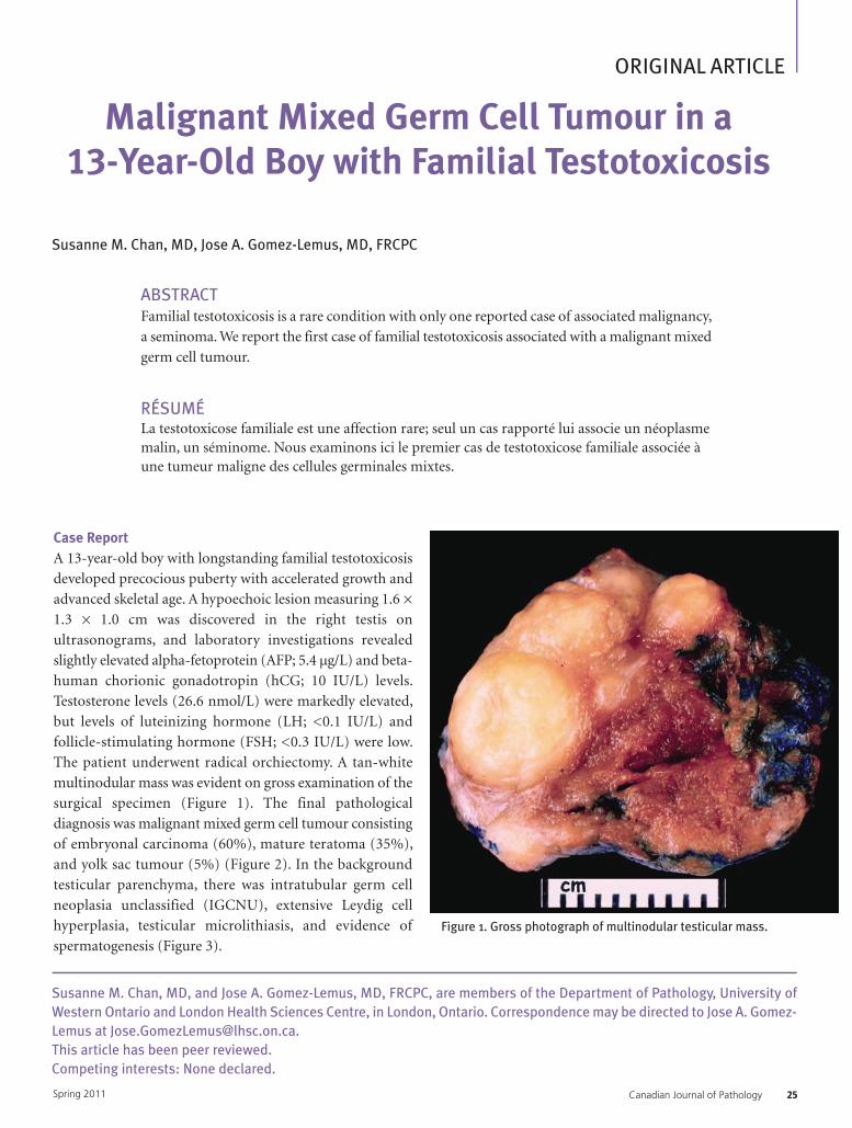
Canadian Journal of P athology 25Spring 2011
ORIGINAL ARTICLE
Malignant Mixed Germ Cell Tumour in a 13-Year-Old Boy with Familial Testotoxicosis
Susanne M. Chan, MD, and Jose A. Gomez-Lemus, MD, FRCPC, are members of the Department of Pathology, University ofWestern Ontario and London Health Sciences Centre, in London, Ontario. Correspondence may be directed to Jose A. Gomez-Lemus at [email protected] article has been peer reviewed.Competing interests: None declared.
Susanne M. Chan, MD, Jose A. Gomez-Lemus, MD, FRCPC
ABSTRACTFamilial testotoxicosis is a rare condition with only one reported case of associated malignancy,
a seminoma. We report the first case of familial testotoxicosis associated with a malignant mixed
germ cell tumour.
RÉSUMÉLa testotoxicose familiale est une affection rare; seul un cas rapporté lui associe un néoplasmemalin, un séminome. Nous examinons ici le premier cas de testotoxicose familiale associée àune tumeur maligne des cellules germinales mixtes.
Case ReportA 13-year-old boy with longstanding familial testotoxicosis
developed precocious puberty with accelerated growth and
advanced skeletal age. A hypoechoic lesion measuring 1.6 ×
1.3 × 1.0 cm was discovered in the right testis on
ultrasonograms, and laboratory investigations revealed
slightly elevated alpha-fetoprotein (AFP; 5.4 µg/L) and beta-
human chorionic gonadotropin (hCG; 10 IU/L) levels.
Testosterone levels (26.6 nmol/L) were markedly elevated,
but levels of luteinizing hormone (LH; <0.1 IU/L) and
follicle-stimulating hormone (FSH; <0.3 IU/L) were low.
The patient underwent radical orchiectomy. A tan-white
multinodular mass was evident on gross examination of the
surgical specimen (Figure 1). The final pathological
diagnosis was malignant mixed germ cell tumour consisting
of embryonal carcinoma (60%), mature teratoma (35%),
and yolk sac tumour (5%) (Figure 2). In the background
testicular parenchyma, there was intratubular germ cell
neoplasia unclassified (IGCNU), extensive Leydig cell
hyperplasia, testicular microlithiasis, and evidence of
spermatogenesis (Figure 3). Figure 1. Gross photograph of multinodular testicular mass.

Spring 201126 Canadian Journal of P athology
MALIGNANT MIXED GERM CELL TUMOUR
DiscussionIn familial testotoxicosis, an autosomal-dominant mutation
in the LH receptor gene leads to early and constitutive
activation of the receptors, resulting in elevated testosterone
levels and precocious puberty.1 The most common mutation
in the United States is the Asp578Gly mutation, and occasional
sporadic mutations do occur.1 Although testosterone is
elevated, LH and FSH levels remain at prepubertal levels,
making testotoxicosis a form of gonadotropin-independent
precocious puberty. Testotoxicosis is also sex limited in that
it affects only males since females require both FSH- and
LH-receptor stimulation for gonadal activation.2 Clinically,
males present with precocious puberty starting as early as
age 1–3 years, with premature testicular enlargement, penile
growth, development of secondary sexual characteristics,
and accelerated skeletal development.2,3 Early treatment in
the form of anti-androgens and aromatase inhibitors may
help delay or slow down the onset of puberty.2
Histologically, testicular Leydig cell hyperplasia, premature
spermatogenesis, Sertoli cells with complex cytoplasmic
differentiation, and Charcot-Böttcher crystals have been
described in testotoxicosis.3 Hyperplastic Leydig cell nodules
and adenomas are also not uncommon.4 However, only a
single case of associated malignancy, a seminoma in a 37-
year-old patient, has been previously reported.5
Our report is the first to describe a malignant mixed germ
cell tumour associated with testotoxicosis. In our case, the
patient had a background of ICGNU and microlithiasis,
both of which are predisposed to testicular germ cell
malignancies. Given that the average presenting age of
malignant mixed germ cell neoplasms is 30 years,6 the
development of malignancy in this patient at age 13 may
have been accelerated by early and sustained exposure to
testosterone resulting from his underlying testotoxicosis.
References1. Kremer H, Martens JW, van Reen M, et al. A limited repertoire of mutations
of the luteinizing hormone (LH) receptor gene in familial and sporadic
patients with male LH-independent precocious puberty. J Clin Endocrinol
Metab 1999;84:1136–40.
2. Wu S, Leschek EW, Rennert OM, et al. Luteinizing hormone receptor
mutations in disorders of sexual development and cancer. Pediatr Pathol Mol
Med 2000;19:21–40.
3. Gondos B, Egli CA, Rosenthal SM, et al. Testicular changes in gonadotropin-
independent familial male sexual precocity. Familial testotoxicosis. Arch Pathol
Lab Med 1985;109:990–5.
4. Leschek EW, Chan WY, Diamond DA, et al. Nodular Leydig cell hyperplasia
in a boy with familial male-limited precocious puberty. J Pediatr 2001;
38:949–51.
5. Martin MM, Wu SM, Martin AL, et al. Testicular seminoma in a patient with
a constitutively activating mutation of the luteinizing hormone/chorionic
gonadotropin receptor. Eur J Endocrinol 1998;139:101–6.
6. Adham WK, Raval BK, Uzquiano MC, et al. Best cases from the AFIP: bilateral
testicular tumors: seminoma and mixed germ cell tumor. Radiographics
2005;25:835–9.
Figure 2. Embryonal carcinoma and teratoma within the mixedgerm cell tumour. (Hematoxylin and eosin)
Figure 3. Background testicular parenchyma with intratubulargerm cell neoplasia unclassified (green arrowheads), extensiveinterstitial Leydig cell hyperplasia, spermatogenesis, andmicrolithiasis. (Hematoxylin and eosin)

Canadian Journal of P athology 27Spring 2011
Mathematical Modelling of Lymph Node Grossing Techniques
Etienne Mahe is a member of the Department of Pathology and Molecular Medicine at McMaster University Medical Centre,in Hamilton, Ontario. Correspondence may be directed to [email protected] article has been peer reviewed.Competing interests: None declared
Etienne Mahe, BSc, MD
ABSTRACTThe optimal histopathological handling of sentinel lymph nodes remains the subject of debate.
There is surprisingly little evidence in support of the manner in which sentinel lymph nodes
are grossed. Some authors recommend that a lymph node be sectioned parallel to the long axis
of the node, while others recommend that the short axis be used. In either case, the manner in
which a sentinel lymph node is grossed determines the cut surface area exposed for histological
examination and, in turn, the amount of area examinable for the presence of metastases. In this
article, mathematical models of each of these approaches are presented and compared from the
perspective of optimization of cut surface area. Computer modelling and simulations are also
performed in order to assess how variously sized lymph nodes should be optimally grossed and
to assess how frequently an optimal approach improves theoretical tumour focus detection.
The results favour the use of the long-axis approach.
RÉSUMÉLe débat fait toujours rage à propos de la préparation histopathologique optimale du ganglion
sentinelle. Étonnamment, peu de données probantes déterminent le mode de préparation de
ce ganglion. Des auteurs recommandent de le trancher en parallèle à son axe long, tandis que
d’autres privilégient la coupe perpendiculaire à cet axe. Quoi qu'il en soit, l’angle de coupe
détermine la surface exposée qui fera l’objet de l’examen histologique et, de ce fait, la taille de
la zone examinée pour détecter la présence de métastases. L’article présente des modèles
mathématiques des deux méthodes et les compare sur le plan de l’optimisation de la surface de
la coupe sectionnée. La modélisation et les simulations informatiques permettent d’évaluer la
préparation optimale de ganglions sentinelles de différentes tailles et de déterminer à quelle
fréquence la méthode optimale améliore la détection théorique de cellules tumorales. Les
résultats penchent pour la coupe parallèle à l’axe long.
ORIGINAL ARTICLE

Spring 201128 Canadian Journal of P athology
MATHEMATICAL MODELLING OF LYMPH NODE GROSSING TECHNIQUES
The microscopic examination of lymph nodes is often an
exercise in detection of tumour metastases. As outlined
in the TNM staging system, lymph node metastasis, as part
of the overall staging, is an indicator of tumour burden and
outcome.1 The effort devoted to the detection of small
tumour foci has increased because the standard of care in
many instances now requires sensitivity to isolated tumour
cells.2 Although the prognostic utility of a positive diagnosis
of isolated tumour cells in a lymph node generally, and a
sentinel lymph node specifically, remains debatable,3–5 a
recent meta-analysis has noted that the identification of low-
volume tumour metastasis (i.e., isolated tumour cells or
micrometastases) in a sentinel lymph node in breast cancer
cases is associated with non-sentinel lymph node disease in
10–15% of cases. It is logical, therefore, from both a patient
care and completeness perspective, that the identification of
anymetastases, whether in the form of isolated tumour cells,
micrometastases, or macroscopic foci, should be the
ultimate goal of the pathological examination of a sentinel
lymph node. As the literature indicates, however,
maximizing the probability that this goal is achieved, and in
the greatest number of instances, is both heuristically
difficult and open to debate on the basis of merit.3–7
As Weaver aptly notes in his review, a pathologist can never
be absolutely confident that a lymph node is negative for
disease, especially if he or she equates the presence of
isolated tumour cells with “positive” for metastasis.4 It
follows that a certain level of error must be accepted as
unavoidable, with the understanding that steps be taken to
mitigate this error whenever possible. It is this mitigation of
error that has sparked much research into the optimal
histopathological evaluation of a sentinel lymph node. As
long ago as 1948, it was recognized that a combination of
deeper sectioning and retrospective review of slides could
reduce the false-negative rate of lymph node evaluation.8
More recently, immunohistochemistry has become
important in detecting otherwise “occult” lymph node
metastases. Weaver et al. noted that a combination of deeper
sectioning and immunohistochemistry could improve the
sensitivity of lymph node evaluation, with 75% of false-
negative sentinel lymph nodes in their series containing
isolated tumour metastases.5 As much as deeper sections and
immunohistochemical stains have been shown to reduce
false-negative rates, the literature looking at the effect of the
specific grossing techniques employed on the detection of
nodal metastases is sparse.
Some sources have recommended that a lymph node be
grossed by means of serial sections parallel to the long axis
of the node; such a technique should maximize the
histological visualization of the lymphatic channels serving
the node and minimize the number of necessary tissue
blocks.3–9 Although intuitively logical, these arguments are
incomplete. First, although serial sections through the long
axis of a node may capture a number of lymphatic channels
in a single section, there is insufficient evidence to say that
multiple sections cut perpendicular to the long axis (and
submitted in the same cassette moreover) would not do the
same job as effectively. Furthermore, although the natural
desire to reduce the total number of tissue blocks submitted
for histological analysis is beneficial from a resource
management perspective, the orientation of grossing (i.e.,
whether cuts are made parallel or perpendicular to the long
axis) does not necessarily dictate the number of blocks since
multiple smaller slices submitted when cutting
perpendicular to the long axis may be placed in the same
cassette. Moreover, a single slice taken parallel to the long
axis may be so large as to necessitate multiple blocks. Finally,
the localization and identification of lymphatic channels
does not equate to the localization and identification of a
tumour deposit since the very biology of a lymph node sees
it acting as a sieve through which a tumour cell may trace
out a path before taking root and forming a tumour focus.
This latter point is exemplified by the seemingly random
scattering of nevus cells that may periodically be observed
in lymph nodes.
Still other authors have recommended that a sentinel lymph
node be grossed by means of sections cut parallel to the
short axis. In his recent review, Shidham noted that sentinel
lymph nodes in cases of melanoma need to be grossed
parallel to the short axis in order to maximize the delineable
circumference on histological sections.10 It is stated that,
since melanoma tends to form subcapsular tumour foci, an
extensive examination of the nodal circumference is
warranted, and Shidham provides a mathematical
derivation in support of his technique. Unfortunately, both
of these arguments can be easily refuted. First, the argument

Canadian Journal of P athology 29Spring 2011
MAHE
that visualization of the nodal circumference should take
precedence is faulted by the reality that tumour foci are not
linear but, rather, three dimensional; maximizing the
visibility of a uni-dimensional aspect of a tumour is
therefore illogical when histological slides present tumour
foci in two dimensions. Secondly, the mathematical
derivations presented in his article are incomplete, and the
summative effect of the circumference of multiple sections
is not adequately accounted for. It would seem, therefore,
that greater evidence in support of either of the grossing
techniques described above is needed.
Since a lymph node cannot be examined in its entirety, it
seems logical to try to maximize the cut surface area of the
lymph node upon grossing to maximize the area that may
be visible on a slide (and on subsequent deeper sections if
needed). Thus, it may be surmised that the optimal
detection of lymph node metastases may be improved if the
grossing technique maximizes the cut surface area of a
lymph node. From first principles there are three general
methods in which a lymph node may be sectioned: with
slices parallel to the long, short or intermediate axis. The
intermediate axis approach is generally not chosen since,
very simply, each slice taken in this manner will undercut
the maximal surface area and will fail to produce the
maximal number of slices. With respect to the other two
methods, one (the “long axis” approach; Figure 1) will
maximize the surface area of a given slice while sacrificing
the total number of slices; the other (the “short axis”
approach; Figure 2) will do the opposite. These two
techniques are the extremes that are compared in this article.
In either case, based on the College of American
Pathologists’ guidelines, sectioning begins with a central cut
followed by further cuts moving outwards from the centre,
with each tissue section taken at a thickness of 2 mm.3,4,9
MethodsA lymph node can be modelled mathematically by the
ellipsoid centred at the Cartesian origin with semi-axes a, b,
and c according to the following:
Figure 1. The long axis approach; sections taken parallel to thelong axis.
Figure 2. The short axis approach; sections taken parallel to theshort axis.

Spring 201130 Canadian Journal of P athology
MATHEMATICAL MODELLING OF LYMPH NODE GROSSING TECHNIQUES
This translates to a lymph node with long, short, and
intermediate axis lengths, respectively, 2c, 2a, and 2b. This
model need not take into account the perfect sphere since
in this case the node would be grossed identically from any
perspective. Other shapes such as the oval or the kidney-
bean shape are not rigorously subsumed within this model
since their equations are more complicated and cannot be
easily described in Cartesian coordinates. In real life,
however, the oval or bean-shaped lymph nodes can be
approximated as ellipsoids since they usually have some
amount of surrounding fat; in these cases the long, short,
and intermediate axes can be measured and the shape
approximated as an ellipsoid.
When sectioning the lymph node parallel to the short axis,
surfaces with traces parallel to the x,y-plane are obtained;
likewise, when sectioning the lymph node perpendicular to
the long axis, surfaces parallel to the y,z-plane are obtained
(see Appendix 1 at http://cap-acp.org/).
For each section in the short axis approach, the area of the
trace ellipse is given by equation (6) of Appendix 1; the
converse long axis approach is given by equation (7). When
the cut surface areas exposed upon sectioning the entire
node are summed, the following are obtained from
equations (9) and (10):
Total Cut Surface Area, Short Axis Approach =
Total Cut Surface Area, Long Axis Approach =
(The angled brackets here refer to the floor function; see
Appendix 1 at http://cap-acp.org/ for details.)
By comparing the values of the two equations for an
individual node, the theoretical optimal surface area for each
grossing technique can be determined. The specific
technique producing the larger theoretical surface area
should be chosen to maximize the optimal cut surface area
available for histological evaluation.
To further analyze the optimal surface areas for each
technique for nodes of various sizes, a computer analysis was
performed to assess the optimal cut surface area of a
hypothetical lymph node using either of the grossing
techniques. In most laboratories, grossing is performed with
a metric precision of 1 mm (i.e., the smallest gradations of
grossing room rulers can be assumed to be 1 mm).
Furthermore, most histopathology tissue cassettes can
contain a maximal linear length of approximately 4 cm.
Thus, using a range of axis lengths from 1 mm to 40 mm in
increments of 1 mm, a reasonable approximation of possible
lymph node sizes was generated. This was performed with
the aid of the mathematical software MATLAB (see
Appendix 2 at http://cap-acp.org/ for the code script).
In order to test the potential improvement that the optimal
grossing strategies noted above might have on tumour focus
detection, a series of computer simulations were performed
to test each grossing technique relative to the other (see code
script from Appendix 3 at http://cap-acp.org/). It was
surmised that the optimal grossing method should more
frequently permit transection of a hypothetical tumour
focus, thus closely approximating the likelihood the tumour
focus will be detected on histological examination. In each
simulation, 1,000 mathematically modelled lymph nodes
were created with random measurements obtained using
MATLAB’s built-in random number generator. This
number of lymph nodes per simulation, by the law of
averages, should adequately account for most possible
lymph node sizes as set forth in the matrix of lymph node
sizes above. In each lymph node, a focus of metastasis was
simulated and assigned a random location (excluding the
lymph node’s surface) and random size (<0.2 mm to limit
the discussion to tumour foci smaller than
micrometastases). The program actively excluded a node
that was exactly spherical since, in such a case, the computer
would be unable to discern an optimal grossing technique.
The lymph node then underwent simulated grossing with
cuts parallel either to the long axis or short axis starting from
the centre and moving out. The program then determined
which of the two methods was optimal based on the above
calculations, calculated whether a grossing cut using either
method would transect the tumour focus, and tallied

Canadian Journal of P athology 31Spring 2011
MAHE
whether the optimal method, the non-optimal method, or
either method worked best. The simulations were repeated
a total of 1,000 times to permit the generation of statistical
data.
ResultsBy comparing the total optimal surface area for each of the
short axis and long axis approaches, a 40 × 40 diagonally
symmetric matrix was generated. As Figure 3 indicates, of
the 820 unique lymph node sizes, the long axis approach
(pink) was optimal in 802 cases (98%). The remaining cases
were either equivalent by either approach (grey) or showed
optimal surface area using the short axis approach (blue).
Furthermore, a specific pattern of repetition was noted in
the few cases of short axis preference: the short axis
technique was preferred when the long and short axes had
measurements 4n + 1 and 4n, respectively, or 4n+2 and 4n,
respectively (for n ≥ 1).
One thousand simulations, with 1,000 simulated lymph
nodes each, were conducted with each node bearing a
randomly positioned and sized tumour deposit (smaller
than a microfocus). An average of 87.7 lymph node tumour
foci showed grossing transection using the optimal
approach; an average of 86.7 lymph node tumour foci
showed grossing transection using the non-optimal
approach. The two-tailed t-test showed this to be a
statistically significant difference of means (p =.010).
Furthermore, on average 13.7 lymph nodes were found to
be equivocal to either technique. Notably, the number of
lymph nodes whose tumour focus was not transected by
either grossing technique was quite high, averaging at 811.9
(81%). This latter result is in keeping with the previous
studies in which the frequency of missed tumour foci
increased when the size cutoff was reduced.5
DiscussionAlthough debate persists as to the extent to which efforts to
identify low-volume tumour foci in sentinel and non-
sentinel lymph nodes should proceed, on a case-by-case
basis, most pathologists will be loath to miss a metastasis,
however small. Research into the best-practice handling of
lymph nodes persists, and the focus of this research tends to
be centred on maximizing the efficiency of histological and
ancillary techniques at detecting metastatic foci, with the
caveat that, from a practical perspective, a lymph node
cannot be fully and completely examined microscopically.
Little research has examined the manner in which a lymph
node is grossed, however, and its potential impact on
maximizing the detection of metastases.
The present study uses a mathematical model of a lymph
node to calculate the maximum optimal cut surface area
available for histological examination when the grossing
technique is varied from either parallel to the long axis or
parallel to the short axis. A comparison of these two
approaches reveals that the optimal grossing technique is the
long axis approach in 98% of simulated lymph nodes of
varying sizes. In the great majority of cases, therefore, the
approach described as optimal by Weaver and others is
sound. A small number of predictable cases, however, do
seem to show optimal grossing using the short axis approach,
as described above. Nonetheless, when a simulation of
mathematically modelled lymph nodes is performed to test
the optimal and non-optimal grossing techniques head to
head, only a marginal improvement in theoretical detection
is noted, although this is statistically significant.
Figure 3. A 40 × 40 matrix of lymph node measurements; pinkindicates optimal long axis approach, blue indicates optimal shortaxis approach, and grey indicates no difference in taking eitherapproach.

Spring 201132 Canadian Journal of P athology
MATHEMATICAL MODELLING OF LYMPH NODE GROSSING TECHNIQUES
A “real-life” study to verify the above conclusions would be
ideal but is highly unlikely. The resources required to
adequately study even one lymph node in its entirety are a
significant impediment. Furthermore, as pointed out in
Weaver’s application of the Heisenberg uncertainty
principle, real-life limitations make the impact of an
individual lymph node’s questionable diagnosis of benignity
impossible to assess; simulation, therefore, is the only
plausible mode of study in this vein. The current study
appears to be the strongest evidence thus far supporting the
lymph node grossing practices described by Weaver et al.5
References1. Edge S, Byrd DR, Comptom CC, et al. Purposes and principles o f cancer
staging. In: AJCC Cancer Staging Manual, 7th edition. New York: Springer;
2010:3–14.
2. Tan JT, Bagnell M, Morgan JW, et al. The identification and treatment of
isolated tumor cells reflect disparities in the delivery of breast cancer care. Am
J Surg 2009;198:508–10.
3. Weaver DL. Pathology evaluation of sentinel lymph nodes in breast cancer:
protocol recommendations and rationale. Mod Pathol 2010;23:S26–32.
4. Weaver DL. The prognostic importance of isolated tumor cell clusters and
micrometastases in sentinel lymph nodes. Cancer Investig 2009;27:121–8.
5. Weaver DL, Krag D, Manna EA, et al. Detection of occult sentinel lymph node
micrometastases by immunohistochemistry in breast cancer: an NSABP
protocol B-32 quality assurance study. Cancer 2006;107:661–7.
6. Cserni G, Gregori D, Merletti F, et al. Meta-analysis of non-sentinel node
metastases associated with micrometastatic sentinel nodes in breast cancer. Br
J Surg 2004;91:1245–52.
7. Park D, Karesen R, Naume B, et al. The prognostic impact of occult nodal
metastasis in early breast carcinoma. Breast Cancer Res Treat 2009;118:57–66.
8. Saphir O, Amromin GD. Obscure axillary lymph node metastases in
carcinoma of the breast. Cancer 1948;1:238–41.
9. Lester SC. Lymph nodes, spleen and bone marrow. In: Manual of Surgical
Pathology, 3rd edition. Philadelphia (PA): Elsevier Saunders; 2010:513–24.
10. Shidham VB. Evaluation of sentinel lymph nodes for melanoma metastases
by pathologists. Clin Plast Surg 2010;37:101–12.
McGill Cytopathology Review Course - NEW PROGRAM
April 30 - May 3, 2011Montréal, Québec, Canada
Course Director: Dr. Manon Auger
For further information contact:Email: [email protected]
McGillUniversityHealth CentreContinuing Education Office
Tel: (514) 934-8253Fax: (514) 934-1779
web site: www.muhc-cme.mcgill.ca

Canadian Journal of P athology 33Spring 2011
Interpretation of Diagnostic Muscle Biopsies
Michael Farrell is a consultant neuropathologist in the Department of Clinical Neurological Sciences at the Royal College ofSurgeons in Ireland and Beaumont Hospital, in Dublin, Ireland. Correspondence may be directed to [email protected] article has been peer reviewed.Competing interests: None declared.
Michael Farrell, FRCPI, FRCPC, FRCPath
ABSTRACTThis review of muscle biopsy interpretation is directed at surgical pathologists who are willing
to take up the challenge. The approach to muscle biopsy interpretation follows an inside-out
algorithm designed to achieve maximum diagnostic information from a well-prepared
specimen, rather than providing the specific diagnostic label, which, even after detailed
biochemical and genetic investigations, may elude even the most experienced myologists.
RÉSUMÉL’examen de l’interprétation des résultats de la biopsie musculaire s’adresse aux pathologistes
spécialisés en pathologie chirurgicale qui sont disposés à relever le défi. La démarche
d’interprétation est balisée par un algorithme conçu pour obtenir l’information diagnostique
optimale du prélèvement effectué dans les règles, plutôt que pour déboucher sur le diagnostic
précis, lequel peut échapper même au myologiste le plus chevronné après les analyses
biochimiques et génétiques approfondies.
INVITED REVIEW
The past 10 years have witnessed spectacular advances in
the characterization of many hitherto unclassified
muscle disorders, largely due to an expanding range of
antibodies directed against muscle membrane and
sarcoplasmic targets.1–4 At the outset, it is vital to stress that
isolated specific diagnostic muscle biopsy appearances are
rare. Instead, one tries to build a composite picture using a
wide panel of stains and immunohistochemical reactions
that allows a stratification of the biopsy into one of a small
number of clinically relevant diagnostic categories (Table 1).
Regular clinical-pathological muscle review meetings are
essential for feedback, learning, and diagnosis refinement.
Choice of Biopsy Site and Transportation to LaboratoryClinicians regularly seek advice from the laboratory on the
optimum biopsy site and biopsy size. In patients with chronic
muscle disease, it is best to avoid very weak muscles.
Ultrasonography is useful for ensuring the muscle is not
completely replaced by fat. In the absence of ultrasonography,
a Medical Research Council (MRC) scale three-quarter strength
muscle is preferred. If muscle disease is acute, a profoundly
weak muscle is most likely to yield maximum diagnostic
information. The gastrocnemius is best avoided as clinically
irrelevant neuropathic change from lumbar spinal degenerative
disc disease may be present, and this muscle often shows fatty
replacement especially in patients with so-called calf
hypertrophy. The decision to carry out open or needle biopsy
is determined by operator experience, but considerable
technical laboratory expertise is required to orientate the small
muscle samples obtained by needle. Small sample size may limit
the range of any additional investigations required, and some
muscles, because of proximity to nerves and blood vessels, are
unsuitable for needle biopsy. Obviously, injection or needle
electromyography (EMG) sites must be avoided. At Beaumont
Hospital, in Dublin, Ireland, we prefer open biopsy and
recommend that a cylinder of muscle 5 × 10 mm be obtained.
The muscle should be received in the laboratory within 2–3
hours of biopsy and is best transported on saline-moistened
(not soaked) gauze without pinning or stretching. Even if
delayed, it is still possible to obtain high-quality histochemistry
for up to 24 hours, provided that muscle freezing, soaking, or
exposure to formalin is avoided. It may be very helpful to

Spring 201134 Canadian Journal of P athology
INTERPRETATION OF DIAGNOSTIC MUSCLE BIOPSIES
orientate the muscle transversely using a dissecting microscope.
Ice crystal artifact is avoided by dusting the muscle in talcum
powder prior to immersion in liquid nitrogen. We do not find
it necessary to use pre-cooled isopentane. A small portion of
muscle is dissected and immersed in glutaraldehyde (prior to
freezing) for electron microscopy (EM). The archived frozen
tissue block is suitable for respiratory chain studies provided
citrate synthase levels have not fallen too low. In general,
formalin should be avoided, but when vasculitis or
granulomatous disease is suspected, all of the muscle must be
processed for microscopy. In this situation, we usually place any
non-frozen muscle fragments in formalin for serial sectioning.
Reading the Biopsy: A Systematic Approach Unless an orderly approach is adopted, one can easily miss
important changes. It is vital to remind oneself that the muscle
biopsy only provides a single snapshot of an evolving dynamic
process. A well-stained hematoxylin and eosin (H & E) section
often provides the key diagnostic information, which is
subsequently endorsed by additional histochemical or
Table 1. Muscle Biopsy Interpretation: Category-Based Diagnosis
NormalUniform fibre size; zero inflammation, necrosis, fibrosis, and intactmosaic pattern on fibre typing.
End-Stage Muscle Fat and fibrous tissue with rare muscle fibres.i. Depending on history, might be suitable for membraneimmunohistochemistry; otherwise, sign out as “inadequatespecimen”
NeuropathicFibre atrophy: check fibre typing and determine if atrophy is oneof the following: i. Random – i.e., both fibres types involvedii. Type specific
1. Type 2 only: steroids – disuse2. Type 1 only: myopathy (see “Myopathic,” below)
InflammatoryDetermine composition (T and B lymphocytes, eosinophils, andmacrophages), distribution of infiltrate, and consider effects ofprevious modifiers, e.g., steroids. Inflammation may be patchy.Scrutinize non-inflammatory areas to eliminate underlyingdystrophy. Proliferating sarcolemmal nuclei may simulateinflammation.i. Targeted at fibres – polymyositisii. Perifascicular atrophy – dermatomyositisiii. Not easily categorized – mixed connective tissue disease;
check for myositis specific antibodiesiv. Fascial – fasciitisv. Vessel based – vasculitisvi. Think of IBM – but remember IBM is not an inflammatory disease
DystrophicEndomysial fibrosis; fibre necrosis with regeneration; variableinflammation (see discussion of fibre atrophy in “Neuropathic,”above); central nucleation; fibre hypertrophy and splitting.Variable features include rods, cores, vacuoles, sarcoplasmicbodies, and ring fibres. Immunohistochemistry ± Western blot.
i. Sarcolemmal membrane1. Dystrophin 2. Sarcoglycans3. Merosin4. Dysferlin5. Caveolin
ii. Sarcolemmal nuclei 1. Emerin2. Lamin A/C 3. Myotubular if many central nucleated fibres; also think
of myotonic dystrophyiii. Sarcoplasm
1. Myofibrillar – cytoskeletal, structurala. Actinopathies, core disease, nemaline, telethionin,
myosin heavy chain, desminiv. Ion channel/transporter (unlikely to be biopsied)
1. Cl–; CA++; K+; Na+ ; ryanodine receptor; R-adenosinetriphosphatase
Myopathic Fibre size variation; absence of main features in “End-StageMuscle,” “Neuropathic,” and “Inflammatory,” above. Histologydictated by speed of disease progression ranging from widespreadfibre necrosis in acute rhabdomyolysis to minimal variation in fibresize in “non-specific minimal change myopathy.” Look for raggedred fibres, cytochrome oxidase depletion and glycogen or lipidexcess. Check myophosphorylase. Think of process.i. Energy provision
1. Glycogen metabolism; lipid transport and metabolism; mitochondrial structure, function and metabolism
ii. Drug induced1. Consider statins in every myopathy
a. Vacuoles: amiodarone, colchicine, chloroquine cyclosporine, and drugs causing low K+
b. Ragged red fibres: zidovudinec. Type II fibre atrophy: steroidsd. Neuropathic features: colchicine, chloroquine, and
hydroxychloroquineiii. Biopsy from intensive care – critical care
myopathy/neuropathy iv. Toxinsv. Infectionvi. Ischemic
1. Compartment syndrome

Canadian Journal of P athology 35Spring 2011
FARRELL
immunohistochemical stains. We favour an “inside out”
approach in which fibres are examined first, followed in order
by the endomysium, perimysium, and epimysium and lastly by
the blood vessels.
Fibre SizeIn the assessment of fibre size, care must be taken to ensure that
the biopsy is orientated transversely. Oblique sectioning results
in non-pathological fibre size variation. Physiological fibre size
variation is found close to muscle fascia. In general, an absence
of fibre size variation usually indicates minimal or no
significant muscle disease.
The presence of randomly distributed atrophic angulated fibres
is suggestive of early denervation, but confirmation of the
random nature of fibre atrophy must be established (see below).
Clusters of angulated atrophic fibres, (grouped atrophy) are
diagnostic of denervation even without recourse to fibre typing.
Fibre size variation may also be due to hypertrophy, a feature
usually observed in the dystrophies. Hypertrophied fibres may
undergo splitting, which exaggerates the degree of fibre size
variation; but the split fibres are always of the same type as the
parent hypertrophied fibre, which helps to distinguish split
fibres from truly atrophic fibres. When atrophy is prominent at
the edge of muscle fascicles, dermatomyositis (DM) should be
considered, the perifascicular atrophy in this instance being
attributed to peripheral fascicular ischemia secondary to small-
vessel immune-mediated disease (Figure 1).
Fibre typing is essential to determine if the atrophic process is
random, that is, affecting both type I and II fibres, or selective.
Random fibre atrophy is diagnostic of denervation without re-
innervation. Selective type II fibre atrophy may be seen in disuse
or following prolonged steroid therapy or sometimes in early
denervation. Selective type I fibre atrophy is a feature of
myotonic dystrophy and the congenital myopathies.5
An eyepiece graticule may be used to assess lesser fibre diameter.
In general, precise measurement of fibre size is not necessary
in adult muscle. In children, especially up to 3 years of age, it is
advisable to calculate the mean lesser fibre diameter for at least
100 fibres. Diagnosis of denervation in these early years is
difficult, and the implications of such a diagnosis is so serious
that biopsies from children of this age should be processed in a
centre that has experience in the interpretation of children’s
muscle biopsies. A mistaken diagnosis of spinal muscle atrophy
(SMA) could result in withdrawal of life support systems from
an infant whose muscle weakness and respiratory failure are
incorrectly attributed to denervation. However, availability of
rapid testing for the survival motor neurone (SMN) gene has
replaced muscle biopsy as the first line of investigation in
children with possible SMA.6
Re-innervation resulting in enlarged motor units and due to
sprouting of distal intramuscular nerve terminals is a diagnostic
feature of denervation on EMG and is identified
microscopically as fibre type grouping. Antibodies to myosin
heavy-chain fast and slow isoforms have been refined to the
point where even fibres of intermediate type can be discerned
using routine immunohistochemistry. Additionally, both
antibodies are capable of identifying fibre types in formalin-
fixed paraffin-embedded (FFPE) muscle. Normal fibre type
distribution has a mosaic or checkerboard pattern. Loss of the
mosaic pattern may be due to a predominance of one type over
another, and in some muscles this is physiological. Fibre type
grouping is the hallmark of re-innervation, and in our hands a
group of fibres usually includes at least 16 fibres. Fibre grouping
is also considered present if two touching fibres are completely
surrounded on all sides by fibres of the same type. Grouping of
both type I and type II fibres is essential for a definitive
diagnosis of denervation. A diagnosis of denervation cannot be
established when grouping involves one fibre type only, that is,
the process is non-random.
Sarcolemmal NucleiAs fetal muscle matures, sarcolemmal nuclei migrate to a
peripheral location beneath the sarcolemmal membrane.
Figure 1. Perifascicular atrophy in the fibres located along theperimysial seam (top left to bottom right). (Hematoxylin andeosin)

Spring 201136 Canadian Journal of P athology
INTERPRETATION OF DIAGNOSTIC MUSCLE BIOPSIES
Persistence of central nuclei is abnormal when more than 4%
of fibres are involved. In myotubular myopathy, the vast
majority of fibres have central-located nuclei, though a similar
predominance of centrally nucleated fibres may be seen in
myotonic dystrophy (Figure 2). Central nucleation is not
specific and may be seen in chronic denervation, myopathy, and
dystrophy. Increased numbers of sarcolemmal nuclei are
frequent in atrophic fibres and may be exaggerated by the
increased packing density associated with fibre atrophy (Figure
3). Perhaps the most common mistake in reading muscle
biopsy is to interpret the densely packed, small, deeply
basophilic nuclei as a lymphocytic infiltrate, but the issue is
easily resolved by application of a lymphocytic marker such as
CD45.
SarcoplasmType I fibres are usually darker than type II fibres because of
the increased numbers of mitochondria. Deeply basophilic
sarcoplasm is typical of muscle fibre regeneration, which can
be confirmed with fetal myosin immunohistochemistry.
Regenerating fibres usually have large, pale vesicular nuclei.
Myofibre necrosis is, like many muscle biopsy appearances,
non-specific. However, its histological recognition is critical.
Sarcoplasmic pallor is suggestive of necrosis, and although
necrosis is typically a feature of myopathy, it may also be present
in acutely denervated muscle. As necrosis progresses, fibres
become intensely eosinophilic and the integrity of the
sarcolemmal membrane is compromised (Figure 4). Spectrin
immunohistochemistry confirms membrane disruption. Later,
necrotic fibres are replaced by macrophages, a finding that can
easily be over-interpreted as evidence for a primary
inflammatory process. The presence of several necrotic fibres
lying close together suggests either a dystrophic process or
perhaps a severe ischemic insult, provided non-necrotic fibres
elsewhere look normal. Isolated spotty myofibre necrosis is
more likely to reflect a toxic-metabolic process. Polymyositis
(PM) and DM often have necrotic fibres as one element in an
overall inflammatory process, the extent of which may have
been modified by prior steroid therapy.7,8 The relative
prevalence of necrotic and regenerating fibres varies, depending
on the phase of the primary disease process; for instance,
myofibre regeneration may be minimal in late Duchenne’s
muscular dystrophy (DMD).
Sarcoplasmic VacuolesPathological sarcoplasmic vacuolar change must be
distinguished from ice crystal vacuoles, which are usually
empty, non-membrane bound, and involve the entire muscle
fascicle. Any sarcoplasmic structure may undergo dilatation and
give rise to vacuoles. At light microscopic examination, it is not
possible to differentiate between vacuolar change due to
dilatation of the T tubules, sarcoplasmic reticulum, or
lysosomes. Stains for lipid and glycogen help to determine the
contents of vacuoles. Vacuoles may vary in size from single and
Figure 2. Virtually all fibres show central nucleation in this patientwith myotubular myopathy. Note the slight increase in endomysialconnective tissue. (Hematoxylin and eosin)
Figure 3. Angulated, atrophic fibres with very prominentsarcolemmal nuclei simulating an inflammatory infiltrate.(Hematoxylin and eosin)

Canadian Journal of P athology 37Spring 2011
FARRELL
large, for example, in hypokalemic periodic paralysis, to
multiple and small, characteristic of lipid excess. Rimmed
vacuoles (Figure 5) have an irregular configuration with
increased peripheral staining using Gomori’s modified
trichrome (GMT). Large vacuoles, which react with acid
phosphatase and periodic acid–Schiff (PAS) stain, are
characteristic of acid maltase deficiency (Figure 6). The most
frequently encountered vacuoles are autophagic vacuoles,
which contain sarcoplasmic degradation products and are best
demonstrated with acid phosphatase. These are non-specific
indicators of muscle fibre degeneration.
Endomysium, Perimysium, and EpimysiumInflammation and fibrosis are the two principal pathological
findings in the endomysium, perimysium, and epimysium.
Endomysial fibrosis is always abnormal and is a dominant
feature of muscular dystrophy, especially congenital muscular
dystrophy, but may also be seen in end-stage denervation and
end-stage myopathy. Congenital muscular dystrophy, an
imprecise term, refers to infants, weak from birth, often with
contractures and usually with delayed motor milestones, and
in whom a neurogenic cause has been excluded. Perimysial
fibrosis is of lesser diagnostic importance, especially if not
accompanied by endomysial fibrosis. An epimysial
inflammatory infiltrate raises the possibility of fasciitis. Fascial
inflammation may suggest eosinophilic fasciitis or eosinophilic
myalgia syndrome.9 When the inflammatory cells are centred
on small endomysial vessels, DM is probable; when centred on
individual myofibres, PM is likely (Figure 7). Lymphocytes
dominate the inflammatory process in PM and DM with
endomysial CD8+ T cells prominent in PM and B cells
prominent in DM.7,8 Demonstration of the complement
membrane attack complex (MAC) C5b-9 in the vasculature
favours DM rather than PM.10,11 Steroid treatment and regional
variability in disease expression may influence the intensity of
the inflammatory infiltrate, and in some biopsies there may be
little or no inflammation. In such situations, expression of
sarcolemmal major histocompatibility complex (MHC) class I
antigens provides supportive evidence of an immunological
process at work.12 Endomysial inflammation may occur in
certain dystrophies, especially in facioscapulohumeral
dystrophy.13 Careful examination of fibres remote from the
inflammation reveals other supportive dystrophic features not
present in PM or DM. Great care must be taken to separate
inclusion body “myositis” (IBM) from other true inflammatory
myopathies. IBM is a degenerative rather than an immune-
mediated disease, and it does not respond to steroids, which are
actually contraindicated.14,15 The temptation to lump all
biopsies with an inflammatory component into a “myositis”
category must be resisted and other supportive features of IBM
sought – including rimmed vacuoles and elements of
mitochondrial dysfunction such as cytochrome oxidase (COX)
depletion and ragged red fibres (RRFs). A prominent
eosinophilic component suggests a drug-induced myopathy,
Figure 4. At least four necrotic fibres are present, and in all, thereis peripheral sarcoplasmic basophilia, indicating earlyregeneration. Note the minor degree of ice crystal artifact in intactfibres. (Hematoxylin and eosin)
Figure 5. Rimmed vacuoles characterized by peripheral granularbasophilic stippling in a patient with inclusion body myositis.(Hematoxylin and eosin)

Spring 201138 Canadian Journal of P athology
INTERPRETATION OF DIAGNOSTIC MUSCLE BIOPSIES
eosinophilic myalgia syndrome, or parasitic infestation.
Myositis dominated by eosinophils should trigger an
examination of multiple levels for the presence of parasitic
larvae, especially trichinosis and cysticercosis. Foci of
calcification and granulomatous inflammation may be all that
remains. Granulomatous inflammation16 suggests either
tuberculosis or sarcoidosis.17,18 Mycobacterium avium-
intracellulare infection of muscle is described in human
immunodeficiency virus infection.19 Granulomas may be
present in up to 60% of patients with sarcoid but are usually
asymptomatic. Similarly, asymptomatic granulomas may be
present in muscles obtained from patients with Crohn’s disease.
When an inflammatory biopsy is not easily characterized as
either PM or DM, less common inflammatory disorders such
as anti-synthetase syndrome should be considered and
supported by screening for myositis-specific antibodies.20,21
Blood VesselsA diagnosis of vasculitis is easy when classic features of vasculitis
are present in an endomysial blood vessel. Typically, however,
patients may already have been treated with steroids and the
inflammatory process may be extremely patchy and vary in its
severity and extent. Nevertheless, whenever a perivascular
lymphocytic infiltrate encroaches on a vessel wall, vasculitis
must be suspected. In polyarteritis nodosa, the infiltrate
comprises lymphocytes, histiocytes, eosinophils, and
neutrophils.22 Frequently, the perivascular infiltrate is less
pleocytic. Multiple levels must be examined and evidence of
vessel wall destruction sought. Where tissue is available,
demonstration of immune complexes in the vessels may be
helpful.
Having carefully examined the H & E–stained section, one
already has a firm view of the categories of muscle disease most
appropriate for the biopsy findings: normal; atrophic process
awaiting fibre typing; inflammatory awaiting characterization
of the inflammatory infiltrate; dystrophic awaiting
identification of deficient proteins in muscle membrane or
myonuclei; and non-specific myopathic awaiting further
characterization with special stains.
Special StainsIn a non-specialist laboratory, a combination of modified GMT,
PAS with and without diastaste, oil red O, myosin
immunohistochemistry, nicotinamide adenine dinucleotide
dehydrogenase tetrazolium reductase (NADH-TR) and COX
with or without succinic dehydrogenase (SDH) is adequate for
most diagnoses.
Gomori’s Modified TrichromeGMT is a capricious stain that is considered satisfactory when
fine sarcoplasmic brown-red granular staining is observed. A
well-executed GMT stain can be judged by its ability to allow a
distinction between type I and type II fibres. RRFs are irregular,
dark red-brown subsarcolemmal deposits representing an
accumulation of mitochondria (either normal or abnormal)
beneath the membrane and are typical of underlying
Figure 6. A, Acid maltase deficiency. Vacuolar change in which at least three centrally located non-rimmed vacuoles are present. Notethe single-rimmed vacuole (top right of centre) (hematoxylin and eosin). B, The vacuoles seen in panel A are periodic acid–Schiff positive.
A B

Canadian Journal of P athology 39Spring 2011
FARRELL
mitochondrial dysfunction (Figure 8). Sometimes there is a
mild increase in subsarcolemmal staining with GMT that is
above normal but that has not yet resulted in RRF formation.
The significance of this change remains to be determined.
Further investigation for possible mitochondrial disease is a
highly specialized task that includes analysis of respiratory chain
function and mitochondrial deoxyribonucleic acid (mtDNA),
as well as genes involved in mtDNA synthesis.23–26 Archived
frozen muscle is perfectly adequate for all of these specialized
studies. RRF must be distinguished from tubular aggregates,
which also accumulate in the subsarcolemmal location and
stain red with GMT. Tubular aggregates do not have the ragged
appearance of RRF, and their tubular structure is readily evident
on electron microscopy (EM). They are not specific for any
disease process.
Though nemaline rods are said to stain red with GMT, they are
usually so dark that it becomes impossible to say whether they
are dark green or maroon. Typically located in the
subsarcolemmal zone, they vary in number and distribution
(see Figure 9 at http://cap-acp.org/). EM shows continuity
between the rods and the Z lines. Rods are diagnostic of
nemaline myopathy, a heterogenous condition that may be
congenital, with up to seven forms identified.27–29 In the most
common form, a-actin accumulates in the rods and mutations
in the ACTA1 gene must be sought. Sporadic forms of nemaline
myopathy range from a congenital, rapidly fatal form through
to a non-progressive adult form with minimal disability. In the
majority of these sporadic forms, the rogue muscle protein has
not been identified. Rarely isolated rods may occur in the
absence of any significant muscle disease.
Periodic Acid–Schiff Stain The assessment of glycogen content of muscle is difficult as
there can be tremendous variation in the staining intensity of
the routine PAS stain. Like GMT, a satisfactory PAS allows
separation of type I and type II fibres. Acid maltase deficiency
(type II glycogenosis) is characterized by a variable number of
PAS-positive vacuoles (see Figure 6).30Acid phosphatase activity
is prominent close to the vacuoles. In McArdle disease (type V
glycogenosis), glycogen accumulation may be minimal; and
unless myophosphorylase staining is carried out routinely, the
diagnosis of McArdle disease is missed. The normal
myophosphorylase reaction fades quickly but not in blood
vessels, which serve as an internal control. Normal muscle
should also be included as a control. Nevertheless, the freshly
stained section should be examined immediately; the absence
of myophosphorylase except in blood vessels establishes the
diagnosis. The glycogen storage disorders usually present with
exercise intolerance or muscle cramps and fatigue and must
always be included in the differential diagnosis of
myoglobinuria.
Oil Red OLarge droplets overlying muscle fibres are frequently
encountered with oil red O but are of no significance. Typically,
sarcoplasmic lipid is visible as small droplets. Excess lipid may
be present in muscle as a result of obesity, but the lipid that
accumulates because of dysfunctional lipid transportation
across membranes (carnitine or carnitine palmityltransferase
deficiency), defective acyl-coenzyme A dehydrogenase, or
reduced functionality of the respiratory chain is typically
intense, striking, and not easily missed (see Figure 10 at
http://cap-acp.org/).31 Confirmation of lipid excess by EM
prompts an investigation of fatty acid metabolism, which is best
carried out in a specialist metabolic unit.
Nicotinamide Adenine Dinucleotide Dehydrogenase Tetrazolium Reductase The NADH-TR stain should not be used for assessment in fibre
type differentiation as blurring of the distinction between type
I and type II fibres may be present. This stain is very useful for
the assessment of mitochondrial myopathies, where increased
subsarcolemmal staining represents an accumulation of
Figure 7. Polymyositis in which lymphocytes are targeted on andbeginning to invade a single muscle fibre. (Hematoxylin andeosin)

Spring 201140 Canadian Journal of P athology
INTERPRETATION OF DIAGNOSTIC MUSCLE BIOPSIES
mitochondria corresponding in location to the RRFs (ragged
blue fibres). The use of NADH-TR is the method of choice for
the demonstration of cores, the hallmark of core myopathy.32,33
Tubular aggregates are also characterized by increased
subsarcolemmal staining. Where it is difficult to establish a
diagnosis of denervation with immunocytochemistry, the
presence in the NADH-TR stain of small, darkly stained,
angulated fibres or of target fibres provides supporting evidence
for denervation. Patchy loss of myofibrillary meshwork on the
NADH-TR reaction, when frequent, suggests a myopathic
process. Lobulated fibres due to clustering of mitochondria are
identified using NADH-TR and, though not specific, are
suggestive of mitochondrial dysfunction (see Figure 11 at
http://cap-acp.org/).
Cytochrome Oxidase with Succinic Dehydrogenase Diminished COX staining may prove difficult to assess, but
when COX is combined with SDH (nuclear encoded), fibres
deficient in COX will, through activation of the mitochondria-
to-nucleus feedback loop, appear blue as a result of increased
activation of the nuclear-encoded SDH (see Figure 12 at
http://cap-acp.org/). The COX-SDH is an excellent screen for
mitochondrial dysfunction.34,35 Apart from the many well-
characterized mitochondrial disorders affecting muscle, COX
depletion may be acquired as result of normal aging and may
be seen in IBM or in patients in receipt of cholesterol-lowering
drugs.
Immunohistochemistry As previously stated, immunocytochemistry has replaced
adenosinetriphosphatase-based histochemistry for fibre typing
and can be easily carried out in a non-specialist laboratory.
Antibodies are available for virtually all of the proteins in
sarcoplasm, muscle membrane, and extracellular matrix, but
only some are required for day-to-day practice.36,37
Demonstration of a membrane protein deficiency usually
requires confirmation by Western blot in a specialist muscle
protein laboratory. Automated immunohistochemistry reduces
workload and day-to-day variability in staining. We favour the
Bond Polymer Detection Kit with a diamin-obenzidine
chromogen. Others favour fluorescence-based antibodies that,
it is claimed, are more sensitive in demonstrating minor
changes in antigen expression. The development of close links
with the regional muscle protein laboratory is essential for
feedback of the results of Western blot studies.
SpectrinIt is always advisable to assess the integrity of muscle
membranes with spectrin; otherwise, “false-positive”
immunostaining is obtained with necrotic or pre-necrotic fibres
(see Figure 13 at http://cap-acp.org/). The pattern of
membranous immunostaining with spectrin is virtually
identical to the normal dystrophin, sarcoglycan (SG),
dystroglycan (DG), and merosin staining.
DystrophinAntibodies to the NH2, COOH, and rod components of the
dystrophin molecule must be used (see Figure 14 at http://cap-
acp.org/). In DMD, there is usually a complete absence of
staining with each antibody in all of the fibres, reflecting a
complete disruption of the DMD gene reading frame.38
Occasional immunopositive revertant fibres may be seen and
reflect a correction of the reading frame with some, albeit
abnormal, dystrophin expression (see Figure 15 at http://cap-
acp.org/). In Becker’s muscular dystrophy, the DMD gene
reading frame is intact, and there is great variation in the extent
of dystrophin staining with a majority of fibres showing some
staining, albeit reduced.
Sarcoglycans a, b, g, and δDeficiencies in the SGs comprise the largest group of recessively
inherited limb girdle dystrophies.39 Because of the pivotal
transmembranous position of the SGs, the loss of one SG may
lead to the secondary loss of other SGs, and indeed secondary SG
loss may also be observed in DMD. It is crucial that any diminution
Figure 8. Mitochondrial myopathy characterized by ragged redfibre highlighted by staining with Gomori’s modified trichrome.

Canadian Journal of P athology 41Spring 2011
FARRELL
of SG staining be verified with a Western blot. Although we use
the four SG antibodies, careful evaluation of just two (a and b�) is
probably sufficient for the needs of most laboratories.
Dystroglycans a and b �The a and bDGs occupy a pivotal position linking the muscle
membrane with the extracellular matrix, and of the two, aDG
is the more important. However, sourcing a reliable aDG
antibody capable of giving reproducible results is difficult. Both
DGs are encoded by the DAG1 gene, but DAG1 mutations are
exceptionally rare as a cause of muscle disease. However,
glycosylation is a key event in normal aDG function, and an
increasing number of congenital muscular dystrophies are due
to genetically determined defects in aDG glycosylation.40,41
Because of the widespread distribution of aDG in other organs,
this group of congenital muscular dystrophies is often
characterized by structural changes in the brain or eye.
Merosin (Laminin a2)Merosin is a heterotrimer composed of laminin subunits �a2,
�b1, and g1 and is the main laminin found in muscle fibres.42,43
Of the extracellular matrix proteins linked to aDG, merosin is
the most important, and its absence is characterized by severe
congenital muscular dystrophy in which affected children have
profound weakness often with contractures, seizures ,and
changes in cerebral white matter reflective of delayed myelin
maturation. As merosin is widely expressed, merosin staining
of the skin may obviate the need for muscle biopsy.
Dysferlin Located in apparent isolation in the muscle membrane,
dysferlin has major role to play in the repair of muscle
membrane defects, and this can be reflected in alterations in
the distribution of immunolabelled dysferlin so that instead of
nice linear membranous staining, sarcoplasmic expression may
be observed with or without reduced membranous staining.44,45
Interpretation of dysferlin immunopositivity may be
challenging, and, as with other muscle proteins but particularly
with dysferlin, it is essential that confirmatory Western blot be
carried out. Over 300 mutations in the DYSF gene have been
reported, and the clinical expression of these is highly variable,
with distal muscle involvement a frequent finding.
CaveolinEncoded by the CAV3 gene, caveolin, like its near neighbour
dysferlin, is fixed in the muscle membrane caveolae remote
from the main dystrophin-dystroglycan-laminin complex and,
as with dysferlin, it may play a role in membrane repair.46
Normal labelling of caveolin-3 is membranous just like
spectrin, but indentation of the sarcolemmal membrane,
reflecting localization in caveolae, is often observed. Defective
caveolin function is associated with an unusual cluster of
muscle disorders that includes rippling muscle disease and
distal myopathy and should also be considered in patients with
persistent creatine kinase–emia.
EmerinEmerin contrasts with the other membranous proteins
described above in that it is expressed in myonuclei (see Figure
16 at http://cap-acp.org/) together with lamin A/C. Encoded
respectively by theEDMD1 gene on Xq28 and the EDMD2 gene
on chromosome 1q21.2, these myonuclear myopathies are
characterized by early contractures, often before much
weakness has developed, and by frequent cardiac
involvement.47,48Normal myonuclear staining is easily assessed,
but muscle pathology is highly variable, with some patients
even showing features indistinguishable from IBM.
Desmin and Myofibrillar MyopathyEncoded by the DES gene on chromosome 2q35, desmin, as all
surgical pathologists are aware, is a muscle-specific,
intermediate filament protein present in all muscle types. What
surgical pathologists may not realize is that mutations in the
DES gene may give rise to an increasingly important group of
myofibrillary myopathies characterized by huge clinical
variability and by accumulation of immunopositive desmin in
muscle (see Figure 17 at http://cap-acp.org/),49,50 Predictably,
cardiac muscle involvement with arrhythmias and conduction
block is frequent and may lead to sudden death. Distal
involvement may simulate peripheral nerve disease. Many
myofibrillary myopathies are not desmin-related, and in such
desmin-negative cases, labelling with ab-crystallin and myotilin
may confirm the identity of the sarcoplasmic hyaline material.
ConclusionReading a muscle biopsy is not difficult provided the biopsy has
been well prepared and observations are made carefully and
systematically. It may be difficult to arrive at “the definitive
diagnosis” until the biopsy is discussed with the clinician. Even
after discussion, a definitive diagnosis may depend on

Spring 201142 Canadian Journal of P athology
INTERPRETATION OF DIAGNOSTIC MUSCLE BIOPSIES
additional specialist investigations and provided the biopsy has
been processed speedily and carefully, these can be
accomplished. The laboratory methods required for optimum
preparation of a muscle biopsy are detailed in the third edition
of the classic seminal work on muscle biopsy by Dubowitz and
Sewry.51
References 1. Muntoni F, Voit T. The congenital muscular dystrophies in 2004: a century of
exciting progress. Neuromuscul Disord 2004;14:635–49. 2. Sewry CA, Jimenez-Mallebrera C, Muntoni F. Congenital myopathies. Curr
Opin Neurol 2008;21:569–75. 3. Muntoni F, Guicheney P, Voit T. 158th ENMC international workshop on
congenital muscular dystrophy (Xth international CMD workshop) 8th–10thFebruary 2008, Naarden, The Netherlands. Neuromuscul Disord 2009;19:229–34.
4. Sewry CA. Immunocytochemical analysis of human muscular dystrophy. Microsc Res Tech 2000;48:142–54.
5. Laing NG. Congenital myopathies. Curr Opin Neurol 2007;20:583–9.6. Farrar MA, Johnston HM, Grattan-Smith P, et al. Spinal muscular atrophy:
molecular mechanisms. Curr Mol Med 2009;9:851–62.7. Bornemann A, Heitmann S, Lindner A. Myositides. Pathologe 2009;30:352–6.8. Mammen AL. Dermatomyositis and polymyositis: clinical presentation,
autoantibodies, and pathogenesis. Ann N Y Acad Sci 2010;1184:134–53.9. Varga J, Kähäri VM. Eosinophilia-myalgia syndrome, eosinophilic fasciitis,
and related fibrosing disorders. Curr Opin Rheumatol 1997;9:562–70.10. Sakuta R, Murakami N, Jin Y, et al. Diagnostic significance of membrane
attack complex and vitronectin in childhood dermatomyositis. J Child Neurol2005;20:597–602.
11. Greenberg SA. Inflammatory myopathies: disease mechanisms. Curr Opin Neurol 2009;22:516–23.
12. Sallum AM, Kiss MH, Silva CA, et al. MHC class I and II expression in juveniledermatomyositis skeletal muscle. Clin Exp Rheumatol 2009 ;27:519–26.
13. Padberg GW, van Engelen BG. Facioscapulohumeral muscular dystrophy. Curr Opin Neurol 2009;22:539–42.
14. Askanas V, Engel WK, Nogalska A. Inclusion body myositis: a degenerative muscle disease associated with intra-muscle fiber multi-protein aggregates, proteasome inhibition, endoplasmic reticulum stress and decreased lysosomaldegradation. Brain Pathol 2009;19:493–506.
15. Amato AA, Barohn RJ. Inclusion body myositis: old and new concepts. J Neurol Neurosurg Psychiatr 2009;80:1186–93.
16. Le Roux K, Streichenberger N, Vial C, et al. Granulomatous myositis: a clinicalstudy of thirteen cases. Muscle Nerve 2007;35:171–7.
17. Yamada H, Ishii W, Ito S, et al. Sarcoid myositis with muscle weakness as a presenting symptom. Mod Rheumatol 2007;17:243–6.
18. Wrzolek MA, Rao C, Kozlowski PB, Sher JH. Muscle and nerve involvement in AIDS patient with disseminated Mycobacterium avium-intracellulare infection. Muscle Nerve 1989;12:247–9.
19. Wrzolek MA, Sher JH, Kozlowski PB, Rao C. Skeletal muscle pathology in AIDS: an autopsy study. Muscle Nerve 1990;13:508–15.
20. Gunawardena H, Betteridge ZE, McHugh NJ. Myositis-specific autoantibodies: their clinical and pathogenic significance in disease expression. Rheumatology (Oxford) 2009;48:607–12.
21. Mimori T, Imura Y, Nakashima R, Yoshifuji H. Autoantibodies in idiopathic inflammatory myopathy: an update on clinical and pathophysiological significance. Curr Opin Rheumatol 2007;19:523–9.
22. Lupoli S, Gargiulo A, Rossi R, Ames PR. Myositis as a presenting feature of polyarteritis nodosa. Clin Exp Rheumatol 2004;22:507–8.
23. McFarland R, Taylor RW, Turnbull DM. Mitochondrial disease--its impact, aetiology, and pathology. Curr Top Dev Biol 2007;77:113–55.
24. Tuppen HA, Blakely EL, Turnbull DM, Taylor RW. Mitochondrial DNA mutations and human disease. Biochim Biophys Acta 2010;1797:113–28.
25. Taylor RW, Turnbull DM. Mitochondrial DNA mutations in human disease.Nat Rev Genet 2005; 6:389–402.
26. Filosto M, Tomelleri G, Tonin P, et al. Neuropathology of mitochondrial diseases. Biosci Rep 2007;27:23–30.
27. Laing NG, Wallgren-Pettersson C. 161st ENMC International Workshop on nemaline myopathy and related disorders, Newcastle upon Tyne, 2008. Neuromuscul Disord 2009;19:300–5.
28. Fernandez C, Figarella-Branger D, Meyronet D, et al. Electron microscopy inneuromuscular disorders. Ultrastruct Pathol 2005;29:437–50.
29. Ochala J. Thin filament proteins mutations associated with skeletal myopathies: defective regulation of muscle contraction. J Mol Med 2008;86:1197–204.
30. DiMauro S, Bruno C. Glycogen storage diseases of muscle. Curr Opin Neurol1998;11:477–84.
31. Bruno C, DiMauro S. Lipid storage myopathies. Curr Opin Neurol 2008;21:601–6.
32. Sewry CA, Müller C, Davis M, et al. The spectrum of pathology in central core disease. Neuromuscul Disord 2002;12:930–8.
33. Quinlivan RM, Muller CR, Davis M, et al. Central core disease: clinical, pathological, and genetic features. Arch Dis Child 2003;88:1051–5.
34. DiMauro S, Lombes A, Nakase H, et al. Cytochrome c oxidase deficiency. Pediatr Res 1990;28:536–41.
35. Sciacco M, Bonilla E. Cytochemistry and immunocytochemistry of mitochondria in tissue sections. Methods Enzymol 1996;264:509–21.
36. Brown SC, Torelli S, Jimenez C, et al. Immunopathology and molecular genetics of dystrophinopathies. Suppl Clin Neurophysiol 2004;57:313–21.
37. Sewry CA. Immunocytochemical analysis of human muscular dystrophy. Microsc Res Tech 2000;48:142–54.
38. Bushby K, Finkel R, Birnkrant DJ, et al., DMD Care Considerations WorkingGroup. Diagnosis and management of Duchenne muscular dystrophy, part
1: diagnosis, and pharmacological and psychosocial management. Lancet Neurol2010;9:77–93.
39. Bushby K. Diagnosis and management of the limb girdle muscular dystrophies. Pract Neurol 2009;9:314–23.
40. Muntoni F, Torelli S, Brockington M. Muscular dystrophies due to glycosylation defects. Neurotherapeutics 2008;5:627–32.
41. Brockington M, Muntoni F. The modulation of skeletal muscle glycosylationas a potential therapeutic intervention in muscular dystrophies. Acta Myol 2005;24:217–21.
42. Mercuri E, Longman C. Congenital muscular dystrophy. Pediatr Ann 2005;34:560–2, 564–8.
43. Jimenez-Mallebrera C, Brown SC, Sewry CA, Muntoni F. Congenital musculardystrophy: molecular and cellular aspects. Cell Mol Life Sci 2005;62:809–23.
44. Han R, Campbell KP. Dysferlin and muscle membrane repair. Curr Opin CellBiol 2007;19:409–16.
45. Bansal D, Campbell KP. Dysferlin and the plasma membrane repair in muscular dystrophy. Trends Cell Biol 2004;14:206–13.
46. Gazzerro E, Sotgia F, Bruno C, et al. Caveolinopathies: from the biology of caveolin-3 to human diseases. Eur J Hum Genet 2010;18:137–45.
47. Sewry CA, Brown SC, Mercuri E, et al. Skeletal muscle pathology in autosomaldominant Emery Dreifuss muscular dystrophy with lamin A/C mutations. Neuropathol Appl Neurobiol 2001;27:281–90
48. Brown SC, Piercy RJ, Muntoni F, Sewry CA. Investigating the pathology of Emery-Dreifuss muscular dystrophy. Biochem Soc Trans 2008;36:1335–8.
49. Goebel HH, Fardeau M, Olivé M, Schröder R. 156th ENMC International Workshop: desmin and protein aggregate myopathies, 9–11 November 2007,Naarden, The Netherlands. Neuromuscul Disord 2008;18:583–92.
50. Schröder R, Schoser B. Myofibrillar myopathies: a clinical and myopathological guide. Brain Pathol 2009;19:483–92.
51. Dubowitz V, Sewry CA. Muscle Biopsy: A Practical Approach, 3rd edition. Saunders Elsevier; 2007.

“ ”

A TRUSTED NAME IN INNOVATIVE LABORATORY SOLUTIONS
■ Cytology■ Histology■ Immunohistochemistry■
Allergy testing
■
Flow Cytometry■
Esoteric testing■
Drug testing/Toxicology
(SAMSHA Certified)
Make our laboratories an extension of your hospital; our pathologists a part of your team.
www.gamma-dynacare.com