Camp en 9781405104418
21
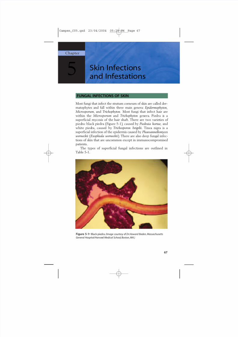
Ch. 5: Skin Infections and Infe stations • 67 FUNGAL INFECTIONS OF SKIN Most fungi that infect the stratum corneum of skin are called der- matophytes and fall within three main genera: Epidermophyton, Microsporum, and Trichophyton. Most fungi that infect hair are within the Microsporum and Trichophyton gen era . Pie dra i s a superficial mycosis of the hair shaft. There are two varie ties of pied ra: black pi edra (Fi gure 5-1) , cause d by Piedraia hortae, and white pied ra, cause d by Trichosporon beigelii. Tinea nigra is a superficial infection of the epidermis caused by Phaeoannellomyces werneckii (Exophiala werneckii ). There are also deep fung al infec- tions of skin that are uncommon except in immunocompromised patients. The types of superficial fungal infections are outlined in Table 5-1. 5 Chapter Skin Infections and Infestations Figure 5-1• Black piedra. (Image courtesy o f Dr.Howard Baden, Massachusetts General Hospital/Harvar d Medical School , Boston, MA.) 67 Campen_C05.qxd 23/04/2004 05:25 PM Page 67
-
Upload
franky-zepplin -
Category
Documents
-
view
219 -
download
0
Transcript of Camp en 9781405104418
8/3/2019 Camp en 9781405104418
http://slidepdf.com/reader/full/camp-en-9781405104418 1/21
Ch. 5:Skin Infections and Infestations • 67
FUNGAL INFECTIONS OF SKIN
Most fungi that infect the stratum corneum of skin are called der-matophytes and fall within three main genera: Epidermophyton,
Microsporum, and Trichophyton. Most fungi that infect hair arewithin the Microsporum and Trichophyton genera. Piedra is asuperficial mycosis of the hair shaft. There are two varieties of piedra: black piedra (Figure 5-1), caused by Piedraia hortae, andwhite piedra, caused by Trichosporon beigelii.Tinea nigra is asuperficial infection of the epidermis caused by Phaeoannellomyceswerneckii (Exophiala werneckii). There are also deep fungal infec-tions of skin that are uncommon except in immunocompromisedpatients.
The types of superficial fungal infections are outlined inTable 5-1.
5Chapter
Skin Infectionsand Infestations
Figure 5-1• Black piedra. (Image courtesy of Dr.Howard Baden, MassachusettsGeneral Hospital/Harvard Medical School, Boston, MA.)
67
Campen_C05.qxd 23/04/2004 05:25 PM Page 67
8/3/2019 Camp en 9781405104418
http://slidepdf.com/reader/full/camp-en-9781405104418 2/21
68 • Blueprints Dermatology
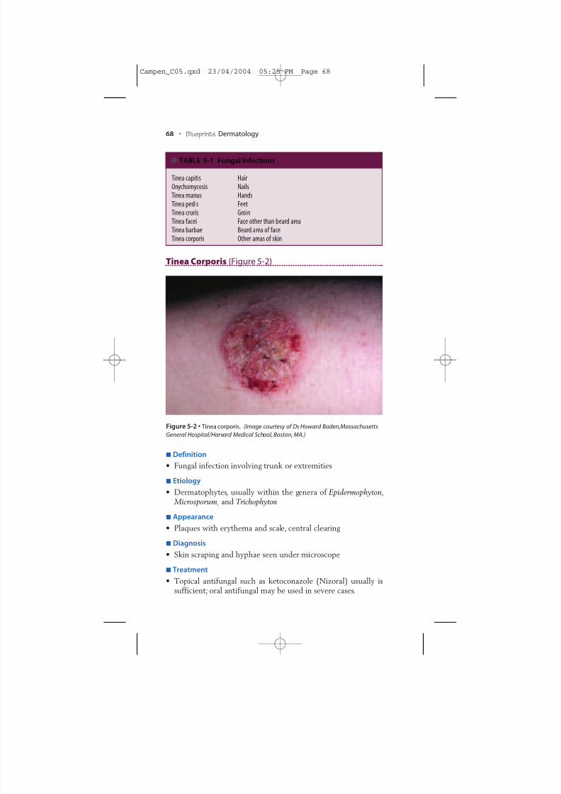
Tinea Corporis (Figure 5-2)
Definition• Fungal infection involving trunk or extremities
Etiology• Dermatophytes, usually within the genera of Epidermophyton,
Microsporum, and Trichophyton
Appearance• Plaques with erythema and scale, central clearing
Diagnosis
• Skin scraping and hyphae seen under microscopeTreatment
• Topical antifungal such as ketoconazole (Nizoral) usually issufficient; oral antifungal may be used in severe cases.
TABLE 5-1 Fungal Infections
Tinea capitis HairOnychomycosis NailsTinea manus HandsTinea pedis FeetTinea cruris GroinTinea facei Face other than beard areaTinea barbae Beard area of faceTinea corporis Other areas of skin
Figure 5-2 • Tinea corporis. (Image courtesy of Dr. Howard Baden,MassachusettsGeneral Hospital/Harvard Medical School, Boston, MA.)
Campen_C05.qxd 23/04/2004 05:25 PM Page 68
8/3/2019 Camp en 9781405104418
http://slidepdf.com/reader/full/camp-en-9781405104418 3/21
Ch. 5:Skin Infections and Infestations • 69
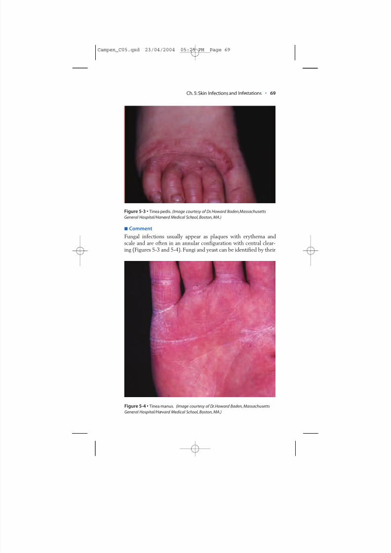
CommentFungal infections usually appear as plaques with erythema andscale and are often in an annular configuration with central clear-ing (Figures 5-3 and 5-4).Fungi and yeast can be identified by their
Figure 5-3 • Tinea pedis. (Image courtesy of Dr. Howard Baden,MassachusettsGeneral Hospital/Harvard Medical School, Boston, MA.)
Figure 5-4 • Tinea manus. (Image courtesy of Dr.Howard Baden, MassachusettsGeneral Hospital/Harvard Medical School, Boston, MA.)
Campen_C05.qxd 23/04/2004 05:25 PM Page 69
8/3/2019 Camp en 9781405104418
http://slidepdf.com/reader/full/camp-en-9781405104418 4/21
70 • Blueprints Dermatology
morphology in tissue (skin scrapings), by growth morphology onSabouraud’s agar and other special media, and by responsivenessto certain laboratory tests. In practice, observing hyphae in skinscrapings under the microscope or receiving positive cultureresults is usually sufficient for treatment. Specific typing is not usu-ally necessary except in persistent or otherwise unusual cases.
Topical antifungals are usually sufficient for treating infectionswith limited distribution.For more widespread involvement or forinfection of hair or nails, oral antifungals may be necessary. If oralantifungals are used, liver functions should be monitored beforeand during treatment.
YEAST INFECTIONS OF SKIN
Yeast can also infect skin. Candida albicans is pathogenic for skinif it gains a foothold. Some of the normally saphrophytic speciesof Candida can also cause infection if overgrowth occurs becauseof erosion, occlusion, and other factors that promote yeast growth.
Pityriasis Versicolor
Definition• Pityriasis versicolor (tinea versicolor): superficial fungal infection
Etiology• Yeast: Malassezia furfur
• Most cases occur in moist, warm climatesAppearance
• Hypopigmentation or hyperpigmentation, salmon-colored ma-cules and patches with scale
• Most often found on the upper back and upper chest • Occasionally pruritic (itchy), otherwise asymptomatic
Diagnosis• Scraping and examination with KOH preparation: “spaghetti
and meatballs” appearance of short hyphae and round yeast forms
Treatment• 2.5% selenium sulfide lotion (Selsun lotion), or shampoo con-
taining zinc pyrithione (Zincon shampoo) applied for 5 to 10minutes prior to showering every day for 2 weeks• Itraconazole 200 or 400 mg orally once or twice a month use-
ful prophylactically
Campen_C05.qxd 23/04/2004 05:25 PM Page 70
8/3/2019 Camp en 9781405104418
http://slidepdf.com/reader/full/camp-en-9781405104418 5/21
Ch. 5:Skin Infections and Infestations • 71
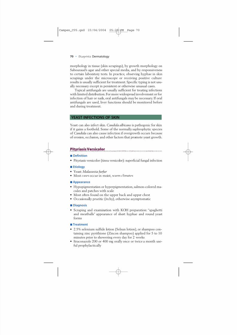
Intertrigo (Figure 5-5)
Figure 5-5 • Intertrigo. (Image courtesy of Dr. Howard Baden,MassachusettsGeneral Hospital/Harvard Medical School, Boston, MA.)
Definition• Irritation of intertriginous areas (buttock creases, groin, bet-
ween fingers and toes, under breasts)
Etiology• Yeast likes to grow in moist, warm intertriginous areas,especially when the skin is chapped or macerated for anyreason.
• Intertrigo can also be caused by bacterial infection, especiallyif between the fingers and toes. Corynebacterium are often thebacterial agents involved.
Appearance• Erythematous plaques and patches in intertriginous areas
Diagnosis• Clinical appearance usually sufficient for diagnosis
Treatment• Antifungal cream such as topical ketoconazole cream.• Mild corticosteroid can be added if there is much irritation.
Note: Vytone cream, containing both hydrocortisone and iodo-quinone (antifungal) is helpful.
Campen_C05.qxd 23/04/2004 05:25 PM Page 71
8/3/2019 Camp en 9781405104418
http://slidepdf.com/reader/full/camp-en-9781405104418 6/21
• If bacterial infection is suspected (between fingers and toes),use topical erythromycin ointment 2% bid.
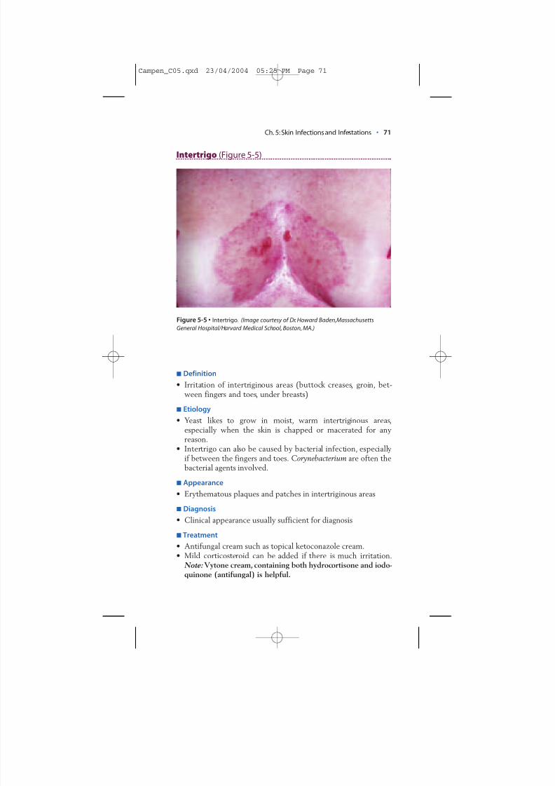
Angular Cheilitis (Figure 5-6)
Definition• Inflammation of commissures of lips (sides of the mouth)
Etiology• Irritation of skin folds can occur from drooling or lip licking.• Yeast ( Candida ) superinfection can occur.
Appearance• Cracks, fissures, and erythema of commissures of lips
Diagnosis
• Clinical appearanceTreatment
• Ketoconazole cream to affected areas bid
72 • Blueprints Dermatology
Figure 5-6 • Angular cheilitis. (Image courtesy of Dr. Howard Baden,MassachusettsGeneral Hospital/Harvard Medical School, Boston, MA.)
Campen_C05.qxd 23/04/2004 05:25 PM Page 72
8/3/2019 Camp en 9781405104418
http://slidepdf.com/reader/full/camp-en-9781405104418 7/21
Ch. 5:Skin Infections and Infestations • 73
BACTERIAL INFECTIONS OF SKIN
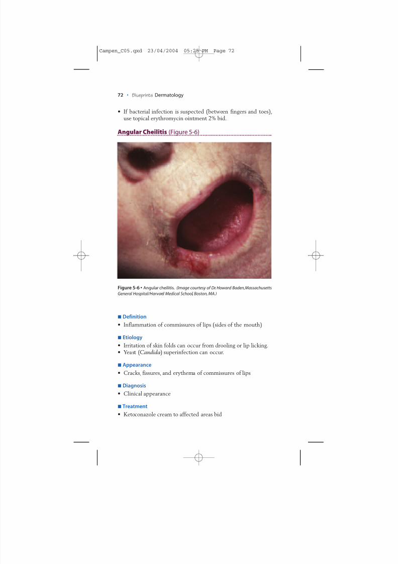
Impetigo (Figure 5-7)
Definition• Skin infection caused by Staphylococcusor Streptococcusorganisms
Etiology• Usually Staphylococcus aureus or Streptococcus pyogenes.• A bullous form of impetigo, characterized by vesicles and bul-
lae, can result from certain strains of S. aureus (including phageII, group 71, which produces a toxin causing separation in thegranular layer of the epidermis).
Appearance• Plaques and erosions, usually with yellowish crust • Bullae, if bullous form
Diagnosis• Infection is superficial in skin, and diagnosis of impetigo can
usually be made on the basis of clinical appearance.
Figure 5-7 • Impetigo. (Image courtesy of Dr. Howard Baden,MassachusettsGeneral Hospital/Harvard Medical School, Boston, MA.)
Campen_C05.qxd 23/04/2004 05:25 PM Page 73
8/3/2019 Camp en 9781405104418
http://slidepdf.com/reader/full/camp-en-9781405104418 8/21
74 • Blueprints Dermatology
• Histopathologic examination shows vesicopustule formation just below the stratum corneum.
Treatment• Impetigo is contagious and needs to be treated with antibiotics.• Oral dicloxacillin (500 mg bid), erythromycin (500 mg bid),or
a first-generation cephalosporin such as cephalexin (500 mgbid) will usually clear the lesions.
• If only a few lesions are present, topical mupiricin may be suc-cessful in clearing the problem.
• Caution: Some strains of S. pyogenes that cause impetigo cancause poststreptococcal glomerulonephritis; early treatment istherefore important.
Furuncles (Boils) and Carbuncles
Definition• Furuncle (boil): deep inflammation and infection of the hair
follicle; individual lesion with one follicular orifice• Carbuncle: coalescing of infection from several adjacent folli-
cles; several follicular orifices
Etiology• Usually staphylococci
Appearance• Furuncle: nodule with pustular formation and inflammation;
most commonly on the legs, face, and groin• Carbuncle: larger than furuncle, larger pustular component,
often found on the posterior neck or buttocks
Diagnosis• Culture may be helpful.
Treatment• Incision, drainage, and oral antibiotics
Chancroid
Definition• Sexually transmitted disease caused by contact with an infected
partner
Etiology• Haemophilus ducreyi
Appearance• Very painful erosions and ulcers on the penis or genital area,
usually with a gray base
Campen_C05.qxd 23/04/2004 05:25 PM Page 74
8/3/2019 Camp en 9781405104418
http://slidepdf.com/reader/full/camp-en-9781405104418 9/21
Diagnosis• Scraping base of an ulcer and Gram staining the scrapings:
microscopic examination will show gram-negative coccobacil-li singly or in “schools of fish.”
• Culture of the organism possible, but difficult, as the organismon a swab dies at room temperature within 2 to 4 hours.
• Swabs should be transported quickly to the laboratory orrefrigerated.
Treatment• Erythromycin 500 mg po qid for 2 weeks
VIRAL INFECTIONS OF SKIN
Warts (Figure 5-8)
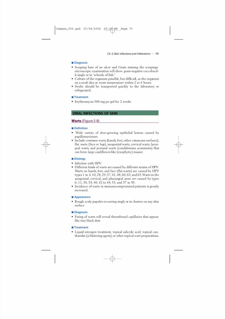
Definition• Wide variety of slow-growing epithelial lesions caused by
papillomaviruses• Include common warts (hands, feet, other cutaneous surfaces),
flat warts (face or legs), anogenital warts, cervical warts, laryn-geal warts, and perianal warts (condylomata acuminata) that can form large cauliflower-like (exophytic) masses
Etiology• Infection with HPV.
• Different kinds of warts are caused by different strains of HPV. Warts on hands, feet, and face (flat warts) are caused by HPVtypes 1 to 4, 10, 28,29,37,41, 48,60,63, and 65.Warts in theanogenital, cervical, and pharangeal areas are caused by types6, 11, 30, 34, 40, 42 to 44, 55, and 57 to 59.
• Incidence of warts in immunocompromised patients is greatlyincreased.
Appearance• Rough, scaly papules occurring singly or in clusters on any skin
surface
Diagnosis• Paring of warts will reveal thrombosed capillaries that appear
like tiny black dots
Treatment• Liquid nitrogen treatment, topical salicylic acid, topical can-
tharidin (a blistering agent), or other topical wart preparations.
Ch. 5:Skin Infections and Infestations • 75
Campen_C05.qxd 23/04/2004 05:25 PM Page 75
8/3/2019 Camp en 9781405104418
http://slidepdf.com/reader/full/camp-en-9781405104418 10/21
76 • Blueprints Dermatology
• Anogenital warts can be treated by painting with podophyllin,which is washed off 6 hours after application.Continued treat-ment every 2 to 3 weeks is necessary until the wart is com-pletely resolved.
• Immunotherapy using topical immune modulators (e.g.,
Imiquimod) is useful for treatment of genital warts.• Note: Some papillomaviruses may affect progression to carci-noma in lesions.- Verrucous carcinoma: low-grade squamous cell carcinoma
Figure 5-8 • Warts. (Image courtesy of Dr.Howard Baden,Massachusetts General Hospital/Harvard Medical School, Boston, MA.)
Campen_C05.qxd 23/04/2004 05:25 PM Page 76
8/3/2019 Camp en 9781405104418
http://slidepdf.com/reader/full/camp-en-9781405104418 11/21
Ch. 5:Skin Infections and Infestations • 77
- Epithelioma cuniculatum: verrucous carcinoma on plantarsurface of the foot
- Bowenoid papulosis: small papules on the external male andfemale genitalia infected with HPV type 16; histologicallyshows cellular atypia
• Warts in the anogenital, cervical, and pharangeal areas aresometimes caused by “high-risk” types (16, 18, 31, 33, 35, 39,45, 51, 52, 56) that may be a factor in carcinogenic progressionof some lesions.
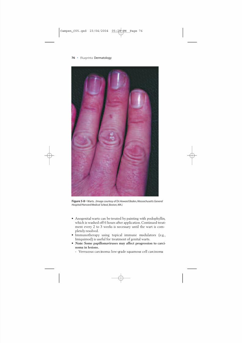
Herpes Simplex (Figure 5-9)
Definition• Herpes simplex virus infection causing “fever blisters,” genital
lesions, and other skin lesions, often around the mouth or onthe buttocks.
Figure 5-9 • Herpes simplex. (Image courtesy of Dr.Howard Baden, MassachusettsGeneral Hospital/Harvard Medical School, Boston, MA.)
Campen_C05.qxd 23/04/2004 05:25 PM Page 77
8/3/2019 Camp en 9781405104418
http://slidepdf.com/reader/full/camp-en-9781405104418 12/21
78 • Blueprints Dermatology
Etiology• Herpesvirus I (mouth, skin) or II (genital lesions, skin)
Appearance• Clusters of vesicles on erythematous base.• Vesicle superficial with fragile roof, resulting in erosion.• Lesions painful with prodrome of stinging and burning.• Lesions tend to recur in the same areas previously infected.• Lesions usually resolve in 2 to 3 weeks.• Virus remains dormant, usually within the trigeminal nerve
root ganglion.• Stress, menstrual periods, illness, and sun exposure can reacti-
vate virus.
Diagnosis• Tzanck test: scraping skin from the base of a vesicle, staining
with Wright-Giemsa stain, examining for multinucleated“ghost cells” (nuclei of keratinocytes have dissolved)
Treatment• Oral acyclovir, valcyclovir, and famcyclovir can shorten course
of active infection.• For those with frequent recurrences, daily suppressive therapy
with these medications can lengthen time between attacks.• Note: Other human herpesvirus that cause infection include
varicella zoster virus (shingles), Epstein-Barr virus (Burkitt’slymphoma, infectious mononucleosis), cytomegalovirus (cyto-megalovirus inclusion disease), human herpesvirus 6 (exanthemsubitum), human herpesvirus 7 (preferentially affects CD4 +
lymphocytes), and human herpesvirus 8 (Kaposi’s sarcoma).
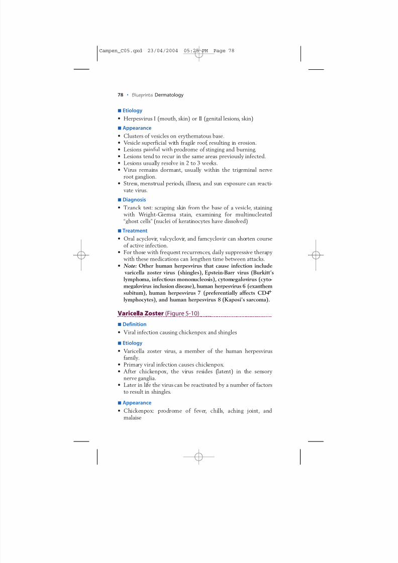
Varicella Zoster (Figure 5-10)
Definition• Viral infection causing chickenpox and shingles
Etiology• Varicella zoster virus, a member of the human herpesvirus
family.• Primary viral infection causes chickenpox.• After chickenpox, the virus resides (latent) in the sensory
nerve ganglia.• Later in life the virus can be reactivated by a number of factors
to result in shingles.Appearance
• Chickenpox: prodrome of fever, chills, aching joint, andmalaise
Campen_C05.qxd 23/04/2004 05:25 PM Page 78
8/3/2019 Camp en 9781405104418
http://slidepdf.com/reader/full/camp-en-9781405104418 13/21
Ch. 5:Skin Infections and Infestations • 79
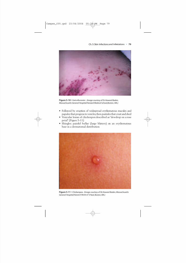
• Followed by eruption of widespread erythematous macules andpapules that progress to vesicles, then pustules that crust and shed
• Vesicular lesion of chickenpox described as “dewdrop on a rosepetal” (Figure 5-11)
• Shingles: painful bullae (large blisters) on an erythematousbase in a dermatomal distribution
Figure 5-10 • Varicella zoster. (Image courtesy of Dr.Howard Baden,Massachusetts General Hospital/Harvard Medical School,Boston, MA.)
Figure 5-11 • Chickenpox. (Image courtesy of Dr.Howard Baden, MassachusettsGeneral Hospital/Harvard Medical School, Boston, MA.)
Campen_C05.qxd 23/04/2004 05:25 PM Page 79
8/3/2019 Camp en 9781405104418
http://slidepdf.com/reader/full/camp-en-9781405104418 14/21
Figure 5-12 • Molluscum contagiosum. (Image courtesy of Dr.Howard Baden,Massachusetts General Hospital/Harvard Medical School,Boston, MA.)
80 • Blueprints Dermatology
Diagnosis• Tzanck preparation:Scrape skin from base of vesicle, stain with
Wright-Giemsa stain, examine for giant cells (intracytoplasmicedema in keratinocytes).
Treatment• Oral acyclovir, valcyclovir, or famcyclovir.• Note: Postherpetic neuralgia is a common, but painful, after
effect of shingles. The risk for postherpetic neuralgia may bereduced by early treatment with oral antiviral medications.
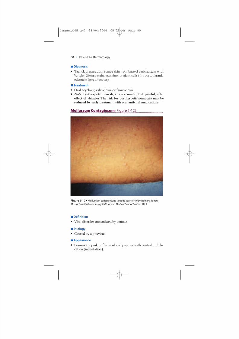
Molluscum Contagiosum (Figure 5-12)
Definition• Viral disorder transmitted by contact
Etiology• Caused by a poxvirus
Appearance• Lesions are pink or flesh-colored papules with central umbili-
cation (indentation).
Campen_C05.qxd 23/04/2004 05:25 PM Page 80
8/3/2019 Camp en 9781405104418
http://slidepdf.com/reader/full/camp-en-9781405104418 15/21
Ch. 5:Skin Infections and Infestations • 81
• Often found on young children on arms, legs, and abdomenafter contact with other children.
• Sometimes found on adults, often in the lower abdominal area,after contact with a sexual partner.Diagnosis
• Usually diagnosed by clinical appearance; biopsy if uncertain asto diagnosis
Treatment• Liquid nitrogen, curettage, or application of topical tretinoin
(Retin A).• Note: Lesions will eventually resolve on their own. In children
with numerous lesions, one option is to not treat, recognizingthat additional lesions may occur before resolution occurs.
Insect and Arachnoid Bites
Definition• Bites by insects or arachnoids
Etiology• Insects (six legs): mosquitos, bees, fleas, fire ants, lice• Arachnoids (eight legs): mites, spiders
Appearance• Typically erythematous papule with a central punctum.• Lesion can occur singly, in groups, or in linear configurations.• Occasionally vesicles or hemorrhagic bullae are present.
• Lesion can be extensive, as in the bite of a brown recluse spider:urticaria, bullae, severe necrosis of bite site and surrounding area.• Can cause a variety of symptoms including itching, stinging
and burning, edema, and/or erythema.• Systemic effects including anaphylaxis can sometimes occur.• Bites can sometimes result in systemic disease.• Tick bites can be vectors for disease; bites by certain tick
species can result in Lyme disease, erlichiosis, or Rocky Mountainspotted fever with systemic symptoms.Tick bites in some casescan also result in tick paralysis, causing ascending paralysis andpotential death, unless the tick is removed. Babesiosis, causedby the intracellular red blood cell parasite Babesia microti, canalso result from tick bites.
• Scorpion bites and black widow spider bites can result in bothlocal and potentially fatal systemic effects. Bites by somespecies of caterpillars can also result in severe reactions.Diagnosis
• Diagnosis by history; if lice, examine hair for nits (lice eggs).
Campen_C05.qxd 23/04/2004 05:25 PM Page 81
8/3/2019 Camp en 9781405104418
http://slidepdf.com/reader/full/camp-en-9781405104418 16/21
82 • Blueprints Dermatology
Treatment• Bites that itch can be treated with topical corticosteroid applied
twice a day.• Persistent itching with edema and erythema can be treated
with oral antihistamines.• Severe reactions with edema and difficulty breathing are med-
ical emergencies that require subcutaneous epinephrine (1:1000solution) injection, corticosteroid injection, and other life sup-port measures.
• Epinephrine emergency kits are available for persons with beeallergy.
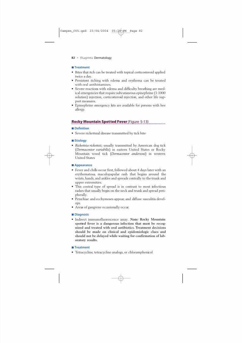
Rocky Mountain Spotted Fever (Figure 5-13)
Definition• Severe rickettsial disease transmitted by tick bite
Etiology• Rickettsia rickettsii; usually transmitted by American dog tick
(Dermacentor variabilis ) in eastern United States or RockyMountain wood tick ( Dermacentor andersoni) in westernUnited States
Appearance• Fever and chills occur first, followed about 4 days later with an
erythematous, maculopapular rash that begins around thewrists, hands, and ankles and spreads centrally to the trunk andupper extremities.
• This central type of spread is in contrast to most infectiousrashes that usually begin on the neck and trunk and spread peri-pherally.
• Petechiae and ecchymoses appear, and diffuse vasculitis devel-ops.
• Areas of gangrene occasionally occur.
Diagnosis• Indirect immunofluorescence assay. Note: Rocky Mountain
spotted fever is a dangerous infection that must be recog-nized and treated with oral antibiotics. Treatment decisionsshould be made on clinical and epidemiologic clues andshould not be delayed while waiting for confirmation of lab-
oratory results.
Treatment• Tetracycline, tetracycline analogs, or chloramphenicol
Campen_C05.qxd 23/04/2004 05:25 PM Page 82
8/3/2019 Camp en 9781405104418
http://slidepdf.com/reader/full/camp-en-9781405104418 17/21
Ch. 5:Skin Infections and Infestations • 83
PARASITIC INFESTATIONS
Lice
Definition• Blood-sucking insects that infest hair-bearing areas (head lice,
pubic lice) or live in seams of clothes (body lice)Etiology
• Pediculus humanus capitis: head louse.
Figure 5-13 • Rocky Mountain spotted fever. (Image courtesy of Dr.Howard Baden,Massachusetts General Hospital/Harvard Medical School, Boston, MA.)
Campen_C05.qxd 23/04/2004 05:25 PM Page 83
8/3/2019 Camp en 9781405104418
http://slidepdf.com/reader/full/camp-en-9781405104418 18/21
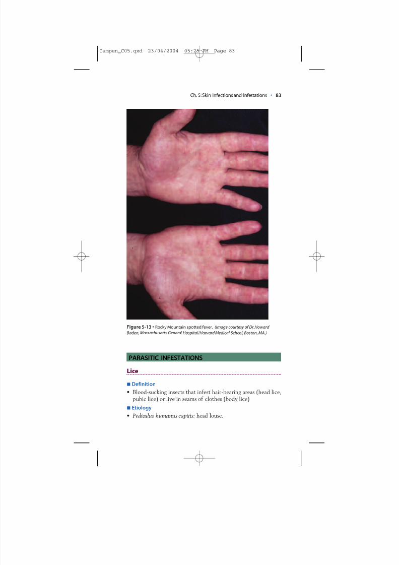
• Pthirus pubis: pubic louse (Figure 5-14).• Pediculus humanus corporis: body louse.• Note: Rarely, lice can transmit epidemic typhus ( Rickettsia
prowazekii ), trench fever ( Rickettsia quintana ), and relapsingfever ( Borrelia recurrentis ).
Appearance• Erythematous puncta, sometimes with surrounding erythema.• Bites can be pruritic (itchy) and/or painful.• There may be only a few lice or hundreds.
Figure 5-14 • Lice (pubic louse). (Image courtesy of Dr.Howard Baden,Massachusetts General Hospital/Harvard Medical School,Boston, MA.)
84 • Blueprints Dermatology
Campen_C05.qxd 23/04/2004 05:26 PM Page 84
8/3/2019 Camp en 9781405104418
http://slidepdf.com/reader/full/camp-en-9781405104418 19/21
Ch. 5:Skin Infections and Infestations • 85
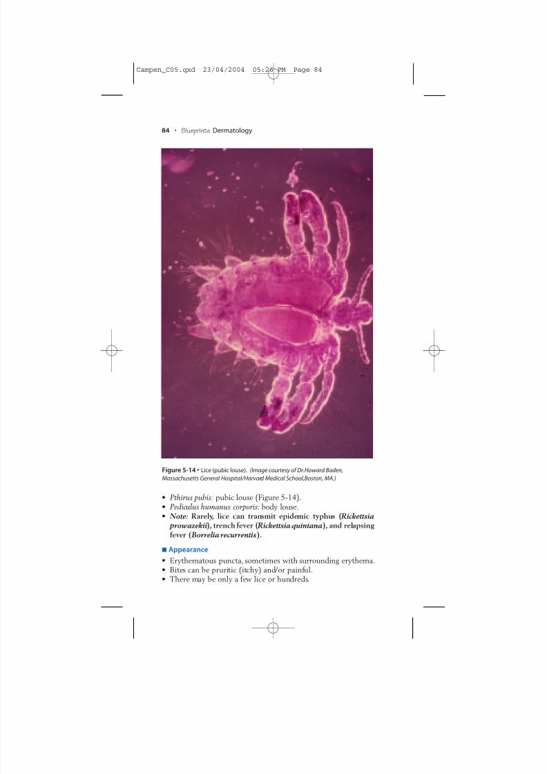
Diagnosis• Clinical examination of scalp and clothes for lice• Examination of scalp for nits (white dots on hair shaft)• Microscopic examination of nits (eggs glued to hair shafts)
(Figure 5-15) to confirm diagnosis
Treatment• Lice can be difficult to eradicate.• For hair-bearing areas infested with lice, use permethrin (Nix)
shampoo or pyrethrin (Rid) shampoo.
Figure 5-15 • Lice (nit). (Image courtesy of Dr. Howard Baden,MassachusettsGeneral Hospital/Harvard Medical School, Boston, MA.)
Campen_C05.qxd 23/04/2004 05:26 PM Page 85
8/3/2019 Camp en 9781405104418
http://slidepdf.com/reader/full/camp-en-9781405104418 20/21
• Follow shampoo with vinegar rinse and fine-toothed combingto remove nits.
• For treatment of skin infested with body lice, use lindane(Kwell) or permethrins (Elimite).
• Clothes of patients with body lice should be cleaned and ironed.• Sulfur (4%–10%) in petrolatum can also be applied to skin or
scalp infested with lice.• Note: Lice infestation can quickly spread through a classroom
of children, and children with nits should not attend classuntil treatment has been started. The CDC recommends thatan infested child be kept from a child-care setting until 24hours after treatment has started. Many states and local healthdepartments require that the child be free of nits before read-mission to school or child care. Children should be checkeddaily for evidence of new or continued infection for 10 daysafter treatment. Retreatment in 7 to 10 days may be necessary.
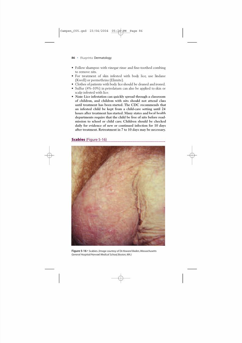
Scabies (Figure 5-16)
Figure 5-16 • Scabies. (Image courtesy of Dr.Howard Baden, MassachusettsGeneral Hospital/Harvard Medical School, Boston, MA.)
86 • Blueprints Dermatology
Campen_C05.qxd 23/04/2004 05:26 PM Page 86
8/3/2019 Camp en 9781405104418
http://slidepdf.com/reader/full/camp-en-9781405104418 21/21
Definition• Scabies mites are arthropods that live, lay eggs within the skin
(stratum corneum), and bite.
Etiology• Sarcoptes scabiei, variation hominis
Appearance• Scaly papules and excoriations (from scratching) as well as tiny
linear or wavy “burrows” in warm areas such as between fingersand toes, in the umbilicus, around the waistline, and on thepenile shaft.
• Skin infested with scabies is extremely itchy.
Diagnosis• Microscopic examination of papule, erosion, or burrow for sca-
bies (six-legged) mite, mite eggs, or mite feces
Treatment• Lindane (Kwell) or permethrin (Elimite) applied from the
neck down, left on overnight, and washed off the followingmorning; repeat procedure 1 week later.
• The morning after treatment, launder sheets, bedclothes, andtowels.
• Entire family should be treated in confirmed cases.• Sulfur (4%–10%) in petrolatum can also be used to treat skin
infested with scabies.• Note: Infestation with hundreds of mites is called Norwegian
scabies and can occur in nursing home patients or otherwisedebilitated patients. Oral ivermectin (single dose of 200 µg/kgbody weight) alone or combined with topical treatment hasbeen successful in treating patients with Norwegian scabies.
Ch. 5:Skin Infections and Infestations • 87
Campen_C05.qxd 23/04/2004 05:26 PM Page 87


![[Code Camp 2009] Utilizando Silverlight en aplicaciones de Social Networking (Adrián Eidelman + Rubén Altman)](https://static.fdocuments.us/doc/165x107/54dbb6854a7959ef358b46cb/code-camp-2009-utilizando-silverlight-en-aplicaciones-de-social-networking-adrian-eidelman-ruben-altman.jpg)


![REFERENCE SECTION · 2017-08-23 · Camp David Accords [Acuerdos de Camp David] s. acuerdos de paz históricos entre Israel y Egipto, negociados en Camp David, Maryland, en 1978.](https://static.fdocuments.us/doc/165x107/5e892f10b6398a6d1d464248/reference-section-2017-08-23-camp-david-accords-acuerdos-de-camp-david-s-acuerdos.jpg)