Cambridge International Examinations Cambridge ... International... · for each cell that shows any...
16
READ THESE INSTRUCTIONS FIRST Write your Centre number, candidate number and name on all the work you hand in. Write in dark blue or black pen. You may use an HB pencil for any diagrams or graphs. Do not use staples, paper clips, glue or correction fluid. DO NOT WRITE IN ANY BARCODES. Answer all questions. Electronic calculators may be used. You may lose marks if you do not show your working or if you do not use appropriate units. At the end of the examination, fasten all your work securely together. The number of marks is given in brackets [ ] at the end of each question or part question. *6673773990* BIOLOGY 9700/36 Advanced Practical Skills 2 October/November 2015 2 hours Candidates answer on the Question Paper. Additional Materials: As listed in the Confidential Instructions. For Examiner’s Use 1 2 Total This document consists of 14 printed pages and 2 blank pages. DC (ST/SW) 89024/4 © UCLES 2015 [Turn over Cambridge International Examinations Cambridge International Advanced Subsidiary and Advanced Level
Transcript of Cambridge International Examinations Cambridge ... International... · for each cell that shows any...

READ THESE INSTRUCTIONS FIRST
Write your Centre number, candidate number and name on all the work you hand in.Write in dark blue or black pen.You may use an HB pencil for any diagrams or graphs.Do not use staples, paper clips, glue or correction fluid.DO NOT WRITE IN ANY BARCODES.
Answer all questions.Electronic calculators may be used.You may lose marks if you do not show your working or if you do not use appropriate units.
At the end of the examination, fasten all your work securely together.The number of marks is given in brackets [ ] at the end of each question or part question.
*6673773990*
BIOLOGY 9700/36
Advanced Practical Skills 2 October/November 2015
2 hours
Candidates answer on the Question Paper.
Additional Materials: As listed in the Confidential Instructions.
For Examiner’s Use
1
2
Total
This document consists of 14 printed pages and 2 blank pages.
DC (ST/SW) 89024/4© UCLES 2015 [Turn over
Cambridge International ExaminationsCambridge International Advanced Subsidiary and Advanced Level
2
9700/36/O/N/15© UCLES 2015
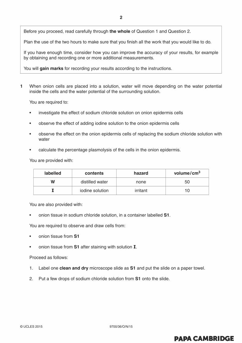
Before you proceed, read carefully through the whole of Question 1 and Question 2.
Plan the use of the two hours to make sure that you finish all the work that you would like to do.
If you have enough time, consider how you can improve the accuracy of your results, for example by obtaining and recording one or more additional measurements.
You will gain marks for recording your results according to the instructions.
1 When onion cells are placed into a solution, water will move depending on the water potential inside the cells and the water potential of the surrounding solution.
You are required to:
• investigate the effect of sodium chloride solution on onion epidermis cells
• observe the effect of adding iodine solution to the onion epidermis cells
• observe the effect on the onion epidermis cells of replacing the sodium chloride solution with water
• calculate the percentage plasmolysis of the cells in the onion epidermis.
You are provided with:
labelled contents hazard volume / cm3
W distilled water none 50
I iodine solution irritant 10
You are also provided with:
• onion tissue in sodium chloride solution, in a container labelled S1.
You are required to observe and draw cells from:
• onion tissue from S1
• onion tissue from S1 after staining with solution I.
Proceed as follows:
1. Label one clean and dry microscope slide as S1 and put the slide on a paper towel.
2. Put a few drops of sodium chloride solution from S1 onto the slide.

3
9700/36/O/N/15© UCLES 2015 [Turn over
3. Remove a piece of the onion tissue from S1 and, using forceps or fingers, peel off the inner epidermis as shown in Fig. 1.1.
Fig. 1.1
4. Cut one piece of the epidermis that will fit under a coverslip. Replace the remaining epidermis into S1.
5. Place the epidermis on the slide as shown in Fig. 1.2. If the epidermis is folded, you may need to add more drops from S1 so that it floats and uncurls.
6. To prevent the epidermis from drying out add more drops from S1 if needed.
epidermis
solution
slide
paper towel
Fig. 1.2
7. Cover the epidermis with a coverslip so that one edge of the epidermis is close to the edge of the coverslip. Use a paper towel to remove any excess liquid that is outside the coverslip.
8. View the slide using the microscope.
You may need to reduce the amount of light entering the microscope to observe the cells clearly.
4
9700/36/O/N/15© UCLES 2015
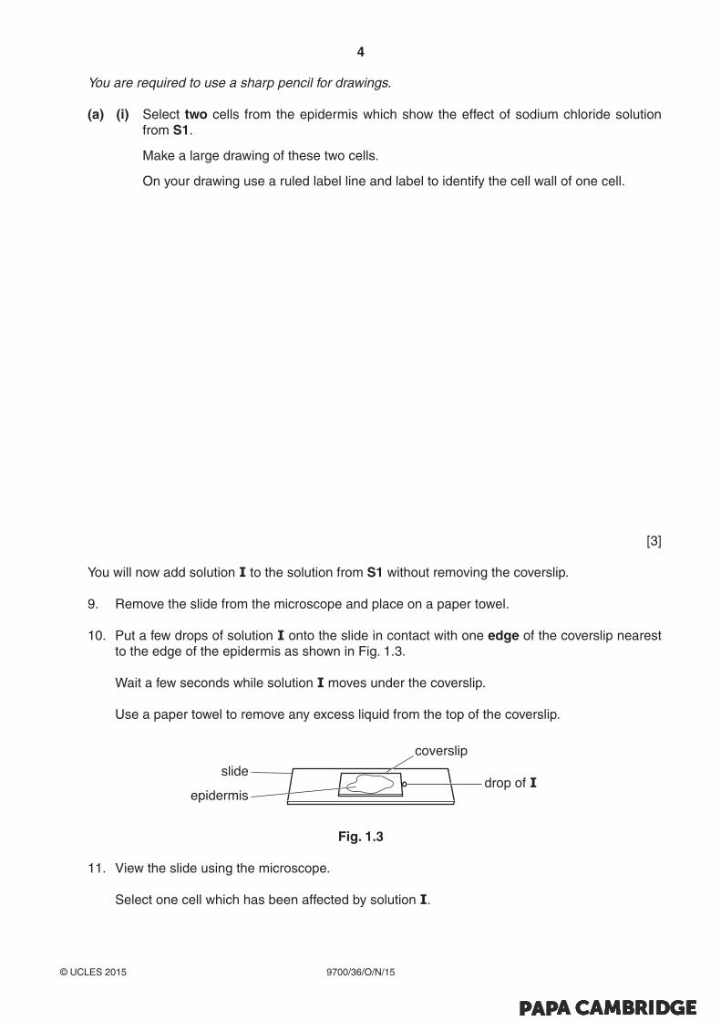
You are required to use a sharp pencil for drawings.
(a) (i) Select two cells from the epidermis which show the effect of sodium chloride solution from S1.
Make a large drawing of these two cells.
On your drawing use a ruled label line and label to identify the cell wall of one cell.
[3]
You will now add solution I to the solution from S1 without removing the coverslip.
9. Remove the slide from the microscope and place on a paper towel.
10. Put a few drops of solution I onto the slide in contact with one edge of the coverslip nearest to the edge of the epidermis as shown in Fig. 1.3.
Wait a few seconds while solution I moves under the coverslip.
Use a paper towel to remove any excess liquid from the top of the coverslip.
epidermis
slidedrop of I
coverslip
Fig. 1.3
11. View the slide using the microscope.
Select one cell which has been affected by solution I.

5
9700/36/O/N/15© UCLES 2015 [Turn over
(ii) Make a large drawing of the cell that you have selected.
Annotate your drawing to describe one feature that is different between this cell and the cells drawn in (a)(i).
[3]
You are now required to:
• investigate the effect of replacing the solution from S1 with water
• calculate the percentage of the cells that are plasmolysed in the onion tissue.
12. Label one clean and dry microscope slide as W and put the slide on a paper towel.
13. Repeat steps 3 and 4 (page 3).
14. Put the piece of the cut epidermis into the container labelled W (which contains distilled water) and leave for two minutes.
15. After two minutes, put a few drops of distilled water from W onto slide W and then put the epidermis on the slide as shown in Fig. 1.2 on page 3.
16. Cover the epidermis with a coverslip and use a paper towel to remove any excess liquid that is outside the coverslip.
17. View the slide using the microscope.
(b) Look at an area of cells using the ×10 and ×40 objective lenses.
Select the objective lens which allows you to count a suitable number of cells in a field of view so that you can calculate the percentage of cells showing plasmolysis.
(i) State the magnification of the objective lens you will use.
magnification × ..............................................[1]
You will need to decide the total number of cells to count as your sample.
(ii) State the total number of cells you decided to count.
total number of cells ......................................[1]

6
9700/36/O/N/15© UCLES 2015
The onion cells may show degrees of plasmolysis, from no plasmolysis to complete plasmolysis as shown in Fig. 1.4.
no plasmolysis slight plasmolysis more plasmolysis complete plasmolysis
Fig. 1.4
18. Select one field of view which includes cells showing any degree of plasmolysis and cells showing no plasmolysis.
19. Observe and record on page 7 each of the cells in your sample, recording:
• ‘✓’ for each cell that shows any degree of plasmolysis (see Fig. 1.4)
• ‘✗’ for each cell that shows no plasmolysis.
Consider how you will obtain results which are as accurate as possible.
(iii) Using your results calculate the percentage of cells that are plasmolysed.
You may lose marks if you do not show your working.
[1]
7
9700/36/O/N/15© UCLES 2015 [Turn over
(iv) Prepare the space below and record the number of cells showing any degree of plasmolysis and the number of cells showing no plasmolysis (raw results) and record processed results for the percentage of cells that are plasmolysed in the onion tissue.
[5]
(v) Identify one significant source of error in measuring the dependent variable.
..........................................................................................................................................
...................................................................................................................................... [1]
(vi) Explain, in terms of the movement of water and water potential, the effect of water replacing the sodium chloride solution on the cells of the epidermis.
..........................................................................................................................................
..........................................................................................................................................
..........................................................................................................................................
..........................................................................................................................................
..........................................................................................................................................
..........................................................................................................................................
...................................................................................................................................... [3]
(vii) Suggest how you would modify this investigation to find the sodium chloride concentration of an unknown solution.
..........................................................................................................................................
..........................................................................................................................................
..........................................................................................................................................
..........................................................................................................................................
..........................................................................................................................................
..........................................................................................................................................
...................................................................................................................................... [3][Total: 21]

8
9700/36/O/N/15© UCLES 2015
2 Plants lose water through a process called transpiration. This involves the flow of water from the roots to the leaves, where water evaporates from the surfaces of the cells in the leaf.
Some plants have features to reduce water loss. For example their leaves may be rolled or their stomata are located mainly on the underside of their leaves.
These features reduce transpiration and such plants are called xerophytes.
Fig. 2.1 shows diagrams of a transverse section and surface view of the same leaf in humid conditions and in dry conditions.
exposed lowerepidermis of leaf
(unrolled)
exposed lowerepidermis of leaf
(rolled)
in humid conditions
in dry conditions
transverse section of leaf(unrolled)
surface view oflower epidermis
(unrolled)
surface view oflower epidermis
(rolled)
transverse section of leaf(rolled)
Fig. 2.1
9
9700/36/O/N/15© UCLES 2015 [Turn over
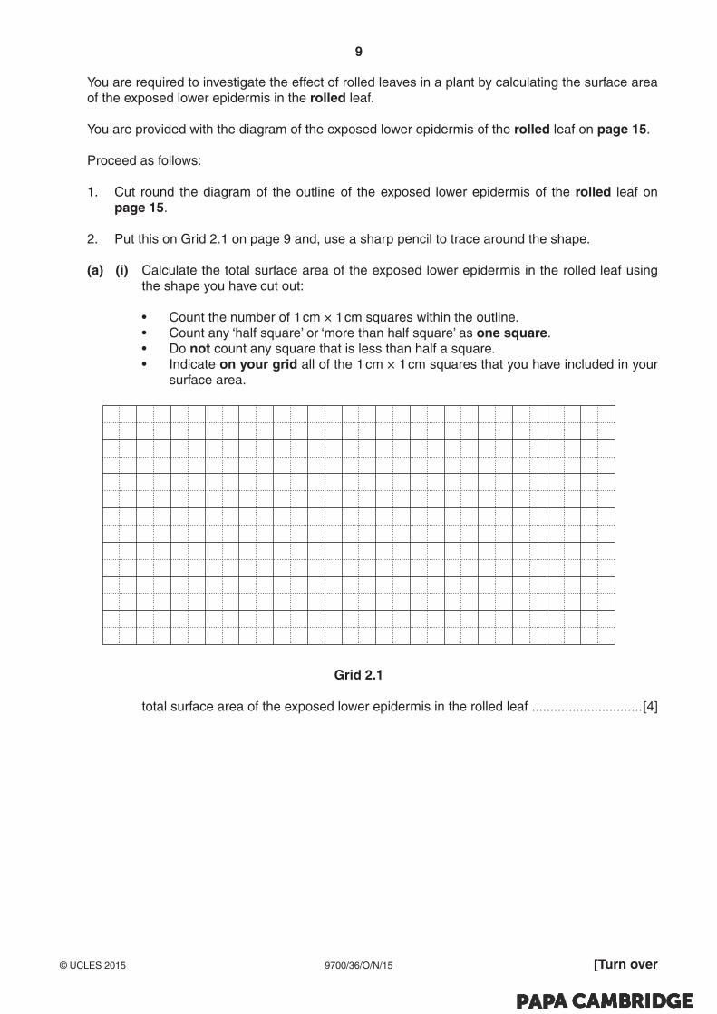
You are required to investigate the effect of rolled leaves in a plant by calculating the surface area of the exposed lower epidermis in the rolled leaf.
You are provided with the diagram of the exposed lower epidermis of the rolled leaf on page 15.
Proceed as follows:
1. Cut round the diagram of the outline of the exposed lower epidermis of the rolled leaf on page 15.
2. Put this on Grid 2.1 on page 9 and, use a sharp pencil to trace around the shape.
(a) (i) Calculate the total surface area of the exposed lower epidermis in the rolled leaf using the shape you have cut out:
• Count the number of 1 cm × 1 cm squares within the outline. • Count any ‘half square’ or ‘more than half square’ as one square. • Do not count any square that is less than half a square. • Indicate on your grid all of the 1 cm × 1 cm squares that you have included in your
surface area.
Grid 2.1
total surface area of the exposed lower epidermis in the rolled leaf .............................. [4]
10
9700/36/O/N/15© UCLES 2015
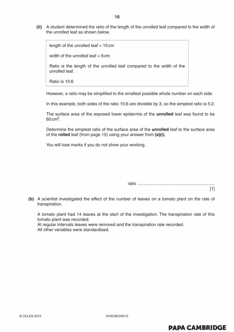
(ii) A student determined the ratio of the length of the unrolled leaf compared to the width of the unrolled leaf as shown below.
length of the unrolled leaf = 15 cm
width of the unrolled leaf = 6 cm
Ratio is the length of the unrolled leaf compared to the width of the unrolled leaf.
Ratio is 15:6
However, a ratio may be simplified to the smallest possible whole number on each side.
In this example, both sides of the ratio 15:6 are divisible by 3, so the simplest ratio is 5:2.
The surface area of the exposed lower epidermis of the unrolled leaf was found to be 60 cm2.
Determine the simplest ratio of the surface area of the unrolled leaf to the surface area of the rolled leaf (from page 15) using your answer from (a)(i).
You will lose marks if you do not show your working.
ratio ................................................................ [1]
(b) A scientist investigated the effect of the number of leaves on a tomato plant on the rate of transpiration.
A tomato plant had 14 leaves at the start of the investigation. The transpiration rate of this tomato plant was recorded.
At regular intervals leaves were removed and the transpiration rate recorded. All other variables were standardised.

11
9700/36/O/N/15© UCLES 2015 [Turn over
The results of the investigation are shown in Table 2.1.
Table 2.1
number of leaves transpiration rate/ μl min–1
14 9.5
10 4.3
8 3.9
5 3.5
2 1.8
You are required to use a sharp pencil for graphs.
(i) Plot a graph of the data shown in Table 2.1.
[4]
(ii) Estimate the transpiration rate of the plant with 12 leaves.
Show on the graph how you estimated the transpiration rate.
transpiration rate ....................................... [2]

12
9700/36/O/N/15© UCLES 2015
You are required to use a sharp pencil for drawing.
(c) Fig. 2.2 is a photomicrograph showing a stained transverse section through a leaf.
You are not expected to be familiar with this specimen.
mid-rib
magnification ×200
Fig. 2.2
Draw a large plan diagram of the mid-rib as shown in Fig. 2.2.
Use one ruled label line and label to identify the xylem.
[5]

13
9700/36/O/N/15© UCLES 2015 [Turn over
(d) Fig. 2.3 is a photomicrograph showing a stained transverse section of a mid-rib of a different plant species.
You are not expected to be familiar with this specimen.
magnification ×100
Fig. 2.3
Prepare the space below so that it is suitable for you to record observable differences between the mid-rib of Fig. 2.2 and the mid-rib of Fig. 2.3.
Record your observations in the space you have prepared.
[3]
[Total: 19]

14
9700/36/O/N/15© UCLES 2015
BLANK PAGE

15
9700/36/O/N/15© UCLES 2015
Diagram for Question 2 (a)(i)
Outline of exposed lower epidermis of the rolled leaf

16
9700/36/O/N/15© UCLES 2015
BLANK PAGE
Permission to reproduce items where third-party owned material protected by copyright is included has been sought and cleared where possible. Every reasonable effort has been made by the publisher (UCLES) to trace copyright holders, but if any items requiring clearance have unwittingly been included, the publisher will be pleased to make amends at the earliest possible opportunity.
To avoid the issue of disclosure of answer-related information to candidates, all copyright acknowledgements are reproduced online in the Cambridge International Examinations Copyright Acknowledgements Booklet. This is produced for each series of examinations and is freely available to download at www.cie.org.uk after the live examination series.
Cambridge International Examinations is part of the Cambridge Assessment Group. Cambridge Assessment is the brand name of University of Cambridge Local Examinations Syndicate (UCLES), which is itself a department of the University of Cambridge.